Y-Box Binding Protein 1 Stabilizes Hepatitis C Virus NS5A via
Phosphorylation-Mediated Interaction with NS5A To Regulate Viral
Propagation
Wei-Ting Wang,aTsung-Yuan Tsai,a,bChi-Hong Chao,a*Bo-Ying Lai,aYan-Hwa Wu Leea,b
Institute of Biochemistry and Molecular Biology, School of Life Sciences, National Yang-Ming University, Taipei, Taiwana
; Department of Biological Science and Technology, College of Biological Science and Technology, National Chiao-Tung University, Hsinchu, Taiwanb
ABSTRACT
Replication of hepatitis C virus (HCV) is dependent on virus-encoded proteins and numerous cellular factors. DDX3 is a
well-known host cofactor of HCV replication. In this study, we investigated the role of a DDX3-interacting protein, Y-box binding
protein 1 (YB-1), in the HCV life cycle. Both YB-1 and DDX3 interacted with the viral nonstructural protein NS5A. During HCV
infection, YB-1 partially colocalized with NS5A and the HCV replication intermediate double-stranded RNA (dsRNA) in
HCV-infected Huh-7.5.1 cells. Despite sharing the same interacting partners, YB-1 participated in HCV RNA replication but was
dis-pensable in steady-state HCV RNA replication, different from the action of DDX3. Moreover, knockdown of YB-1 in
HCV-in-fected cells prevented infectious virus production and reduced the ratio of hyperphosphorylated (p58) to hypophosphorylated
(p56) forms of NS5A, whereas DDX3 silencing did not affect the ratio of the p58 and p56 phosphoforms of NS5A. Interestingly,
silencing of YB-1 severely reduced NS5A protein stability in NS5A-ectopically expressing, replicon-containing, and
HCV-in-fected cells. Furthermore, mutations of serine 102 of YB-1 afHCV-in-fected both YB-1–NS5A interaction and NS5A-stabilizing activity of
YB-1, indicating that this Akt phosphorylation site of YB-1 plays an important role in stabilizing NS5A. Collectively, our results
support a model in which the event of YB-1 phosphorylation-mediated interaction with NS5A results in stabilizing NS5A to
sus-tain HCV RNA replication and infectious HCV production. Overall, our study may reveal a new aspect for the development of
novel anti-HCV drugs.
IMPORTANCE
Chronic hepatitis C virus (HCV) infection induces liver cirrhosis and hepatocellular carcinoma. The viral nonstructural protein
NS5A co-opting various cellular signaling pathways and cofactors to support viral genome replication and virion assembly is a
new strategy for anti-HCV drug development. NS5A phosphorylation is believed to modulate switches between different stages
of the HCV life cycle. In this study, we identified the cellular protein YB-1 as a novel NS5A-interacting protein. YB-1 is a
multi-functional protein participating in oncogenesis and is an oncomarker of hepatocellular carcinoma (HCC). We found that YB-1
protects NS5A from degradation and likely regulates NS5A phosphorylation through its phosphorylation-dependent interaction
with NS5A, which might be controlled by HCV-induced signaling pathways. Our observations suggest a model in which HCV
modulates NS5A level and the ratio of the p58 and p56 phosphoforms for efficient viral propagation via regulation of cellular
signaling inducing YB-1 phosphorylation. Our finding may provide new aspects for developing novel anti-HCV drugs.
H
epatitis C virus (HCV) chronically infects millions of people
worldwide (
1
). Chronic HCV infection induces chronic
hep-atitis, liver cirrhosis, and hepatocellular carcinoma. HCV
infec-tion has become a serious health problem due to the unavailability
of an effective vaccine and limited clinical treatment protocols (
2
).
HCV is a positive-stranded RNA virus that contains a 9.6-kb
genome consisting of a single open reading frame flanked by 5
=
and 3
=
nontranslated regions (NTR). An internal ribosome entry
site (IRES) in the 5
=
NTR directs the translation of a polyprotein,
which is processed co- and posttranslationally into 10 or more
viral proteins (
3
,
4
). HCV infection is sustained by spatiotemporal
interplay between viral proteins and a panel of cellular cofactors to
coordinate translation of the viral genome, viral RNA replication,
and the production of infectious viral particles. However, there is
still limited understanding of the molecular mechanisms
under-lying the coordinated interactions of these events.
The nonstructural protein 5A (NS5A) is a phosphoprotein
highly variable among genotypes of HCV (
5
). NS5A is recognized
as a key modulator of the HCV life cycle, and the factor has
emerged as a new target of drug development (
2
). NS5A,
consist-ing of three domains (
6
), is a component of the HCV replication
complex (
7–10
) required for infectious virus production (
11–13
).
Domain I of NS5A is essential for HCV RNA replication (
14
),
while most of domain II is not involved (
12
). Domain III
partici-Received10 June 2015 Accepted1 September 2015
Accepted manuscript posted online9 September 2015
CitationWang W-T, Tsai T-Y, Chao C-H, Lai B-Y, Wu Lee Y-H. 2015. Y-box binding protein 1 stabilizes hepatitis C virus NS5A via phosphorylation-mediated interaction with NS5A to regulate viral propagation. J Virol 89:11584 –11602.
doi:10.1128/JVI.01513-15.
Editor:J.-H. J. Ou
Address correspondence to Yan-Hwa Wu Lee, [email protected].
*Present address: Chi-Hong Chao, Department of Biological Science and Technology, College of Biological Science and Technology, National Chiao-Tung University, Hsinchu, Taiwan.
Copyright © 2015, American Society for Microbiology. All Rights Reserved.
on November 7, 2019 by guest
http://jvi.asm.org/
pates in virion assembly (
12
,
13
,
15
). NS5A has also been reported
to either positively or negatively regulate HCV IRES-mediated
translation (
16–18
). By regulating activity of cellular lipid kinase
phosphatidylinositol 4-kinase type III alpha (PI4KIII-␣), NS5A
has been demonstrated to modulate the formation of a
membra-nous web to support HCV RNA replication (
19
,
20
). A recent
study on stilbene 1,2-diamines, small anti-HCV compounds,
re-vealed that NS5A may have a role in the initiation of HCV RNA
replication, which is distinct from steady-state HCV RNA
replica-tion (
21
). Moreover, a transient HCV RNA replication occurring
early after infection was later recognized and characterized by the
colocalization of negative-strand HCV RNA with NS5A but not
another replicase component, NS3 (
22
), underscoring the unique
role of NS5A in the early stage of HCV RNA replication. To
facil-itate HCV propagation, NS5A also regulates multiple cellular
sig-naling pathways, including the phosphoinositol 3-kinase
(PI3K)-Akt survival pathway (
23
). Although NS5A is involved in many
steps in the HCV life cycle and host signaling pathways, it does not
have known enzymatic activity. NS5A is believed to exhibit its
different functions via interactions with specific viral proteins and
various host proteins (
2
). On the other hand, NS5A has been
re-ported to be regulated by ubiquitin-proteasome degradation (
24
).
Administration of zinc mesoporphyrin (ZnMP), a synthetic
non-heme metalloporphyrin, induces NS5A ubiquitination and
pro-teasome degradation and, hence, inhibition of HCV RNA
replica-tion (
24
).
Among the cellular factors reported to be involved in the HCV
life cycle, DDX3 gets much attention. DDX3 is a multifunctional
DEAD box RNA helicase that regulates transcription and
transla-tion and may functransla-tion as either a tumor suppressor or an
onco-gene in different cell types (
25–30
). We and others have reported
that DDX3 interacts with the HCV core (
31–33
). Further studies
demonstrated that DDX3 is involved in the HCV life cycle and
positively regulates HCV RNA replication (
34
,
35
). As DDX3 also
accomplishes its multiple functions via intracellular formation of
different complexes with a variety of proteins (
25
,
36
), we were
interested to know whether DDX3 would act in concert with other
cellular factors to modulate HCV replication or viral assembly.
The yeast two-hybrid assay was performed to investigate the
DDX3 interactome, and Y-box binding protein 1 (YB-1) was
identified as one of DDX3-interacting partners (C. H. Chao and
Y. H. Wu Lee, unpublished data).
YB-1 has DNA and RNA binding activities and interacts with a
number of proteins to participate in almost every aspect of DNA
and mRNA metabolism (
37
). Our recent study demonstrated that
YB-1 inhibits mismatch repair through interactions with PCNA
(
38
). Moreover, YB-1 is one of the main packing proteins of
mRNPs, positively or negatively regulates cap-dependent
transla-tion (
37
), and activates
myc
family IRES (
39
,
40
). Several studies
have revealed that YB-1 has a role in oncogenesis (
37
). Many of the
YB-1 functions have been demonstrated to be regulated by
phos-phorylation of the protein at serine 102 (S102) by Akt, p90
ribo-somal S6 kinase (RSK) and protein kinase C
␣
(PKC
␣
) (
41
).
Co-incident with its RNA binding activity, YB-1 has been identified as
a HCV 3
=
NTR-binding protein (
42
) and interacts with HCV RNA
during infection (
43
). Recent studies suggested that YB-1 interacts
and colocalizes with NS3/4A, core, and several HCV cellular
co-factors, including DDX3 (
43
,
44
). YB-1 also supports HCV RNA
replication while transiently suppressing virion release in an early
stage of a plasmid-driven infection system (
43
,
44
).
In this study, we investigated the interaction between YB-1 and
the pivotal viral protein NS5A and demonstrated that YB-1 and its
cellular interacting partner, DDX3, are novel NS5A-interacting
proteins. However, YB-1 regulates HCV RNA replication but not
steady-state HCV RNA replication, a function different from that
of DDX3. YB-1 silencing also inhibits HCV virus production as
well as NS5A phosphorylation. Moreover, NS5A–YB-1
interac-tion is mediated by the phosphorylainterac-tion of YB-1 at serine 102. The
phosphorylation-mediated interaction between YB-1 and NS5A is
essential for sustaining NS5A stability. Findings in the present
work suggest a model in which YB-1 maintains the level and
phos-phorylation state of NS5A in HCV infection to support HCV RNA
replication and infectious virus production, which is possibly
reg-ulated by HCV-induced cell signaling.
MATERIALS AND METHODS
Cell culture and virus stock preparation.Huh-7.5.1 cells (kindly pro-vided by Francis V. Chisari, Scripps Research Institute) (45) were cultured in Dulbecco’s modified Eagle’s medium (DMEM) with 10% fetal bovine serum (FBS). Ava5 cells containing the subgenomic HCV replicon of ge-notype 1b (kindly provided by Charles M. Rice, Rockefeller University) (46) were cultured in this medium with 1 mg/ml of G418. J6/JFH virus stock was prepared in Huh-7.5.1 cells and titers were determined as pre-viously described (45).
Plasmids and siRNAs. pFL-J6/JFH (47), J6/JFH(p7-Rluc2A), J6/ JFH(p7-Rluc2A)GNN, and J6/JFH(p7-Rluc2A)K33A/R35A (48) plas-mids were kindly provided by Charles M. Rice (Rockefeller University). Plasmid pcDNA3/HA-YB-1 or pGFP-YB-1 was generated by inserting the corresponding full-length DNA fragment of YB-1 into BamHI/XhoI-di-gested pcDNA3-HA (Invitrogen) or BamHI/XbaI-diBamHI/XhoI-di-gested pEGFP-C1 (Clontech), respectively. Hemagglutinin (HA)-YB-1(S102A)-, HA-YB-1(S102D)-, green fluorescent protein (GFP)-YB-1(S102A)-, and GFP-YB-1(S102D)-expressing plasmids in which YB-1 serine 102 was replaced by either alanine (S102A mutant) or aspartic acid (S102D mutant) were constructed by site-directed mutagenesis of pcDNA3/HA-YB-1 or pGFP-YB-1 using the QuikChange site-directed mutagenesis system (Strat-agene). Small interfering RNA (siRNA)-resistant 1-, HA-YB-1(S102A)-, HA-YB-1(S102D)-, GFP-YB-1-, GFP-YB-HA-YB-1(S102A)-, and GFP-YB-1(S102D)-expressing plasmids bearing silent mutations in the target sequence of YB-1-specific siRNA (see below) were generated using QuikChange site-directed mutagenesis system (Stratagene). For con-struction of plasmid p5=Rluc, a DNA fragment of the EcoRI site to the first 63 nucleotides (nt) of the core coding sequence amplified from pFL-J6/ JFH, and theRenillaluciferase (Rluc) coding sequence amplified from pRL-TK (Promega) using a primer containing an XbaI site, were fused by overlapping PCR. The DNA cassette was inserted between the EcoRI and XbaI sites of pFL-J6/JFH to replace the HCV genome. A similar strategy was used to construct plasmid p5=Rluc3=in which the fused fragment containing 5=NTR and Rluc coding sequence was further fused with a DNA fragment of the last 13 nt of NS5B to the XbaI site amplified from pFL-J6/JFH by overlapping PCR and inserted into pFL-J6/JFH to replace the HCV genome. Plasmid pcDNA-CMV/FLAG was constructed by in-serting a Flag tag into KpnI/EcoRI-digested pcDNA3 (Invitrogen). To generate plasmid NS5A, pcDNA-CMV/FLAG-NS3, or pcDNA-CMV/FLAG-core, a DNA fragment corresponding to NS5A, NS3, or core, respectively, was amplified from pFL-J6/JFH (geno-type 2a) and cloned into EcoRI/XbaI-treated pcDNA-CMV/FLAG. Plas-mids pFlag-NS5A (49), pFlag-NS3/4A (49), and pFlag-core (genotype 1b, version gi:329763; GenBank accession no.M84754) were kindly provided by Lih-Hwa Hwang (National Yang-Ming University). YB-1-specific siRNA (Silencer Select Validated siRNA s9731) and paired nontargeting negative-control siRNA (Silencer Select Negative Control No. 1) were purchased from Ambion. Pools of siRNAs targeting DDX3 (ON-TARGETplus SMARTpool)
on November 7, 2019 by guest
http://jvi.asm.org/
and paired control siRNA (ON-TARGETplus Nontargeting siRNA 2) were purchased from Dharmacon.
Antibodies and reagents.The primary antibodies used for Western blotting or immunofluorescence were YB-1 (rabbit; Abcam), anti-DDX3 (rabbit [26]), anti-NS5A (mouse monoclonal IgG1; Austral Bio-logicals), anti-NS3 (mouse monoclonal; Abcam), anti-core (mouse monoclonal; Abcam), anti-E2 (mouse monoclonal; Thermo Scientific), anti-double-stranded RNA (dsRNA) (J2 mouse monoclonal IgG2a; Eng-lish and Scientific Consulting Bt.), anti-GFP (rabbit; Abcam), anti-Flag-horseradish peroxidase (HRP) (Sigma-Aldrich), anti-HA-HRP (Roche), and anti-glyceraldehyde-3-phosphate dehydrogenase (GAPDH) (mouse monoclonal; Sigma-Aldrich) antibodies. The following secondary anti-bodies were used for immunofluorescence: anti-mouse IgG1 DyLight649 (Jackson ImmunoResearch Laboratories), anti-mouse IgG2a Alexa Fluor 488, and anti-rabbit Alexa Fluor 555 (Molecular Probes, Invitrogen) an-tibodies. Sodium arsenite, cycloheximide, and MG132 were purchased from Sigma-Aldrich, dimethyl sulfoxide (DMSO) was purchased from Merck, and 4=,6-diamidino-2-phenylindole (DAPI) was purchased from Roche.
Plasmid and siRNA transfection.Huh-7.5.1 cells were transfected with plasmids by TransIT-LT1 (Mirus) according to the manufacturer’s recommendations. Knockdown of YB-1 was done with 100 nM siRNA (unless otherwise indicated), while DDX3 silencing was achieved with 60 nM siRNA using Lipofectamine 2000 (Invitrogen). For both plasmid and siRNA transfections, cells were washed with phosphate-buffered saline (PBS) once at 4 h posttransfection.
In vitrotranscription and RNA transfection.Plasmids encoding the monocistronic reporter RNAs and reporter genomes were linearized by XbaI and purified.In vitrotranscription was performed with commercial kits (MEGAscript T7 [Ambion] and RiboMAX T7 [Promega]) according to the manufacturers’ protocols. The RNA products were purified with either a MEGAclear kit (Ambion) or TRI reagent (Sigma-Aldrich). Puri-fied monocistronic reporter RNAs were transfected with Lipofectamine 2000 (Invitrogen), whereas reporter genomes were transfected by electro-poration as previously described (45).
Western blotting.Cell lysates or immunoprecipitates were subjected to SDS-PAGE, probed with the desired antibodies, and detected by im-munoblotting using enhanced chemiluminescence.
Coimmunoprecipitation.Cell extracts of Huh-7.5.1 cells cotrans-fected with either the HA vector or plasmids encoding HA-YB-1 along with the Flag vector, Flag-tagged HCV core, NS3, or NS5A expression plasmids (genotype 2a) were prepared in lysis buffer (0.5% NP-40 and 1⫻ protease inhibitor cocktail in PBS). Cell lysates (800g of protein) were immunoprecipitated with 20l of anti-Flag M2 magnetic beads (Sigma-Aldrich) in the presence of RNase A at 50g/ml. Proteins bound to the beads were then suspended in 60l of 2⫻SDS sample buffer, boiled, and subjected to Western blotting with anti-Flag-HRP and anti-HA-HRP an-tibodies.
For coimmunoprecipitation with replicon cells, Ava5 cells were lysed in the above-mentioned lysis buffer and incubated (400g of protein) with either anti-NS5A antibodies or control anti-Flag-HRP antibodies in the presence of RNase A (50g/ml),followed by 20l of bovine serum albumin (BSA)-blocked protein G-Sepharose beads (GE Healthcare). Proteins bound to the beads were then suspended in 60l of 2⫻SDS sample buffer, boiled, and subjected to Western blotting with anti-NS5A, anti-YB-1, anti-DDX3, and anti-GAPDH antibodies.
For coimmunoprecipitation with HCV-infected cells, extracts of Huh-7.5.1 cells infected with J6/JFH (genotype 2a) were prepared in the above-mentioned lysis buffer and incubated (2.5 mg of protein) with either anti-YB-1 antibodies or control anti-HA-HRP antibodies in the presence of RNase A (50g/ml) followed by 20l of BSA-blocked protein G-Sepharose beads (GE Healthcare). Proteins bound to the beads were then suspended in 200l of 2⫻SDS sample buffer, boiled, and subjected to Western blotting with core, E2, NS3, NS5A, anti-YB-1, and anti-GAPDH antibodies.
To examine interactions between NS5A and YB-1 variants, extracts of Huh-7.5.1 cells cotransfected with either the HA vector or plasmids en-coding HA-YB-1 variants along with the Flag vector or Flag-tagged HCV NS5A expression plasmid (genotype 2a) were prepared in the lysis buffer described above. Cell lysates (1.2 mg of protein) were immunoprecipi-tated with 20l of anti-Flag M2 magnetic beads (Sigma-Aldrich) in the presence of RNase A at 50g/ml. Proteins bound to the beads were then suspended in 90l of 2⫻SDS sample buffer, boiled, and subjected to Western blotting with anti-Flag-HRP and anti-HA-HRP antibodies.
In situPLA.Naive Huh-7.5.1 or Huh-7.5.1 cells infected with J6/JFH (multiplicity of infection [MOI]⫽0.5) for 48 h were fixed with 4% para-formaldehyde, permeabilized with 0.1% Triton X-100, and blocked with 3% BSA in PBS. After blocking, cells were incubated with anti-YB-1 (rab-bit) and either anti-NS5A (mouse) or anti-E2 (mouse) antibodies diluted in PBS with 1% BSA. Recognition of primary antibodies by the two prox-imity ligation assay (PLA) probes (anti-mouse MINUS and anti-rabbit PLUS) and subsequent ligation and amplification reactions were per-formed with the Duolinkin situkit (Sigma-Aldrich) according to the manufacturer’s instructions. Images were acquired using a Leica DM 6000B fluorescence microscope.
Infection assays.To examine the role of YB-1 in the HCV life cycle and in HCV replication complex formation, control- or YB-1 siRNA-transfected cells were inoculated with J6/JFH virus at an MOI of 0.5 for 6 h and rinsed twice with fresh medium. Forty-eight hours after inocula-tion, intracellular protein and RNA samples as well as culture media were collected for Western blotting, quantitative real-time reverse transcrip-tion-PCR (RT-PCR), or focus-forming unit analysis (45).
For YB-1 knockdown/rescue experiments, Huh-7.5.1 cells transfected with control or YB-1 siRNA were further transfected 24 h later with either the HA vector or a plasmid encoding siRNA-resistant 1, HA-YB-1(S102A), or HA-YB-1(S102D). For examining intracellular viral protein expression and infectious virus production, transfected cells were then inoculated with J6/JFH virus (MOI⫽0.5) 24 h after plasmid transfection. Forty-eight hours later, samples were collected and analyzed as described above. For assessing intracellular HCV RNA levels, siRNA- and plasmid-transfected cells were inoculated with J6/JFH virus at 24, 40, or 48 h after plasmid transfection. Forty-eight hours postinoculation, intracellular RNA was extracted for quantitative real-time RT-PCR.
To knock down YB-1 or DDX3 after HCV infection, naive Huh-7.5.1 cells were infected with J6/JFH at an MOI of 5 to 7.5 a day after seeding. Six hours after inoculation, cells were washed twice with fresh medium. Two hours later, the infected cells were transfected with control, YB-1, or DDX3 siRNA. Seventy-two hours after transfection, intracellular protein and RNA samples and culture media were collected for Western blotting, quantitative real-time RT-PCR, or focus-forming unit analysis (45), re-spectively, or the cells were fixed for immunofluorescence.
RNA extraction and quantitative real-time RT-PCR.Total RNA was extracted using TRI reagent (Invitrogen). cDNAs were synthesized using RevertAid First Strand cDNA synthesis kit (Fermentas). Primers used for reverse transcription were oligo(dT)18and a previously described primer
specific for HCV (50) (5=-CTCCCGGGGCACTCGCAAGC-3=). Real-time PCR was performed using LightCycler FastStart DNA Master SYBR green I (Roche) and LightCycler 2.0 System (Roche). Primers used were as follows: for HCV subgenomic replicon (genotype 1b), 5=-GTCTAGCCA TGGCGTTAGTA-3=(sense) and 5=-CTCCCGGGGCACTCGCAAGC-3=
(antisense) (50); for J6/JFH RNA, 5=-GCCTAGCCATGGCGTTAGTA-3=
(sense) and 5=-CTCCCGGGGCACTCGCAAGC-3= (antisense), previ-ously described primers (50) with modifications to match the JFH-1 se-quence in the 5=NTR; and for GAPDH, 5=-CAACTACATGGTTTACAT GTTC-3=(sense) and 5=-GCCAGTGGACTCCACGAC-3=(antisense).
HCV translation assays.Huh-7.5.1 cells were transfected with control or YB-1 siRNA as described above. siRNA-transfected cells were pre-treated with distilled water or sodium arsenite (40M) for 1 h and were then transfected with a monocistronic reporter (5=Rluc or 5=Rluc3=) RNA at 96 h post-siRNA transfection. Two hours later, cell lysates were pre-Wang et al.
on November 7, 2019 by guest
http://jvi.asm.org/
pared and luciferase activities were detected using theRenillaluciferase assay system (Promega).
HCV replication assay.Control- or YB-1 siRNA-transfected Huh-7.5.1 cells were transfected with a reporter genome [J6/JFH(p7-Rluc2A), J6/JFH(p7-Rluc2A)K33A/R35A, or J6/JFH(p7-Rluc2A)GNN] by electro-poration at 72 h post-siRNA transfection as described above. Cell lysates were collected and assayed for luciferase activities at the desired time points using theRenillaluciferase assay system (Promega).
Immunofluorescence and confocal microscopy.J6/JFH-infected or naive Huh-7.5.1 cells grown on coverslips were fixed with 4% paraformal-dehyde at 4°C for 30 min, permeabilized (0.1% Triton X-100 in PBS), and incubated with 3% BSA in PBS at 4°C for 30 min. Cells were then incu-bated with primary antibodies diluted in PBS with 1% BSA at 4°C over-night and subsequently secondary antibodies at room temperature for 1 h. After a washing, nuclei were stained with DAPI (Roche). Cells were then mounted in fluorescence mounting medium (Dako). Images were ac-quired using a Zeiss LSM 700 confocal microscope or a Leica DM 6000B
fluorescence microscope. Colocalization coefficients, the ratios of colocal-ized pixels to all pixels of the proteins examined, were calculated using the Zen 2009 software (Carl Zeiss) and were represented as percentages of colocalization.
Protein half-life assay.To determine the half-life of the ectopically expressed Flag-tagged NS5A, control- or YB-1 siRNA-transfected Huh-7.5.1 cells were further transfected with the Flag-NS5A-expressing plas-mid at 48 h post-siRNA transfection. Forty-eight hours later, siRNA- and plasmid-transfected cells were treated with 100g/ml of cycloheximide and collected at the desired time points after cycloheximide treatment.
To assess the half-life of NS5A in Ava5 cells, cycloheximide (100g/ ml) was added at 72 h after control or YB-1 siRNA transfection, and the protein samples were collected at the desired time points after cyclohexi-mide treatment.
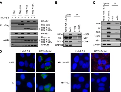
For examining the roles of different YB-1 variants in modulating NS5A half-life, control- or YB-1 siRNA-transfected Huh-7.5.1 cells were further transfected with the Flag-NS5A-expressing plasmid (JFH1, geno-FIG 1YB-1 is a novel NS5A-interacting cellular protein. (A) Coimmunoprecipitation was performed in Huh-7.5.1 cells cotransfected with either an HA vector (⫺) or expression plasmids for HA-YB-1 (⫹) along with a Flag vector or Flag-tagged HCV core, NS3, or NS5A of HCV genotype 2a. Immunoprecipitation was performed with lysates treated with RNase A and anti-Flag beads. For detection of HA-YB-1, 10-g cell lysates (1.25% of total input protein) and one-third of the precipitates (IP) were subjected to Western blotting with an anti-HA antibody. To detect Flag-tagged viral proteins, 40-g cell lysates (5% of total input protein) and a quarter of the precipitates were subjected to Western blotting with an anti-Flag antibody. GAPDH was included as an internal control. (B) Coimmunoprecipitation was performed in replicon cells (Ava5, genotype 1b). Immunoprecipitation was performed with lysates treated with RNase A and an anti-NS5A antibody. For detection of YB-1, DDX3, and GAPDH, 10g of Ava5 cell lysate (2.5% of total input protein) and two-thirds of the precipitates were subjected to Western blotting with anti-YB-1, anti-DDX3, and anti-GAPDH antibodies, respectively. To detect NS5A, 10-g Ava5 cell lysates (2.5% of total input protein) and a quarter of the precipitates were subjected to Western blotting with an anti-NS5A antibody. (C) Coimmunoprecipitation was performed in J6/JFH-infected Huh-7.5.1 cells (genotype 2a). Immunoprecipitation was performed with lysates treated with RNase A and an anti-YB-1 antibody. For detection of core, E2, NS3, and GAPDH, 10g of HCV-infected cell lysate (0.4% of total input protein) and 5% of the precipitates were subjected to Western blotting with anti-core, anti-E2, anti-NS3, and anti-GAPDH antibodies, respectively. To detect NS5A, 10-g HCV-infected cell lysates (0.4% of total input protein) and 20% of the precipitates were subjected to Western blotting with an anti-NS5A antibody. To detect YB-1, 20-g HCV-infected cell lysates (0.8% of total input protein) and 15% of the precipitates were subjected to Western blotting with an anti-YB-1 antibody. (D) (Left side) Huh-7.5.1 cells were infected with J6/JFH (genotype 2a) at an MOI of 0.5 for 48 h. Naive or infected Huh-7.5.1 cells were then immunostained for NS5A (green) or E2 (green) as indicated and were observed under a confocal or a fluorescence microscope, respectively. (Right side) Detection of interactions between YB-1 and NS5A or E2 in Huh-7.5.1 cells infected with J6/JFH (genotype 2a) byin situproximity ligation assay with anti-YB-1 and anti-NS5A or anti-E2 antibodies, as indicated. Images were acquired by a fluorescence microscope. Nuclei were stained with DAPI (blue).
on November 7, 2019 by guest
http://jvi.asm.org/
[image:4.585.95.491.64.364.2]FIG 2Knockdown of YB-1 inhibits the HCV life cycle. Huh-7.5.1 cells transfected with control siRNA (siCtrl) or YB-1 siRNA (siYB-1) for 48 h were infected with J6/JFH (genotype 2a) at an MOI of 0.5. Cells and culture media were collected at 48 h postinfection. (A) Intracellular protein levels were assessed by Western blotting for core, NS3, NS5A, and YB-1, and GAPDH was used as an internal control. HCV core, NS3, and NS5A levels were quantified by ImageJ and normalized to GAPDH. A representative immunoblot is shown at the bottom. (B) Intracellular HCV RNA levels were analyzed by quantitative RT-PCR and normalized to GAPDH RNA. (C) Infectious virus titers in the culture media were determined by focus-forming unit analysis. Means⫾SDs from three independent experiments are shown as amount relative to that of control siRNA-treated cells. Huh-7.5.1 cells transfected with control siRNA (siCtrl) or YB-1 siRNA (siYB-1) for 24 h were transfected with an HA vector or a plasmid encoding siRNA-resistant HA-YB-1 (HA-YB-1R) and infected with J6/JFH (genotype 2a) at an MOI of 0.5 24 h later. Cells and culture media were collected at 48 h postinfection. (D) Intracellular protein expression was detected by Western blotting for core, NS3, NS5A, and YB-1, and GAPDH was used as an internal control. HCV core, NS3, NS5A, and YB-1 levels were quantified by ImageJ and normalized to GAPDH levels. Results are represented as percentages relative to that of cells transfected with control siRNA and the HA vector. (E) Infectious virus titers in the culture media were determined by focus-forming unit analysis in triplicates. Representative results from one of two independent experiments are shown as means⫾SDs relative to control siRNA/HA vector-transfected cells. (F) Huh-7.5.1 cells transfected with control siRNA or YB-1 siRNA for 24 h were transfected with the HA vector or a plasmid encoding siRNA-resistant HA-YB-1. At 24, 40, or 48 h, cells were infected with J6/JFH (genotype 2a) at an MOI of 0.5. Cells were collected at 48 h postinfection. Intracellular HCV RNA levels were analyzed by quantitative RT-PCR and normalized to GAPDH RNA in triplicate. Means⫾SDs are shown as amount relative to that of control siRNA/HA vector-transfected cells. *,P⬍0.05; **,P⬍0.01; ***,P⬍0.001.
Wang et al.
on November 7, 2019 by guest
http://jvi.asm.org/
type 2a) together with plasmids encoding GFP-YB-1 variants at 48 h post-siRNA transfection. Forty-eight hours later, cells were treated with 100
g/ml of cycloheximide and collected at the desired time points after cycloheximide treatment.
Statistical analysis.Statistical analysis was conducted with a two-tailed, unpaired Student’sttest.
RESULTS
Interaction of NS5A with YB-1 and DDX3.
To gain new insights
into the mechanism by which YB-1 regulates the HCV life cycle,
we attempted to identify novel interactions between HCV
pro-teins and YB-1. Among the HCV propro-teins, core protein, NS3, and
NS5A have notably been shown to have a larger number of cellular
interacting partners (
51
). In view of the fact that both core/YB-1
and NS3/YB-1 interactions have been extensively studied (
43
,
44
),
we turned our attention to examining the interaction between
NS5A and YB-1. To this end, coimmunoprecipitation was
per-formed with Huh-7.5.1 cell extracts coexpressing HA-YB-1 and
Flag-tagged NS5A (JFH1) of genotype 2a in the presence of RNase
A. Interaction between NS5A and YB-1 was observed, as were the
known NS3–YB-1 and core–YB-1 interactions (
Fig. 1A
and
refer-ences
43
and
44
. The NS5A–YB-1 interaction was further verified
in the context of HCV RNA replication by immunoprecipitating
NS5A from HCV subgenomic replicon cells (Ava5, genotype 1b)
(
46
). Specific detection of NS5A by NS5A antibody was validated
in naive Huh-7.5.1 and Ava5 cells. Both endogenous YB-1 and its
interacting partner DDX3 were coimmunoprecipitated with
NS5A (
Fig. 1B
), indicating that the NS5A/YB-1 interaction is not
genotype specific. Next, we examined the NS5A–YB-1 interaction
in HCV infection by immunoprecipitating YB-1 from Huh-7.5.1
cells infected with the genotype 2a J6/JFH chimeric virus. Specific
detection of the HCV proteins was verified with naive Huh-7.5.1
cells. Although both the known NS3–YB-1 and core–YB-1
inter-actions were detected, interinter-actions between YB-1 and NS5A or E2
were not observed (
Fig. 1C
). To further investigate whether NS5A
or E2 interacts with YB-1 in HCV infection, the highly sensitive
in
situ
proximity ligation assay (PLA; see Materials and Methods)
was performed with HCV-infected Huh-7.5.1 cells (genotype 2a).
Specific detection of NS5A and E2 by immunofluorescence with
NS5A and E2 antibodies, respectively, was confirmed with naive
and HCV-infected Huh-7.5.1 cells (
Fig. 1D
, left side). The NS5A–
YB-1 interaction was observed in HCV-infected cells (red
fluores-cent dots in
Fig. 1D
, right side), while interaction between E2 and
YB-1 was not detected (
Fig. 1D
, right side), demonstrating that
YB-1 is an authentic NS5A-interacting partner. Collectively,
be-sides the known association with core and NS3 (
31–33
,
43
,
44
),
both YB-1 and DDX3 interact with the pivotal viral protein NS5A.
The results reveal that YB-1 and DDX3 not only interact with each
other but also share similar HCV interacting partners.
YB-1 silencing inhibits the HCV life cycle.
To validate the role
of YB-1 in the HCV life cycle, YB-1 siRNA- or control siRNA (100
nM)-transfected Huh-7.5.1 cells were infected with J6/JFH at the
low MOI of 0.5. Intracellular HCV protein and RNA levels as well
as infectivity in the supernatants were analyzed at 48 h
postinfec-tion. YB-1 silencing resulted in downregulation of core, NS3, and
NS5A protein expression to 14%, 34% and 8%, respectively,
com-pared with that of control siRNA-transfected cells (
Fig. 2A
).
In-tracellular HCV RNA levels and infectious virus titers in the
cul-ture supernatant were also reduced by 49% and 52%, respectively,
by the knockdown of YB-1 (
Fig. 2B
and
C
). YB-1 downregulation
was confirmed by immunoblotting, and specific detection of the
HCV proteins was validated with naive Huh-7.5.1 cells (
Fig. 2A
).
To confirm the specificity of YB-1 siRNA silencing, HCV
in-fection was examined by overexpressing siRNA-resistant YB-1 in
YB-1 siRNA-transfected cells (see Materials and Methods).
Ex-pression of siRNA-resistant YB-1 restored core, NS3, and NS5A
protein levels from 28% to 76%, 39% to 120%, and 2% to 50%,
respectively, in YB-1 knockdown cells compared with the control
(
Fig. 2D
). Infectious virus production levels in YB-1 knockdown
cells also recovered from 51% to 100% by expressing
siRNA-re-sistant YB-1 (
Fig. 2E
). Likewise, intracellular HCV RNA levels
recovered from 49% to 61%, 57% to 76%, and 81% to 123% with
increased intervals (24 to 48 h) between the time points for
intro-ducing plasmid encoding siRNA-resistant YB-1 and virus
inocu-lation (
Fig. 2F
). The results substantiate that YB-1 is essential for
the HCV life cycle, as reported by Chatel-Chaix et al. (
43
,
44
).
The effect of YB-1 on HCV IRES-mediated translation does
not account for the crucial role of YB-1 in the HCV life cycle.
To
examine whether YB-1 participates in the HCV life cycle via
reg-ulating HCV IRES-mediated translation,
in vivo
translation assay
was performed with monocistronic reporter RNAs containing the
5
=
NTR alone (5
=
Rluc) or both the 5
=
and 3
=
NTRs (5
=
Rluc3
=
), as
the 3
=
NTR promotes HCV translation (
52
,
53
) (
Fig. 3A
).
Inter-estingly, in contrast to the critical role of YB-1 in HCV
propaga-tion, YB-1 silencing moderately enhanced translation of 5
=
Rluc3
=
by 25% (
Fig. 3C
) but not 5
=
Rluc (
Fig. 3B
).
HCV has been reported to induce translational stress at various
periods of infection (
54
) and uses different translation initiation
factors under stress than normal conditions (
55
,
56
). To further
explore the role of YB-1 in HCV translation, an
in vivo
translation
assay was performed under stress conditions induced by sodium
arsenite. Relative to the translation of 5
=
Rluc, translation of
5
=
Rluc3
=
was more resistant to stress (78% versus 55% retention,
Fig. 3B
and
C
), implying a previously undefined role of the HCV
FIG 3YB-1 silencing moderately enhances and slightly inhibits HCV IRES-mediated translation under normal and stress conditions, respectively. (A) Diagram of the reporter RNAs. 5=Rluc consists of the HCV 5=NTR (genotype 2a) andRenillaluciferase. 5=Rluc3=contains the same structure at the 5=end but with the HCV 3=NTR (genotype 2a) followingRenillaluciferase. Huh-7.5.1 cells transfected with control or YB-1 siRNA were pretreated with dis-tilled water or 40M sodium arsenite (SA)for 1 h before transfection with 5=Rluc (B) or 5=Rluc3=(C) RNA. Luciferase activities were analyzed at 2 h posttransfection. Results are presented as means⫾SDs from three indepen-dent experiments relative to cells treated with the control siRNA. *,P⬍0.05; ***,P⬍0.001.
on November 7, 2019 by guest
http://jvi.asm.org/
[image:6.585.299.543.63.219.2]3
=
NTR in the stress resistance of HCV IRES translation.
Knock-down of YB-1 had no effects on HCV IRES-mediated translation
of 5
=
Rluc (
Fig. 3B
), and there was only a 10% decrease of
transla-tion of 5
=
Rluc3
=
under stress conditions (
Fig. 3C
). Taken together,
our results suggest that YB-1 has either a moderate or a minor
effect that inhibits or promotes HCV IRES-mediated translation
under normal or stress conditions, respectively, which are
insuf-ficient to account for the crucial role of YB-1 in the HCV life cycle.
YB-1 is involved in HCV RNA replication.
The effects of YB-1
on HCV RNA replication were next investigated by luciferase
as-say with the infectious HCV genome J6/JFH(p7-Rluc2A) (
Fig. 4A
)
(
48
). The incorporated
Renilla
luciferase reporter allows sensitive
detection of primary translation of HCV genome and
establish-ment of HCV RNA replication in the HCV life cycle. These two
stages of the viral life cycle can be distinguished by comparing the
replication kinetics of J6/JFH(p7-Rluc2A) with that of the
repli-cation-defective genome harboring mutations in NS5B [J6/
JFH(p7-Rluc2A)GNN] (
Fig. 4A
) (
48
). To exclude the effects of
YB-1 on HCV infectivity, we further included an
assembly-defec-tive variant, J6/JFH(p7-Rluc2A)K33A/R35A, with mutations in
p7 (
Fig. 4A
), which leads to defective virion morphogenesis
with-out impairing HCV RNA replication (
48
).
YB-1 transient knockdown cells or control cells were
trans-fected with each of the reporter genome variants, and luciferase
activities were determined at various time points. Knockdown of
YB-1 led to a slight increase in luciferase activity in cells
trans-fected with each of three genomes at 2 and/or 4 h posttransfection
(
Fig. 4B
and
C
[left side] and
D
), implying that YB-1 might repress
FIG 4Downregulation of YB-1 reduces HCV RNA replication. (A) Diagram of the reporter genome (genotype 2a). The p7 mutation, K33A/R35A, and the GNN mutation are shown. Huh-7.5.1 cells transfected with control siRNA (siCtrl) or YB-1 siRNA (siYB-1) were electroporated with the wild-type reporter genome [J6/JFH(p7-Rluc2A)] (B), the reporter genome with the p7 mutation [J6/JFH(p7-Rluc2A)K33A/R35A)](C), or the replication-deficient reporter genome [J6/JFH(p7-Rluc2A)GNN] (D). Cell lysates were harvested at the indicated time points posttransfection, and luciferase activities were analyzed. For panels B and C, graphs of luciferase activity along with the posttransfection time points are shown on the right, while the luciferase activity at 2 to 12 h is focused on in left graphs. Means⫾SDs for triplicate wells are shown. *,P⬍0.05; **,P⬍0.01; ***,P⬍0.001.
Wang et al.
on November 7, 2019 by guest
http://jvi.asm.org/
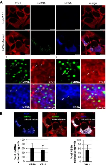
[image:7.585.91.487.66.483.2]FIG 5YB-1 colocalizes with HCV replication complexes in HCV-infected cells. (A) Huh-7.5.1 cells were infected with J6/JFH (genotype 2a) at an MOI of 0.5. Forty-eight hours postinfection, cells were immunostained for dsRNA (green), NS5A (blue), and YB-1 (red) and were observed under a confocal microscope. The lower portion shows the enlarged images of the boxed regions. The colocalization sites of YB-1, dsRNA, and NS5A are indicated by white arrows. (B) Colocalization of NS5A and dsRNA (left), YB-1 and dsRNA (middle), and YB-1 and NS5A (right) as analyzed by Zen 2009 software (Zeiss). Colocalization sites are indicated in white. Percentages of dsRNA that colocalized with NS5A or YB-1 or of NS5A that colocalized with YB-1 are shown as means⫾SDs (graphs).n⫽
44; scale bars⫽10m.
on November 7, 2019 by guest
http://jvi.asm.org/
[image:8.585.112.474.66.629.2]Wang et al.
on November 7, 2019 by guest
http://jvi.asm.org/
primary translation of the HCV genome, consistent with the
re-sults from the
in vivo
translation assay (
Fig. 3C
). Notably,
lucifer-ase activities were elevated at 10 to 12 h posttransfection in cells
transfected with J6/JFH(p7-Rluc2A) (
Fig. 4B
) or
J6/JFH(p7-Rluc2A)K33A/R35A (
Fig. 4C
), but not in cells transfected with
J6/JFH(p7-Rluc2A)GNN (
Fig. 4D
), indicating that the initiation
of HCV RNA replication had occurred at 10 to 12 h
posttransfec-tion. YB-1 silencing inhibited luciferase activities in cells
trans-fected with J6/JFH(p7-Rluc2A) and J6/JFH(p7-Rluc2A)K33A/
R35A to up to 48% and 52%, respectively (
Fig. 4B
and
C
) at 10 to
30 h posttransfection, suggesting that YB-1 is required for HCV
RNA replication.
YB-1 colocalizes with HCV replication complexes.
To
exam-ine whether YB-1 colocalizes with the HCV replication markers,
dsRNA and NS5A, immunofluorescence analysis was performed
on J6/JFH-infected Huh-7.5.1 cells. dsRNA is the intermediate of
HCV RNA replication representing the site of replication complex
(
57
), while NS5A is a component of the HCV replication complex
(
7–10
). Triple staining with antibodies against YB-1, dsRNA, and
NS5A revealed that YB-1 did colocalize with NS5A in the HCV
replication complex (
Fig. 5A
). Quantitative analysis showed that
about 52% of the dsRNA colocalized with YB-1, while
approxi-mately 60% of dsRNA colocalized with NS5A (
Fig. 5B
).
Consis-tent with results of coimmunoprecipitation and
in situ
proximity
ligation assays (
Fig. 1
), 53% of NS5A colocalized with YB-1 in
both the replication complex and the cytoplasm (
Fig. 5B
),
con-firming that YB-1 is an NS5A-interacting protein.
To further confirm the role of YB-1 in HCV RNA replication,
control- or YB-1 siRNA-transfected Huh-7.5.1 cells were infected
with J6/JFH (genotype 2a) for 48 h, and the cells were subjected to
immunofluorescence detection with dsRNA, NS5A, and YB-1
an-tibodies. YB-1 was found to be downregulated to 13% that of
control cells by siRNA-mediated silencing (
Fig. 6A
). Consistent
with the positive regulatory role of YB-1 in HCV RNA replication
(
Fig. 4
), YB-1 knockdown significantly reduced dsRNA and NS5A
levels, to 51% and 42%, respectively, compared with that of the
control cells (
Fig. 6A
).
The inhibitory effect of YB-1 silencing on HCV replication
com-plex formation was then affirmed by quantitative analysis of dsRNA
and NS5A colocalization, which was reduced to 65% of the control by
YB-1 silencing (
Fig. 6B
), further substantiating that YB-1 silencing
disturbs the formation of HCV replication complexes. Colocalization
of dsRNA and NS5A with YB-1 was also analyzed. The percentages of
dsRNA and NS5A that colocalized with YB-1 were decreased to 27%
and 12% of the control, respectively, by knockdown of YB-1 (
Fig. 6B
),
as a consequence of a more dramatic decline of YB-1 levels than that
of dsRNA and NS5A (
Fig. 6A
). Taken together, these results reveal
that YB-1 colocalizes with HCV replication complexes in
HCV-in-fected cells, which further substantiates the role of YB-1 in HCV RNA
replication.
YB-1 is dispensable for steady-state HCV RNA replication
but is involved in infectious virus production.
In addition to
steady-state HCV RNA replication, the initial phase of HCV RNA
replication has previously been identified (
21
,
22
). Moreover,
be-sides its role in the steady-state HCV RNA replication, unique
roles of NS5A in the early stage of HCV RNA replication,
includ-ing membranous web formation and transient RNA replication
after infection, have been recognized (
19–22
). Given that YB-1 is
an NS5A-interacting protein, we examined whether YB-1
func-tions in the initiation of HCV RNA replication or in steady-state
HCV RNA replication by investigating the effects of silencing
YB-1 in HCV-infected cells (
Fig. 7A
). The HCV replication
com-plex is protected by a membrane (
8
,
58
,
59
) and is stable, with
limited interchange of internal and periphery proteins (
60
,
61
). To
verify the elimination of preincorporated YB-1 in the replication
complex in HCV-infected cells upon YB-1 silencing,
colocaliza-tion of dsRNA and YB-1 was first examined (
Fig. 7B
). The
per-centage of dsRNA that colocalized with YB-1 was severely
re-duced, from 60% to 8%, by YB-1 silencing (
Fig. 7C
, left graph),
confirming siRNA-mediated knockdown of YB-1 in the HCV
rep-lication complex. The formation of HCV reprep-lication complex was
also evaluated by analyzing the colocalization of dsRNA with
NS5A. Notably, different from that of YB-1 silencing before HCV
infection (
Fig. 6B
), knockdown of YB-1 in HCV-infected cells did
not affect the colocalization of dsRNA with NS5A (
Fig. 7B
and
C
,
right graph), suggesting that eliminating YB-1 from the assembled
HCV replication complex does not disrupt the replication
com-plex. The effect of YB-1 silencing in HCV-infected cells on HCV
propagation was further investigated. The YB-1 interacting
part-ner, DDX3, which is also an NS5A-interacting protein (
Fig. 1B
)
involved in HCV RNA replication (
34
), was silenced in parallel.
siRNA-mediated individual knockdown of YB-1 and DDX3
re-sulted in comparable reductions, 74% and 78%, respectively, by
immunoblotting.
Interestingly, in contrast to the effects of silencing of YB-1
be-fore reporter genome transfection (
Fig. 4B
and
C
), knockdown of
YB-1 in HCV-infected cells had no significant impact on the
in-tracellular HCV RNA levels (
Fig. 7D
, left graph), while silencing of
DDX3 had a pronounced (30%) reduction in the amount of HCV
RNA (
Fig. 7D
, right graph). However, the infectious virus
produc-tion was more severely repressed by YB-1 silencing (49%) (
Fig. 7E
,
left graph) than by DDX3 silencing (37%) (
Fig. 7E
, right graph).
As the intracellular HCV RNA level was unaltered, the YB-1
si-lencing-mediated reduction of infectivity suggests a regulatory
role for YB-1 in infectious virus production. Notably, knockdown
of YB-1 resulted in a marked (62%) reduction of the NS5A level
(
Fig. 7F
), while effects were absent on the core and modest on NS3
(32% reduction) (
Fig. 7F
). Preferential reduction of the NS5A
level was also observed in similar DDX3-silencing experiments,
albeit less severely (
Fig. 7F
).
NS5A exists in two phosphorylation forms,
hyperphosphory-lated (p58) and hypophosphoryhyperphosphory-lated (p56) (
62
). The
phosphory-lation status of NS5A has been proposed to regulate the
equilib-rium between HCV RNA replication and virus assembly (
63
).
FIG 6YB-1 silencing reduces HCV replication complexes. (A) Huh-7.5.1 cells transfected with control siRNA (siCtrl) or YB-1 siRNA (siYB-1) for 48 h were infected with J6/JFH (genotype 2a) at an MOI of 0.5. Cells were fixed at 48 h postinfection and subjected to immunostain for YB-1 (red), dsRNA (green), and NS5A (blue) and were observed under a confocal microscope. Intensities of YB-1, dsRNA, and NS5A were analyzed by Zen 2009 software (Zeiss) and are shown as means⫾SDs relative to that of control siRNA-treated cells (graphs). (B) Colocalizations of NS5A and dsRNA (left), YB-1 and dsRNA (middle), and YB-1 and NS5A (right) from panel A were analyzed by Zen 2009 software (Zeiss) and are displayed in white. Percentages of dsRNA that colocalized with NS5A or YB-1 or of NS5A that colocalized with YB-1 are shown as means⫾SDs relative to control siRNA-treated cells (graphs).n⫽45. ***,P⬍0.001.
on November 7, 2019 by guest
http://jvi.asm.org/
Interestingly, our results showed that knockdown of YB-1 in
HCV-infected Huh-7.5.1 cells preferentially downregulated p58
over p56, resulting in a 44% decline in the p58/p56 ratio (
Fig. 7G
,
left graph). In contrast, DDX3 silencing-mediated reduction of
NS5A had no apparent preference for either NS5A
phosphoryla-tion form and hence did not affect the p58/p56 ratio (
Fig. 7G
, right
graph). These results indicate that YB-1 knockdown not only
re-duces the NS5A level but also alters the ratio of p58 to p56, which
is different from that of DDX3.
To validate that YB-1 functions in the initial stage of HCV RNA
replication rather than in steady-state HCV RNA replication,
YB-1 was silenced with different siRNA doses in the
replicon-containing cells supporting persistent HCV RNA replication
(Ava5, genotype 1b), and the replicon RNA levels were analyzed.
FIG 7Knockdown of YB-1 does not affect steady-state HCV RNA replication but represses production of infectious particles. (A) The process of YB-1 or DDX3 silencing on HCV infection. Huh-7.5.1 cells infected with J6/JFH (genotype 2a) at a high MOI (5 to 7.5) were transfected with control (siCtrl), YB-1 (siYB-1), or DDX3 (siDDX3) siRNA at 8 h postinfection. Three days posttransfection, cells and culture media were fixed or harvested. (B) Immunostaining for YB-1 (red), dsRNA (green), and NS5A (blue) was observed by confocal microscopy. The colocalization images show the results as analyzed by Zen 2009 software (Zeiss); colocalization sites of dsRNA with YB-1 or NS5A are indicated in white. (C) Quantitative results from panel B are shown as means⫾SDs relative to those for control siRNA-treated cells (siCtrl,n⫽128; siYB-1,n⫽153). For panels D to G, results are presented as means⫾SDs from three independent experiments relative to those for control siRNA-transfected cells. (D) Intracellular HCV RNA levels were quantified by real-time RT-PCR normalized to the GAPDH data. (E) Infectious virus titers in culture media were determined by focus-forming unit analysis. (F) Intracellular protein levels were detected by Western blotting for NS5A, NS3, and core, and GAPDH was used as an internal control. NS5A, NS3, and core protein expression levels were quantified by ImageJ and normalized to GAPDH levels. A representative immunoblot is shown on the right. YB-1 and DDX3 were detected to confirm knockdown efficiency. (G) Quantitation of the p58/p56 ratios of NS5A. *,P⬍0.05; **,P⬍0.01; ***,P⬍0.001.Wang et al.
on November 7, 2019 by guest
http://jvi.asm.org/
[image:11.585.90.498.63.511.2]The different degrees of YB-1 silencing were verified by
immuno-blotting (79% and 90% reduction) (
Fig. 8B
). Consistent with that
observed in the HCV-infected cells (
Fig. 7D
), knockdown of YB-1
in replicon cells had no impact on the HCV RNA levels (
Fig. 8A
),
even at the dose of siRNA inhibiting HCV life cycle when
admin-istered before HCV infection (
Fig. 2
). Remarkably, while the NS3
level was almost not affected by YB-1 knockdown, the amount of
NS5A was reduced by YB-1 silencing in a dose-dependent manner
from 22% to 62% (
Fig. 8B
), in accordance with that observed in
HCV-infected cells (
Fig. 7F
). Thus, our results suggest that YB-1 is
not involved in the steady-state HCV RNA replication but
partic-ipates in the production of infectious particles.
YB-1 knockdown promotes NS5A degradation.
As the NS5A
protein level was shown to be more susceptible to YB-1 silencing
than those of NS3 and core (
Fig. 2A
,
7F
, and
8B
), we further
investigated whether YB-1 regulates the NS5A protein level.
YB-1-or control siRNA-transfected Huh-7.5.1 cells were transfected
in-dividually with Flag-tagged NS5A, NS3, or core of HCV genotype
2a. Knockdown of YB-1 markedly reduced the Flag-NS5A protein
level but had no discernible effects on the expression of Flag-NS3
and Flag-core, even in the context of the absence of HCV
replica-tion (
Fig. 9A
). Similar results were also obtained with Flag-tagged
NS5A, NS3, and core from genotype 1b (
Fig. 9B
), indicating that
among the three interacting viral proteins, YB-1 specifically and
positively modulates the NS5A expression level.
To clarify whether the decrease of Flag-NS5A protein level in
YB-1 silencing cells is due to protein degradation, the half-life of
Flag-NS5A was assessed with the translation inhibitor
cyclohexi-mide. In the presence of cycloheximide, knockdown of YB-1
ac-celerated the degradation of Flag-NS5A compared with that of
control cells, as demonstrated by the reduction of NS5A half-life
from 4.4 h to 2.4 h (
Fig. 9C
). The positive regulatory effect of YB-1
on NS5A protein stability was then examined in the context of
HCV RNA replication. To this end, replicon cells (Ava5, genotype
1b) transfected with YB-1 or control siRNA were treated with
cycloheximide; the NS5A half-life was found to be dramatically
reduced, from 19.2 h to 0.6 h, by YB-1 silencing (
Fig. 9D
). The
results suggest that YB-1 protects NS5A from protein degradation.
Variations between the half-life of NS5A expressed ectopically and
in replicon cells were similarly observed in previous studies of core
and NS5B (
64–67
).
We next investigated whether the rapid degradation of NS5A
evoked by YB-1 silencing is proteasome dependent by treating
YB-1 knockdown or control cells expressing Flag-NS5A with a
proteasome inhibitor, MG132. MG132 clearly abolished YB-1
si-lencing-mediated Flag-NS5A reduction (
Fig. 9E
). To confirm this
effect in the context of HCV infection, control or YB-1
knock-down cells were infected with J6/JFH (MOI of 0.5) for 48 h and
treated with MG132 for 4 h before analysis. MG132 partially
re-stored the NS5A protein level, while recovery of NS3 and core
protein levels was not observed (
Fig. 9F
). Taken together, our
results indicate that knockdown of YB-1 leads to NS5A instability.
Phosphorylation of YB-1 at serine 102 (S102) modulates
NS5A–YB-1 interaction and NS5A stability.
We were also
inter-ested to know whether NS5A–YB-1 interaction is crucial for
maintaining NS5A stability and whether this interaction is elicited
by HCV-induced signaling. Along this line, it is noted that both
HCV infection and NS5A alone activate the PI3K/Akt pathway
(
23
,
68
), and phosphorylation at the YB-1 S102 residue by Akt is
known to modulate multiple functions of YB-1 (
69
). To examine
if the YB-1–NS5A interaction is dependent on the
phosphoryla-tion of YB-1 at the S102 residue, coimmunoprecipitaphosphoryla-tion was
per-formed with Huh-7.5.1 cell extracts coexpressing Flag-NS5A and
either 1, the nonphosphorylatable mutant
HA-YB-1(S102A), or the phospho-mimic mutant HA-YB-1(S102D).
Interactions between the YB-1(S102A) mutant and NS5A were
disturbed, resulting in a 62% reduction compared to that with
wild-type YB-1, while binding between the YB-1(S102D) mutant
and NS5A was comparable to wild-type YB-1–NS5A interaction
(
Fig. 10A
).
Next, we investigated the importance of the
phosphorylation-mediated NS5A–YB-1 interaction for YB-1 to stabilize NS5A.
NS5A protein levels were examined in YB-1 knockdown
Huh-7.5.1 cells transfected with Flag-NS5A and either siRNA-resistant
1, the 1(S102A) mutant, or the
GFP-1(S102D) mutant. Compared with wild-type 1, the
YB-1(S102A) mutant, which only partially interacted with NS5A,
failed to maintain the NS5A level in showing a 67% reduction
compared to that with the wild type, while the phospho-mimic
YB-1(S102D) mutant not only interacted with NS5A but also
maintained the NS5A level in YB-1 knockdown cells (
Fig. 10B
).
The diverse NS5A levels in YB-1 knockdown cells expressing
dif-ferent YB-1 variants were then verified to reflect the altered NS5A
stability. A decreased NS5A half-life from 2.2 h to 0.8 h was
ob-served in YB-1 knockdown cells expressing the siRNA-resistant
FIG 8YB-1 silencing has no effect on steady-state HCV RNA replication inreplicon cells but specifically reduces NS5A levels. Ava5 cells harboring the subgenomic replicon (genotype 1b) were transfected with control siRNA (siCtrl; 100 nM) or 50 or 100 nM YB-1 siRNA (siYB-1) and harvested 72 h later. (A) Intracellular HCV replicon RNA levels were analyzed by quantitative RT-PCR and normalized to GAPDH RNA. Means⫾SDs from three indepen-dent experiments are shown as amount relative to that of control siRNA-transfected cells. (B) Intracellular protein levels were detected by Western blotting for NS3, NS5A, YB-1, and GAPDH, quantified by ImageJ, and nor-malized to GAPDH. Results are represented as percentages relative to that of control siRNA-transfected cells. A representative immunoblot of three inde-pendent experiments is shown.
on November 7, 2019 by guest
http://jvi.asm.org/
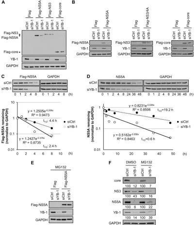
[image:12.585.92.235.67.314.2]FIG 9YB-1 knockdown promotes NS5A degradation. Control (siCtrl) or YB-1 (siYB-1) siRNA-transfected Huh-7.5.1 cells were transfected with expression plasmid for Flag-NS5A, Flag-NS3, or Flag-core of HCV genotype 2a (A) or Flag-NS5A, Flag-NS3/4A, or Flag-core of HCV genotype 1b (B). Twenty-four hours (A) or 16 h (B) posttransfection, protein levels were detected by Western blotting for viral proteins with anti-Flag antibody, GAPDH as an internal control, and YB-1 to confirm silencing efficiency. (C) (Top) Control- or YB-1 siRNA-transfected Huh-7.5.1 cells were transfected with Flag-NS5A (genotype 2a) expression plasmid. Forty-eight hours later, cells were treated with cycloheximide at 100g/ml. Cells were harvested and analyzed for Flag-NS5A levels by Western blotting with an anti-Flag antibody at the indicated time points after cycloheximide addition. GAPDH was used as an internal control. (Bottom) Flag-NS5A levels were quantified by ImageJ and normalized to GAPDH levels. The half-life of Flag-NS5A was determined by regression analysis. Protein levels of Flag-NS5A without cycloheximide treatment (0 h) are set as 1. (D) (Top) Ava5 replicon cells (genotype 1b) were transfected with control or YB-1 siRNA. At 72 h posttransfection, cells were treated with cycloheximide at 100g/ml. Cells were harvested and analyzed for NS5A levels by Western blotting at the indicated time points after cycloheximide addition. GAPDH was used as an internal control. (Bottom) NS5A levels were quantified by ImageJ and normalized to GAPDH levels, and the half-life of NS5A was determined by regression analysis. Protein levels of NS5A without cycloheximide treatment (0 h) are set as 1. (E) Control or YB-1 siRNA-transfected Huh-7.5.1 cells were transfected with Flag or Flag-NS5A (genotype 2a) expression plasmid. At 20 h posttransfection, cells were treated with MG132 at 10M for 4 h to inhibit proteasome activities. Protein levels were detected by Western blotting for NS5A with an anti-Flag antibody; GAPDH was used as an internal control, and YB-1 was included for confirming knockdown efficiency. (F) Huh-7.5.1 cells transfected with control or YB-1 siRNA were infected with J6/JFH (genotype 2a) at an MOI of 0.5 as described in the legend toFig. 2. Four hours before harvest, infected cells were treated with DMSO or MG132 (10
M). Intracellular protein levels were detected by Western blotting for core, NS3, NS5A, YB-1, and GAPDH, quantified by ImageJ, and normalized to GAPDH. Results are represented as percentages relative to that of control siRNA-transfected cells.
Wang et al.
on November 7, 2019 by guest
http://jvi.asm.org/
[image:13.585.91.493.68.532.2]GFP-YB-1(S102A) mutant compared to that in cells transfected
with wild-type GFP-YB-1, while a comparable half-life of NS5A
was shown with expression of the GFP-YB-1(S102D) mutant (1.7
h) and wild-type GFP-YB-1 (
Fig. 10C
). Thus, NS5A–YB-1
inter-action is mediated by phosphorylation of YB-1 at the S102 residue,
which is important for NS5A stabilization.
To validate the role of YB-1 S102 phosphorylation in HCV
infection, Huh-7.5.1 cells coexpressing YB-1 siRNA and either
siRNA-resistant wild-type YB-1, YB-1(S102A), or
HA-YB-1(S102D) were infected with J6/JFH virus. Consistent with
their NS5A-interacting activities and their abilities to stabilize
ec-topically expressed NS5A, both wild-type 1 and the
YB-1(S102D) mutant rescued the NS5A levels from 5% to 64% and
52%, respectively, compared to control cells, in HCV infection
(
Fig. 10D
). However, the phosphorylation-null HA-YB-1(S102A)
mutant led to 24% and, therefore, a much lower restoration of
NS5A expression (
Fig. 10D
). Taken together, our findings indicate
that YB-1 stabilizes the pivotal viral protein NS5A via
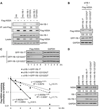
phosphory-FIG 10YB-1 stabilizes NS5A through its serine 102 phosphorylation-mediated interaction with NS5A. (A) Coimmunoprecipitation was performed with Huh-7.5.1 cells cotransfected with a Flag vector (⫺) or expression plasmids for Flag-NS5A (⫹) (genotype 2a) and either an HA vector, 1, HA-YB-1(S102A), or HA-YB-1(S102D) in the presence of RNase A with anti-Flag beads. For detection of HA-YB-1, 20-g cell lysates (1.67% of total input protein) and four-ninths of the precipitates (IP) were subjected to Western blotting with an anti-HA antibody. To detect Flag-NS5A, 20-g cell lysates (1.67% of total input protein) and two-ninths of the precipitates were subjected to Western blotting with an anti-Flag antibody. GAPDH was included as an internal control. Levels of immunoprecipitated HA-YB-1 variants were quantified by ImageJ and are shown as percentages of wild-type HA-YB-1. (B) YB-1 siRNA-transfected Huh-7.5.1 cells were cotransfected with plasmids encoding Flag-NS5A (genotype 2a) and either siRNA-resistant GFP-YB-1 (GFP-YB-1R), GFP-YB-1(S102A) [GFP-YB-1(S102A)R], or GFP-YB-1(S102D) [GFP-YB-1(S102D)R]. Twenty-four hours posttransfection, levels of Flag-NS5A and mutants for GFP-YB-1 or GFP-YB-1 were analyzed by Western blotting with anti-Flag and anti-GFP antibodies, respectively. GAPDH was used as an internal control. Flag-NS5A levels were quantified by ImageJ and normalized to GAPDH levels and are shown as percentages of that of YB-1 knockdown cells cotransfected with Flag-NS5A and wild-type GFP-YB-1. (C) (Top) YB-1 siRNA-transfected Huh-7.5.1 cells were cotransfected with plasmids encoding Flag-NS5A (genotype 2a) and either siRNA-resistant GFP-YB-1 (GFP-YB-1R), GFP-YB-1(S102A) [GFP-YB-1(S102A)R], or GFP-YB-1(S102D) [GFP-YB-1(S102D)R]. Forty-eight hours later, cells were treated with cycloheximide at 100g/ml. Cell lysates were prepared and analyzed for Flag-NS5A levels by immunoblotting with an anti-Flag antibody at the indicated time points after cycloheximide addition. GAPDH was used as an internal control. (Bottom) Flag-NS5A levels were quantified by ImageJ and normalized to GAPDH levels, and the half-life of Flag-NS5A was determined by regression analysis. Protein levels of Flag-NS5A without cycloheximide treatment (0 h) are set as 1. (D) Huh-7.5.1 cells transfected with control siRNA (siCtrl) or YB-1 siRNA (siYB-1) for 24 h were transfected with the HA vector or plasmid encoding either siRNA-resistant HA-YB-1 (HA-YB-1R), HA-YB-1(S102A) [HA-YB-1(S102A)R], or HA-YB-1(S102D) [HA-YB-1(S102D)R] and infected with J6/JFH (genotype 2a) at an MOI of 0.5 24 h later. Cells lysates prepared at 48 h postinfection were subjected to Western blotting for NS5A, endogenous YB-1, and HA-tagged YB-1 variants, and GAPDH was used as an internal control. NS5A levels were quantified by ImageJ and normalized to GAPDH levels, and are shown as percentages of that of cells transfected with control siRNA and HA vector.on November 7, 2019 by guest
http://jvi.asm.org/
[image:14.585.135.451.62.412.2]lation-dependent interaction with NS5A through which YB-1
plays a role in HCV infection and HCV RNA replication.
DISCUSSION
NS5A is a pivotal viral protein involved in multiple steps in the
HCV life cycle, including HCV RNA replication (
14
,
63
),
infec-tious virus production (
12
,
13
,
15
), and the recently suggested
early stage of HCV RNA replication (
19–22
). In this study, we
revealed that YB-1 is a novel NS5A-interacting cellular factor (
Fig.
1
) and that such an interaction stabilizes NS5A (
Fig. 10
). The
colocalization of YB-1 and NS5A in the HCV replication
com-plexes and also in the cytoplasm in HCV-infected cells (
Fig. 5
)
implies that YB-1 may modulate various steps of the HCV life
cycle via maintaining the cellular NS5A level. Supporting this
no-tion, knockdown of YB-1 before HCV infection and reporter
ge-nome transfection reduced HCV replication complex formation
and RNA replication (
Fig. 4
and
6
), while YB-1 silencing in
HCV-infected and replicon cells did not affect intracellular HCV RNA
levels (
Fig. 7D
, left graph, and
8A
), indicating YB-1 involvement
in the establishment of HCV replication complex formation.
Moreover, as the intracellular HCV RNA level was not altered by
YB-1 knockdown in HCV-infected cells (
Fig. 7D
), reduced
infec-tivity in the culture medium (
Fig. 7E
) conceivably suggests that
YB-1 is important for the production of infectious HCV particles.
Interestingly, by using a plasmid-derived JFH-1-expressing
sys-tem, Chatel-Chaix et al. showed that knockdown of YB-1
pro-moted infectious virus production (
43
,
44
). Notably, the kinetics
of the plasmid-derived JFH-1 expression is much slower than that
of cell culture-derived HCV (HCVcc) infection adopted in our
study (
22
,
43
). While Chatel-Chaix et al. assessed the role of YB-1
silencing in virus production at a defined early stage of the
plas-mid-derived infection system (
43
), we detected the inhibitory
ef-fect of YB-1 knockdown on the production of HCV inef-fectious
virus at the late stage of the HCV life cycle (
Fig. 2C
,
2E
, and
7E
and
reference
22
). Results from the use of different infection systems
and at different time windows have hence revealed distinctive
roles for YB-1 in virus production at different stages of the HCV
life cycle.
HCV co-opts ribonucleoprotein complexes via virus-host
pro-tein-protein and RNA-protein interactions to facilitate its
propa-gation (
70
). Several studies, including our own, have shown that
host factors YB-1 and DDX3 not only participate in the HCV life
cycle (
Fig. 2
and
7
and references
34
,
35
, and
43
) but also associate
with HCV RNA (
42
,
43
) and interact with HCV core, NS3, and
NS5A (
Fig. 1
and references
31
to
33
,
43
, and
44
). Although
shar-ing the same viral interactshar-ing partners, YB-1 and DDX3 seem to
play several different roles in the HCV life cycle. While DDX3 is
required for HCV IRES-mediated translation (
29
,
71
), YB-1 plays
either a moderate or a minor role that suppresses or promotes
HCV translation under normal or stress conditions, respectively
(
Fig. 3C
). The regulatory effects of DDX3 on HCV IRES are
inde-pendent of the HCV 3
=
NTR (
71
). However, the effects of YB-1 on
HCV IRES-mediated translation are dependent on the presence of
the HCV 3
=
NTR (
Fig. 3
). While YB-1 is a positive regulator
re-quired in HCV RNA replication but dispensable in maintaining
the steady-state HCV RNA replication (
Fig. 4
,
7D
, left side, and
8A
), DDX3 participates in steady-state HCV RNA replication
(
Fig. 7D
, right graph, and reference
34
). Moreover, unlike YB-1,
DDX3 is not involved the equilibrium between HCV RNA
repli-cation and infectious virus production under defined conditions
(
44
). The comparable reductions of HCV RNA levels and
infec-tivity after DDX3 knockdown in HCV-infected cells (
Fig. 7D
,
right graph, and
7E
, right graph) indicate that such infectivity
decreases by DDX3 silencing is a consequence of impaired HCV
RNA replication, which might be different from the case with
YB-1.
Besides participating in HCV RNA translation, RNA
replica-tion, and virus production of the HCV life cycle, NS5A has further
been proposed to regulate switches between these life cycle stages
(
13
,
72
). The HCV genome RNA is used as the template for both
IRES-mediated translation and negative-strand RNA synthesis,
two essential but opposing processes (
3
). To avoid functional
con-flicts, a mechanism which coordinates these two processes for
ef-ficient HCV replication is necessary and is believed to be
accom-plished by cooperation between viral and host proteins (
70
,
73
,
74
). NS5A interacts with both the HCV 5
=
and 3
=
NTRs and hence
has been proposed to regulate the switch from HCV translation to
RNA replication (
72
). Given that YB-1 associates with NS5A to
sustain the NS5A level (
Fig. 10
), and that YB-1 has been identified
as the HCV 5
=
and 3
=
NTR-interacing protein (
42
,
75
,
76
), it is
likely that YB-1 regulates the translation-replication switch in the
HCV life cycle through the NS5A–YB-1 complex. Interestingly, in
the reporter genome assay, the observation that YB-1 moderately
inhibits primary translation (
Fig. 3C
, normal condition [i.e.,
with-out sodium arsenite], and
Fig. 4
) while playing a critical role in
HCV RNA replication may further support this notion. Recently,
NS3, another YB-1-interacting viral protein, has been reported to
regulate the switch from translation to replication of HCV RNA
via replacing the human La protein, an HCV IRES
trans
-acting
factor, from the HCV IRES (
73
). It is thus possible that YB-1 may
interexchange with viral proteins, such as NS3 and NS5A, to
par-ticipate in the translation-replication switch in the HCV life cycle.
Phosphorylation of NS5A has been suggested to modulate
HCV RNA replication. A study based on HCV genotype 1b has
revealed that hyperphosphorylation of NS5A represses HCV RNA
replication (
77
). Nevertheless, hyperphosphorylation of genotype
2a NS5A either enhances or inhibits HCV RNA replication,
de-pending on the serine residues examined (
13
,
78
,
79
). Since NS5A
phosphorylation also regulates virion assembly (
13
),
phosphory-lation of NS5A has been suggested to control the switch from HCV
RNA replication to virus production (
13
), and the ratio of the
hyperphosphorylated (p58) to hypophosphorylated (p56) forms
of NS5A may be im