Murine Gammaherpesvirus Lytic Transactivator RTA
Brian S. Wakeman,aL. Steven Johnson,bClinton R. Paden,aKathleen S. Gray,aHerbert W. Virgin,bSamuel H. Specka
Emory Vaccine Center and Department of Microbiology & Immunology, Emory University School of Medicine, Atlanta, Georgia, USAa; Department of Pathology and Immunology, Washington University School of Medicine, St. Louis, Missouri, USAb
ABSTRACT
The essential immediate early transcriptional activator RTA, encoded by gene 50, is conserved among all characterized gamma-herpesviruses. Analyses of a recombinant murine gammaherpesvirus 68 (MHV68) lacking both of the known gene 50 promoters (G50DblKo) revealed that this mutant retained the ability to replicate in the simian kidney epithelial cell line Vero but not in permissive murine fibroblasts following low-multiplicity infection. However, G50DblKo replication in permissive fibroblasts was partially rescued by high-multiplicity infection. In addition, replication of the G50DblKo virus was rescued by growth on
mouse embryonic fibroblasts (MEFs) isolated from IFN-␣/Rⴚ/ⴚmice, while growth on Vero cells was suppressed by the
addi-tion of alpha interferon (IFN-␣). 5=rapid amplification of cDNA ends (RACE) analyses of RNAs prepared from G50DblKo and
wild-type MHV68-infected murine macrophages identified three novel gene 50 transcripts initiating from 2 transcription initia-tion sites located upstream of the currently defined proximal and distal gene 50 promoters. In transient promoter assays, neither
of the newly identified gene 50 promoters exhibited sensitivity to IFN-␣treatment. Furthermore, in a single-step growth analysis
RTA levels were higher at early times postinfection with the G50DblKo mutant than with wild-type virus but ultimately fell be-low the levels of RTA expressed by wild-type virus at later times in infection. Infection of mice with the MHV68 G50DblKo virus demonstrated that this mutant virus was able to establish latency in the spleen and peritoneal exudate cells (PECs) of C57BL/6 mice with about 1/10 the efficiency of wild-type virus or marker rescue virus. However, despite the ability to establish latency, the G50DblKo virus mutant was severely impaired in its ability to reactivate from either latently infected splenocytes or PECs. Consistent with the ability to rescue replication of the G50DblKo mutant by growth on type I interferon receptor null MEFs,
infection of IFN-␣/Rⴚ/ⴚmice with the G50DblKo mutant virus demonstrated partial rescue of (i) acute virus replication in the
lungs, (ii) establishment of latency, and (iii) reactivation from latency. The identification of additional gene 50/RTA transcripts highlights the complex mechanisms involved in controlling expression of RTA, likely reflecting time-dependent and/or cell-spe-cific roles of different gene 50 promoters in controlling virus replication. Furthermore, the newly identified gene 50 transcripts may also act as negative regulators that modulate RTA expression.
IMPORTANCE
The viral transcription factor RTA, encoded by open reading frame 50 (Orf50), is well conserved among all known gammaher-pesviruses and is essential for both virus replication and reactivation from latently infected cells. Previous studies have shown that regulation of gene 50 transcription is complex. The studies reported here describe the presence of additional alternatively initiated, spliced transcripts that encode RTA. Understanding how expression of this essential viral gene product is regulated may identify new strategies for interfering with infection in the setting of gammaherpesvirus-induced diseases.
H
erpesviruses are large double-stranded DNA viruses. The hallmark of all herpesvirus infections is their ability to persist latently for the lifetime of the host, which is marked by sporadic virus reactivation, replication, and shedding. The herpesvirus family is divided into three classes, the alpha-, beta-, and gamma-herpesviruses. While both alpha- and betaherpesviruses generally exhibit broad tropism and the ability to infect a range of cell types, gammaherpesviruses are unique in that they are mainly lympho-tropic, infecting and establishing latency in T or B lymphocytes. Gammaherpesviruses are also unique in that they are associated with a variety of lymphomas. Kaposi’s sarcoma-associated virus (KSHV) is associated with tumor development in its namesake Kaposi’s sarcoma, as well as multicentric Castleman’s disease and primary effusion lymphoma. Epstein-Barr virus (EBV) is tightly associated with the development of several human malignancies, including nasopharyngeal carcinoma, Hodgkin’s lymphoma, Burkitt’s lymphoma, and posttransplant lymphoproliferative dis-ease (1). Though both KSHV- and EBV-associated malignanciesare rare, their prevalence is elevated in people who are immuno-compromised via either immunosuppressive therapies or HIV in-fection (2–4).
Whilein vitrocell culture systems exist to study the two human gammaherpesviruses EBV and KSHV, these viruses have a narrow host tropism which severely limitsin vivostudies. This has gener-ated a need for robust small-animal models to further investigate primary viral infection, establishment of latency, and reactivation in the context of natural infection. Murine gammaherpesvirus 68
Received23 October 2013Accepted22 February 2014
Published ahead of print26 February 2014
Editor:R. M. Longnecker
Address correspondence to Samuel H. Speck, [email protected].
Copyright © 2014, American Society for Microbiology. All Rights Reserved.
doi:10.1128/JVI.03110-13
on November 7, 2019 by guest
http://jvi.asm.org/
(MHV68) has emerged as the leading animal model for character-izing gammaherpesvirus pathogenesisin vivo. MHV68 is a natural pathogen of wild murid rodents and readily infects laboratory strains of mice (5). Complete sequencing of the MHV68 genome revealed extensive homology to those of both EBV and KSHV (6). MHV68 infects multiple organs, establishes latency in the spleen and lymph nodes, is primarily lymphotropic, and is associated with development of lymphoproliferative disease and lymphomas (7–9).
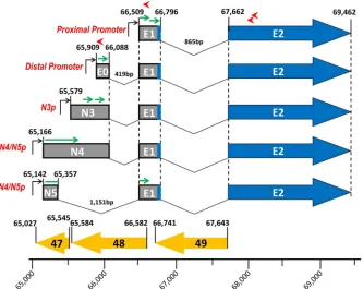
An area of the MHV68 genome that is well conserved in both sequence homology and function is the gene 50 region, which encodes the essential immediate early viral transcriptional activa-tor RTA (also known as R or BRLF1). Notably, the MHV68, KSHV, and EBV gene 50 transcripts exhibit similar splicing pat-terns, promoters, and genome locations (Fig. 1). Furthermore, ectopic expression of RTA in cells latently infected with MHV68, herpesvirus saimiri (HVS), KSHV, and EBV is capable of driving virus reactivation (10,11), mediated through RTA activation of viral lytic cycle-associated gene promoters (12,13–15). RTA feeds into many different lytic and latent gene pathways, acting as both a positive and negative regulator of gene transcription. The many important functions exhibited by RTA make it one of the most tightly regulated genes in gammaherpesviruses where control is established through the use of alternative splicing, epigenetic modifications, feedback loops, and multiple promoters.
Recently we identified a new RTA promoter, which we named the distal gene 50 promoter (16). Notably, we previously showed that this promoter is also present in KSHV and EBV (Fig. 1). The transcript initiated from the distal promoter encodes a 181-bp exon (E0) at coordinates bp 65909 to 66089 in the MHV68 ge-nome, which splices to the E1 exon located at coordinates bp 66509 to 66796 and then splices to exon 2 at coordinates bp 67662 to 69462. Notably, we have shown that a proximal gene 50 mutant virus is able to replicate, establish latency in both splenocytes and
peritoneal exudate cells (PECs), and reactivate from PECs, sug-gesting that the distal promoter is functional (16). However, the proximal-promoter mutant is defective in reactivation from splenocytes (16).
Here we demonstrate that the expression of RTA can be driven from multiple previously unknown promoters upstream of the distal promoter. These promoters encode three new exons, two of which represent extensions of exon 0 and the other a completely unique exon. To further characterize these promoters, we gener-ated an MHV68 mutant lacking both the proximal and distal gene 50 promoters (G50DblKo). We show the G50DblKo virus retains the ability to replicate to levels similar to those for the wild-type (WT) virus, but only in the absence of a type I interferon (IFN) response. Furthermore, we show that the G50DblKo virus is ex-tremely sensitive to type I interferons bothin vitroandin vivo. We also show that despite the replication defect observed in the pres-ence of a type I interferon response, the G50DblKo virus estab-lished a latent infection in both splenocytes and PECs, although it was unable to reactivate from either splenocytes or PECs. Impor-tantly, the replication and latency defects exhibited by the G50DblKo mutant were largely rescued by infection of mice lack-ing a type I interferon response. Finally, despite the sensitivity of the G50DblKo mutant virus to type I IFNs, we show that the newly identified promoters themselves are not directly sensitive to IFN-␣, indicating that type I IFNs likely act downstream of RTA transcription.
MATERIALS AND METHODS
Generation of G50DblKo and G50DblKo.MR viruses.The MHV68. G50DblKo virus was generated using the galK-mediated Red recom-bineering method as described previously (17,18). Briefly, we introduced the MHV68 bacterial artificial chromosome (BAC) into the galK bacterial strain SW102, where thegalKwas inserted into the Orf50 locus. To generate the Orf50-galKswap, we amplified thegalKgene from the plasmid pGalK using the primers GalK-G50-F (5=-CTTAAAGGAGGAG CTGTTGGCAGTGCTTGCTGAAAATACCTCATCAAACTCCTGTTG ACAATTAATCATCGGCA-3=) and GalK-G50-R (5=-CCTCAGCCTTT GAAGGGACATTTCATGACTCATGTAGGAGGAGCTACCAGTCAGC ACTGTC CTGCTCCTT-3=); this PCR product generated from pGalK includes 50 bp of homologous sequence upstream and downstream from the Orf50 locus. The PCR product was then introduced into the SW102 cells through electroporation, and those cells expressing recombinants were selected through the use of minimal medium with galactose. The ORF50/GalK recombinant BAC mutants (MHV68.⌬50galK) were con-firmed through analyses using multiple restriction endonucleases.
To generate the G50DblKo virus, two separate PCRs with 25-l reaction mixtures were performed on MHV68 WT BAC with 30 cycles using Phusion polymerase (New England BioLabs) and the follow-ing cyclfollow-ing conditions: 94°C for 30 s, 58°C for 30 s, and 72°C for 1 min. The primers used for reaction one amplification were forward
primer Orf50F65040_65089 (5=-CTTAAAGGAGGAGCTGTTGGCA
GTGCTTGCTGAAAATACCTCATCAAACT-3=) with reverse primer
Orf50DpKo100RHindIII (5=-AATGGACTCCAGCTGAAGCTTAGT
CATAGAACATACCATGA-3=), and the primers used for reaction two amplification were reverse primer Orf50R66641_66690 (5=-CCTCAG CCTTTGAAGGGACATTTCATGACTCATGTAGGAGGAGCTACCA G-3=) with forward primer Orf50DpKo100FHindIII (5=-GTATGTTC
TATGACTAAGCTTCAGCTGGAGTCCATTATTCT-3=), generating
overlapping PCR products to be used in round two amplification (of note, each internal primer contained a HindIII restriction enzyme site to be used for diagnostic purposes). Two microliters of each round one amplification product was then used as the template for a round two reaction using the external forward primer from reaction one
FIG 1Genomic alignment of Orf50/BRLF1/Rta regions from MHV68, EBV, KSHV, and HVS gammaherpesviruses, illustrating the conserved organization of gene 50 transcription. A schematic diagram of the Orf50 region with exon 2, exon 1, and recently discovered exon 0 is shown. The antisense coding regions Orf48/BRRF2 and Orf49/BRRF1 are shown to demonstrate the conserved or-ganizational context. The boxes around exon 1 and exon 0 shown their relative positions within the genome. The nucleotide positions of exon 0 (E0), exon 1 (E1), and exon 2 (E2) are given.
on November 7, 2019 by guest
http://jvi.asm.org/
[image:2.585.43.284.64.250.2]Orf50F65040_65089 and the reverse external primer from reaction two Orf50R66641_66690. The resulting product, containing a 50-bp deletion within the distal promoter region of the Orf50 locus, was gel purified and used as the template for a second overlapping PCR using the same conditions as the first PCR but this time targeting a deletion within the proximal promoter region. The primers used for reaction one amplification were forward primer Orf50F65040_65089 (5=-CTT AAAGGAGGAGCTGTTGGCAGTGCTTGCTGAAAATACCTCATC AAACT-3=) with reverse primer Orf50PpKORHindIII (5=-GAACAGT
ATGAGAAAAAGCTTCAGGGAATTTTGTTATGTGC-3=), and the
primers used for reaction two amplification were reverse primer
Orf50R66641_66690 (5=-CCTCAGCCTTTGAAGGGACATTTCATG
ACTCATGTAGGAGGAGCTACCAG-3=) with forward primer
Orf50PpKoFHindIII (5=-CCCTGGAATCATAGAAAGCTTTTTCTCAT
ACTGTTCCTTTT-3=). As before, 2l of each round one amplification product was used as the template for a round two reaction using the same external primers as before, Orf50F65040_65089 and Orf50R66641_66690. The resulting PCR product was gel purified and now contained a 50-bp deletion within the Orf50 distal promoter region, a 70-bp deletion within the Orf50 proximal promoter region, and 50 bp of homology to the region directly external to the MHV68.⌬50galK Orf50galK region. A WT Orf50 PCR product containing the 50-bp ho-mology arms was also generated in order to create an Orf50 marker rescue of the MHV68.⌬50galK parent BAC to ensure that no spontaneous mu-tations had arisen. This mutant or WT PCR product was electroporated into SW102/MHV68.⌬50galK and after recombination was selected on minimal medium plates containing glycerol and 2-deoxy-D
-galactose. The colonies were then screened by colony PCR using primers
ORF50E0F (5=-CACAACCCAGCACATGTTCAAACAT-3=) and
ORF50E0R (5=-CTGTGTCTCACTGAAAACACTC-3=). Colonies were then further verified by restriction digestion using the HindIII diagnostic sites, as well as confirmation by PCR of the ORF50 region and DNA sequencing of the PCR product (Macrogen Sequencing). The integrity of the nonmutated BAC was confirmed through further restriction endonu-clease digestion using PstI and EcoRI. Restriction endonuendonu-clease digests were then subjected to Southern blotting analyses using a PCR-generated fragment spanning the entire Orf50 region as the probe.
Production of virus.Both the G50DblKo and G50DblKo.MR viruses were generated from yellow fluorescent protein (YFP) BAC (19). In order to ensure proper viral growth of the G50DblKo virus, a protocol was developed in which the virus would be passaged and titers determined in the absence of type I interferons. Both mutant and marker rescue BACs were transfected using LT-1 transfection reagent (Mirus) into Vero cells expressing Cre-recombinase (Vero-Cre cells). When cells reached 70% cytopathic effect (CPE), cells and supernatants were collected and freeze-thaw lysed three times. The resulting stock was used to infect new Vero-Cre cells to ensure the excision of the BAC. At 70% CPE, cells and super-natants were once again collected, lysed, and used to infect large quantities of Vero-Cre cells to scale up viral stocks. Viral titers were determined as described below.
Tissue culture.Mouse embryonic fibroblasts (MEFs) and NIH 3T12, Raw264.7, and Vero-Cre cells were maintained in Dulbecco’s modified Eagle’s medium (DMEM) supplemented with 10% fetal calf serum, 2 mM
L-glutamine, 100 U of streptomycin per ml, and 100 U of penicillin per ml
(cMEM). Vero-Cre cells used for virus generation were passaged with the addition of 300g of hygromycin B/ml. Cells were maintain at 37°C in a tissue culture incubator with 5% CO2. MEFs were obtained from C57BL/6
mouse embryos, and IFN-␣/R⫺/⫺MEFs were obtained from 129S2/ SvPas.IFN-␣/R⫺/⫺mice as previously described (20).
Growth curves, plaque assays, and viral titers.Growth curves were performed using Vero-Cre, 3T12, IFN-␣/R⫺/⫺MEF, or C57BL/6 MEF cells plated in six-well plates at a concentration of 1.75⫻105cells per well
24 h prior to infection. The cellular concentration after 24 h was deter-mined, and viral stocks were diluted to a 200l volume in cMEM at multiplicities of infection (MOIs) of 0.01, 0.1, and 10. These inoculums
were added to the cellular monolayer, and plates were rocked every 15 min at 37°C for 1 h. After 1 h, fresh cMEM at a volume of 2 ml was added back to each well. The contents of the wells were collected at the indicated time points and frozen at⫺80°C until plaque assays were performed to deter-mine titers. Growth curves with the addition of mouse recombinant IFN-␣(Miltenyi Biotec) were performed in a similar manner, but IFN-␣ was added to the plated cells at a concentration of 10,000 IU/ml 1 h before infection. IFN-␣was subsequently added at a concentration of 5,000 IU/ml every 24 h for the duration of the infection. Plaque assays were performed by plating Vero-Cre cells in six-well plates at a concentration of 1.75⫻105cells per well 24 h prior to infection. Viral stocks or growth
curve stocks at the indicated time points were freeze-thawed 3 times, and a 10-fold serial dilution was generated, 200l of each dilution was added to the Vero-Cre monolayer, and plates were rocked every 15 min at 37°C for 1 h. After 1 h, cells were overlaid with 2% fetal bovine serum (FBS) complete medium containing 20 g/liter methylcellulose (Sigma). Plaques were visualized at 14 days postinoculation by staining with neutral red (Sigma) at a concentration of 6% overnight.
RACE analysis. One-hundred-millimeter plates were seeded with Raw 264.7, Vero-Cre, or IFN-␣/R⫺/⫺MEF cells in cMEM and 24 h later were infected with WT-YFP or G50DblKo virus at an MOI of 5. Total RNA was isolated from these cells at 24 or 48 h postinfection using TRIzol reagent (Invitrogen). Five micrograms of RNA was DNase I treated (In-vitrogen) and subjected to 5=rapid amplification of cDNA ends (RACE) performed using the GeneRacer system (Invitrogen). RACE-ready cDNA was generated using Superscript III (Invitrogen) reverse transcription us-ing random primers as per the manufacturer’s instructions. RACE-ready cDNA was used to look for additional 5=transcripts through the use of nested PCR utilizing Phusion high-fidelityTaq(NEB) and PlatinumTaq (Invitrogen). Nested PCR was performed using the 5=universal forward primer (round 1), the 5=universal forward primer (round 2), and various reverse primers located in the Orf50 region: E2R1 (5=-ATTTCGAACAG
ACTGCAGGCCAGAGGTTGA-3=), E2R2 (5=-CGAACATGGGGCAGT
CAGAAACAGC-3=), E1R (5=-TTCAATTCTCATGGTCACATCT-3=), and E0R (5=-TTTGAACATGTGCTGGGTTGTG-3=). The following cy-cling conditions for PhusionTaqwere used with 1l of cDNA in a 50-l PCR mixture: 98°C for 30 s; 30 cycles of denaturing at 98°C for 10 s, annealing at 60°C for 30 s, and extension at 72°C for 1 min; and a final extension of 72°C for 10 min. Round 2 nested amplification was per-formed using 2l of round 1 product in a 50-l reaction mixture using the same cycling conditions. The following cycling conditions for Plati-numTaqwere used with 1l of cDNA in a 50-l PCR mixture: 95°C for 5 min; 30 cycles of denaturing at 94°C for 30 s, annealing at 60°C for 30 s, and annealing at 72°C for 1 min 30 s; and a final extension at 72°C for 7 min. Round 2 nested amplification was performed using 2l of round 1 product in a 50-l reaction mixture using the same PCR conditions. PCR products were visualized through running on a 1% ethidium bromide gel, and excised bands were purified using a Geneclean II kit (MP Bio). Puri-fied PCR products from Phusion PCR were ligated into a pCR-Blunt II TOPO vector (Invitrogen), and purified PCR products from Platinum TaqPCR were ligated into either a pCR4-TOPO vector (Invitrogen) or a pGEMT-Easy vector (Promega) and analyzed by DNA sequencing (Mac-rogen USA).
Cell transfections and luciferase assays.Transfection of Vero-Cre and RAW 264.7 cells was done in 6-well plates, where at 1 day prior to transfection, cells were plated at 2⫻105cells per well in cMEM.
Trans-fection mixtures were prepared using 2.5g of reported plasmid and 5 ng of pHR-LUCRenillaluciferase vector as a transfection control. Transfec-tions were performed using LT-1 transfection reagent (Mirus) according to the manufacturer’s instructions. pGL4.13[Luc] was used as a positive control, pGL4.10[luc] was used as a negative control, and the green fluo-rescent protein pMaxGFP was used to determine transfection efficiency. For assay mixtures treated with IFN-␣, 5,000 IU/ml was added at 24 h posttransfection, and all cells were collected at 48 h posttransfection and lysed. All dual-luciferase assays were performed using a dual-luciferase kit
on November 7, 2019 by guest
http://jvi.asm.org/
(Promega) according to manufacturer’s instructions. Single-luciferase as-says were performed using lab-made luciferase agent (1.5 mM HEPES [pH 8], 80M MgSO4, 0.4 mM dithiothreitol [DTT], 2M EDTA, 10.6
M ATP, 5.4M coenzyme A, and 9.4M beetle Luciferin), where 10l of cell lysate was added to 50l of luciferase agent. Both single- and dual-luciferase assays were read using a TD-20/20 luminometer (Turner Biosystems). All transfections were repeated in triplicate, and results are presented as a fold ratio over that for empty pGL4.10 vector.
Reporter plasmids and cloning.DNA from the Orf50 region was am-plified from the MHV68 WT BAC and cloned into a luciferase reporter construct. PhusionTaq(NEB) was used with the following cycling parame-ters for all amplifications: 95°C for 5 min; 30 cycles of denaturing at 94°C for 30 s, annealing at 58°C for 30 s, and extension at 72°C for 30 s; and a final 72°C extension for 10 min. Overlapping PCR was used with the following primers to generate E0 luciferase promoter constructs with 50-bp deletions. Round one PCR used forward primer E0-250F (5=-GATCGGCTAGCTTAATCCTA TATGGAGAT-3=) with the following reverse primers: del 65822-65872R
(5-CATGTCTCAGCCAACAGCTCGACACTTCGAGTACC-3=), del
65772-65822R (5=-AGAATAATGGACTCCAGCTGAGTCATAGAACATAC-3=),
and del 65722-65772R (5=-GCACGTATTGCTGAAAAGGAATGATCAGG AATTCT-3=). Reverse primer E0ATGR (5=-GATCGAAGCTTGTGCTGGG TTGTGAAG-3=) was used with the following forward primers: del
65822-65872F (5=-GGTACTCGAAGTGTCGAGCTGTTGGCTGAGACATG-3=),
del 65772-65822F (5=-GTATGTTCTATGACTCAGCTGGAGTCCATTAT
TCT-3=), and del 65722-65772F (5=-AGAATTCCTGATCATTCCTTTTCA GCAATACGTGC-3=). After round one PCR, 2l of PCR products from del 65822-65872, del 65772-65822, and del 65722-65772 was used as the template for a round two PCR following the same cycling conditions as for round one with forward primer E0-250F and reverse primer E0ATGR. These second-round PCR products were gel purified and cloned into pCR-Blunt (Invitro-gen) for vector shuttling and sequence confirmation. The pCR-Blunt plasmid was then cut with HindIII and NheI to excise the E0 fragments, and the luciferase reporter construct pGL4.10[luc2] (Promega) was also digested with HindIII and NheI. The digested E0 products and pGL4.10[luc2] vector were then gel purified, resuspended in Tris-EDTA (TE), and ligated together using T4 DNA ligase (NEB) overnight at 16°C. Ligation products were transformed into Top 10 chemically competent cells, and colonies were screened for the presence of correctly oriented E0 pGL4.10[luc2] vectors through restriction digestion and DNA sequencing. Clones containing correct E0 deletion and expression were cultured, and plasmid DNA was isolated using an Endofree Maxikit (Qiagen). The process of generating Orf50 N3 and N4/N5 promoter luciferase constructs followed a method similar to that described above, with only a single PCR amplification required to generate 1,000-bp-long PCR frag-ments. The primers used for this single PCR were as follows: the reverse primer N4_N5RBgl2 (5=-CGATAGATCTAAGCCGTGGTCAGCAGGT-3=) was used with the forward primer N4_N5F1000Nhe1 (5=-AGTCGCTAGCA ATCGTCCGGGGGGTTAA-3=), and the reverse primer N3RBgl2 (5=-CGA TAGATCTAGCCTGGGGCATAGTTCTT-3=) was used with the forward primer N3FNhe1 (5=-AGTCGCTAGCTCAGGATGCAGTTAAGCA-3=). These products were gel purified, shuttled through pCR-blunt, digested with NheI and BglII, and then ligated into pGL4.10[luc2]. The generation of the proximal promoter expression constructs followed the same protocol as de-scribed above. Forward primer ProxPromF (5=-GATCGCTAGCTCTTTAT AGGTACCAGGGAA-3=) was used with reverse primer ProxPromR (5=-TA GCAGATCTGGTCACATCTGACAGAGAAA-3=) to generate the 410-bp proximal promoter luciferase construct. Overlapping PCR using forward primer ProxPromDelF (5=-CCCTGGAATCATAGATTTCTCATACTGTTC CTTTT-3=) with primer ProxPromR and reverse primer ProxPromDelR (5=
-GAACAGTATGAGAAATCTATGATTCCAGGGAATTT-3=) with primer
ProxPromF was used to generate the proximal promoter 70-bp deletion con-struct. Digestion, ligation, and purification also followed the same protocol as described above.
Limiting-dilution PCR and limiting-dilution reactivation assays.
Limiting-dilution PCR to determine the frequency of viral genomes and a limiting-dilution CPE assay to determine the number of cells reactivating
from latency were performed as previously described (20,21). Briefly, to determine the frequency of viral genomes, splenocytes or PECs were serial diluted in 96-well plates and subjected to protease K digestion. After di-gestion, cells were used in a two-round nested PCR using primers found in the G50 region. PCR products were resolved on a 2% agarose gel and analyzed. To determine the frequency of infected cells reactivating from latency, splenocytes or PECs were counted and diluted in cMEM. Cells were then plated as serial 2-fold dilutions onto an MEF or IFN-␣/R⫺/⫺ MEF monolayer in 96-well plates. In parallel, mechanically disrupted cells also were plated as a serial 2-fold dilution to detect any preformed infec-tious virus. At 21 days postplating, each well was assessed for CPE and scored as a percentage. For both limiting-dilution PCR and limiting-di-lution reactivation, the Poisson distribution was used to determine fre-quencies.
Mice, infections, and tissue preparation.Female C57BL/6 mice (The Jackson Laboratory) and 129S2/SvPas.IFN-␣/R⫺/⫺mice 6 to 8 weeks of age were maintained at Emory University. Mice were housed under sterile conditions and maintained in accordance with Emory University School of Medicine (Atlanta, GA), as well as all federal guidelines. C57BL/6 or 129S2/SvPas.IFN-␣/R⫺/⫺ mice were infected intranasally (i.n.) with 1,000 PFU of WT-YFP, G50DblKo, or G50DblKo.MR virus in 20l cMEM following isoflurane anesthetization. 129S2/SvPas.IFN-␣/R⫺/⫺ mice were weighed at the time of infection, and weight was monitored daily; mice were sacrificed if they lost 20% of their original body weight. C57BL/6 mice were also infected intraperitoneally with 1,000 PFU of WT-YFP, G50DblKo, or G50DblKo.MR virus in 200l cMEM. With both routes of infection, C57BL/6 mice were sacrificed at day 18 postin-fection or at 7 days postinpostin-fection for lung titers. Surviving 129S2/ SvPas.IFN-␣/R⫺/⫺mice were sacrificed at day 28 postinfection. Mice were sacrificed by isoflurane and cervical dislocation. PECs were collected by peritoneal lavage using 10 ml cMEM, spleens were harvested, and splenocytes were prepared by manual homogenization, while lungs were harvested and prepared by mechanical bead disruption using 1.0-mm silica beads. Splenocytes were treated with Tris-ammonium chloride to eliminate red blood cells. All prepared cells were counted using a Cellom-eter Auto T4 (Nexcelom) and immediately used for lung titer, reactiva-tion, or genome analysis. Cells not used immediately were stored in cMEM–10% dimethyl sulfoxide at⫺80°C.
Immunoblotting.Infections were carried out as described above for growth curves. At 24, 48, and 72 h postinfection, cells were harvested, washed with 1⫻phosphate-buffered saline (PBS), and then resuspended in 40l of lysis buffer containing 150 mM NaCl, 50 mM Tris-HCl, 1 mM EDTA, and 0.1% Triton X-100 supplemented with 1 mM NaF, 1 mM Na3VO4, and an EDTA-free protease inhibitor tablet (Roche). Protein quantifications were carried out using a DC protein assay (Bio-Rad). For all blots, 30g of protein was mixed with 6⫻SDS loading buffer, boiled for 5 min at 100°C, and resolved by SDS-PAGE. SDS-polyacrylamide gels were transferred to a nitrocellulose membrane using a semidry apparatus (Bio-Rad). After transfer, membranes were blocked in 5% milk in Tris-buffered saline (TBS)–Tween for 1 h at room temperature. After 1 h, membranes were washed three times with TBS-Tween, and the primary antibody rabbit anti-MHV68 RTA (22) diluted 1:1,000 in blocking buffer was added and left overnight with rocking at 4°C. Following overnight incubation, the primary antibody was removed and the membrane washed three times with TBS-Tween. The membrane was then incubated with the secondary donkey anti-rabbit antibody (Jackson Immunore-search) diluted 1:2,000 in blocking buffer for 1 h at room temperature. This was followed by three additional washes with TBS-Tween. The blot was then developed using SuperSignal West Pico chemiluminescent sub-strate (Pierce). Membranes were stripped using Restore PLUS Western blot stripping buffer (Thermo Scientific). Membranes were then sub-jected to the same protocol using the primary mouse monoclonal-actin antibody (Sigma) at 1:5,000 and the secondary donkey mouse anti-body (Jackson Immunoresearch) at 1:5,000. Membranes were also sub-jected to the same protocol to blot for v-cyclin, where primary rabbit
on November 7, 2019 by guest
http://jvi.asm.org/
monoclonal v-cyclin antibody was used at 1:2,000 overnight and the sec-ondary donkey anti-rabbit antibody (Jackson Immunoresearch) was used at 1:2,000 for 1 h at 4°C.
RESULTS
Characterization of the Orf50 distal promoter activityin vitro.
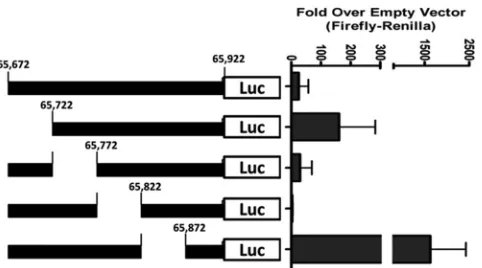
We previously reported the generation and characterization of a gene 50 proximal promoter knockout virus (G50pKo), which led to the identification of an additional promoter upstream of the proximal promoter, now referred to as the distal promoter (16). The additional distal promoter drives the expression of a new exon (E0), and the core promoter region corresponds to the first 250 bp upstream of the E0 transcriptional start site. To further character-ize the activity of this newly defined promoter region, we gener-ated serial 50-bp deletions (⌬65672-65722, ⌬65722-65772, ⌬65772-65822, and⌬65822-65872) in the core 250-bp distal pro-moter region (Fig. 2). These serial 50-bp deletion fragments were cloned into the pGL4.10 luciferase reporter vector, and the result-ing reporter plasmids transfected into RAW 264.7 cells. It was previously reported that the gene 50 distal promoter was most active in RAW 264.7 cells with the addition of lipopolysaccharide (LPS) (16).
Luciferase assays confirmed the previous finding that the 250-bp distal promoter region drives significant gene expression, ⬃30-fold over that with empty vector in the RAW 264.7 cells with the addition of LPS (Fig. 2). Notably, deletion of the sequences from bp 65672 to 65722 resulted in a substantial increase in pro-moter activity over that with empty vector (⬃4.6-fold above that observed with the full-length 250-bp promoter construct), indi-cating the presence of negativeciselements in this region. Simi-larly, deletion of the sequences from bp 65822 to 65872 also re-sulted in a strong enhancement of activity (⬃40-fold above that observed with the full-length 250-bp promoter construct). This would indicate that both of these regions play an important role in limiting gene 50 transcription. Conversely, deletion of the se-quences from bp 65772 to 65822 resulted in nearly complete
si-lencing of distal promoter activity (ca.16-fold below that observed with the full-length 250-bp promoter construct), arguing that es-sential positiveciselements map to this region. Therefore, we chose to delete the latter region in the context of the viral genome to ablate distal gene 50 promoter activity.
Generation of a recombinant MHV68 lacking both the
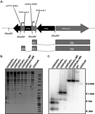
prox-imal and distal gene 50 promoters.With the identification of a
second promoter driving RTA expression, we set out to generate a gene 50 functionally null virus through the deletion of both the distal and proximal promoters. We targeted the promoter regions because previous attempts to propagate gene 50 null viruses har-boring mutations within the coding sequence were confounded by the generation of wild-type revertant viruses upon growth on complementing cell lines (i.e., recombination of wild-type gene 50 sequences into the viral genome) (23). Since the gene 50 null mu-tants grow slowly on complementing cell lines, any wild-type re-vertant virus quickly overtakes growth of the gene 50 null virus and ends up dominating the viral stock generated. In a previous attempt to overcome this problem, we deleted a 183-bp region of the proximal gene 50 promoter (the only known gene 50 pro-moter at the time), and it was the analysis of this mutant virus that led to the identification of the distal gene 50 promoter (16). No-tably, the deletion introduced into the viral genome to generate the proximal promoter mutant virus (G50pKo) also deleted the splice acceptor site utilized by the distal gene 50 promoter-driven gene 50 transcript. To circumvent this issue, we redesigned the proximal promoter deletion to introduce a 70-bp deletion (corre-sponding to the region immediately upstream of the exon 1 tran-script at coordinates bp 66412 to 66482), leaving the splice accep-tor site intact (Fig. 3A). This new gene 50 proximal promoter knockout virus (G50PpKo) was used as the backbone for the gen-eration of the G50DblKo virus.
To disrupt the distal gene 50 promoter, we targeted the 50-bp region from bp 65772 to 65822, which was determined to be es-sential for promoter activity in thein vitroanalyses carried out in the RAW 264.7 cell line (Fig. 2). To confirm the generation of the G50DblKo virus, we sequenced the areas of interest to ensure that the desired deletions were present in the viral genome (data not shown). To further confirm that the virus was intact and that the deletions were inserted into the gene 50 locus, as well as the ab-sence of spontaneous rearrangements and insertions in the viral ge-nome, we digested the mutant BAC with the restriction endonu-cleases HindIII, PstI, and EcoRI. Notably, in the process of generating the desired deletions, we introduced diagnostic HindIII sites (Fig. 3B andC). HindIII digestion of the G50DblKo mutant yielded the three expected digestion fragments (590 bp, 874 bp, and 1,059 bp), while HindIII digestion of wild-type virus and the marker rescue BAC DNAs resulted in the expected 2,643-bp product. To further assess the presence of the deletions in the gene 50 locus, we per-formed Southern blotting of the HindIII-digested BAC DNAs (Fig. 3C). We probed the blot with a 1,651-bp probe correspond-ing to genome coordinates from bp 65040 to 66690, which hybrid-ized to all three fragments generated. Notably, the band intensities varied and were dependent on the extent that the probe over-lapped with the fragments generated. Importantly, the wild-type-sized fragment was absent from the G50DblKo and G50PpKo viral genomes, eliminating the possibility of a genome duplication of the gene 50 locus or recombination of the targeting sequences into another region of the viral genome. The final confirmation that the mutant generated was specific to the area of interest was
FIG 2Promoter deletions within the MHV68 E0 250-bp promoter region. Reporter constructs were generated within the context of the 250-bp pro-moter through overlapping PCR. The 50-bp E0 propro-moter mutants were cloned into the pGL4.10[luc] luciferase report construct. RAW 264.7 cells were cotransfected with pGL4.10[luc] luciferase report constructs contain-ing 50-bp deletions as illustrated and phRL-Luc (Renillaluciferase). RAW 264.7 cells were stimulated with LPS (5g/ml) at 24 h after transfection, and at 48 h after transfection luciferase assays were performed. Data are presented as the fold difference in the ratio of firefly toRenilla luciferase versus the pGL4.10[luc] empty vector control. The data were compiled from 3 indepen-dent transfections, each done in triplicate. Standard errors of the means are shown.
on November 7, 2019 by guest
http://jvi.asm.org/
[image:5.585.45.286.65.199.2]the generation and confirmation of the G50DblKo.MR and G50PpKo.MR viruses, in which the Orf50 GalK region was re-placed with the WT Orf50 region. These marker rescue viruses were all analyzed by sequencing, restriction enzyme digestion, and Southern blotting, and all three methods confirmed reversion back to WT virus. To ensure that the phenotypes observed with the G50pDblKo virus were in fact the result of the desired muta-tions, this process was conducted four independent times, result-ing in the isolation of four independently generated mutant clones. These clones were used throughout the experiments inter-changeably, and each experiment was conducted using at least two if not all of the clones. The use of multiple independent clones, in conjunction with the G50pDblKo.MR virus, makes it very un-likely that the observed phenotypes were the result of spontaneous mutations arising elsewhere within the genome.
Replication of the MHV68 G50DblKo mutant, but not
wild-type MHV68, is inhibited by wild-type I interferons.To generate the
G50DblKo virus, the mutated BAC was first transfected into Vero cells expressing Cre recombinase (Vero-Cre cells) (24) to excise the BAC from the MHV68 BAC. The removal of the BAC se-quence is necessary since virus containing BAC sese-quences is sig-nificantly attenuatedin vivo(25). Upon successful removal of the BAC in Vero-Cre cells, viral stocks are generated by growth on murine NIH 3T12 cells (24). Surprisingly, Vero-Cre cells trans-fected with the G50DblKo BAC DNA supported growth of this mutant, indicating that the proximal and distal gene 50 promoters are not required for RTA expression. However, upon low-MOI infection of NIH 3T12 fibroblasts with the resulting viral stock generated from growth in Vero-Cre cells, we failed to observe any replication of the G50DblKo mutant. To further investigate this
FIG 3Generation of MHV68 G50DblKo and G50DblKo.MR viruses. (A) Schematic diagram of alternatively initiated gene 50 transcripts. The locations of the known proximal and distal gene 50 promoters are shown. The two hatched areas depict the regions deleted within the mutant G50DblKo virus: a 50-bp deletion extending from bp 65772 to 65822 in the distal promoter region and a 70-bp deletion extending from bp 66412 to 66482 in the proximal promoter region. There are two HindIII sites in the WT genome located at bp 64898 and 67541; the G50DblKo virus has two additional HindIII sites introduced within the deletions, located at bp 65797 and 66447. The splicing of E1 to E2 driven by the proximal promoter and the splicing of E0 to E1 to E2 driven by the distal promoter are shown. (B) HindIII restriction endonuclease digests of G50DblKo, G50PpKo, G50DblKo.MR, G50PpKo.MR, G50GalK, and WT-YFP BAC. The diagnostic restriction fragments are depicted (). (C) Southern blot of the HindIII digest shown in panel B using a probe specific for the Orf50 region. Three unique digestion fragments are shown for the G50DblKo virus, the WT and MR rescue viruses are shown to be the same, and thegalKparent fails to hybridize the Orf50 probe due to the Orf50 region replacement with the G50PpKo cassette.
on November 7, 2019 by guest
http://jvi.asm.org/
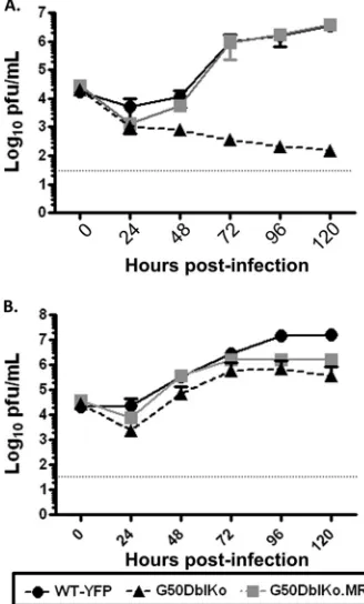
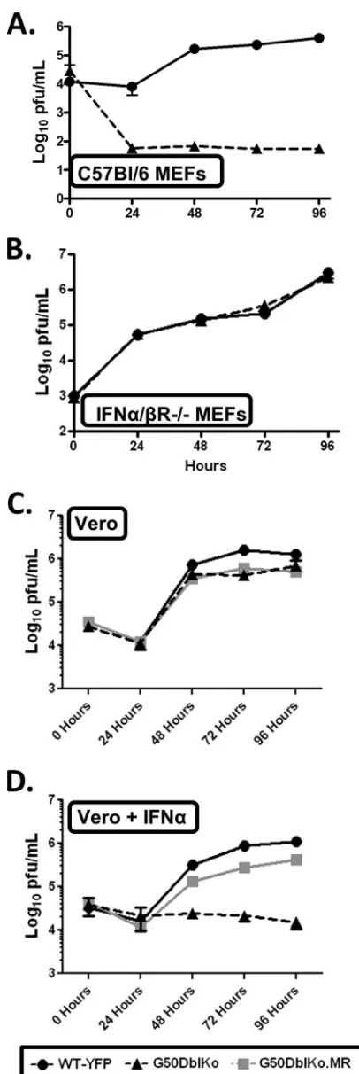
[image:6.585.135.446.67.443.2]phenomenon, multistep growth curves were performed on both NIH 3T12 and Vero-Cre cells (Fig. 4). Consistent with the initial analyses, the G50DblKo mutant was able to grow in Vero-Cre cells, exhibiting only a mild replication defect (Fig. 4B), indicating that both the proximal and distal gene 50 promoters are dispens-able for RTA expression in these cells. However, we failed to ob-serve any growth of the G50DblKo mutant on NIH 3T12 fibro-blasts (ca. a 4- to 5-log defect in viral titers between the marker rescue virus and the G50DblKo mutant at late times postinfec-tion) (Fig. 4A).
Since a major difference between Vero cells and NIH 3T12 fibroblasts is that Vero cells lack the ability to generate type I interferons, we assessed whether a type I IFN response can block replication of the G50DblKo mutant virus. We infected mouse embryo fibroblasts (MEFs) generated from C57BL/6 mice with the G50DblKo virus, and once again we failed to see efficient rep-lication (Fig. 5A). The G50DblKo virus exhibited nearly a 4-log defect in viral titers at late times postinfection. To determine whether type I interferons were responsible for this large defect, we infected MEFs generated from 129S2/SvPas.IFN-␣/R⫺/⫺
mice. Similar to the results obtained in Vero cells, the inability of the MEFs from mice lacking the IFN-␣/receptor to respond to a type I IFN response rescued the replication defect seen in the C57BL/6 MEFs (Fig. 5B). Notably, in the 129S2/SvPas.IFN-␣/ R⫺/⫺MEFs, the G50DblKo virus replication was
indistinguish-able from that of wild-type virus (Fig. 5B). To directly examine the
impact of type I IFNs on replication of the G50DblKo mutant, we compared virus replication in Vero cells in the absence and pres-ence of IFN-␣. Vero-Cre cells were pretreated with IFN-␣and then treated every 24 h postinfection. Cells that were not treated with IFN-␣, as seen before, showed normal growth and kinetics of the G50DblKo virus, similar to those of WT and G50DblKo.MR viruses (Fig. 5C). However, adding IFN-␣severely inhibited rep-lication of the G50DblKo mutant (Fig. 5D).
These analyses revealed an impact of type I interferons onin vitro replication of MHV68 (although it has previously been shown that acute viral titers in the lungs of IFN-␣/R⫺/⫺mice are
significant higher than those in wild-type mice and that the ab-sence of a type I IFN response renders mice highly susceptible to lethal MHV68 infection [26–28]). These analyses also reveal that, despite replication of the G50DblKo virus being attenuated in the presence of a type I IFN response, there appears to be little or no replication defect in comparison to wild-type virus when a type I interferon response is absent, due to either the lack of the IFN-␣/R or the absence of type I IFN expression. In addition, these analyses demonstrate that despite the deletion of the known gene 50 promoters, the G50DblKo mutant is replication competent under some experimental conditions, suggesting that there are alternative mechanisms for expression of the essential immediate early RTA.
Notably, upon higher-MOI infections, although there was a partial rescue of the G50DblKo growth defect, this mutant still exhibited a pronounced growth defect (Fig. 6A). Consistent with the observed growth defect, we observed a small-plaque pheno-type (Fig. 6C), which was observed with 4 independently derived G50DblKo mutant BAC clones, making it unlikely that any of the observed phenotypes are the result of secondary mutations in the viral genome. However, when we examined the levels of RTA pro-tein as a function of time postinfection, we observed a faster ki-netics and higher levels of RTA at early times postinfection (Fig. 6B, 8-hour time point). This correlated with higher levels of the v-cyclin expression as well at 8 hours postinfection (Fig. 6B). This argues that the defect in virus replication is not directly attribut-able to failure to express RTA protein, although it does suggest that entry into the lytic cycle may be dysregulated and that this could lead to a defect in virion production.
Identification of three additional gene 50 transcripts driven by 2 promoters mapping upstream of the distal gene 50
pro-moter.We have previously shown that transcription of MHV68
gene 50 is driven by two distinct promoters: (i) the proximal pro-moter, encoding a short 288-bp exon, E1, which splices to the large 1,800-bp exon, E2 and (ii) the distal promoter, which en-codes a short 181-bp exon, E0, which splices to E1, which in turn splices to E2 (6, 12, 16) (Fig. 1). We further showed that this organization of gene 50 transcription was conserved in both EBV and KSHV (16). The unexpected ability of the G50DblKo mutant to replicate under some experimental conditions indicated that there must exist alternative mechanisms for driving gene 50 tran-scription, since RTA is known to be absolutely required for virus replication (this has been shown for EBV, KSHV, and MHV68) (23,29–32). To determine if this hypothesis was indeed correct, we performed 5=RACE analysis of RNAs from Vero-Cre, RAW 264.7, and IFN-␣/R⫺/⫺ MEFs infected with either wild-type
MHV68 or the G50DblKo mutant. From this analysis, we were able to detect three previously unidentified gene 50 transcripts
FIG 4G50DblKo virus replicatesin vitroin Vero-Cre cells but fails to replicate in NIH 3T12 fibroblasts. (A) Multistep growth curve of NIH 3T12 cells in-fected with an MOI of 0.1. Cells were inin-fected with G50DblKo, G50DblKo.MR, and WT-YFP viruses and collected for viral titer analysis at the indicated times postinfection. (B) Multistep growth curve of Vero-Cre cells infected at an MOI of 0.1. Cells were also infected with G50DblKo, G50DblKo.MR, and WT-YFP
viruses and collected for viral titer analysis at the indicated times postinfection.
on November 7, 2019 by guest
http://jvi.asm.org/
[image:7.585.78.242.69.341.2]which initiated from two distinct upstream transcription initia-tion sites (Fig. 7).
The three additional transcripts were identified in all three cell types examined and also were identified in both wild-type MHV68- and G50DblKo virus-infected cells. It is important to note that the 5=RACE analyses conducted using the G50DblKo virus failed to detect the presence of any E1-E2 or E0-E1-E2 tran-scripts, further confirming that G50DblKo is indeed a true
knock-FIG 5G50DblKo virus fails to replicatein vitrowhen IFN-␣is present. (A) Multistep growth curve of C57BL/6 mouse embryonic fibroblasts (MEFs) in-fected with G50DblKo or WT-YFP virus at an MOI of 0.01. (B) Multistep growth curve of 129S2/SvPas.IFN-␣/R⫺/⫺ mouse embryonic fibroblasts (IFN-␣/R⫺/⫺MEFs) infected with G50DblKo or WT-YFP virus at an MOI of
0.01. (C) Multistep growth curve of Vero-Cre cells infected with G50DblKo, G50DblKo.MR, or WT-YFP virus at an MOI of 0.1. (D) Multistep growth curve of Vero-Cre cells, treated with IFN-␣(10,000IU/ml) at the time of in-fection and every 24 h thereafter, infected with G50DblKo, G50DblKo.MR, or WT-YFP virus at an MOI of 0.1. All cells were collected at the times indicated, and experiments were repeated at least in triplicate.
FIG 6Single-step growth analyses of the G50pDblKo mutant and analysis of the kinetics of RTA expression. (A) Single-step growth curve of G50pDblKo or WT-YFP virus in NIH 3T12 fibroblasts infected at an MOI of 10. (B) Western blot staining for RTA, v-cyclin, and-actin expression from the same cells used in the single-step growth curve shown in panel A. (C) Representative sample of the small-plaque phenotype exhibited under all experimental con-ditions when using different G50pDblKo clones.
on November 7, 2019 by guest
http://jvi.asm.org/
[image:8.585.318.521.63.465.2] [image:8.585.58.261.65.671.2]out of the proximal and distal gene 50 promoters. The first new exon, which we have termed N3, is a 330-bp extension of the previously characterized E0 exon. This new N3 exon is 511 bp long and maps to coordinates bp 65579 to 66090 in the MHV68 ge-nome. The 3=end of this exon is identical to the 3=end of the E0 exon. Like E0, this newly identified N3 exon splices to the E1 exon, leading to removal of a 419-bp intron, which in turn splices to the E2 exon, removing an 865-bp intron (Fig. 7). The second new exon, which we have termed N4, is a 743-bp extension of the E0 exon. The N4 exon map to bp 65166 to 66090 in the MHV68 genome and is 924 bp long (Fig. 7). The 3=end of this exon is also identical to the 3=end of E0 and splices to the E1 exon and then to the E2 exon. The final alternatively spliced transcript contains a new exon, which we have termed N5, which is 216 bp long and maps to bp 65142 to 65358 in the MHV68 genome (Fig. 7). This new exon consists mainly of the 5=end of the newly identified N4 exon, with a 24-bp extension, indicating that the N4 and N5 exons share a promoter region. However, unlike N4, which is an exten-sion of E0, the N5 exon exhibits a completely unique splicing event in which the 3=end at bp 65358 splices to E1, directly eliminating a large 1,151-bp intron. Like the other known exons, the E1 exon then splices to the E2 exon.
It is important to note that none of the newly identified exons have been observed to splice directly to the E2 exon, but rather all of them splice to the E1 exon, which contains the RTA translation initiation codon. Thus, to date, there is no evidence for alternative
RTA translation initiation sites. This, however, does not exclude the possibility of unique splicing events from the newly identified transcripts to a novel position within the E2 exon or elsewhere within the viral genome. It should be noted that there are several short ATG-initiated open reading frames within the newly iden-tified exons, which may play an important role (i.e., encoding novel viral gene products and/or interfering with RTA transla-tion) (Fig. 7).
Neither the N3 promoter nor the N4/N5 promoter exhibits
sensitivity to IFN-␣, but RTA levels from the G50DblKo mutant
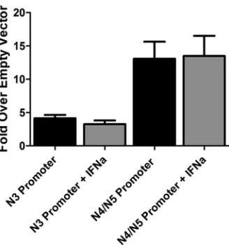
are diminished in the presence of a type I IFN response at low MOI.The identification of alternative gene 50 transcripts pro-vides a clear mechanism by which the G50DblKo virus is appar-ently able to generate sufficient levels of RTA to drive virus repli-cation, at least in the absence of a type I IFN response. However, it does not address the issue of why replication of the G50DblKo mutant is suppressed by type I IFNs. To begin to address this issue, we cloned 1,000-bp fragments immediately upstream of the 5= ends of the N3 and N4/N5 transcripts into the pGL4.10 luciferase reporter vector (Promega Biotech) and transfected the resulting reporter constructs into Vero-Cre cells. While the N3 promoter resulted in expression⬃4-fold over that with empty vector, the N4/N5 promoter resulted in slightly higher levels of luciferase expression (⬃13-fold over empty vector) (Fig. 8). These data demonstrate that the region immediately upstream of the newly identified gene 50 transcripts exhibits modest promoter activity.
FIG 7RACE analyses reveal three additional G50 exons upstream of E0. RACE analyses were performed using cDNAs generated from WT-YFP- and G50DblKo-infected Vero-Cre, Raw 264.7, and IFN-␣/R⫺/⫺MEFs at 24 and 48 h postinfection. 5=RACE analysis using reverse primers located in E2, E1, and E0 were used in conjunction with the universal 5=RACE forward primer. All three experiments identified three additional exons upstream of E0: exon N3, representing a 330-bp extension of E0; exon N4, representing a 743-bp extension of E0; and exon N5, representing a small novel 216-bp exon. All three new exons follow canonical splicing in which they splice to E1, which in turn splices to E2. Green arrows denote short ATG-initiated open reading frames that lie upstream of the RTA-coding sequences, which are indicated in blue. The locations of the major open reading frames antisense to gene 50 are also shown. Red arrows denote primers used in the 5=RACE analyses. The positions of the primers used were as follows: E2-1, bp 68095 to 68066; E2-2, bp 68051 to 68027; E1, bp 66530 to 66509;
and E0, bp 65930 to 65909.
on November 7, 2019 by guest
http://jvi.asm.org/
[image:9.585.127.460.68.333.2]To determine if the type I IFN-sensitive replication of the G50DblKo mutant virus reflects type I IFN-mediated suppression of gene 50 transcription from the G50DblKo mutant, we exam-ined N3 and N4/N5 promoter activity in the presence of IFN-␣ treatment (Fig. 8). Since Vero cells fail to produce IFN-␣but retain the ability to respond to exogenously added IFN-␣(Fig. 5D), IFN-␣was added to the transfected Vero cells and promoter activity assessed (Fig. 8). Similar luciferase assays of previously identified Orf50 transfected promoters have shown them to be sensitive to IFN-␥(33). However, our analysis showed no evi-dence of IFN-␣suppression of reporter gene activity, suggesting that IFN-␣inhibition of G50DblKo replication acts downstream of gene 50 transcription. This is consistent with the absence of any consensus IFN-stimulated response elements (ISREs) in the re-gion upstream of the N3 and N4/N5 transcription initiation sites. Since it appeared that the newly identified gene 50 promoters are not sensitive to IFN-␣, we examined the levels of RTA protein expression in wild-type MHV68- and G50DblKo mutant-infected cells. To assess RTA levels following infection, immunoblots were probed at several time points following low-MOI infection (Fig. 9). Consistent with the observed replication defect of the G50DblKo mutant virus in either NIH 3T12 fibroblasts or C57BL/6 MEFs, RTA levels were lower at all time points assessed (Fig. 9AandB). However, equivalent levels of RTA were detected in 129S2/SvPas.IFN-␣/R⫺/⫺ MEFs infected with either
wild-type MHV68 or the G50DblKo mutant virus. It is also notable that RTA was readily detectable by 24 h postinfection of either C57BL/6 MEFs or 129S2/SvPas.IFN-␣/R⫺/⫺MEFs but not NIH
3T12 fibroblasts (compareFig. 9BandCwithA). We have previ-ously noted that MEFs are more sensitive to MHV68 infection as determined by a limiting-dilution CPE analysis which can detect between 0.1 and 0.2 PFU of virus with titers determined on NIH 3T12 fibroblasts (34), and this likely accounts for the earlier de-tection of RTA expression (i.e., effectively a higher-MOI infec-tion).
When looking at RTA levels from growth curves following high-MOI infection, in which there is a partial rescue of the G50DblKo growth defect (Fig. 6A), the early kinetics and expres-sion of RTA appear to be at similar or slightly higher levels than in WT infection (Fig. 6B). RTA was detectable at significant levels at 8 h postinfection in the G50DblKo virus-infected fibroblasts, while RTA was detectable at 12 h postinfection in WT virus-in-fected fibroblasts. However, the levels of RTA begin to wane by 24 h postinfection with the G50DblKo mutant, while WT RTA con-tinues to increase. This may help explain the partial rescue of the growth phenotype at high MOI. Notably, the induction of v-cyclin expression parallels that of RTA expression for both the WT and G50DblKo infections. These results demonstrate that despite an increased sensitivity to type I interferons, as well as a partial growth defect at high MOI, the G50DblKo virus is able to generate functional RTA capable of driving transcription as well as launch-ing expression of downstream viral targets such as v-cyclin.
The G50DblKo mutant establishes latency in the spleen and
in PECs but is severely impaired for virus reactivation.To assess
infection in mice with the G50DblKo mutant, we initially infected C57BL/6 mice intranasally with 1,000 PFU of either the G50DblKo virus, WT MHV68, or the G50DblKo.MR virus. We then assessed viral latency at day 18 postinfection, the peak of
FIG 8Promoter activity in the region immediately 5=to MHV68 N3, N4, and N5 exons. Vero cells were transfected with pGL4.10[luc] luciferase report con-structs containing either 1,000 bp upstream of N3 or 1,000 bp upstream of the N4/N5 exon. Vero cells were stimulated with IFN-␣(10,000 IU/ml) at 24 h after transfection, and at 48 h after transfection luciferase assays were per-formed. Data are presented as the fold difference of firefly luciferase activity versus the pGL4.10[luc] empty vector control. The data represent triplicates of at least three independent transfections.
FIG 9Immunoblot analyses of RTA protein expression levels in WT- and G50DblKo-infected NIH 3T12 fibroblasts, C57BL/6 MEFs, and 129S2/ SvPas.IFN-␣/R⫺/⫺MEFs. (A) NIH 3T12 cells were infected with wild-type MHV68 or the G50DblKo mutant at an MOI of 0.1, and cells were harvested at 24, 48, and 72 h postinfection. Cells were lysed, and 30g of protein was used for the immunoblot analyses to assess RTA expression levels. (B and C) MEFs prepared from either C57BL/6 (B) or 129S2/SvPas.IFN-␣/R⫺/⫺(C) were infected at an MOI of 0.1, and cells were harvested at 24, 48, and 72 h postin-fection. As for panel A, cells were then lysed and 30g of protein used in a immunoblot analysis to detect RTA expression levels. All immunoblots were stripped and then reprobed for-actin levels to ensure equal protein loading.
on November 7, 2019 by guest
http://jvi.asm.org/
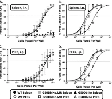
[image:10.585.82.246.65.240.2] [image:10.585.319.524.66.335.2]latency in the spleen, and determined both the frequency of splenocytes harboring the viral genome and the frequency of splenocytes able to reactivate from latency. Splenocyte reactiva-tion was determined by the previously described limiting-dilureactiva-tion CPE assay (20) modified by plating splenocytes on IFN-␣/R⫺/⫺
MEF monolayers to ensure the ability to detect any reactivated G50DblKo mutant virus. The reactivation analyses revealed the nearly complete absence of detectable reactivation of the G50DblKo virus from splenocytes (Fig. 10A). In contrast, spleno-cytes harvested from wild-type virus-infected animals reactivated at a frequency of 1 in 7,035 cells, while the frequency of reactiva-tion from the G50DblKo.MR virus was 1 in 9,549 cells (Fig. 10A andTable 1). No preformed infectious virus was detected in the harvested splenocyte samples, as measured by mechanical disrup-tion of the cells as previously described (20) (data not shown).
To determine if this failure to reactivate from splenocytes ob-served with the G50DblKo mutant virus was the result of a failure to establish latency, limiting-dilution PCR was performed to de-termine the frequency of splenocytes harboring viral genomes
(20). We observed that the frequency of splenocytes harboring latent virus was roughly equivalent for the wild-type virus and the G50DblKo.MR virus at 1 in 223 and 1 in 305 cells, respectively, while the G50DblKo mutant virus was present in splenocytes at a frequency of ca. 1 in 1,941 cells (Fig. 10BandTable 1). While the frequency of viral genome-positive splenocytes in the G50DblKo virus-infected animals was around 8.5-fold lower than that in the wild-type virus-infected animals, these analyses demonstrate that this mutant virus is able to get to the spleen and establish latency. Thus, the lack of detectable reactivation of the G50DblKo mutant reflects a significant defect in virus reactivation from B cells, the dominant latently infected cell population in the spleen (19,35). We extended these analyses to address whether the inability of the G50DblKo mutant virus to reactivate from splenocytes was dependent on the route of inoculation and to assess latency and reactivation from PECs. To do this we assessed virus reactivation from splenocytes and PECs following intraperitoneal (i.p.) inoc-ulation. C57BL/6 mice were infected by intraperitoneal inocula-tion with 1,000 PFU of G50DblKo, wild-type, or G50DblKo.MR
FIG 10G50DblKo virus exhibits a severe reactivation defect and a moderate latency defectin vivo. Female C57BL/6 mice were infected with 1,000 PFU i.n. (A and B) or with 1,000 PFU i.p. (C and D). Splenocytes and PECs were harvested at 18 days postinfection and assessed for the establishment of latency and reactivation from latency by limiting-dilution CPE and PCR assays. (A) Splenocytes from i.n. infections were plated in serial dilutions onto an IFN-␣/R⫺/⫺MEF monolayer, and at 21 days postplating wells were individually scored for CPE. The percentage of these wells was used to calculate the frequency of virally reactivating cells. (B) Splenocytes from i.n. infections were plated in serial dilutions and subjected to nested PCR to detect gene 50 copies. The percentage of genome-positive cells in each dilution was used to calculate the frequency of latency. (C) PECs from i.p. infections were plated in serial dilutions onto an IFN-␣/R⫺/⫺MEF monolayer, and 21 days postplating wells were individually scored for CPE. The percentage of these wells was used to calculate the frequency of virally reactivating cells. (D) PECs from i.p. infections were plated in serial dilutions and subjected to nested PCR to detect gene 50 copies. The percentage of genome-positive cells in each dilution was used to calculate the frequency of latency. For all reactivation assays (A and C), mechanically disrupted cells were plated in parallel for each virus shown to control for preformed infectious virus, and all mechanically disrupted cells were negative for reactivation (data not shown). Data are representative of at least two independent experiments consisting of 5 mice per group. Error bars indicate the standard error of the mean.
on November 7, 2019 by guest
http://jvi.asm.org/
[image:11.585.114.473.67.383.2]virus, and splenocytes and PECs were harvested at 18 days postin-fection. We first examined the ability of the virus to reactivate from splenocytes, as described above. As observed following in-tranasal inoculation, the G50DblKo mutant exhibited a large de-fect in reactivation, although a low but significant level of virus reactivation was detectable (it was estimated by extrapolation that the G50DblKo mutant reactivates at a frequency of 1 in 422,863 splenocytes) (Table 1). In these analyses, wild-type virus reacti-vated at a frequency of 1 in 36,107 splenocytes, while the G50DblKo.MR virus reactivated at a frequency of 1 in 65,962 splenocytes (Table 1). Notably, the frequency of splenocytes reac-tivating virus at day 18 following intraperitoneal inoculation was 5- to 7-fold lower than that observed at the same time point fol-lowing intranasal inoculation. This likely reflects faster kinetics of establishment and contraction of splenic latency following intra-peritoneal inoculation. This effect, coupled with the observed low-level reactivation of the G50DblKo mutant, provides evi-dence that the route of inoculation does have an impact on reac-tivation of this mutant virus, which could be linked to better es-tablishment of B cell latency (see discussion below). We then assessed whether the observed reactivation defects correlated with a defect in the establishment of latency. Notably, the 8.5-fold de-fect in establishment of latency in splenocytes observed following intranasal inoculation was largely rescued following intraperito-neal inoculation (Table 1). The frequency of viral genome-posi-tive cells in the splenocytes following intraperitoneal inoculation was 1 in 388 cells for wild-type MHV68, 1 in 1,047 cells for the G50DblKo.MR virus, and 1 in 627 cells for the G50DblKo mutant (Table 1).
While nearly all virus reactivation from splenocytes arises from latently infected B cells (21,36), macrophages represent the major latently infected cell type in PECs. Thus, to address whether the observed defect in G50DblKo reactivation from splenocytes re-flects a B cell-specific defect, we determined the frequency of PECs reactivating virus. In contrast to the previous observation that the G50pKo virus retained the ability to reactivate from PECs (16), G50DblKo exhibited no detectable reactivation from PECs (Fig. 10CandTable 1). As expected, both wild-type MHV68 and the G50DblKo.MR virus reactivated from PECs, at fre-quencies of 1 in 17,782 cells and 1 in 14,417 cells, respectively (Fig. 10CandTable 1). These results indicate that despite the ability to drive viral replication, the absence of both the prox-imal and distal gene 50 promoters results in a substantial im-pairment in the ability of MHV68 to reactivate from infected
cells in vivo. Despite rescue of establishment of latency in splenocytes following intraperitoneal inoculation, a ca. 6-fold defect in the establishment of latency in PECs was observed (the frequency of viral genome-positive PECs in wild-type-infected animals was 1 in 401 cells, in G50DblKo.MR-infected animals it was 1 in 562 cells, and in G50DblKo-infected animals it was ca. 1 in 2,137 cells) (Fig. 10DandTable 1). Despite the modest impair-ment of the G50DblKo mutant in the establishimpair-ment of latency in PECs, it is clear that this mutant is significantly impaired in reac-tivation from latency from both splenic B cells and peritoneal macrophages.
The G50DblKo phenotypein vivois partially rescued in mice
lacking the IFN-␣/ receptor. Since we have shown that the
G50DblKo mutant virus retains the ability to grow with normal kinetics and to high titer in cells that lack a type I IFN response (Fig. 4Band5B), we assessed whether the absence of a type I IFN responsein vivowould rescue the G50DblKo replication and re-activation defects. Thus, we infected 129S2/SvPas.IFN-␣/R⫺/⫺
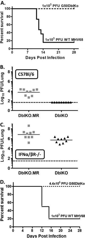
mice with 1,000 PFU of either the G50DblKo mutant or wild-type MHV68 via intranasal inoculation. It has previously been shown that MHV68 infection of mice lacking the IFN-␣/receptor re-sults in the majority of mice succumbing during the acute phase of infection (26–28,37). Surprisingly, while the G50DblKo mutant replicated to high titers in the lungs of IFN-␣/R⫺/⫺mice (Fig.
11C), all infected mice survived (Fig. 11A). However, as expected, only 3 of 19 mice infected with 1,000 PFU wild-type MHV68 WT virus survived past 2 weeks postinfection (Fig. 11A). Parallel anal-yses in C57BL/6 mice confirmed the severe replication defect ob-served with the G50DblKo mutant (virus replication in the lungs was below the limit of detection in all infected mice). Notably, acute replication of the G50DblKo mutant in the lungs of IFN-␣/ R⫺/⫺mice was rescued to near-wild-type MHV68 levels (Fig.
11C). To further address the absence of lethality in IFN-␣/R⫺/⫺
[image:12.585.39.551.77.200.2]mice with the G50DblKo mutant, we extended these studies using higher inoculating doses. We did not observe any lethality, even when mice were infected with a ca. 400-fold-higher dose of the G50DblKo mutant (5 of 5 mice infected survived for greater than 4 weeks postinfection) (Fig. 11D). Thus, while acute virus replica-tion of the G50DblKo mutant in the lungs following intranasal inoculation was largely rescued by loss of a type I IFN response, MHV68-associated lethality was not observed, indicating a re-quirement for the gene 50 proximal and/or distal promoter for RTA expression in some anatomical site during the acute phase of virus infection.
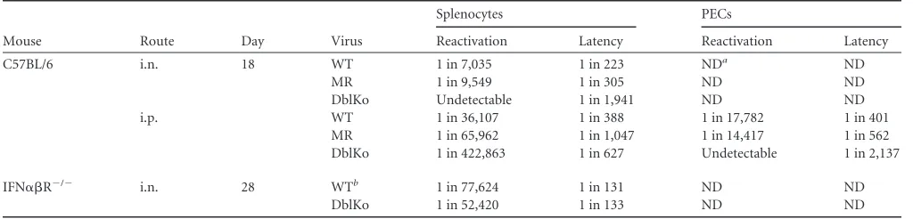
TABLE 1Summary of virus latency and reactivation
Mouse Route Day Virus
Splenocytes PECs
Reactivation Latency Reactivation Latency
C57BL/6 i.n. 18 WT 1 in 7,035 1 in 223 NDa ND
MR 1 in 9,549 1 in 305 ND ND
DblKo Undetectable 1 in 1,941 ND ND
i.p. WT 1 in 36,107 1 in 388 1 in 17,782 1 in 401
MR 1 in 65,962 1 in 1,047 1 in 14,417 1 in 562
DblKo 1 in 422,863 1 in 627 Undetectable 1 in 2,137
IFN␣R⫺/⫺ i.n. 28 WTb 1 in 77,624 1 in 131 ND ND
DblKo 1 in 52,420 1 in 133 ND ND
a
ND, not determined.
bLatency and reactivation were determined by pooling splenocytes from the 3 IFN␣R⫺/⫺mice that survived wild-type MHV68 infection.
on November 7, 2019 by guest
http://jvi.asm.org/
Having shown that the severe defect in acute replication of the G50DblKo mutant observed in C57BL/6 mice was largely rescued in mice lacking the ability to respond to type I IFNs, we assessed establishment of latency and virus reactivation in IFN-␣/R⫺/⫺
mice. Spleens were harvested at day 28 from IFN-␣/R⫺/⫺mice
intranasally infected with 1,000 PFU of either wild-type or
G50DblKo virus. It is important to note that the latency and reac-tivation analyses for wild-type virus-infected IFN-␣/R⫺/⫺mice
were carried out using the subset of infected animals that survived acute virus replication (Fig. 10A). Like the rescue of acute virus replication in the lungs, the frequency of viral genome-positive splenocytes went from a ca. 8-fold defect in i.n. infected C57BL/6 mice (Fig. 10B) to an almost identical frequency of virus-infected splenocytes in IFN-␣/R⫺/⫺mice (1 in 131 cells for wild-type
MHV68 and 1 in 133 for the G50DblKo mutant) (Fig. 12Aand Table 1). More importantly, the defect in virus reactivation ob-served in C57BL/6 mice was completely rescued in IFN-␣/R⫺/⫺
mice (1 in 77,624 splenocytes in WT-infected mice compared to 1 in 52,420 splenocytes in G50DblKo virus-infected mice) (Fig. 12B andTable 1). The latter result argues in favor of a model in which the sole defect in G50DblKo virus reactivation from C57BL/6 splenocytes is the inability to overcome type I IFN suppression of virus replication.
One possible explanation for the ability of the G50DblKo virus to replicate in the absence of type I interferons would be an in-creased expression of E1-E2 transcripts. This would make the G50DblKo virus not a true double promoter knockout virus, as E1-E2 transcripts would be responsible for the ability of the virus to replicate. However, this does not appear to be the case because (i) as shown inFig. 6, RTA expression from the G50DblKo in
FIG 11Infection of 129S2/SvPas.IFN-␣/R⫺/⫺mice with G50DblKo virus does not result in lethality as seen with WT virus, despite the presence of high viral titers in the lungs. (A) Kaplan-Meier curve depicting survival of 129S2/ SvPas.IFN-␣/R⫺/⫺mice when challenged with 1,000 PFU i.n. of WT or G50DblKo virus. (B) Lung titers at day 7 from C57BL/6 mice infected with 1,000 PFU i.n. of G50DblKo or G50DblKo.MR virus. (C) Lung titers at day 7 from 129S2/SvPas.IFN-␣/R⫺/⫺ mice infected with 1,000 PFU i.n. of G50DblKo or G50DblKo.MR virus. (D) Kaplan-Meier curve depicting sur-vival of 129S2/SvPas.IFN-␣/R⫺/⫺mice when challenged with 440,000 PFU i.n. of G50DblKo virus compared to survival of mice when challenged with 1,000 PFU G50DblKo.MR virus.
FIG 12G50DblKo virus establishes latency and reactivates to similar levels as WT virus when used to infect 129S2/SvPas.IFN-␣/R⫺/⫺micein vivo. Female 129S2/SvPas.IFN-␣/R⫺/⫺mice were infected with 1,000 PFU of G50DblKo virus, WT virus, or G50DblKo.MR virus by intranasal injection. Mice surviv-ing at day 28 postinfection (n ⫽ 20 for G50DblKo; n ⫽ 3 for WT/ G50DblKo.MR) were harvested for splenocytes and assessed for establishment of latency and reactivation from latency. (A) As forFig. 10A, splenocytes were serially diluted and used in a CPE assay to look for the frequency of viral reactivating cells. (B) As forFig. 10B, splenocytes were serially diluted and used in a nested PCR assay to look for the frequency of viral genome-positive cells.