Cite this:Mol. BioSyst.,2016,
12, 3032
Biological processing of dinuclear ruthenium
complexes in eukaryotic cells
Xin Li,aKirsten Heimann,*bcXuyen Thi Dinh,dF. Richard Keene*cefand J. Grant Collins*a
The biological processing – mechanism of cellular uptake, effects on the cytoplasmic and mitochondrial membranes, intracellular sites of localisation and induction of reactive oxygen species – of two dinuclear polypyridylruthenium(II) complexes has been examined in three eukaryotic cells lines. Flow cytometry was used to determine the uptake of [{Ru(phen)2}2{m-bb12}]4+(Rubb12) and [Ru(phen)2(m-bb7)Ru(tpy)Cl]3+ {Rubb7-Cl, where phen = 1,10-phenanthroline, tpy = 2,20:60,200-terpyridine and bb
n = bis[4(40
-methyl-2,20-bipyridyl)]-1,n-alkane} in baby hamster kidney (BHK), human embryonic kidney (HEK-293) and liver carcinoma (HepG2) cell lines. The results demonstrated that the major uptake mechanism for Rubb12 and Rubb7-Cl was active transport, although with a significant contribution from carrier-assisted diffusion for Rubb12 and passive diffusion for Rubb7-Cl. Flow cytometry coupled with Annexin V/TO-PRO-3 double-staining was used to compare cell death by membrane damage or apoptosis. Rubb12 induced significant direct membrane damage, particularly with HepG2 cells, while Rubb7-Cl caused considerably less membrane damage but induced greater levels of apoptosis. Confocal microscopy, coupled with JC-1 assays, demonstrated that Rubb12depolarises the mitochondrial membrane, whereas Rubb7-Cl had a much smaller affect. Cellular localisation experiments indicated that Rubb12did not accumulate in the mitochondria, whereas significant mitochondrial accumulation was observed for Rubb7-Cl. The effect of Rubb12 and Rubb7-Cl on intracellular superoxide dismutase activity showed that the ruthenium complexes could induce cell deathviaa reactive oxygen species-mediated pathway. The results of this study demonstrate that Rubb12 predominantly kills eukaryotic cells by damaging the cytoplasmic membrane. As this dinuclear ruthenium complex has been previously shown to exhibit greater toxicity towards bacteria than eukaryotic cells, the results of the present study suggest that metal-based cationic oligomers can achieve selective toxicity against bacteria, despite exhibiting a non-specific membrane damage mechanism of action.
Introduction
There is considerable interest in developing ruthenium complexes as therapeutic agents, and a variety of coordinatively-labile mono-nuclear ruthenium(II) and ruthenium(III) complexes have shown
significant promise as anticancer drugs.1–6Furthermore, a range
of inert mononuclear ruthenium(II) complexes have shown
pro-mising in vitro anticancer and/or antimicrobial properties.7–15 More recently, there has been increasing interest in ‘‘tuning’’ the structure of the ruthenium complexes to increase their activity and selectivity for bacterial or cancer cells. One such strategy, the synthesis of cationic oligomers,16–19 takes advantage of the greater presence of negatively-charged components in the bacterial membrane compared to eukaryotic cells.20,21 The cell wall and outer membrane of bacteria contain a higher proportion of negatively-charged phospholipids –e.g. phosphatidyl-glycerol, as well as a variety of negatively-charged macro-molecules such as teichoic acids and lipopolysaccharides – than do eukaryotic cells.20,21
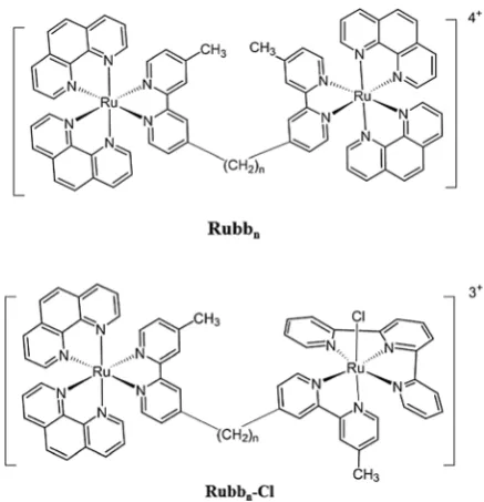
We have synthesised and examined the antibacterial activities of a series of dinuclear polypyridylruthenium(II) complexes
[{Ru(phen)2}2{m-bbn}]4+(Rubbn, see Fig. 1) where (phen = 1,10-phenanthroline and bbn = bis[4(40-methyl-2,20-bipyridyl)]-1,n -alkane).22–25The Rubbn complexes with a short linking chain
aSchool of Physical, Environmental and Mathematical Sciences,
University of New South Wales, Australian Defence Force Academy, Canberra, ACT 2600, Australia. E-mail: [email protected]
b
College of Science & Engineering, James Cook University, Townsville, QLD 4811, Australia. E-mail: [email protected]
cCentre for Biodiscovery and Molecular Development of Therapeutics,
James Cook University, Townsville, QLD 4811, Australia
dComparative Genomics Centre, James Cook University, Townsville,
QLD 4811, Australia
eCollege of Science, Technology & Engineering, James Cook University, Townsville,
QLD 4811, Australia
fSchool of Physical Sciences, University of Adelaide, Adelaide, SA 5005, Australia.
E-mail: [email protected]
Received 31st May 2016, Accepted 19th July 2016
DOI: 10.1039/c6mb00431h
www.rsc.org/molecularbiosystems
Molecular
BioSystems
PAPER
Open Access Article. Published on 25 July 2016. Downloaded on 04/04/2017 00:47:07.
This article is licensed under a
Creative Commons Attribution 3.0 Unported Licence.
(bb2, bb5 and bb7) showed little or no antibacterial activity,
whereas Rubb12showed excellent activity and was 40–100 fold
less toxic towards eukaryotic cells.22,25However, it was noted that Rubb12 showed very similar 24-hour IC50 values towards
three significantly different eukaryotic cell lines – a baby hamster kidney (BHK), a human embryonic kidney (HEK-293) and a human liver carcinoma (HepG2).25The similar cytotoxi-city data between the eukaryotic cell lines suggest a common, non-specific, mechanism of action. Previous studies with bacteria demonstrated that although Rubb12 preferentially localises at
ribosomes within a bacterial cell,26it also significantly permea-bilises the cytoplasmic membrane.27 Non-specific membrane
damage would likely result in the ruthenium complex exhibiting very similar IC50 values against the three eukaryotic cell lines.
However, a previous study suggested that the Rubbn complexes enter L1210 murine leukaemia cells largely by passive diffusion, accumulate in the mitochondria and induce cell death by apoptosis.28 By contrast, the results of a confocal microscopy study with the BHK, HEK-293 and HepG2 cell lines demon-strated that Rubb12 did not accumulate in the mitochondria,
but predominantly localised in the nucleolus of the cell.25 Taken together, the combined results suggest that the bio-logical processing of the Rubbncomplexes in white blood cells (L1210) is significantly different to that in kidney and liver cells (BHK, HEK-293 and HepG2). In order to gain a deeper under-standing of the effect of the Rubbn complexes on organ cells, and in particular the organs responsible for processing drugs, it was of interest to determine the cellular uptake mechanism and mechanism of cell death of the Rubb12 complex in the
BHK, HEK-293 and HepG2 cells.
Although this study was aimed at understanding the bio-logical processing of the Rubbncomplexes in eukaryotic cells, it also provides important information for the development of cationic antimicrobial or anticancer oligomers in general.
As previously noted, there is considerable current interest in the use of cationic oligomers/polymers for combating bacterial resistance to current drugs.16,19However, because many of these
potential drugs disrupt the bacterial membrane, their clinical application could be limited as they would also interact with eukaryotic membranes in a similar manner. As alkyl chains are commonly used to link the monomeric units of antimicrobial (and anticancer) cationic oligomers/polymers,16–19the results of this study could well be applicable to a wider range of drugs than the Rubbncomplexes.
In addition to the Rubbn complexes, we have also synthe-sised and examined the biological activities of a series of dinuclear complexes that contain one inert metal centre and one coordinatively-labile metal centre (the Rubbn-Cl complexes, see Fig. 1).29 Interestingly, while each of the Rubb12-Cl and
Rubb16-Cl complexes showed similar toxicities across the BHK,
HEK-293 and HepG2 cell lines, Rubb7-Cl showed surprising
differences in cytotoxicities.29Against the BHK cells, Rubb7-Cl
was by far the least toxic of the Rubbn-Cl and Rubbncomplexes (24 h-IC50 = 238mM); whereas by contrast, against the HepG2
cell line it was by far the most toxic (24 h-IC50= 3.7mM).29The
significantly different response exhibited by the BHK, HEK-293 and HepG2 cells to Rubb7-Cl suggests that the ruthenium
complex does not act in a non-specific manner (e.g.membrane damage), but through a specific pathway that leads to apop-tosis. Consequently, the cellular uptake mechanism and mecha-nism of cell death for Rubb7-Cl against the BHK, HEK-293 and
HepG2 cells was also examined and compared to that obtained for Rubb12.
Results
Cellular uptake mechanism of the ruthenium complexes
Cellular uptake studies {described in (a)–(d) below} were carried out using flow cytometry for the Rubb12 and Rubb7-Cl
com-plexes in the kidney and liver cell lines. These studies aimed to differentiate between three major uptake pathways: (1) active transport, which is energy-dependent; (2) passive diffusion, an energy-independent form of molecular transport; and (3) carrier-assisted diffusion, an energy-independent pathway, facilitated by membrane protein carriers such as organic cation trans-porters (OCT).
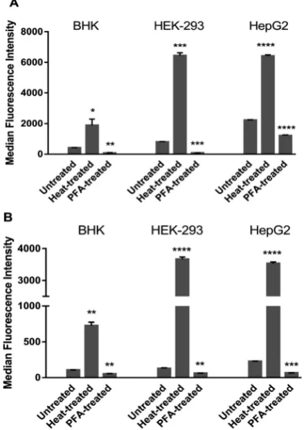
(a) Effect of cell membrane permeability on uptake of Rubb12and Rubb7-Cl.To differentiate between passive uptake and active transport/carrier-facilitated diffusion, the cellular uptake of Rubb12and Rubb7-Cl were initially examined in fully
permeabilised (heat treated, representing the maximum cellular uptake possible) and PFA-fixed (active transport/carrier-impaired) cells (Fig. 2). PFA-fixation causes cross-linking of primary amino groups in proteins that results in protein related uptake of the ruthenium complex being blocked. Consequently, the cellular uptake of the ruthenium complexes after heat treatment or PFA-fixation provides useful comparative data to that obtained under control conditions (371C in the absence of metabolic
inhibitors or activators). Fig. 1 Structure of the Rubbnand Rubbn-Cl complexes (n= 7, 12 and 16).
Open Access Article. Published on 25 July 2016. Downloaded on 04/04/2017 00:47:07.
This article is licensed under a
[image:2.595.57.276.47.274.2]The Rubb12and Rubb7-Cl complexes have different
lumines-cence signal strengths. Therefore, the median fluoreslumines-cence inten-sity (MFI) values cannot be used to compare cellular uptake of different complexes, but only for analysing changes in the uptake of one complex under different conditions in different cells.
Both complexes accumulated significantly more in heat-treated cells than in untreated and PFA-fixed cells irrespective of the cell line. In control and PFA-fixed samples, the uptake generally followed HepG2ZHEK-2934BHK for both complexes. However, in heat-treated cells, MFIs in HEK-293 cells were slightly higher than HepG2, and the MFIs in both human cell lines were more than 3-fold higher than for the baby hamster kidney BHK cells. After PFA-fixation, the cellular uptake of Rubb12and Rubb7-Cl
was strongly impeded by 50–90% compared with the untreated controls in the three cell lines.
(b) Temperature-dependent cellular uptake of Rubb12and Rubb7-Cl.To differentiate between temperature-dependent and -independent transport routes, the cellular uptake of Rubb12
and Rubb7-Cl were investigated at 4, 24 and 371C by flow
cyto-metry in the three cell lines. Relative MFI values (percentage of MFI at each temperatureversusMFI at 371C) are summarised in
Fig. 3. Uptake increased with temperature for both ruthenium complexes in the three cell lines. At 41C, almost no uptake was
observed in BHK and HEK-293 cells, with a slightly higher
uptake in HepG2. At 24 1C, the uptake of Rubb12 increased
sharply in BHK cells, as it did for Rubb7-Cl in HEK-293, and
moderately increased (40–50%) in the other cell lines. These data suggest a larger contribution to uptake of Rubb12by an
energy-independent process in BHK, but predominantly an active uptake mechanism in HEK-293. However, the opposite was observed for Rubb7-Cl, where a larger contribution to uptake by an
energy-independent process was seen in HEK-293 cells. By contrast, the uptake of both ruthenium complexes in HepG2 cells appeared to be predominantly by an active process.
(c) Dependence of cellular uptake of Rubb12and Rubb7-Cl on ATP production. Flow cytometry experiments were con-ducted to confirm the ATP requirement for the cellular uptake of the ruthenium complexes (see Fig. 4). Cells were pretreated with the glycolysis inhibitor 2-deoxy-glucose30 and the mito-chondria inhibitor oligomycin to inhibit ATP production,31and withD-glucose to stimulate ATP production.
The ATP-production stimulator D-glucose did not enhance
uptake of the ruthenium complexes in BHK and HEK-293 cells, with uptake of Rubb12in BHK even slightly lower. This suggested
that the cellular ATP quota was optimal, preventing further accu-mulation by the treatment. However, for HepG2 cells,D-glucose treatment did induce slightly greater accumulation for Rubb12and
Rubb7-Cl. The inhibition of ATP production caused a significant
decrease in the uptake of both ruthenium complexes in all three cell lines, indicating that uptake was predominantly through an energy-dependent (active) process.
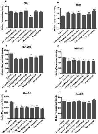
(d) Effect of organic cation transport (OCT) inhibitors on the uptake of Rubb12and Rubb7-Cl.Organisms use polyspecific organic cation (OCT) transporters for facilitating the diffusion of endogenous organic cations, cationic drugs and toxins across the cell membrane.32 The possibility of energy-independent Fig. 3 Relative cellular uptake (MFIs at each temperature to MFIs at 371C) of (A) Rubb12and (B) Rubb7-Cl at 4, 24 and 371C in BHK, HEK-293 and HepG2 cells after a 4-hour incubation. MFIs: medium fluorescence intensities.
Fig. 2 Cellular uptake as median fluorescence intensity (MFI) values of 20mM ruthenium complexes (A, Rubb12and B, Rubb7-Cl) after a 4-hour incubation at 371C in untreated, heat-treated and PFA-fixed cells for the three cell lines BHK, HEK-293 and HepG2.
Molecular BioSystems Paper
Open Access Article. Published on 25 July 2016. Downloaded on 04/04/2017 00:47:07.
This article is licensed under a
Creative Commons Attribution 3.0 Unported Licence.
[image:3.595.57.277.52.363.2] [image:3.595.309.546.54.277.2]carrier-assisted diffusion via OCTs was investigated by flow cytometry (see Fig. 5). To determine if the ruthenium complexes were transported by OCTs, cells were pre-treated with tetraalkyl-ammonium salts or procainamide to inhibit the function of organic cation transporters then incubated with the ruthenium complexes. In BHK cells, uptake of Rubb12was inhibited slightly but
signifi-cantly by the tetra-n-butylammonium, tetramethylammonium and tetraethylammonium salts, suggesting OCT1, OCT2 and OCTN1 could be involved in transportation. Interestingly, uptake appeared to be stimulated for Rubb7-Cl, suggesting that these OCTs were not
involved in the uptake of Rubb7-Cl. A significant decrease in uptake
of both ruthenium complexes was observed in OCT-inhibited HEK-293 cells. This observation suggests that HEK-293 cells can transport both ruthenium complexes by OCT-facilitated diffusion as an additional pathway to active transport mechanism. Rubb12
uptake was significantly lower (by 32–42%) in the OCT-inhibited HepG2 cells, suggesting a significant contribution of OCTs in the transportation process. By contrast, OCTs do not appear to be involved in the uptake of Rubb7-Cl in HepG2 cells.
Detection of apoptosis and membrane damage
Annexin V/TO-PRO-3 double-staining assays were carried out with BHK and HepG2 cells to compare cell death by membrane
damage or apoptosis for Rubb12and Rubb7-Cl. Early stages of
apoptosis are characterised by the translocation of phosphati-dylserine from the inner leaflet of the plasma membrane to the outer leaflet, which can be visualised by binding to the Annexin V fluorescent conjugate.33As cell death progresses, TO-PRO-3
(a cell-impermeant dye) diffuses into the cells with compromised membrane integrity and binds to DNA.33 Alternatively, if the cytoplasmic membrane is directly damaged by the ruthenium complex, TO-PRO-3 will enter the cell and bind to DNA without any translocation of phosphatidylserine from the inner leaflet of the plasma membrane to the outer leaflet. Accordingly, in the flow cytometry assays, apoptosis is demonstrated by cells ‘‘moving’’ from Q4 (bottom-left quadrant) to Q3 (bottom-right) and then finally to Q2 (top-right). On the other hand, direct membrane damage is detected by cells moving from Q4 to Q1 (top-left), and then to Q2 as the cell becomes totally permeabilised (allowing the large Annexin V fluorescent conjugate into the cell) and then possibly to Q3 as TO-PRO-3 is displaced from the DNA by the ruthenium complex.
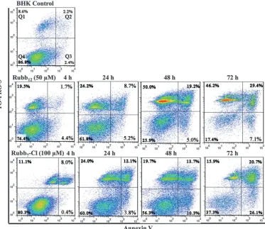
Time-course (4-, 24-, 48- and 72-hours) flow cytometry was conducted using BHK cells with Rubb12and Rubb7-Cl at half of
their 24 h-IC50 concentrations (50 and 100 mM, respectively).
A small percentage of the BHK cells (5–10%) were always damaged (observed in Q1) during the trypsin treatment required for cell harvesting; however, little membrane damage was observed for HepG2 cells. After incubation with Rubb12, viable BHK cells
(bottom-left quadrant, Q4) decreased gradually with incubation time (see Fig. 6). Noticeably, there was a sharp increase in the top-left quadrant, indicating that half the cells had significant membrane damage, while the early apoptotic cells (bottom-right quadrant, Q3) only increased slowly and to a lesser extent. BHK cells treated with Rubb7-Cl showed a different cell
popula-tion distribupopula-tion to that observed with Rubb12. The percentage
of cells that exhibited direct membrane damage was consider-ably smaller (Q1), but the percentage of cells exhibiting apop-tosis (Q2 and Q3) was significantly higher than was observed with Rubb12.
Time-course Annexin V/TO-PRO-3 double-staining assays were also carried out with HepG2 cells treated with either Rubb12or
Rubb7-Cl (see Fig. 7). For Rubb12, extensive membrane damage
was observed after a 4-hour incubation, with over 50% of the cell population observed in Q1 after 24 hours. Interestingly, after a 72-hour incubation a significant percentage of the cells were Annexin positive and TO-PRO-3 negative (Q3). Given that most of the cells were non-viable due to membrane damage at 4 and 24 hours, the significant percentage of cells in Q3 after a 72-hour incubation suggests the DNA was saturated with Rubb12, rather
being indicative of early apoptosis. Saturation with Rubb12would
inhibit TO-PRO-3 from binding to DNA, leading to the observed low levels of TO-PRO-3 fluorescence.
HepG2 cells treated with 10mM Rubb7-Cl appeared to induce
relatively weak responses, with few cells exhibiting significant membrane damage (Q1) and even fewer cells being early-apoptotic (Q3). To further amplify cell responses, the assays were repeated with a Rubb7-Cl concentration of 20 mM. The increased
concen-tration of Rubb7-Cl did slightly enhance the responses, with the
Fig. 4 Cellular uptake (MFIs) of (A), Rubb12and (B), Rubb7-Cl (4-hour incuba-tion) in ATP-inhibitor-treated, glucose-treated and untreated BHK, HEK-293 and HepG2 cells. MFI: medium fluorescence intensity.
Open Access Article. Published on 25 July 2016. Downloaded on 04/04/2017 00:47:07.
This article is licensed under a
[image:4.595.75.258.51.388.2]percentage of cells exhibiting direct membrane damage doubling (Q1); however there was only a very slight increase in the number of early apoptotic cells (Q3). As the 24 h-IC50is
3.7mM, the flow cytometry results suggest that Rubb7-Cl is not
cytotoxic against HepG2 cells due to direct membrane damage or any process leading to apoptosis.
Mitochondrial membrane potential
Mitochondrial dysfunction plays an important role in different cell death mechanisms, and the depolarisation of the mito-chondrial membrane potential (MMP) due to changes in the mitochondrial permeability has been demonstrated to be an early stage of mitochondrial dysfunction.34 To evaluate the disruption of mitochondrial function induced by Rubb12 and
Rubb7-Cl, the change in the mitochondrial membrane potential
was determined by confocal microscopy using JC-1 assays.35 Functional polarised mitochondria have a high MMP promoting
the formation of red fluorescent JC-1 aggregates, while depolarised mitochondria with low MMP show a diffuse green fluorescence from JC-1 monomers.
BHK cells incubated with 50 mM of Rubb12 and Rubb7-Cl
showed different changing patterns in MMP (Fig. 8). After a 4-hour incubation, Rubb12treatment affected the distribution
of mitochondria in BHK cells, with low MMP mitochondria spreading throughout the cytoplasm. Mitochondria with high MMP appeared to be confined to the cell periphery. Moreover, some polarised mitochondria appeared elongated. By contrast, in Rubb7-Cl treated BHK cells very few low MMP mitochondria
were observed, with the high MMP mitochondria red fluores-cence being maintained.
HepG2 cells incubated with 20mM of Rubb12and Rubb7-Cl
displayed some differences in the staining pattern (Fig. 9). Treatment with Rubb12 led to the localisation of depolarised
mitochondria in the perinuclear region. The presence of several Fig. 5 Effect of OCT inhibition on Rubb12(A–C) and Rubb7-Cl (D–F) uptake in BHK, HEK-293 and HepG2 cells after a 4-hour incubation.
Molecular BioSystems Paper
Open Access Article. Published on 25 July 2016. Downloaded on 04/04/2017 00:47:07.
This article is licensed under a
Creative Commons Attribution 3.0 Unported Licence.
[image:5.595.138.457.47.482.2]condensed green hotspots could be indicative of condensed mitochondria with low MMP in response to Rubb12treatment.
On the other hand, mitochondria with high MMP displayed condensed red fluorescence. After incubation with Rubb7-Cl at
the concentration of 20mM, HepG2 cells displayed only slightly decreased red fluorescence of high MMP mitochondria, with only a few low MMP mitochondria observed in the perinuclear region. Additionally, time-course MMP changes (0-, 4- and 20-hour incubations) were monitored in BHK cells with Rubb12 and
Rubb7-Cl. As expected, longer incubation time led to increased
depolarisation of mitochondria (data not shown).
Cellular localisation
As the JC-1 assays demonstrated that Rubb12 and Rubb7-Cl
affected the mitochondrial membrane potential, it was of interest to determine if the ruthenium complexes localised to any extent in the mitochondria. The cellular localisation of high and low concentrations (relative to 24 h-IC50 values) of Rubb12
and Rubb7-Cl in BHK cells has been previously reported.29
Both Rubb12 and Rubb7-Cl preferentially accumulated in the
nucleolus at low concentrations, but exhibited significant DNA binding at higher concentrations. Furthermore, mitochondrial localisation was observed for Rubb7-Cl at high concentration,
but not for Rubb12.29The localisation of both ruthenium
com-plexes at low concentrations (5 mM) in HepG2 cells has also been reported.29However, the localisation of higher concentra-tions of Rubb12 and Rubb7-Cl in HepG2 cells has not been
reported. Although considerable Rubb12phosphorescence was
observed in the cytoplasm of HepG2 cells, no co-localisation was observed in the mitochondria (see Fig. 10). Co-localisation experiments with ERtracker indicated that a significant propor-tion of the cytoplasmic Rubb12 phosphorescence was due to
accumulation in the ER (Fig. 10). By contrast, Rubb7-Cl showed
intense co-localisation with mitochondria (yellow overlay), but little in the ER (Fig. 10).
Reactive oxygen superoxide dismutase assays
The primary superoxide scavenger, superoxide dismutase (SOD), is an attractive target for pharmacological intervention.36–38To
investigate whether the treatment with ruthenium complexes affected intracellular SOD activity, SOD assays were conducted on BHK, HEK-293 and HepG2 cells after a 20-hour incubation with Rubb12and Rubb7-Cl (Fig. 11). Compared with the untreated
controls, BHK cells appeared to slightly increase SOD activity in response to treatment with the ruthenium complexes, with a non-significant increase for Rubb12and Rubb7-Cl being observed.
Fig. 6 Time-course Annexin V/TO-PRO-3 staining of BHK cells (control, top) and with treatment by the ruthenium complexes (middle: Rubb12and bottom: Rubb7-Cl). Cells plotted in each quadrant represent the following: left (Annexin V-negative/TO-PRO-3-negative), viable cells; bottom-right (Annexin V-positive/TO-PRO-3-negative), early apoptotic cells; top-bottom-right (Annexin V-positive/TO-PRO-3-positive), late apoptotic/necrotic cells; top-left (Annexin V-negative/TO-PRO-3-positive), membrane-damaged dead cells.
Open Access Article. Published on 25 July 2016. Downloaded on 04/04/2017 00:47:07.
This article is licensed under a
[image:6.595.111.485.50.373.2]Similarly, SOD activity in HEK-293 cells also showed a slight increase for both ruthenium complexes. In contrast, the cancer cell line HepG2 showed a reverse trend in SOD activity, with a significant down-regulation for Rubb12, but no obvious change
for Rubb7-Cl.
Discussion
Cationic oligomers, such as cationic peptides and di- and oligo-nuclear ruthenium complexes, have considerable potential as selective antibacterial agents due to their preferential inter-actions with the negatively-charged bacterial membrane and cell wall. One approach to the preparation of di- and oligo-nuclear ruthenium complexes is to link the individual metal centres
with a ligand that contains an alkyl chain. The length of the alkyl chain could affect cellular uptake, membrane damage and intracellular localisation in bacterial and eukaryotic cells. It has been previously demonstrated that the alkyl chain length controls the uptake in bacterial and L1210 cells.23,28However, for both bacterial and L1210 cells, the dominant mechanism of cellular uptake was passive diffusion.23,28The aims of this study were to determine if the Rubb12 and Rubb7-Cl complexes were
taken up by passive diffusion in organ cells, and to examine the effect of the ruthenium complexes on the cytoplasmic and mitochondrial membranes.
Heat treatment at 601C destroys membrane integrity
com-pletely39and compounds freely pass into the cells and associate with all available biological ligands. Following 601C treatment,
both Rubb12and Rubb7-Cl uptake was greater in the human cell
Fig. 7 Annexin V/TO-PRO-3 staining of HepG2 cells (control) and with treatment by the ruthenium complexes treatment (from top to bottom: 50mM Rubb12, 10mM Rubb7-Cl and 20mM Rubb7-Cl).
Molecular BioSystems Paper
Open Access Article. Published on 25 July 2016. Downloaded on 04/04/2017 00:47:07.
This article is licensed under a
Creative Commons Attribution 3.0 Unported Licence.
[image:7.595.108.491.54.489.2]Fig. 8 JC-1 fluorescence imaging of mitochondria in BHK cells after a 4-hour incubation with 50mM of Rubb12(middle) and Rubb7-Cl (bottom). Red fluorescence is the sign of high MMP, whereas the green fluorescent signal indicates mitochondrial membrane depolarisation. Scale bar = 20mm.
Fig. 9 JC-1 fluorescence imaging of mitochondria in HepG2 cells after a 4-hour incubation with 20mM of Rubb12(middle) and Rubb7-Cl (bottom). Scale bar = 20mm.
Open Access Article. Published on 25 July 2016. Downloaded on 04/04/2017 00:47:07.
This article is licensed under a
[image:8.595.141.455.47.338.2] [image:8.595.131.455.396.693.2]lines than in the baby hamster cells (HEK-293ZHepG24BHK), suggesting more intracellular binding sites in the two human cell lines than in BHK cells. As chromosomal DNA was one of the main intracellular targets, these results could be partly explained by the different chromosome numbers in HEK-293, HepG2 and BHK cells – with 64, 55 and 44 chromosomes, respectively.40 PFA-fixation causes cross-linking of membrane proteins (including protein carriers),41 leading to diminished uptake, with
com-pounds predominantly entering cellsviapassive diffusion. The observed large loss of Rubb12and Rubb7-Cl uptake in PFA-fixed
cells may suggest that the inhibited active/carrier-assisted transportation routes contribute more than 50% of the total uptake in all cases.
As carrier proteins are inactivated and ATP-production is limited at 41C,42 the small uptake of Rubb12and Rubb7-Cl at
41C compared with the greater uptake at higher temperatures
demonstrated the strong temperature-dependence of the uptake in the eukaryotic cells. This is consistent with the previously reported energy-dependent uptake of the rigidly linked dinuclear complex [{Ru(phen)2}2{m-tpphz}]4+ in MCF-7 cells.43 However,
other ruthenium complexes are transportedvia passive diffu-sion; for instance the mononuclear complex [Ru(DIP)2dppz]2+
showed much higher uptake at 4 1C.44 Due to the decreased
membrane fluidity at 41C (increased rigidity of the
phospho-lipids chains), the cell membrane would be less permeable than it would be at 371C.45,46Consequently, and particularly as the
interactions between the phospholipid alkyl chains and the alkyl chains of the bbnligand are likely to be important, it is possible that the ruthenium complexes might not be able to
enter the cells by passive diffusion at 41C. On the other hand, at
241C, ATP-dependent transport is mostly inhibited, while passive
transport and carrier-facilitated diffusion function normally.47,48 Other studies have demonstrated that strictly ATP-required uptake of peptides was reduced byB75% at 241C compared to 371C,49
and Na–K-ATPase activity is inhibited byB70% in mammalian erythrocytes at 241C.50These findings indicate thatB25–30% of
active uptake occurs at 241C. Accordingly, the actual active uptake
can be estimated. Corrected estimations suggest active transport generally appears to be the dominant cellular uptake mechanism, contributing to more than half of the total uptake. However, for Rubb12in BHK cells and Rubb7-Cl in HEK-239 cells slightly lower
contributions of active uptake (B30%) are estimated, indicating carrier-assisted diffusion could also be involved in the uptake.
To further ascertain the contribution of active uptake, the energy dependence of cellular uptake of Rubb12and Rubb7-Cl
was determined by pretreating cells with the ATP-production stimulatorD-glucose or the metabolic inhibitors 2-deoxy-glucose and oligomycin. The unchanged uptake in theD-glucose-treated
BHK and HEK-293 cells indicate an optimal energy condition, which could not be further improved. However, the increased uptake inD-glucose-treated HepG2 cells was indicative of glucose
deprivation in HepG2 cells. This may be a result of a heavy reliance on glycolysis for ATP production in cancer cells instead of mitochondrial oxidative phosphorylation,51the latter being the primary pathway to produce ATP in healthy cells.52Therefore, it is possible that the external supply of glucose enhanced energy production in HepG2 cells, consequently leading to greater active uptake of ruthenium complexes. In contrast, the significant decrease in uptake in ATP-inhibited cells for both Rubb12 and
Rubb7-Cl confirmed a significantly large contribution of active
uptake of the ruthenium complexes.
The actual contribution of active uptake could be even larger than the observed proportions, considering that ATP-production cannot be completely blocked by the metabolic inhibitors.53,54
These findings, in conjunction with the lower temperature study results, strongly support the conclusion that active uptake is the major mechanism of cell entry for the ruthenium complexes studied in kidney and liver cell lines.
Based on the 24 1C uptake and the ATP-inhibited uptake
[image:9.595.107.491.48.176.2]results, the possible role of polyspecific organic cation transporters Fig. 10 Rubb12(top) and Rubb7-Cl (20 hour incubation) localisation in HepG2 cells at 20mM, co-stained with Mitotracker Green (left) or ERtracker Green (right), where the yellow coloration arises from co-localisation of Mitotracker Green/ERtracker Green and Rubbn-Cl. Scale bar = 20mm.
Fig. 11 Effect of the ruthenium complexes on SOD activity in BHK, HEK-293 and HepG2 cells.
Molecular BioSystems Paper
Open Access Article. Published on 25 July 2016. Downloaded on 04/04/2017 00:47:07.
This article is licensed under a
Creative Commons Attribution 3.0 Unported Licence.
[image:9.595.47.287.226.322.2](OCT)-assisted diffusion of Rubb12and Rubb7-Cl was explored.
The lower uptake of Rubb12 following OCTs inhibition in the
three cell lines, supports the conclusion that the uptake of Rubb12 was partially facilitated by OCTs. In contrast, Rubb7-Cl
displayed no OCTs-facilitated diffusion in BHK and HepG2 cells, but a considerable contribution of OCTs-facilitated uptake was noted in HEK-293 cells. It is possible that the labile complex Rubb7-Cl interacts with the carrier proteins in a covalent mode,
making dissociation harder from the membrane protein, and transport by carriers like OCTs less likely. On the other hand, a correlation between OCT binding with molecules that contain both a positive charge and high lipophilicity has been observed.55 This may explain the greater OCT-assisted uptake of the more positively-charged and possibly more lipophilic Rubb12.
In summary, the major uptake mechanism for Rubb12and
Rubb7-Cl is active transport in kidney and liver cells – with a
significant contribution from carrier-assisted diffusion for Rubb12 but mainly passive diffusion for Rubb7-Cl. However,
previous studies from our research group on Rubbncomplexes demonstrated uptake by passive diffusion in L1210 leukaemia cells and bacteria cells.23,28As the function of kidney and liver
are filtering and detoxification,56uptake of molecules can be expected to be more selective and finely managed, which is only possible by active uptake.
To determine the extent of the damage to the cytoplasmic membrane, and to compare the cellular consequences of mem-brane damage with cell death through an apoptotic pathway, Annexin V/TO-PRO-3 assays were carried out. The results clearly demonstrated that Rubb12caused significant direct membrane
damage to both BHK and HepG2 cells; however, membrane damage was considerably more rapid and extensive with the HepG2 cells. By contrast, Rubb7-Cl caused significantly less
membrane damage than Rubb12 in both cell lines. Of note
however, was the relatively low level of apoptotic cells up to 48 hours for both cell lines.
As an indicator of mitochondrial health, mitochondrial membrane potential (MMP) was monitored using the JC-1 assays. The results clearly demonstrate that Rubb12depolarises
the mitochondrial membrane in both BHK and HepG2 cells; whereas, Rubb7-Cl had a much smaller affect. The cellular
localisa-tion experiments indicated that although Rubb12did not
accumu-late in the mitochondria in either cell line, significant mitochondrial accumulation was observed for Rubb7-Cl in both BHK and HepG2
lines. While DNA damage and ER stress beyond a certain repair-threshold could reduce MMP,55,57,58the combined results (lack of localisation and reduction in MMP) are consistent with Rubb12
damaging the mitochondrial membrane. Given the previously-reported membrane damage induced by Rubbn in bacterial cells,27 the lipophilic ruthenium complexes could lead to
depolarisation of membranes in eukaryotic cells, including the cell membrane and intracellular membrane compartments (e.g.ER, mitochondria, Golgi, endosome and lysosome), result-ing in irreparable membrane damage.
Reactive oxygen species (ROS) have long been known as inducers of programmed cell death.59ER/mitochondrial localisa-tion of the ruthenium complexes makes generalocalisa-tion of ROS likely,
as these two organelles are intricately involved in the overall ROS regulation.60 Many ruthenium complexes have been demon-strated to increase ROS generation and induce oxidative stress and apoptosis.61–64To estimate the oxidative perturbation caused
by the ruthenium complexes Rubb12and Rubb7-Cl, their effects
on the intracellular activity of superoxide dismutase (SOD), the essential enzyme eliminating ROS, was studied. The observed enhanced SOD activity in BHK and HEK-293 cells can be indica-tive of increased ROS stress induced by the ruthenium com-plexes. This hypothesis is supported by the reported increased SOD activity in response to oxidative stress in plants65and other animal cells.66However, in HepG2 cells, SOD activity decreased with treatment by Rubb12but remained unchanged for Rubb7-Cl.
Since HepG2 cells are more susceptible to the ruthenium com-plexes than the other two cell lines, a decrease in SOD activity rather than increased activity was to be expected, due to the direct oxidative damage of SOD enzymes and/or ROS-altered SOD gene expression.67–69 Similar reduction of SOD activity has been reported in cisplatin treated rat ovarian cells70 and in
vitamin-treated breast cancer cells.71 The unchanged SOD activity in
Rubb7-Cl treated HepG2 could be explained by a balance between
up-regulation and the inhibition of the enzyme activity/expression. These results suggest that the ruthenium complexes could induce cell deathviaa ROS-mediated pathway.
The present study demonstrates that the dinuclear ruthenium complex linked by the bb12ligand (Rubb12) extensively damages
the cytoplasmic membrane and probably the mitochondrial membrane. Although it has been established that Rubb12
selec-tively localises with RNA in eukaryotic cells,25 it is likely that membrane damage is the dominant mechanism of cell death. By contrast, Rubb7-Cl has a considerably smaller affect on the
cytoplasmic and mitochondrial membranes. As a consequence, Rubb7-Cl shows greater variability in its 24 h-IC50s towards
eukaryotic cells. After activation by aquation, Rubb7-Cl can
form covalent bonds with biomolecules. Although Rubb7-Cl
only requires 3.5 hours to be 50% aquated in aqueous solution at 25 1C,29 covalent bond formation with nucleotides and
amino acids is very slow.29 This suggests that Rubb7-Cl
pre-dominantly induces its biological effect through non-covalent interactions within eukaryotic cells. HepG2 cells treated with Rubb7-Cl at a concentration more than six-fold the 72 h-IC50
exhibited only a slight cell response, with weak apoptosis and weak membrane damage. These weak cellular responses could be partly due to the low incidence of drug-induced apoptosis reported for some cancer cells.72,73However, Rubb7-Cl targets
nucleic DNA in HepG2 cells,29 which may suggest another predominant cell death pathway. The DNA binding caused by Rubb7-Cl may induce DNA damage (for example condensation),
therefore halting the cell cycle.
The results of this study indicate that Rubb12causes significant
membrane damage – both cytoplasmic and mitochondrial – whereas little membrane damage was observed for Rubb7-Cl. These
conclusions are consistent with the IC50 values observed for
Rubb12and Rubb7-Cl against the three eukaryotic cell lines – very
similar values for Rubb12, and highly divergent values for
Rubb7-Cl. Furthermore, the results of this study suggest that
Open Access Article. Published on 25 July 2016. Downloaded on 04/04/2017 00:47:07.
This article is licensed under a
cationic oligomers joined by alkyl chain lengths of twelve carbons or longer will likely cause significant membrane damage to eukaryotic cells. Alternatively, cationic oligomers joined by alkyl chain lengths of seven carbons are unlikely to cause significant membrane damage, and hence may provide a better scaffold for oligomeric or polymeric antibacterial agents. The mechanism by which Rubb12damages membranes is unknown, but it could be
related to the lipophilicity of the dinuclear ruthenium complex or the actual chain length. Given that the logPfor Rubb12is2.7,22it
seems unlikely that differences in lipophilicity between Rubb12
and Rubb7-Cl is the dominant determinant of membrane damage.
It is noted that the highly non-polar section of a membrane is 25–30 Å in width,74which is approximately the same distance as that between the ruthenium centres (26 Å) in Rubb12. As the
ruthenium metal centres can favourably interact (through p–p interactions of the phenanthroline ligands) and the distance between the ruthenium centres is compatible with the non-polar region of a membrane, it is possible that membrane damage is caused by aggregated Rubb12molecules inserting perpendicularly
into the membrane to form a pore. This could then lead to greater levels of membrane disruption, eventually leading to the complete permeabilisation of the cell.
Conclusions
Dinuclear ruthenium complexes have shown considerable poten-tial as antimicrobial agents; however, in order to rationally design more active and more selective compounds, it is necessary to gain an understanding of the biological processing of the dinuclear complexes in bacterial and eukaryotic cells. This study provides a detailed analysis of the biological processing of the dinuclear complexes in eukaryotic cells, and in particular, an analysis of the effects of these compounds on the membranes of eukaryotic cells. The results indicate that Rubb12 damages the cytoplasmic
membrane, and probably the membranes of internal organelles like the mitochondria. However, despite the fact that Rubb12
kills eukaryotic cells in a non-specific manner, it can still be 40–100 times more active against bacteria than eukaryotic cells.29 By contrast, Rubb7-Cl does not significantly damage membranes
and is cytotoxic through other specific mechanism(s). As a con-sequence of this, Rubb7-Cl exhibits very variable cytotoxicities
against various cell lines. It is commonly believed that potential new antimicrobial drugs should not be active through non-specific mechanisms (as generally evidenced by similar IC50values against
eukaryotic cells), and in particular, should not just damage cell membranes. The results of this study suggest there are sufficient differences, particularly in the charge density, between bacterial and eukaryotic membranes that cationic oligomers (like Rubb12)
could form the basis of new types of antimicrobial agents.
Experimental procedures
Material
The ruthenium complexes Rubb12 and Rubb7-Cl were
synthe-sised as previously described.29,75
Cell culture
Three mammalian cell lines BHK (baby hamster kidney), HEK-293 (embryonic kidney) and HepG2 (liver carcinoma) were used in this study. All cell lines were generously supplied by the Australian Army Malaria Institute (AMI, Enoggera, QLD, Australia), and originated from the American Type Culture Collection (ATCC, Manassas, USA). Cells were cultured as previously described.25 Cells used in the study were in the logarithmic growth phase and were grown to 70% confluence, and then trypsinised with 0.25% trypsin–0.02% EDTA (Sigma-Aldrich) for detachment and used in the assays described below.
Cellular localisation of the ruthenium complexes
The trypsinised cells were seeded in Lumoxsmultiwell plates (Sarstedt, Numbrecht, Germany), and incubated with 20 to 50mM ruthenium complexes at 37 1C with 5% CO2 for 4 or 20 h as
described. Following incubation, cells were stained with 100 nM Mitotrackers
Green FM (Invitrogen, Carlsbad, USA) for mito-chondrial staining and 1mM ER-TrackertGreen (Invitrogen) for endoplasmic reticulum (ER) staining. Staining was carried out in RPMI-1640 medium under standard cultivation conditions as per the manufacturer’s instructions. Following staining, cells were gently rinsed with phosphate buffer solution (PBS; pH = 7.1) prior to confocal laser scanning microscopy.
The cellular localisation of the ruthenium complexes was deter-mined using a Zeiss laser scanning confocal microscope (LSM 700, Carl Zeiss, Go¨ttingen, Germany). Samples were viewed under 63oil immersion using the following excitation (lex) and emission
(lem) wavelengths. Metal complexes (lex= 450 nm,lem= 610 nm),
Mitotracker Green FM (lex = 490 nm, lem = 516 nm) and ER-TrackertGreen (lex= 504 nm,lem= 511 nm) were excited using a blue argon laser (lex= 488 nm), and emissions were collected at lem = 570–650 nm for the ruthenium complexes, lem = 470– 550 nm for Mitotracker and ER-tracker. Image data acquisition and processing was performed using Zen software 2009 (Carl Zeiss).
Cellular uptake of ruthenium complexes
Flow cytometry.Cultures atB85% confluency were treated with ruthenium complexes at the desired concentrations in 6-well plates, for various periods of time. Cells were gently trypsinised and harvested by centrifuging at 1000gfor 3 min, and re-suspended and washed in PBS to remove ruthenium complexes from the culture medium and the cell surface. Cellular uptake of the ruthenium complexes was measured by monitoring the median fluorescence intensity (MFI) using a Guava Easycytet5HT flow cytometer (Merck Milipore, Darmstadt, Germany). Experiments were performed in triplicate. Ruthenium complexes were excited with alex= 488 nm blue argon laser and emissions collected over the rangelem= 570–615 nm. All flow cytometry data were processed with GuavaSoft 2.5 (Merck Milipore).
Effect of cell membrane permeability on the uptake of ruthenium complexes
Cell membrane permeability was modified by heat treatment or fixation with paraformaldehyde (PFA, Sigma-Aldrich, St. Louis, USA).
Molecular BioSystems Paper
Open Access Article. Published on 25 July 2016. Downloaded on 04/04/2017 00:47:07.
This article is licensed under a
Creative Commons Attribution 3.0 Unported Licence.
The cells were cultured in three aliquots. Given the upper temperature limit for eukaryotic cells near 60 1C (due to their
inability to form membranes that are both thermostable and functional),39one aliquot was pre-incubated at 601C for 30 min
before the ruthenium complexes were added. The heat treatment permeabilised and killed the cells, allowing for the maximal interaction between the ruthenium complexes and their intra-cellular targets. The second aliquot was incubated in fresh 4% PFA solution for 10 min at room temperature. The PFA solution was removed and the cell layer was rinsed with PBS before incubation with the ruthenium complexes. The third aliquot served as the control group following standard staining procedures. Ruthenium complexes (20 mM) were added to pretreated and control cells, followed by incubation at 371C for 4 h for all samples.
After the incubation, cells were gently trypsinised, centrifuged and washed twice with PBS, and re-suspended in PBS for flow cytometry. Statistical analysis was performed using at-test in GraphPad Prism 6.0 (GraphPad Software, San Diego, USA). APvalue ofo0.05 was considered significant.
Dependence of cellular uptake of the ruthenium complexes on temperature
Cells were incubated with the ruthenium complexes (50mM for BHK, 20mM for HEK-293 and 10mM for HepG2, reflecting the trend in the IC50values for Rubb7-Cl) for 4 h at 4, 24 or 371C.
Following incubation, the cells were trypsinised, centrifuged and rinsed with PBS before analysis by flow cytometry.t-Tests were performed to evaluate the significance of temperature on the uptake with 371C treatments serving as controls.
Dependence of cellular uptake of the ruthenium complexes on ATP production
Cells were pre-incubated with 2-deoxy-glucose (30 mM, Sigma-Aldrich) and oligomycin (10 mM, Sigma-Aldrich), or the ATP stimulatorD-glucose (30 mM; Sigma-Aldrich) for 2 h at 371C.
Ruthenium complexes (20mM) were added to pretreated cells and untreated cells (controls) and incubated at 371C for 4 h.
After incubation, the cells were typsinised, centrifuged and rinsed twice with PBS for flow cytometry analysis. Results were statisti-cally analysed using at-test.
Effect of organic cation transport inhibitors on the uptake of the ruthenium complexes
Cells were pre-incubated for 1 h at 371C with six organic cation
transport inhibitors32(Table 1; tetramethylammonium chloride,
tetraethylammonium chloride monohydrate, tetrapropylammonium
chloride, tetra-n-butylammonium chloride, tetra-n-pentylammonium chloride and procainamide hydrochloride; Sigma-Aldrich) at 2 mM in RPMI-1640 complete medium. Thereafter, the ruthenium complexes (20mM) were added and incubations continued for 4 h at 371C. The cells were trypsinised, centrifuged and washed for
analysis by flow cytometry. To compare the treated groups with controls, the data were analyzed by multiplet-tests.
Determination of membrane damage and apoptosis induced by the ruthenium complexes
Apoptosis induced by the ruthenium complexes was assessed using the Alexa Fluors488 Annexin V assay (Sigma-Aldrich) for detecting phosphatidyl-serine in the external plasma membrane leaflet and TO-PRO-3 (Molecular Probess, Invitrogen) for asses-sing cell membrane permeability. BHK and HepG2 cells were incubated with the ruthenium complexes for the desired times. Cells were gently trypsinised, washed and collected by centrifuga-tion. Cell pellets were resuspended to 1105to 1106cells mL1 in 100mL Annexin V binding buffer. Alexa Fluors
488 Annexin V (5mL) was added and the samples were incubated at room tem-perature for 15 min in darkness. Annexin V binding buffer (400mL) and TO-PRO-3 (5mL) were added to each sample immediately prior to analysis on a BD LSR Fortessa flow cytometer (Franklin Lakes, USA), with a 100 000 events per sample recorded for each experi-ment. Data were analysed using FlowJo 7.6.1 (FlowJo Software, Ashland, USA).
Detection of mitochondrial membrane potential
Detection of mitochondrial membrane potential (MMP) was carried out using a Zeiss laser scanning confocal microscope (LSM 700, Carl Zeiss, Go¨ttingen, Germany). BHK and HepG2 cells were treated with Rubb12and Rubb7-Cl for 4 h in Lumox
s
multiwell plates (Sarstedt). After rinsing, treated cells were incubated with 5 mM JC-1 dye (mitochondrial membrane potential probe, Invitrogen) in complete RPMI-1640 medium for 30 min at 371C. Cells were washed with PBS before confocal
microscopy. JC-1 monomers were excited with alex= 488 nm argon-ion laser and emissions collected over the range of lem = 515–545 nm. J-aggregate forms were excited with a
568 nm argon–krypton laser and the emission signals collected over the range oflem= 575–625 nm. Time-course MMP tests
were conducted using the BHK cell line. The cells were incubated with 20mM of Rubb12or 50mM of Rubb7-Cl (approximately 25% of
the respective 24 h-IC50values) for 4 or 20 h, and the treated cells
were processed as above.
Reactive oxygen superoxide dismutase (SOD) assays
The SOD activities in the cell lines BHK, HEK-293 and HepG2 after treatment with Rubb12 and Rubb7-Cl were determined
[image:12.595.41.292.634.718.2]using the SOD assay kit (Sigma-Aldrich). The cell lines were incubated with the ruthenium complexes for 20 h at different concentrations (50mM for BHK, 20mM for HEK-293 and 10mM for HepG2, respectively). Cells were detached from the tissue culture flask by trypsinisation and collected by centrifugation. The cell pellets were resuspended in PBS buffer solution to 5106 cells mL1. Glass beads (150–212mm; Sigma-Aldrich) Table 1 Inhibitors of polyspecific organic cation transporters (OCTs)
OCT inhibitors OCTs inhibited
Tetramethylammonium chloride OCT1, OCT2
Tetraethylammonium chloride OCT1, OCT2, OCT3, OCTN1, OCTN2 Tetrapropylammonium chloride OCT1, OCT2
Tetra-n-butylammonium chloride OCT1, OCT2, OCTN1 Tetra-n-pentylammonium
chloride
OCT1, OCT2, OCT3, OCTN1
Procainamide OCT1, OCT2, OCT3, OCTN1, OCTN2
Open Access Article. Published on 25 July 2016. Downloaded on 04/04/2017 00:47:07.
This article is licensed under a
were added to the cell suspension and agitated using a bead beater (BBY24M-CE; Next Advance, Averill Park, USA) for 10 min at 41C to break the cell membrane. Supernatants were collected
after centrifugation, and the SOD activity was determined as per the procedure provided by the manufacturer of the assay kit. Data were statistically analysed usingt-tests, with the SOD activity in untreated cells serving as controls.
Acknowledgements
The authors thank Professor Alan Baxter for open access to the tissue culture and specialist infrastructure in the Comparative Genomics Centre at James Cook University.
References
1 S. Kapitza, M. A. Jakupec, M. Uhla, B. K. Keppler and B. Marian,Cancer Lett., 2005,226, 115.
2 E. Alessio, G. Mestroni, A. Bergamo and G. Sava, inMetal Ions Biological Systems, ed. A. Sigel, H. Sigel and M. Dekker, CRC Press, New York, 2004, pp. 323–351.
3 L. Ronconi and P. J. Sadler, Coord. Chem. Rev., 2007, 251, 1633.
4 Y. K. Yan, M. Melchart, A. Habtemariam and P. J. Sadler,
Chem. Commun., 2005, 4764.
5 C. A. Vock, W. H. Ang, C. Scolaro, A. D. Phillips, L. Lagopoulos, L. Juillerat-Jeanneret, G. Sava, R. Scopelliti and P. J. Dyson,
J. Med. Chem., 2007,50, 2166.
6 G. Sava, E. Alessio, A. Bergamo and G. Mestroni, inBiological Inorganic Chemistry, ed. M. J. Clarke and P. J. Sadler, Springer, Berlin, 1999, vol. 1, pp. 143–169.
7 M. R. Gill and J. A. Thomas,Chem. Soc. Rev., 2012,41, 3179. 8 C. S. Devi, D. A. Kumar, S. S. Singh, N. Gabra, N. Deepika, Y. P. Kumar and S. Satyanarayana,Eur. J. Med. Chem., 2013, 64, 410.
9 F. P. Dwyer, E. C. Gyarfas, W. P. Rogers and J. H. Koch,
Nature, 1952,170, 190.
10 A. Bolhuis, L. Hand, J. E. Marshall, A. D. Richards, A. Rodger and J. Aldrich-Wright,Eur. J. Pharm. Sci., 2011,42, 313. 11 S. V. Kumar, W. K. C. Lo, H. J. L. Brooks and J. D. Crowley,
Inorg. Chim. Acta, 2015,425, 1.
12 V. Pierroz, T. Joshi, A. Leonidova, C. Mari, J. Schur, I. Ott, L. Spiccia, S. Ferrari and G. Gasser,J. Am. Chem. Soc., 2012, 134, 20376.
13 H. Huang, P. Zhang, B. Yu, Y. Chen, J. Wang, L. Ji and H. Chao,J. Med. Chem., 2014,57, 8971.
14 H. Huang, B. Yu, P. Zhang, J. Huang, Y. Chen, G. Gasser, L. Ji and H. Chao,Angew. Chem., Int. Ed., 2015,54, 14049. 15 H. Huang, P. Zhang, H. Chen, L. Ji and H. Chao,
Chem. – Eur. J., 2015,21, 715.
16 A. S. Abd-El-Aziz, C. Agatemor, N. Etkin, D. P. Overy, M. Lanteigne, K. McQuillan and R. G. Kerr,Biomacromolecules, 2015,16, 3694.
17 A. Martin, A. Byrne, C. S. Burke, R. J. Forster and T. E. Keyes,
J. Am. Chem. Soc., 2014,136, 15300.
18 P. Liu, B.-Y. Wu, J. Liu, Y.-C. Dai, Y.-J. Wang and K.-Z. Wang,
Inorg. Chem., 2016,55, 1412.
19 F. Li, J. G. Collins and F. R. Keene,Chem. Soc. Rev., 2015, 44, 2529.
20 H. Jenssen, P. Hamill and R. E. W. Hancock,Clin. Microbiol. Rev., 2006,19, 491.
21 S. K. Vooturi, C. M. Cheung, M. J. Rybak and S. M. Firestine,
J. Med. Chem., 2009,52, 5020.
22 F. Li, Y. Mulyana, M. Feterl, J. M. Warner, J. G. Collins and F. R. Keene,Dalton Trans., 2011,40, 5032.
23 F. Li, M. Feterl, Y. Mulyana, J. M. Warner, J. G. Collins and F. R. Keene,J. Antimicrob. Chemother., 2012,67, 2686. 24 F. Li, M. Feterl, J. M. Warner, A. I. Day, F. R. Keene and J. G.
Collins,Dalton Trans., 2013,42, 8868.
25 X. Li, A. K. Gorle, T. D. Ainsworth, K. Heimann, C. E. Woodward, J. G. Collins and F. R. Keene,Dalton Trans., 2015,44, 3594. 26 F. Li, E. J. Harry, A. L. Bottomley, M. D. Edstein, G. W. Birrell,
C. E. Woodward, F. R. Keene and J. G. Collins,Chem. Sci., 2014, 5, 685.
27 F. Li, M. Feterl, J. M. Warner, F. R. Keene and J. G. Collins,
J. Antimicrob. Chemother., 2013,68, 2825.
28 M. J. Pisani, P. D. Fromm, Y. Mulyana, R. J. Clarke, H. Ko¨rner, K. Heimann, J. G. Collins and F. R. Keene,ChemMedChem, 2011,6, 848.
29 X. Li, K. Heimann, F. Li, J. M. Warner, F. R. Keene and J. G. Collins,Dalton Trans., 2016,45, 4017.
30 E. C. Slater,Methods Enzymol., 1967,10, 48.
31 H. Koepsell and H. Endou,Eur. J. Appl. Physiol., 2004,447, 666. 32 S. Barban and H. O. Schulze,J. Biol. Chem., 1961,236, 1887. 33 I. Vermes, C. Haanen, H. Steffens-Nakken and
C. Reutelingsperger,J. Immunol. Methods, 1995,184, 39. 34 V. Borutaite,Environ. Mol. Mutagen., 2010,51, 406. 35 J. D. Ly, D. R. Grubb and A. Lawen,Apoptosis, 2003,8, 115. 36 B. Halliwell and J. M. C. Gutteridge,Free radicals in biology and medicine, Oxford University Press, New York, 2007, pp. 449–450.
37 L. W. Oberley, G. R. Buettner and G. R. Bueftner, Cancer Res., 1979,39, 1141.
38 I. Fridovich,Acc. Chem. Res., 1972,5, 321.
39 M. R. Tansey and T. D. Brock,Proc. Natl. Acad. Sci. U. S. A., 1972,69, 2426.
40 American Type Culture Collection, http://www.atcc.org/. 41 J. A. Kiernan,Microsc. Today, 2000,1, 8.
42 H. Ginsburg and S. Yeroushalmy, J. Appl. Physiol., 1978, 282, 399.
43 M. R. Gill, J. Garcia-Lara, S. J. Foster, C. Smythe, G. Battaglia and J. A. Thomas,Nat. Chem., 2009,1, 662.
44 C. A. Puckett and J. K. Barton,Biochemistry, 2008,47, 11711. 45 M. Rossignol, T. Uso and P. Thomas,J. Membr. Biol., 1985,
87, 269.
46 M. B. Lande, J. M. Donovan and M. L. Zeidel,J. Gen. Physiol., 1995,106, 67.
47 U. D. Kulkarni, R. Mahalingam, X. Li, I. Pather and B. Jasti,
AAPS PharmSciTech, 2011,12, 579.
48 C. A. Lesch, C. A. Squier, A. Cruchley, D. M. Williams and P. Speight,J. Dent. Res., 1989,68, 1345.
Molecular BioSystems Paper
Open Access Article. Published on 25 July 2016. Downloaded on 04/04/2017 00:47:07.
This article is licensed under a
Creative Commons Attribution 3.0 Unported Licence.
49 J. C. Wolters, R. Abele and R. Tampe´,J. Biol. Chem., 2005, 280, 23631.
50 S. L. Kimzey and J. S. Willis,J. Gen. Physiol., 1971,58, 634. 51 O. Warburg,Science, 1956,123, 309.
52 I. M. Ahmad and M. Y. Abdalla, inOxidative stress in cancer biology and therapy, ed. D. R. Spitz, K. J. Dornfeld, K. Krishnan and D. Gius, Humana Press, New York, 2012, pp. 1–19.
53 A. N. Wick, D. R. Drury, H. I. Nakada and J. B. Wolfe,J. Biol. Chem., 1957,224, 963.
54 M. Jastroch, A. S. Divakaruni, S. Mookerjee, J. R. Treberg and M. D. Brand,Essays Biochem., 2010,47, 53.
55 G. Kroemer, L. Galluzzi and C. Brenner,Physiol. Rev., 2007, 87, 99.
56 N. Ahmed, D. Maureen, C. Smith and E. Wood,Biology of Disease, Taylor & Francis, 2006, p. 600.
57 W. X. Zong, C. Li, G. Hatzivassiliou, T. Lindsten, Q. C. Yu, J. Yuan and C. B. Thompson,J. Cell Biol., 2003,162, 59. 58 L. Filippin, P. J. Magalha˜es, G. Di Benedetto, M. Colella and
T. Pozzan,J. Biol. Chem., 2003,278, 39224.
59 S. C. Gupta, D. Hevia, S. Patchva, B. Park, W. Koh and B. B. Aggarwal,Antioxid. Redox Signaling, 2012,16, 1295. 60 A. A. Alfadda and R. M. Sallam,J. Biomed. Biotechnol., 2012,
936486.
61 W. Li, B.-J. Han, J.-H. Yao, G.-B. Jiang and Y.-J. Liu,RSC Adv., 2015,5, 24534.
62 Z. Zhao, Z. Luo, Q. Wu, W. Zheng, Y. Feng and T. Chen,
Dalton Trans., 2014,43, 17017.
63 Z. Luo, L. Yu, F. Yang, Z. Zhao, B. Yu, H. Lai, K.-H. Wong, S.-M. Ngai, W. Zheng and T. Chen,Metallomics, 2014,6, 1480. 64 C. Tan, S. Lai, S. Wu, S. Hu, L. Zhou, Y. Chen, M. Wang,
Y. Zhu, W. Lian, W. Peng, J. Liangnian and A. Xu,J. Med. Chem., 2010,53, 7613.
65 A. S. Gupta, J. L. Heinen, A. S. Holaday, J. J. Burke and R. D. Allen,Proc. Natl. Acad. Sci. U. S. A., 1993,90, 1629. 66 P. States, T. Promotion and N. Series,Science, 1985,227, 375. 67 M. S. Cooke, M. D. Evans, M. Dizdaroglu and J. Lunec,
FASEB J., 2003,17, 1195.
68 P. V. Limaye, N. Raghuram and S. Sivakami, Mol. Cell. Biochem., 2003,243, 147.
69 C. Gemma, J. Vila, A. Bachstetter and P. Bickford, inBrain aging: models, methods and mechanisms, ed. D. R. Riddle, CRC Press/Taylor & Francis, 2007, pp. 353–374.
70 X. Li, S. Yang, X. Lv, H. Sun, J. Weng, Y. Liang and D. Zhou,
J. Gynecol. Oncol., 2013,24, 177.
71 D. Feldman, J. W. Pike and F. H. Glorieux, Vitamin D, Elsevier Academic Press, 2005, p. 1952.
72 D. Hanahan and R. A. Weinberg,Cell, 2000,100, 57. 73 S. Fulda,Int. J. Cancer, 2009,124, 511.
74 B. A. Lewis and D. M. Engelman,J. Mol. Biol., 1983,166, 211. 75 Y. Mulyana, D. K. Weber, D. P. Buck, C. A. Motti, J. G. Collins
and F. R. Keene,Dalton Trans., 2011,40, 1510.
Open Access Article. Published on 25 July 2016. Downloaded on 04/04/2017 00:47:07.
This article is licensed under a