Japan
bDepartment of Pharmaceutical Sciences, Teikyo Heisei University, Nakano-ku, Tokyo, Japan
cGraduate School of Pharmaceutical Sciences, Osaka University, Suita, Osaka, Japan
dDepartment of Infectious Diseases, Hamamatsu University School of Medicine, Hamamatsu, Shizuoka, Japan
eDepartment of Virology II, National Institute of Infectious Diseases, Shinjuku-ku, Tokyo, Japan
ABSTRACT
Hepatitis C virus (HCV) entry into host cells is a multistep process
re-quiring various host factors, including the tight junction protein occludin (OCLN),
which has been shown to be essential for HCV infection in
in vitro
cell culture
sys-tems. However, it remains unclear whether OCLN is an effective and safe target for
HCV therapy, owing to the lack of binders that can recognize the intact extracellular
loop domains of OCLN and prevent HCV infection. In this study, we successfully
gen-erated four rat anti-OCLN monoclonal antibodies (MAbs) by the genetic
immuniza-tion method and unique cell differential screening. These four MAbs bound to
hu-man OCLN with a very high affinity (antibody dissociation constant of
⬍
1 nM). One
MAb recognized the second loop of human and mouse OCLN, whereas the three
other MAbs recognized the first loop of human OCLN. All MAbs inhibited HCV
infec-tion in Huh7.5.1-8 cells in a dose-dependent manner without apparent cytotoxicity.
Additionally, the anti-OCLN MAbs prevented both cell-free HCV infection and
cell-to-cell HCV transmission. Kinetic studies with anti-OCLN and anti-claudin-1 (CLDN1)
MAbs demonstrated that OCLN interacts with HCV after CLDN1 in the internalization
step. Two selected MAbs completely inhibited HCV infection in human liver chimeric
mice without apparent adverse effects. Therefore, OCLN would be an appropriate
host target for anti-HCV entry inhibitors, and anti-OCLN MAbs may be promising
candidates for novel anti-HCV agents, particularly in combination with direct-acting
HCV antiviral agents.
IMPORTANCE
HCV entry into host cells is thought to be a very complex process
in-volving various host entry factors, such as the tight junction proteins claudin-1 and
OCLN. In this study, we developed novel functional MAbs that recognize intact
ex-tracellular domains of OCLN, which is essential for HCV entry into host cells. The
es-tablished MAbs against OCLN, which had very high affinity and selectivity for intact
OCLN, strongly inhibited HCV infection both
in vitro
and
in vivo
. Using these
anti-OCLN MAbs, we found that anti-OCLN is necessary for the later stages of HCV entry.
These anti-OCLN MAbs are likely to be very useful for understanding the
OCLN-mediated HCV entry mechanism and might be promising candidates for novel HCV
entry inhibitors.
KEYWORDS
antiviral agents, hepatitis C virus, monoclonal antibodies, occludin
H
epatitis C virus (HCV) is an enveloped RNA virus in the
Flaviviridae
family that
possesses a single-stranded, positive-sense RNA genome. An estimated 185 million
people are infected with HCV worldwide (1). Persistent HCV infection can result in liver
Received30 December 2017Accepted1 February 2018
Accepted manuscript posted online7 February 2018
CitationShimizu Y, Shirasago Y, Kondoh M, Suzuki T, Wakita T, Hanada K, Yagi K, Fukasawa M. 2018. Monoclonal antibodies against occludin completely prevented hepatitis C virus infection in a mouse model. J Virol 92:e02258-17.https://doi.org/10.1128/JVI .02258-17.
EditorMichael S. Diamond, Washington University School of Medicine
Copyright© 2018 American Society for Microbiology.All Rights Reserved. Address correspondence to Masuo Kondoh, masuo@phs.osaka-u.ac.jp, or Masayoshi Fukasawa, fuka@nih.go.jp.
on November 6, 2019 by guest
cirrhosis and hepatocellular carcinomas (2). The recent development of direct-acting
antiviral agents (DAAs) against HCV has markedly improved the outcome of antiviral
treatments without serious side effects. The latest generation of DAA therapies is not
prone to drug resistance; however, extensive and long-term use of DAAs might cause
the emergence of drug-resistant viruses, which could be a major obstacle in successful
pharmacological treatment of HCV in the future. Conversely, host-targeting agents
exhibit a high genetic barrier to drug resistance and thus may be candidates for
next-generation HCV therapies, even though there is some concern regarding adverse
effects.
Although the detailed mechanism remains unclear, HCV entry into hepatocytes is a
multistep process involving various host entry factors such as the low-density
lipopro-tein receptor (LDL-R) (3), glycosaminoglycans (GAGs) (4), the high-density lipoprolipopro-tein
receptor scavenger receptor class B type I (SR-BI) (5), the tetraspanin cluster of
differ-entiation 81 (CD81) (6), the cholesterol transporter Niemann-Pick disease type C1 like
1 (7), epidermal growth factor receptor (8), and the tight junction (TJ) proteins claudin-1
(CLDN1) (9) and occludin (OCLN) (10). We previously showed that both CLDN1 and
OCLN are essential for HCV infection of human hepatic cells using
CLDN1
- and
OCLN
-defective hepatic cells (11, 12). Using a pooled CRISPR/Cas9 genetic screening strategy,
Marceau et al. recently demonstrated that among these entry factors,
CD81
,
CLDN1
, and
OCLN
would be essential for HCV infection (13). HCV entry inhibitors targeting host
CD81, SR-BI, CLDN1, Niemann-Pick disease type C1 like 1, and epidermal growth factor
receptor exhibit broad pangenomic activities (12, 14–19). Further, Colpitts et al.
re-ported that anti-CLDN1 monoclonal antibodies (MAbs) inhibited infection of hepatic
cells with DAA-resistant strains of HCV and showed synergistic inhibition with current
DAAs (20). From the genetic studies,
cd81
knockout mice were found to have defects
in development and fertility (21, 22), and
cldn1
knockout mice died within 1 day of birth
with wrinkled skin (23), whereas
OCLN
knockout mice showed no apparent abnormal
phenotypes (24). Hence, among the host entry factors, OCLN may be a promising target
for novel host-targeting anti-HCV agents. However, the lack of OCLN-specific binders
has hindered the development of OCLN-targeting drugs against HCV infection.
In this study, we created anti-human OCLN (hOCLN) MAbs that recognize the intact
extracellular loop domains of OCLN using DNA immunization methods and screening
of differential cell binding. The anti-hOCLN MAbs prevented both
in vitro
and
in vivo
HCV infections without apparent adverse effects. Based on these results, we propose
the use of OCLN-targeting agents as potential anti-HCV drugs and the usefulness of our
anti-hOCLN MAbs for understanding HCV entry mechanisms mediated by OCLN.
RESULTS
Development and characterization of MAbs against extracellular domains of
hOCLN.
To create MAbs that recognize the extracellular domains of intact hOCLN, rats
were subcutaneously immunized with an expression vector encoding hOCLN, and
plasma cells isolated from the immunized rats were fused with mouse myeloma cells
(Fig. 1A). The resultant hybridomas were used to screen differential cell binding using
two sets of cells: Chinese hamster ovary (CHO)-K1 cells, which either express
undetect-able levels of endogenous OCLN (10) or transiently express hOCLN (CHO-K1 and
CHO-K1/hOCLN cells, respectively), and human hepatoma Huh7.5.1-8 cells, which
express intact hOCLN (25) or have defective hOCLN expression (Huh7.5.1-8 and OKH-4
cells, respectively) (11), as described in Materials and Methods. Finally, four hybridoma
clones (1-3, 32-1, 37-5, and 44-10) were established. Flow cytometry analysis showed
that all MAbs from these hybridomas bound to native OCLN-expressing Huh7.5.1-8 cells
but not to
OCLN
knockout OKH-4 cells (Fig. 1B). These established MAbs were also
found to interact with human fibrosarcoma HT1080 cells that stably expressed hOCLN
(HT1080/hOCLN cells) but not with those carrying empty vectors (HT1080/mock cells),
which did not express OCLN (Fig. 1C). OCLN is a member of the MARVEL family of
proteins; however, four clones did not react with the cell surface domains of MARVEL
on November 6, 2019 by guest
http://jvi.asm.org/
FIG 1Establishment of MAbs against extracellular domains of hOCLN. (A) Strategy for development of MAbs against hOCLN. s.c., subcutaneous. (B and C) Huh7.5.1-8 cells or Huh7.5.1-8-derived OKH-4 (OCLNknockout [KO]) cells. HT1080 cells carrying empty vectors (HT1080/mock) (B) and those expressing
(Continued on next page)
on November 6, 2019 by guest
[image:3.585.48.528.79.709.2]family proteins (i.e., tricellulin and MARVELD3) (Fig. 2). These results confirmed that
clones 1-3, 32-1, 37-5, and 44-10 specifically recognized intact OCLN on the cell surface.
Isotype analysis of these MAbs classified clones 1-3, 37-5, and 44-10 as
immuno-globulin IgG2b and clone 32-1 as IgG2a, whereas sequencing data analysis showed that
the homology of each V
Hand V
Lregion among these MAbs was less than 50%, except
for the homology of the V
Hregion between clones 32-1 and 37-5, which was 73%.
Flow cytometry analysis using HT1080 cells stably expressing deletion mutants of
the first or second extracellular loop of hOCLN (referred to as HT1080/ΔEL1 and
HT1080/ΔEL2, respectively) showed that clone 1-3 bound to HT1080/ΔEL1 cells but not
to HT1080/ΔEL2 cells, whereas clones 32-1, 37-5, and 44-10 bound to HT1080/ΔEL2 cells
but not to HT1080/ΔEL1 cells (Fig. 3). These results indicated that clones 32-1, 37-5, and
44-10 recognized the first extracellular loop of hOCLN, whereas clone 1-3 recognized
the second extracellular loop of hOCLN. Meanwhile, only clone 1-3 bound to HT1080
cells stably expressing mouse OCLN (HT1080/mOCLN).
The binding properties of the MAbs were also examined using a cell-based
enzyme-linked immunosorbent assay (ELISA) with HT1080/hOCLN cells, which showed that all
anti-OCLN MAbs bound to HT1080/hOCLN cells in a dose-dependent manner but did
not bind to HT1080/mock cells. All apparent antibody dissociation constant (
K
d) values
were in the picomolar range (580
⫾
31 [clone 1-3], 590
⫾
52 [clone 32-1], 720
⫾
84
[clone 37-5], and 630
⫾
48 [clone 44-10] pM), indicating that these MAbs bind with a
very high affinity to cell surface hOCLN (Fig. 4).
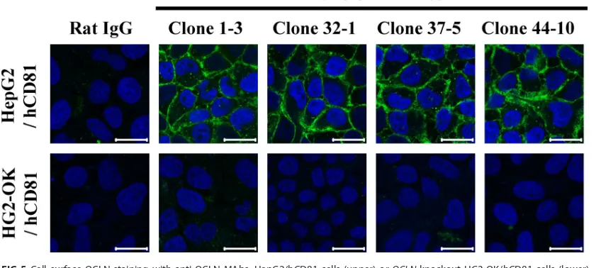
Immunocytochemical analysis under nonpermeabilized conditions revealed that
cell-cell contact regions in polarized human hepatic HepG2/hCD81 cells were mainly
stained with these anti-OCLN MAbs but not those in
OCLN
-knockout HepG2/hCD81
(HG2-OK/hCD81) cells (Fig. 5), suggesting that each of these MAbs can recognize intact
extracellular domains of OCLN.
Anti-OCLN MAbs did not affect cell viability or TJ barrier function.
OCLN is one
of the major proteins localized to TJs and plays an important role in the regulation of
polarity and barrier function in epithelial cells (26). Therefore, we next investigated
whether anti-OCLN MAbs affected the TJ barrier function using human colorectal
adenocarcinoma Caco-2 cells, which form well-constructed TJ barriers in a cell
mono-layer culture system. Indeed, we observed that cell-cell contact regions were stained in
Caco-2 cell monolayers by anti-OCLN MAbs (Fig. 6A), similar to staining of HepG2 cells
(Fig. 5), and these anti-OCLN MAbs had no cytotoxic effects on Caco-2 cells (Fig. 6B).
Commercially available anti-OCLN MAb (clone OC-3F10) that recognizes the cytosolic
domain of OCLN, which is usually used as a TJ marker, showed sharper stains of cell-cell
contact regions than our anti-OCLN MAbs (Fig. 6A), although this may be due to the use
of permeabilized cells. Future studies are needed to assess the possibility that intact
OCLN has broader distributions beyond TJ regions. When polarized Caco-2 cell
mono-layers were treated with control rat IgG, anti-OCLN MAbs, or the TJ breaker C-terminal
fragment of
Clostridium perfringens
enterotoxin (C-CPE), transepithelial electrical
resis-tance (TEER) values were reduced only in C-CPE-treated cells (Fig. 6C). These results
demonstrated that the established anti-OCLN MAbs did not disrupt the TJ barrier of
cultured epithelial cells.
Anti-OCLN MAbs prevented HCV infection
in vitro
.
Studies using the cell culture
infection system have shown that OCLN is involved in HCV infection (10, 27), and our
group confirmed that OCLN is essential for HCV infection of human hepatic cells (11).
We then investigated whether our anti-OCLN MAbs could prevent HCV infection in a
cell culture infection system. We first tested the effect of these MAbs on the viability of
hepatic cells using the XTT
[2,3-bis-(2-methoxy-4-nitro-5-sulfophenyl)-2H-tetrazolium-5-carboxanilide salt] cell viability assay, which showed that none of the MAbs affected
FIG 1Legend (Continued)
full-length hOCLN (HT1080/hOCLN) (C) were incubated with control rat IgG or each anti-OCLN MAb (clone 1-3, 32-1, 37-5, or 44-10) and treated with Alexa Fluor 488-conjugated anti-rat IgG. Stained cells were analyzed by flow cytometry. The vertical dashed lines indicate the peak value of each cell stained with control rat IgG (background levels).
on November 6, 2019 by guest
http://jvi.asm.org/
the viability of HCV-permissive Huh7.5.1-8 cells (Fig. 6D) as well as Caco-2 cells (Fig. 6B),
even at high concentrations (50
g/ml). Huh7.5.1-8 cells next were infected with cell
culture-derived HCV-JFH1 in the presence of control rat IgG or each anti-OCLN MAb,
and cellular HCV RNA levels were measured by quantitative reverse transcription-PCR
FIG 2Established anti-OCLN MAbs were specifically bound to OCLN. HT1080 cells exogenously expressing human MARVEL family proteins OCLN (hOCLN), tricellulin (hTricellulin), MARVELD3 (hMARVELD3), or not expressing any protein (mock) were incubated with control rat IgG or each anti-OCLN MAb (clone 1-3, 32-1, 37-5, or 44-10) and treated with Alexa Fluor 488-conjugated anti-rat IgG. Stained cells were analyzed by flow cytometry. The vertical dashed lines indicate the peak value of each cell stained with control rat IgG.
on November 6, 2019 by guest
[image:5.585.45.545.68.624.2](qRT-PCR) at 4 days postinfection (dpi). As shown in Fig. 7A, all anti-OCLN MAbs
inhibited HCV RNA production in a dose-dependent manner. For clone 1-3, which
recognizes the second extracellular loop of OCLN, the HCV RNA level was strongly
decreased to the background level, and clone 32-1 showed relatively weak inhibition of
FIG 3Clone 1-3 recognized the second loop of hOCLN and mOCLN, whereas the three other clones recognized the first loop of hOCLN. HT1080 cells carrying an empty vector (mock) and those expressing full-length hOCLN (hOCLN), OCLN mutants deleted of either extracellular loop (ΔEL1 and ΔEL2), or mouse OCLN (mOCLN) was incubated with control rat IgG or each anti-OCLN MAb and then treated with Alexa Fluor 488-conjugated anti-rat IgG. Stained cells were analyzed by flow cytometry. The vertical dashed lines indicate the peak value of each cell stained with control rat IgG.
on November 6, 2019 by guest
http://jvi.asm.org/
[image:6.585.44.542.69.621.2]HCV RNA production. The concentration of each MAb that resulted in a 90% inhibition
of cellular HCV RNA production was 0.280
⫾
0.180 (clone 1-3), 33.4
⫾
25.1 (clone 32-1),
5.32
⫾
4.62 (clone 37-5), and 6.54
⫾
5.64 (clone 44-10) nM. Further
immunocytochem-ical analysis was conducted to examine the effect of these MAbs on infection with two
other infectious HCV strains, TNS2J1 (genotype 1b/2a chimera) and Jc1 (genotype 2a/2a
chimera). Similar to the case of the HCV-JFH1 strain, our results showed that anti-OCLN
MAbs decreased the amount of cellular HCV core protein in Huh7.5.1-8 cells infected
with both the TNS2J1 and Jc1 strains (Fig. 7B).
HCV pseudoparticle (HCVpp) infection is a valid model of HCV entry into host cells.
Therefore, we tested whether anti-OCLN MAbs block the entry of various genotypes of
HCVpp into Huh7.5.1-8 cells. As shown in Fig. 8, treatment of cells with control rat IgG
did not reduce HCVpp infection, whereas treatment with each anti-OCLN MAb
de-creased the entry of all tested genotypes of HCVpp (1a, H77c; 1b, Con1 and TH; 2a, JFH1
and J6) in a dose-dependent manner. These anti-OCLN MAbs did not affect the entry
of vesicular stomatitis virus pseudoparticle (VSVpp). These results showed that
anti-FIG 4Binding of anti-OCLN MAbs to cell surface hOCLN with high affinity. After fixation, HT1080 cells carrying an empty vector (HT1080/mock cells) (A) or those expressing hOCLN (HT1080/hOCLN cells) (B) in 96-well plates were incubated with serial dilutions of each anti-OCLN MAb (clone 1-3, 32-1, 37-5, or 44-10) or control rat IgG, followed by horseradish peroxidase-conjugated anti-rat IgG. The binding of each antibody to cell surface hOCLN was detected as described in the cell ELISA section of Materials and Methods. Data are presented as means⫾SD (n⫽3).
FIG 5Cell surface OCLN staining with anti-OCLN MAbs. HepG2/hCD81 cells (upper) orOCLNknockout HG2-OK/hCD81 cells (lower) derived from HepG2/hCD81 cells were fixed and incubated with anti-OCLN MAb (clone 1-3, 32-1, 37-5, or 44-10) or control rat IgG, followed by 4=,6-diamidino-2-phenylindole dihydrochloride (DAPI; blue) and Alexa Fluor 488-conjugated anti-rat IgG (green). Stained cells were observed by confocal microscopy. Scale bars, 20m.
on November 6, 2019 by guest
[image:7.585.45.466.72.234.2] [image:7.585.40.464.508.697.2]OCLN MAbs can specifically prevent HCV infection through the interruption of viral
entry into hepatic cells. Anti-OCLN MAbs, particularly clone 1-3, may be promising
candidates for the development of HCV entry inhibitors.
OCLN is involved in the later step of HCV entry.
HCV entry into hepatocytes is
known to be mediated by various host factors, including SR-BI, CD81, CLDN1, and OCLN
(28), suggesting that these host entry factors are engaged in HCV entry both
cooper-atively and serially. A study of HCV infection with artificial FLAG-tagged OCLN and
FIG 6Anti-OCLN MAbs had no effect on cell viability and TJ barrier function. (A) Fixed Caco-2 cells were incubated with control rat IgG, the established anti-OCLN MAbs, or commercially available MAb that recognizes the cytosolic domain of OCLN (clone OC-3F10) and then were treated with DAPI (blue) and Alexa Fluor 488-conjugated anti-rat IgG or anti-mouse IgG (green). Cells stained with clone 3F10 were pretreated with 0.2% Triton X-100 –PBS for permeabilization. Stained cells were observed by confocal microscopy. Scale bars, 20m. (B and D) Caco-2 (B) and Huh7.5.1-8 (D) cells were treated with the indicated concentrations of control rat IgG, anti-OCLN MAbs, or the TJ barrier breaker C-CPE for 4 days. Cell viability was then determined using the XTT assay. Values are expressed as percentages of control values. (C) Polarized Caco-2 cells in 24-well Transwell filters were cultured until the formation of a confluent cell monolayer and then either left untreated or treated with 5g/ml of control rat IgG or each anti-OCLN MAb (clone 1-3, 32-1, 37-5, or 44-10) or with 50g/ml of C-CPE. TEERs of each cell monolayer were measured at the indicated time points. Data are presented as means⫾SD (n⫽3).on November 6, 2019 by guest
http://jvi.asm.org/
[image:8.585.43.404.70.532.2]anti-FLAG antibody revealed that OCLN seems to act late in the HCV entry process,
during the postbinding stage and after the use of CD81 and CLDN1 (29). To confirm this
finding, the timing of OCLN usage during HCVpp infection was analyzed using our
anti-OCLN and anti-CLDN1 functional MAbs. In brief, HCVpp was incubated with
HCV-permissive HepG2/hCD81 cells at 4°C, and unbound particles were washed with
phosphate-buffered saline (PBS). The culture temperature was then shifted to 37°C to
allow postbinding infection. After 2 days of culture, HCVpp entry was gauged using the
luciferase reporter assay. Anti-OCLN and anti-CLDN1 MAbs were added at different
times before or after the binding phase. Treatment of cells with each MAb at 2 h after
HCVpp binding still inhibited HCVpp entry. Meanwhile, the addition of anti-CLDN1 MAb
(clone 3A2) 3 h after virus binding had no effect on HCVpp entry, but similar addition
of the anti-OCLN MAbs (clones 1-3 and 37-5) significantly inhibited HCVpp entry (Fig.
9). These results showed that OCLN is used after CLDN1 in HCV infection, as previously
suggested (29).
Anti-OCLN MAbs inhibited cell-to-cell HCV infection.
It is well known that there
are two types of HCV invasion into hepatocytes: infection through viruses floating free
FIG 7Anti-OCLN MAbs inhibitedin vitroHCVcc infection. (A) Huh7.5.1-8 cells were preincubated with increasing concentrations of each control rat IgG or anti-OCLN MAb for 30 min at room temperature and then infected with HCV-JFH1 for 2 h at 37°C. Cells were further cultured in the presence of each Ab for 4 days. Cellular HCV RNA was measured by qRT-PCR. Values are expressed as percentages of control values (treatment without Ab). Data are presented as means⫾SD (n⫽4). The limit of detection was 0.021⫾0.027. (B) Huh7.5.1-8 cells were preincubated with 5g/ml of each rat IgG for 30 min at room temperature and then infected with various infectious HCVcc strains (JFH1, TNS2J1, or Jc1) at an MOI of 1.0. At 4 dpi, the cells were fixed and stained with anti-HCV core MAb (green). Scale bars, 100m.
on November 6, 2019 by guest
[image:9.585.43.463.70.464.2]in the medium (cell-free infection) and direct infection between neighboring cells
(cell-to-cell infection [30, 31]). In this context, OCLN is suggested to be important for
cell-to-cell (31, 32) as well as cell-free HCV infection. We also demonstrated that OCLN
is essential for cell-to-cell infection using
OCLN
knockout hepatic cells (11). Here, we
evaluated whether anti-OCLN MAbs can inhibit cell-to-cell HCV transmission. We first
developed a visualization system to monitor cell-to-cell HCV infection using a
mito-chondrial antiviral signaling protein (MAVS)-based reporter. Mitomito-chondrially tethered
MAVS (33), which is also known as IPS-1/VISA/Cardiff, is a known cellular substrate of
FIG 8Anti-OCLN MAbs inhibitedin vitroHCVpp infection. Huh7.5.1-8 cells were preincubated with the indicated amounts of control rat IgG or each anti-OCLN MAb for 30 min at room temperature and then infected with various genotypes of HCVpp (1a, H77c; 1b, Con1 and TH; 2a, JFH1 and J6), VSVpp, or pseudoparticles with no envelope proteins (⫺Env) for 6 h at 37°C. At 2 dpi, cellular luciferase activity was determined as described in Materials and Methods. Values are expressed as percentages of control values (treatment without Ab). Data are presented as means⫾SD (n⫽3).FIG 9Analysis of the timing of entry factor usage during HCV infection. HepG2/hCD81 cells were infected with HCVpp (JFH1) at 0 h. To monitor the relative timing of each step in the entry process, anti-CLDN1 MAb (clone 3A2) or anti-OCLN MAb (clone 1-3 or 37-5) was added to the culture medium at the indicated time points. At 2 dpi, cellular luciferase activity was determined. Values are expressed as percentages of control values (at 5 h without Ab treatment). Data are presented as means⫾SD (n⫽3). *,P⬍0.01.
on November 6, 2019 by guest
http://jvi.asm.org/
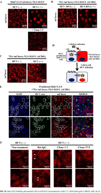
[image:10.585.50.364.70.280.2] [image:10.585.51.360.484.681.2]transfection of HCV RNA (Fig. 10B), but this did not occur after infection with HCV-JFH1
(Fig. 10C). When HCV-preinfected Huh7.5.1-8 cells were mixed with naive 751r/
mCherry-NLS-MAVS cells and cocultured for 4 days, a significant portion of the 751r/
mCherry-NLS-MAVS cells adjacent to the HCV-infected Huh7.5.1-8 cells had nuclear
localization of red fluorescence, indicating cell-to-cell HCV transmission from
Huh7.5.1-8 cells to 751r/mCherry-NLS-MAVS cells (Fig. 10D and E). In this coculture
system, the addition of anti-OCLN MAbs (clones 1-3 and 37-5) completely blocked
nuclear localization of red fluorescence, but the addition of control rat IgG did not (Fig.
10F). Taken together with the above-described data, these results demonstrated that
anti-OCLN MAbs can inhibit not only cell-free infection but also cell-to-cell infection.
Anti-OCLN MAbs completely prevented HCV infection
in vivo
.
We next tested
whether administration of anti-OCLN MAbs could inhibit HCV infection
in vivo
using
human liver chimeric mice, a well-known animal model of HCV infection (35–37). We
focused on clones 1-3 (recognized by EL2) and 37-5 (recognized by EL1) in the
in vivo
HCV infection analysis, because these two MAbs recognize different epitopes of OCLN,
are of the same IgG2b subclass, and sufficiently inhibit HCV infection
in vitro
. Chimeric
mice were intraperitoneally administered clone 1-3 or 37-5 at concentrations of 50, 30,
20, and 10 mg/kg of body weight on days 0, 3, 7, and 10, respectively. On day 0, 6 h
after MAb administration, the mice were challenged with HCV (genotype 1b) (Fig. 11A).
All mice that received the control rat IgG were HCV positive at 14 dpi. Serum HCV RNA
levels in all mice plateaued at
⬎
5
⫻
10
6copies/ml at 21 dpi. Meanwhile, the serum of
all mice that received anti-OCLN MAbs (clone 1-3 or 37-5) was HCV negative even after
35 dpi (Fig. 11B). We also determined serum anti-OCLN MAb levels at 0 to 35 dpi using
cell-based ELISA. The serum MAbs in clone 1-3-treated mice and clone 37-5-treated
mice were detectable (
⬎
0.3
g/ml) until 28 dpi, and those at 35 dpi were under the
limit of detection (Fig. 11C and D). These results indicated that anti-OCLN MAb clones
1-3 and 37-5 completely prevented HCV infection
in vivo
. To monitor the toxic
effects of anti-OCLN MAbs, we measured the body weights and analyzed human
albumin, aspartate aminotransferase (AST), and alanine aminotransferase (ALT)
levels in the sera of MAb-treated mice. As shown in Fig. 11E to H, there were no
significant changes in body weight or human albumin, AST, and ALT levels in mice
that received control rat IgG or anti-OCLN MAbs. These data suggested that
anti-OCLN MAb clones 1-3 and 37-5 had no significant cytotoxic effect on human
hepatocytes at the administered doses.
DISCUSSION
HCV entry into host cells is a complex process involving multiple host factors,
including the TJ protein OCLN. Because OCLN is essential for HCV infection (11), it may
be a promising target for novel host-targeting anti-HCV agents. However, to date, there
are no OCLN-binding probes that can inhibit HCV infection, because OCLN is a multiple
transmembrane protein and MAbs against such proteins are very difficult to produce.
Therefore, MAbs recognizing the extracellular domains of intact hOCLN have not yet
been developed. However, we successfully developed MAbs against the extracellular
on November 6, 2019 by guest
FIG 10Anti-OCLN MAbs prevented cell-to-cell HCV transmission. Huh7.5.1-8/mCherry-NLS-MAVS cells (A) or CD81-defective 751r/mCherry-NLS-MAVS cells (C) were infected with HCV-JFH1 or left uninfected. (B) 751r/mCherry-NLS-MAVS cells were left untransfected or were transfected with HCV subgenomic replicon
(Continued on next page)
on November 6, 2019 by guest
http://jvi.asm.org/
[image:12.585.41.377.70.707.2]All anti-OCLN MAbs prevented infection of Huh7.5.1-8 cells with HCVcc and HCVpp
in a dose-dependent manner without apparent cytotoxicity (Fig. 6D, 7, and 8). In
particular, the MAb clone 1-3 showed much stronger HCV inhibition. Consistent with
this observation, previous reports have suggested that this second extracellular loop of
OCLN is critical for HCV infection (10, 11, 42, 43). Conversely, the first extracellular loop
of OCLN is nonessential for HCV entry into hepatic cells, as we showed previously (11).
Therefore, the inhibitory effects of MAbs that recognize the first loop of OCLN may be
due to steric effects.
To investigate the antiviral effects of the anti-OCLN MAbs
in vivo
, anti-OCLN MAb
clones 1-3 and 37-5 or a control antibody were administered to human liver chimeric
uPA-SCID mice. As shown in Fig. 11, each anti-OCLN MAb completely blocked HCV
infection in human liver chimeric mice without apparent adverse effects and could be
detected in the sera until 28 dpi. It is well known that HCV is quickly inactivated (44)
compared with other viruses, which may be the reason why the anti-OCLN MAbs
completely blocked HCV invasion into hepatocytes of chimeric mice. Furthermore, we
demonstrated that the anti-OCLN MAbs completely blocked cell-to-cell transmission as
well as cell-free infection (Fig. 7 and 10F). The ability of these anti-OCLN MAbs to inhibit
both modes of infection may result in efficient blockage of HCV transmission among
hepatocytes
in vivo
, suggesting that the anti-OCLN MAbs convey antiviral effects in the
hepatocytes of patients with persistent HCV infection.
Hopcraft and Evans recently reported that an HCV variant with a single-amino-acid
mutation could infect cells via a CLDN1-independent cell entry mechanism (45).
Meanwhile, Mailly et al. demonstrated that a single-dose treatment with anti-CLDN1
MAb could cure persistent HCV infection in a humanized mouse model (15).
Further-more, no escape viruses were observed in the mouse model or on primary human
hepatocytes (20). These results indicated that escape via CLDN6 and CLDN9 is a cell
line-specific observation. Based on these studies, CLDN1 is considered a promising
anti-HCV target (12, 19, 20, 46), although
cldn1
knockout mice (23) had wrinkled skin
and died within 1 day of birth.
Ocln
knockout mice were born with no gross phenotypes
(24). We also demonstrated, using a transwell culture system, that the anti-OCLN MAbs
were not cytotoxic to cultured cells (Fig. 6B and D) and did not disrupt the epithelial TJ
barrier in polarized Caco-2 cells (Fig. 6C). The administration of the second
loop-FIG 10Legend (Continued)
RNA. (D) An overview of the detection of cell-to-cell HCV transmission. (E) Huh7.5.1-8 cells were preinfected with HCV-JFH1 and cultured for 1 day, mixed with 751r/mCherry-NLS-MAVS cells (at a cell ratio of 1:1), and plated in a 24-well plate at 1⫻105cells/well. After 4 days, the cells were fixed, stained with anti-HCV core protein MAb (green) and DAPI (blue), and then visualized by confocal microscopy. Arrows, donor Huh7.5.1-8 cells preinfected with HCV. Arrowheads, acceptor 751r/mCherry-NLS-MAVS cells in which nucleus was stained red. Solid line, HCV core-positive Huh7.5.1-8 cells. Broken line, HCV core-positive 751r/mCherry-NLS-MAVS. (F) Huh7.5.1-8 cells were preinfected with HCV-JFH1, or not, and cultured for 1 day, mixed with 751r/mCherry-NLS-MAVS cells (at a cell ratio of 1:1), and plated in a 24-well plate at 1⫻105cells/well in the presence of control rat IgG or anti-OCLN MAb (clone 1-3 or 37-5). (A to C and F) After 4 days, the cells were visualized by confocal microscopy. (F) Arrows indicate HCV-infected 751r/mCherry-NLS-MAVS cells by cell-to-cell transmission. Scale bars, 20m (A to C), 50m (E), and 200m (F).
on November 6, 2019 by guest
recognizing MAb clone 1-3, which reacts with both human and mouse OCLN, showed
no systemic toxicity in human liver chimeric mice (Fig. 11E to H). Furthermore, there
have been no reports of HCV infection via OCLN-independent routes. The attempted
genetic approaches with
OCLN
knockout cells showed that multiple serial passages of
HCV-JFH-1 with
OCLN
knockout cells resulted in no isolation of viruses. Taken together,
we believe that OCLN, in addition to CLDN1, is a promising anti-HCV target.
In conclusion, we developed four MAbs against the intact extracellular domains of
OCLN that strongly prevented HCV infection both
in vitro
and
in vivo
and demonstrated
FIG 11Anti-OCLN MAbs completely blockedin vivoHCV infection. (A) Experimental procedure ofin vivo HCV infection using human liver chimeric mice. i.p., intraperitoneal; i.v., intravenous. (B) Human liver chimeric mice were treated with control rat IgG (n⫽4) or anti-OCLN MAb (clone 1-3 or 37-5;n⫽3), as described in Materials and Methods. HCV (genotype 1b) was used for infections in these experiments. At the indicated time points, serum HCV RNA levels were determined by qRT-PCR. The dotted line indicates the limit of detection (2⫻ 104copies/ml). Arrows show the times of injection of MAbs. (C and D) Anti-OCLN MAb contents in sera were determined using a cell ELISA-based assay, as described in Materials and Methods (clone 1-3-treated mice [C] and clone 37-5-treated mice [D]). Data are presented as means⫾SD (n⫽3). The limit of detection was 0.27⫾0.012g/ml for the clone 1-3-treated mice and 0.33 ⫾ 0.010 g/ml for the clone 37-5-treated mice. Characteristics of Ab-treated chimeric mice, including body weight (E) and human albumin (F), AST (G), and ALT (H) levels in sera.on November 6, 2019 by guest
http://jvi.asm.org/
[image:14.585.48.365.63.509.2]respectively) were constructed by inverse PCR from pCX4-bleo-hOCLN, as described previously (11). The pCX4-bleo-hTricellulin and pCX4-bleo-hMARVELD3-1 vectors were kindly provided by Akihiro Watari (Osaka University, Japan).
ThemCherry-NLS-MAVSgene, which contains amino acids (aa) 1 to 262 of themCherrygene, the nuclear localization signal sequence of SV40 (corresponding to aa 2 to 8), the spacer nucleotides GGT, and the C-terminal region of theMAVSgene (corresponding to aa 503 to 540), provided by Yoshiharu Matsuura (Osaka University, Japan) (48), was cloned into the BamHI/EcoRI sites of the pCX4-pur vector. The constructed vector was referred to as pCX4-pur-mCherry-NLS-MAVS.
Cell lines.We used human hepatoma cells derived from Huh7.5.1 cells, kindly provided by Francis V. Chisari (The Scripps Research Institute, La Jolla, CA, USA) (49), including Huh7.5.1-8 cells (25), which are highly permissive to HCV infection, andCD81-defective 751r (25) andOCLN-defective OKH-4 (11) cells, which are nonpermissive to HCV infection. Human fibrosarcoma HT1080 cells and human hepatoma HepG2 cells were obtained from the Japanese Collection of Research Bioresources (Osaka, Japan). Human embryonic kidney 293T (HEK 293T), human colorectal adenocarcinoma Caco-2, and CHO-K1 cells were obtained from the American Type Culture Collection (Manassas, VA, USA). Huh7.5.1-derived, HepG2, HEK 293T, and HT1080 cells were cultured at 37°C under an atmosphere of 5% CO2in Dulbecco’s modified Eagle’s medium supplemented with 10% fetal bovine serum (FBS), 100 U/ml penicillin G, 100g/ml streptomycin sulfate, and 0.1 mM nonessential amino acids (normal medium). Caco-2 cells were maintained in Eagle’s minimum essential medium supplemented with 60g/ml kanamycin (Nissui Pharmaceutical Co., Ltd., Tokyo, Japan) and 10% FBS. CHO-K1 cells were maintained in Ham’s F-12 medium supplemented with 10% newborn calf serum, 100 U/ml penicillin G, and 100g/ml strepto-mycin sulfate (50).
Various stable transfectants (HT1080/mock, HT1080/hOCLN, ΔEL1, HT1080/hOCLN-ΔEL2, HT1080/mOCLN, HepG2/hCD81, Huh7.5.1-8/mCherry-NLS-MAVS, and 751r/mCherry-NLS-MAVS cells) were established by retroviral-mediated gene transfer and drug selection, as described previously (11). HepG2/hCD81-derivedOCLNknockout cells, referred to as HG2-OK/hCD81, were established by the same method as that used for the generation of OKH-4 cells (11).
Isolation of MAbs against the extracellular domains of OCLN.Six-week-old male Wistar rats were subcutaneously immunized with a mammalian expression vector encodinghOCLNevery 2 weeks for 2 months according to proprietary GENOVAC technology (GENOVAC GmbH, Freiburg, Germany) (40). Plasma cells were removed 7 days after the last immunization and fused with mouse myeloma P3U1 cells in the presence of polyethylene glycol 1000 to generate hybridomas. Hybridomas producing anti-OCLN MAbs were initially screened for the ability of their culture supernatants to bind to CHO-K1 cells transiently expressing hOCLN, but not to wild-type CHO-K1 cells, by cell ELISA and flow cytometry. The hybridomas were then further selected by flow cytometry based on their ability to bind Huh7.5.1-8 cells but not toOCLNknockout OKH-4 cells. After cloning by limiting dilution, four stable hybridomas that produced MAbs binding to the extracellular domains of hOCLN (clones 1-3, 32-1, 37-5, and 44-10) were obtained. Immunoglobulin classes and subclasses of the clones were determined using a rat immuno-globulin isotyping ELISA kit (BD Biosciences, Woburn, MA, USA).
Flow-cytometric analysis.Cells were detached from culture dishes with 0.05% (wt/vol) Trypsin– 0.5 mM EDTA and then treated with antibodies (5g/ml) in PBS containing 2% FBS for 60 min at 4°C. After washing with PBS, the cells were incubated with Alexa Fluor 488-conjugated goat anti-rat IgG (Thermo Fisher Scientific, Waltham, MA, USA) at 2g/ml in PBS containing 2% FBS for 30 min at 4°C. After washing with PBS, the cells were analyzed using a FACSCalibur flow cytometer (BD Biosciences).
Cell ELISA.Cell ELISA was performed as described previously (12), with slight modifications. In brief, confluent HT1080/hOCLN or HT1080/mock cells in 96-well plates, fixed with 3.7% formaldehyde, were incubated with serial dilutions of 100l of each anti-OCLN MAb or control IgG (5g/ml) for 2 h, followed by a 1:5,000 dilution of 100l of horseradish peroxidase-conjugated goat anti-rat IgG (GE Healthcare, Waukesha, WI, USA) for 1 h at room temperature. To detect antibody binding to the cells, -phenylenediamine dihydrochloride and hydrogen peroxide were used as substrates. The absorbance at 492 nm for each well was determined on an OpsysMR plate reader (Dynex Technologies GmbH, Denkendorf, Germany). The half-saturating concentrations (apparentKd[antibody dissociation constants]
values) were determined using GraphPad Prism version 6.0 (GraphPad Software, San Diego, CA) as described previously (51).
on November 6, 2019 by guest
TJ barrier function assay.To determine the TJ barrier function of cell monolayers, we measured the electrical resistance across the cell monolayers (TEER) as described previously (52–54). In brief, Caco-2 cells were seeded in 24-well Transwell filters (Corning, NY) at a subconfluent density. TEER of the cell sheets on the filter was monitored using a Millicell electrical resistance system with a dual electrode (Millipore, Billerica, MA). After TEER values reached a plateau, showing that TJs were well developed in the cell sheets, the cell monolayers were treated with 5.0g of TJ barrier breaker C-CPE (55) or 50g of each anti-OCLN MAb on the basal side of the filter. The TEER values on the filter were then monitored. Cell viability assay.To determine the cytotoxicity of anti-OCLN MAbs, a colorimetric XTT assay was performed with cell proliferation kit II (XTT) (Roche Diagnostics, Mannheim, Germany) according to the manufacturer’s protocol. Briefly, after treatment of the cells with the XTT reagent for 2 h, the superna-tants were transferred to a 96-well plate, and the absorbance at 450 nm was measured using an OpsysMR plate reader (Dynex Technologies GmbH).
In vitroHCV infection.The infectious source of HCV-JFH1 (49, 56) was prepared from culture supernatants of Huh7.5.1-8 cells that had been transfected within vitro-transcribed HCV-JFH1 RNA at a multiplicity of infection (MOI) of 0.1 for 2 h and passaged a few times on Huh7.5.1-8 cells (12). Two other infectious HCV-JFH1 chimera strains, Jc1 (genotype 2a/2a) (57) and TNS2J1 (genotype 1b/2a) (58), were prepared as described previously (11, 59). HCV infection was detected using qRT-PCR and immunohis-tochemical analysis as described previously (12, 25, 60).
HCVpp infection.HCVpps were generated as described previously (11). In brief, agag-polpackaging construct (Gag-Pol 5349), a transfer vector construct (Luc 126), and an envelope glycoprotein expression vector (H77c [genotype 1a], TH [genotype 1b], Con1 [genotype 1b; GenBank accession number
AJ238799], JFH1 [genotype 2a], J6 [genotype 2a], and VSV-G) were transfected into HEK 293T cells. The medium from the transfected cells was collected and used as the HCVpp source. HCVpp infection and a luciferase reporter assay were performed as described previously (12, 25).
Cell-to-cell HCV transmission.Huh7.5.1-8 cells were seeded at a density of 2⫻106cells in a 100-mm dish, cultured for 1 day, and infected with HCV-JFH1 at an MOI of 0.1. The HCV-infected Huh7.5.1-8 cells and naive 751r/mCherry-NLS-MAVS cells were mixed at a 1:1 ratio and plated in each well of a 24-well plate at a density of 8⫻104cells. Control rat IgG or anti-OCLN MAb (clone 1-3 or 37-5; 10g/ml) was immediately added to each well. Cells were cultured for 4 days and then observed under a confocal microscope (LSM700; Carl Zeiss, Jena, Germany).
In vivoHCV infection.Human liver chimeric mice (PXB mice) were prepared as described previously (61). PXB mice (16 or 17 weeks old, male) were intraperitoneally injected with anti-OCLN MAbs (clone 1-3 or 37-5) or control rat IgG at 50 mg/kg (three anti-OCLN-treated mice/group and four control Ab-treated mice/group). At 8 h after Ab administration, the mice were intravenously inoculated with HCV-PBC001 (104copies; genotype 1b; lot 0080811). The total blood volume of a mouse is approximately 80 ml/kg (17); hence, 104HCV RNA copies/mouse corresponds to⬃1.7⫻104HCV RNA copies/ml in serum. Mice were intraperitoneally injected with anti-OCLN MAbs (clone 1-3 or 37-5) or control Ab at 30, 20, and 10 mg/kg on days 3, 7, and 10, respectively. Sera were collected from mice on days 0, 3, 7, 10, 14, 21, 28, and 35 for analyses. HCV RNA in mouse serum was quantified using TaqMan EZ RT-PCR core reagent (Life Technologies Corp., Carlsbad, CA, USA) and an ABI Prism 7500 sequence detector system (Life Technol-ogies Corp.). The lower quantification limit of the assay was 2.0⫻104copies/ml.
Ethics statement.All procedures using PXB mice were approved by the Animal Ethics Committee of PhoenixBio Co., Ltd. (Hiroshima, Japan) (permission number 1564), and all animal experiments were performed after approval by the Committee on the Ethics of Animal Experiments of the National Institute of Infectious Diseases (permission number 815005) under the guidelines of the National Institute of Infectious Diseases, in accordance with theGuidelines for Proper Conduct of Animal Experiments estab-lished by the Science Council of Japan (http://www.scj.go.jp/ja/info/kohyo/pdf/kohyo-20-k16-2e.pdf).
Measurements of anti-OCLN MAbs in mouse serum.Anti-OCLN MAb (clones 1-3 and 37-5) levels in mouse serum were determined by cell ELISA. In brief, confluent HT1080/hOCLN cells in 96-well plates, fixed with 3.7% formaldehyde, were incubated with 100l of each serum sample diluted with PBS containing 5% (wt/vol) skim milk for 2 h, followed by a 1:5,000 dilution of 100l of horseradish peroxidase-conjugated goat anti-rat IgG (GE Healthcare) for 1 h at room temperature. To detect cellular binding of MAbs, 1-Step Ultra TMB-ELISA substrate solution (Thermo Fischer Scientific) was used as the substrate. For each well, the absorbance at 450 nm was determined on an OpsysMR plate reader (Dynex Technologies GmbH). Each purified MAb was used as a standard.
Other methods.Immunohistochemical analysis was performed as described previously (25). Analysis of the timing of entry factor usage during HCV infection was performed as described previously (29). For in vivoexperiments, human albumin, AST, and ALT levels in mouse sera were measured as described previously (12).
Statistical analysis. The data for statistical analyses are presented as the averages from three independent experiments⫾standard deviations (SD). The significance of differences in the means was determined by Student’sttest. A probability (P) value of⬍0.05 was considered statistically significant.
ACKNOWLEDGMENTS
We thank K. Endo (Wako Pure Chemical Industries, Ltd.) and all members of our
laboratory for their useful comments. We thank Y. Matsuura (Osaka University, Osaka,
Japan) and H. Aizaki (National Institute of Infectious Diseases, Tokyo, Japan) for the gift
of an
mCherry-NLS-MAVS
gene-encoding plasmid. We also thank K. Sugiyama (Keio
on November 6, 2019 by guest
http://jvi.asm.org/
1. Messina JP, Humphreys I, Flaxman A, Brown A, Cooke GS, Pybus OG, Barnes E. 2015. Global distribution and prevalence of hepatitis C virus genotypes. Hepatology 61:77– 87.https://doi.org/10.1002/hep.27259. 2. Petruzziello A, Marigliano S, Loquercio G, Cozzolino A, Cacciapuoti C.
2016. Global epidemiology of hepatitis C virus infection: an up-date of the distribution and circulation of hepatitis C virus genotypes. World J Gastroenterol 22:7824 –7840.https://doi.org/10.3748/wjg.v22.i34.7824. 3. Molina S, Castet V, Fournier-Wirth C, Pichard-Garcia L, Avner R, Harats D,
Roitelman J, Barbaras R, Graber P, Ghersa P, Smolarsky M, Funaro A, Malavasi F, Larrey D, Coste J, Fabre JM, Sa-Cunha A, Maurel P. 2007. The low-density lipoprotein receptor plays a role in the infection of primary human hepatocytes by hepatitis C virus. J Hepatol 46:411– 419.https:// doi.org/10.1016/j.jhep.2006.09.024.
4. Barth H, Schafer C, Adah MI, Zhang F, Linhardt RJ, Toyoda H, Kinoshita-Toyoda A, Toida T, Van Kuppevelt TH, Depla E, Von Weizsacker F, Blum HE, Baumert TF. 2003. Cellular binding of hepatitis C virus envelope glycoprotein E2 requires cell surface heparan sulfate. J Biol Chem 278: 41003– 41012.https://doi.org/10.1074/jbc.M302267200.
5. Scarselli E, Ansuini H, Cerino R, Roccasecca RM, Acali S, Filocamo G, Traboni C, Nicosia A, Cortese R, Vitelli A. 2002. The human scavenger receptor class B type I is a novel candidate receptor for the hepatitis C virus. EMBO J 21:5017–5025.https://doi.org/10.1093/emboj/cdf529. 6. Pileri P, Uematsu Y, Campagnoli S, Galli G, Falugi F, Petracca R, Weiner
AJ, Houghton M, Rosa D, Grandi G, Abrignani S. 1998. Binding of hepatitis C virus to CD81. Science 282:938 –941.https://doi.org/10.1126/ science.282.5390.938.
7. Sainz B, Jr, Barretto N, Martin DN, Hiraga N, Imamura M, Hussain S, Marsh KA, Yu X, Chayama K, Alrefai WA, Uprichard SL. 2012. Identification of the Niemann-Pick C1-like 1 cholesterol absorption receptor as a new hepa-titis C virus entry factor. Nat Med 18:281–285.https://doi.org/10.1038/ nm.2581.
8. Lupberger J, Zeisel MB, Xiao F, Thumann C, Fofana I, Zona L, Davis C, Mee CJ, Turek M, Gorke S, Royer C, Fischer B, Zahid MN, Lavillette D, Fresquet J, Cosset FL, Rothenberg SM, Pietschmann T, Patel AH, Pessaux P, Doffoel M, Raffelsberger W, Poch O, McKeating JA, Brino L, Baumert TF. 2011. EGFR and EphA2 are host factors for hepatitis C virus entry and possible targets for antiviral therapy. Nat Med 17:589 –595.https://doi .org/10.1038/nm.2341.
9. Evans MJ, von Hahn T, Tscherne DM, Syder AJ, Panis M, Wolk B, Hatzi-ioannou T, McKeating JA, Bieniasz PD, Rice CM. 2007. Claudin-1 is a hepatitis C virus co-receptor required for a late step in entry. Nature 446:801– 805.https://doi.org/10.1038/nature05654.
10. Ploss A, Evans MJ, Gaysinskaya VA, Panis M, You H, de Jong YP, Rice CM. 2009. Human occludin is a hepatitis C virus entry factor required for infection of mouse cells. Nature 457:882– 886.https://doi.org/10.1038/ nature07684.
11. Shirasago Y, Shimizu Y, Tanida I, Suzuki T, Suzuki R, Sugiyama K, Wakita T, Hanada K, Yagi K, Kondoh M, Fukasawa M. 2016. Occludin-knockout human hepatic Huh7.5.1-8-derived cells are completely resistant to hepatitis C virus infection. Biol Pharm Bull 39:839 – 848.https://doi.org/ 10.1248/bpb.b15-01023.
12. Fukasawa M, Nagase S, Shirasago Y, Iida M, Yamashita M, Endo K, Yagi K, Suzuki T, Wakita T, Hanada K, Kuniyasu H, Kondoh M. 2015. Mono-clonal antibodies against extracellular domains of claudin-1 block hep-atitis C virus infection in a mouse model. J Virol 89:4866 – 4879.https:// doi.org/10.1128/JVI.03676-14.
13. Marceau CD, Puschnik AS, Majzoub K, Ooi YS, Brewer SM, Fuchs G,
Swaminathan K, Mata MA, Elias JE, Sarnow P, Carette JE. 2016. Genetic dissection of Flaviviridae host factors through genome-scale CRISPR screens. Nature 535:159 –163.https://doi.org/10.1038/nature18631. 14. Vercauteren K, Van Den Eede N, Mesalam AA, Belouzard S, Catanese
MT, Bankwitz D, Wong-Staal F, Cortese R, Dubuisson J, Rice CM, Pietschmann T, Leroux-Roels G, Nicosia A, Meuleman P. 2014. Suc-cessful anti-scavenger receptor class B type I (SR-BI) monoclonal antibody therapy in humanized mice after challenge with HCV vari-ants with in vitro resistance to SR-BI-targeting agents. Hepatology 60:1508 –1518.https://doi.org/10.1002/hep.27196.
15. Mailly L, Xiao F, Lupberger J, Wilson GK, Aubert P, Duong FHT, Calabrese D, Leboeuf C, Fofana I, Thumann C, Bandiera S, Lutgehetmann M, Volz T, Davis C, Harris HJ, Mee CJ, Girardi E, Chane-Woon-Ming B, Ericsson M, Fletcher N, Bartenschlager R, Pessaux P, Vercauteren K, Meuleman P, Villa P, Kaderali L, Pfeffer S, Heim MH, Neunlist M, Zeisel MB, Dandri M, McKeating JA, Robinet E, Baumert TF. 2015. Clearance of persistent hepatitis C virus infection in humanized mice using a claudin-1-targeting monoclonal antibody. Nat Biotechnol 33:549 –554. https://doi.org/10 .1038/nbt.3179.
16. Zahid MN, Turek M, Xiao F, Thi VL, Guerin M, Fofana I, Bachellier P, Thompson J, Delang L, Neyts J, Bankwitz D, Pietschmann T, Dreux M, Cosset FL, Grunert F, Baumert TF, Zeisel MB. 2013. The postbinding activity of scavenger receptor class B type I mediates initiation of hepatitis C virus infection and viral dissemination. Hepatology 57: 492–504.https://doi.org/10.1002/hep.26097.
17. Meuleman P, Hesselgesser J, Paulson M, Vanwolleghem T, Desombere I, Reiser H, Leroux-Roels G. 2008. Anti-CD81 antibodies can prevent a hepatitis C virus infection in vivo. Hepatology 48:1761–1768.https://doi .org/10.1002/hep.22547.
18. Syder AJ, Lee H, Zeisel MB, Grove J, Soulier E, Macdonald J, Chow S, Chang J, Baumert TF, McKeating JA, McKelvy J, Wong-Staal F. 2011. Small molecule scavenger receptor BI antagonists are potent HCV entry inhib-itors. J Hepatol 54:48 –55.https://doi.org/10.1016/j.jhep.2010.06.024. 19. Fofana I, Krieger SE, Grunert F, Glauben S, Xiao F, Fafi-Kremer S, Soulier
E, Royer C, Thumann C, Mee CJ, McKeating JA, Dragic T, Pessaux P, Stoll-Keller F, Schuster C, Thompson J, Baumert TF. 2010. Monoclonal anti-claudin 1 antibodies prevent hepatitis C virus infection of primary human hepatocytes. Gastroenterology 139:953–964.https://doi.org/10 .1053/j.gastro.2010.05.073.
20. Colpitts CC, Tawar RG, Mailly L, Thumann C, Heydmann L, Durand SC, Xiao F, Robinet E, Pessaux P, Zeisel MB, Baumert TF. 30 March 2017. Humanisation of a claudin-1-specific monoclonal antibody for clinical prevention and cure of HCV infection without escape. Guthttps://doi .org/10.1136/gutjnl-2016-312577.
21. Rubinstein E, Ziyyat A, Prenant M, Wrobel E, Wolf JP, Levy S, Le Naour F, Boucheix C. 2006. Reduced fertility of female mice lacking CD81. Dev Biol 290:351–358.https://doi.org/10.1016/j.ydbio.2005.11.031. 22. Mordica WJ, Gallagher RJ, Kennedy JL, Chapes SK. 2010. Male CD81
knockout genotype disrupts Mendelian distribution of offspring. Comp Med 60:196 –199.
23. Furuse M, Hata M, Furuse K, Yoshida Y, Haratake A, Sugitani Y, Noda T, Kubo A, Tsukita S. 2002. Claudin-based tight junctions are crucial for the mammalian epidermal barrier: a lesson from claudin-1-deficient mice. J Cell Biol 156:1099 –1111.https://doi.org/10.1083/jcb.200110122. 24. Saitou M, Furuse M, Sasaki H, Schulzke JD, Fromm M, Takano H, Noda T,
Tsukita S. 2000. Complex phenotype of mice lacking occludin, a
on November 6, 2019 by guest
ponent of tight junction strands. Mol Biol Cell 11:4131– 4142.https://doi .org/10.1091/mbc.11.12.4131.
25. Shirasago Y, Sekizuka T, Saito K, Suzuki T, Wakita T, Hanada K, Kuroda M, Abe R, Fukasawa M. 2015. Isolation and characterization of an Huh.7.5.1-derived cell clone highly permissive to hepatitis C virus. Jpn J Infect Dis 68:81– 88.https://doi.org/10.7883/yoken.JJID.2014.231.
26. Paris L, Tonutti L, Vannini C, Bazzoni G. 2008. Structural organization of the tight junctions. Biochim Biophys Acta 1778:646 – 659.https://doi.org/ 10.1016/j.bbamem.2007.08.004.
27. Liu S, Yang W, Shen L, Turner JR, Coyne CB, Wang T. 2009. Tight junction proteins claudin-1 and occludin control hepatitis C virus entry and are downregulated during infection to prevent superinfection. J Virol 83: 2011–2014.https://doi.org/10.1128/JVI.01888-08.
28. Douam F, Lavillette D, Cosset FL. 2015. The mechanism of HCV entry into host cells. Prog Mol Biol Transl Sci 129:63–107.https://doi.org/10.1016/ bs.pmbts.2014.10.003.
29. Sourisseau M, Michta ML, Zony C, Israelow B, Hopcraft SE, Narbus CM, Parra Martin A, Evans MJ. 2013. Temporal analysis of hepatitis C virus cell entry with occludin directed blocking antibodies. PLoS Pathog 9:e1003244.https://doi.org/10.1371/journal.ppat.1003244.
30. Timpe JM, Stamataki Z, Jennings A, Hu K, Farquhar MJ, Harris HJ, Schwarz A, Desombere I, Roels GL, Balfe P, McKeating JA. 2008. Hepatitis C virus cell-cell transmission in hepatoma cells in the presence of neutralizing antibodies. Hepatology 47:17–24.https://doi.org/10.1002/ hep.21959.
31. Liu Z, He JJ. 2013. Cell-cell contact-mediated hepatitis C virus (HCV) transfer, productive infection, and replication and their requirement for HCV receptors. J Virol 87:8545– 8558.https://doi.org/10.1128/JVI .01062-13.
32. Ciesek S, Westhaus S, Wicht M, Wappler I, Henschen S, Sarrazin C, Hamdi N, Abdelaziz AI, Strassburg CP, Wedemeyer H, Manns MP, Pietschmann T, von Hahn T. 2011. Impact of intra- and interspecies variation of occludin on its function as coreceptor for authentic hepatitis C virus particles. J Virol 85:7613–7621.https://doi.org/10.1128/JVI.00212-11. 33. Kawai T, Takahashi K, Sato S, Coban C, Kumar H, Kato H, Ishii KJ, Takeuchi
O, Akira S. 2005. IPS-1, an adaptor triggering RIG-I- and Mda5-mediated type I interferon induction. Nat Immunol 6:981–988.https://doi.org/10 .1038/ni1243.
34. Jones CT, Catanese MT, Law LM, Khetani SR, Syder AJ, Ploss A, Oh TS, Schoggins JW, MacDonald MR, Bhatia SN, Rice CM. 2010. Real-time imaging of hepatitis C virus infection using a fluorescent cell-based reporter system. Nat Biotechnol 28:167–171.https://doi.org/10.1038/nbt .1604.
35. Umehara T, Sudoh M, Yasui F, Matsuda C, Hayashi Y, Chayama K, Kohara M. 2006. Serine palmitoyltransferase inhibitor suppresses HCV replica-tion in a mouse model. Biochem Biophys Res Commun 346:67–73.
https://doi.org/10.1016/j.bbrc.2006.05.085.
36. Hiraga N, Imamura M, Tsuge M, Noguchi C, Takahashi S, Iwao E, Fujimoto Y, Abe H, Maekawa T, Ochi H, Tateno C, Yoshizato K, Sakai A, Sakai Y, Honda M, Kaneko S, Wakita T, Chayama K. 2007. Infection of human hepatocyte chimeric mouse with genetically engineered hepatitis C virus and its susceptibility to interferon. FEBS Lett 581:1983–1987.
https://doi.org/10.1016/j.febslet.2007.04.021.
37. Inoue K, Umehara T, Ruegg UT, Yasui F, Watanabe T, Yasuda H, Dumont JM, Scalfaro P, Yoshiba M, Kohara M. 2007. Evaluation of a cyclophilin inhibitor in hepatitis C virus-infected chimeric mice in vivo. Hepatology 45:921–928.https://doi.org/10.1002/hep.21587.
38. Hashimoto Y, Kawahigashi Y, Hata T, Li X, Watari A, Tada M, Ishii-Watabe A, Okada Y, Doi T, Fukasawa M, Kuniyasu H, Yagi K, Kondoh M. 2016. Efficacy and safety evaluation of claudin-4-targeted antitumor therapy using a human and mouse cross-reactive monoclonal antibody. Phar-macol Res Perspect 4:e00266.https://doi.org/10.1002/prp2.266. 39. Hashimoto Y, Fukasawa M, Kuniyasu H, Yagi K, Kondoh M. 2017.
Claudin-targeted drug development using anti-claudin monoclonal antibodies to treat hepatitis and cancer. Ann N Y Acad Sci 1397:5–16.https://doi .org/10.1111/nyas.13337.
40. Li X, Iida M, Tada M, Watari A, Kawahigashi Y, Kimura Y, Yamashita T, Ishii-Watabe A, Uno T, Fukasawa M, Kuniyasu H, Yagi K, Kondoh M. 2014. Development of an anti-claudin-3 and -4 bispecific monoclonal antibody for cancer diagnosis and therapy. J Pharmacol Exp Ther 351:206 –213.
https://doi.org/10.1124/jpet.114.216911.
41. Takigawa M, Iida M, Nagase S, Suzuki H, Watari A, Tada M, Okada Y, Doi T, Fukasawa M, Yagi K, Kunisawa J, Kondoh M. 2017. Creation of a Claudin-2 binder and its tight junction-modulating activity in a human
intestinal model. J Pharmacol Exp Ther 363:444 – 451.https://doi.org/10 .1124/jpet.117.242214.
42. Liu S, Kuo W, Yang W, Liu W, Gibson GA, Dorko K, Watkins SC, Strom SC, Wang T. 2010. The second extracellular loop dictates occludin-mediated HCV entry. Virology 407:160 –170. https://doi.org/10.1016/j.virol.2010.08 .009.
43. Michta ML, Hopcraft SE, Narbus CM, Kratovac Z, Israelow B, Sourisseau M, Evans MJ. 2010. Species-specific regions of occludin required by hepa-titis C virus for cell entry. J Virol 84:11696 –11708.https://doi.org/10 .1128/JVI.01555-10.
44. Song H, Li J, Shi S, Yan L, Zhuang H, Li K. 2010. Thermal stability and inactivation of hepatitis C virus grown in cell culture. Virol J 7:40.
https://doi.org/10.1186/1743-422X-7-40.
45. Hopcraft SE, Evans MJ. 2015. Selection of a hepatitis C virus with altered entry factor requirements reveals a genetic interaction between the E1 glycoprotein and claudins. Hepatology 62:1059 –1069.https://doi.org/10 .1002/hep.27815.
46. Krieger SE, Zeisel MB, Davis C, Thumann C, Harris HJ, Schnober EK, Mee C, Soulier E, Royer C, Lambotin M, Grunert F, Dao Thi VL, Dreux M, Cosset FL, McKeating JA, Schuster C, Baumert TF. 2010. Inhibition of hepatitis C virus infection by anti-claudin-1 antibodies is mediated by neutralization of E2-CD81-claudin-1 associations. Hepatology 51:1144 –1157.https:// doi.org/10.1002/hep.23445.
47. Akagi T, Sasai K, Hanafusa H. 2003. Refractory nature of normal human diploid fibroblasts with respect to oncogene-mediated transformation. Proc Natl Acad Sci U S A 100:13567–13572.https://doi.org/10.1073/pnas .1834876100.
48. Tanaka Y, Mori Y, Tani H, Abe T, Moriishi K, Kojima H, Nagano T, Okabe T, Suzuki T, Tatsumi M, Matsuura Y. 2010. Establishment of an indicator cell system for hepatitis C virus. Microbiol Immunol 54:206 –220.https:// doi.org/10.1111/j.1348-0421.2010.00209.x.
49. Zhong J, Gastaminza P, Cheng G, Kapadia S, Kato T, Burton DR, Wieland SF, Uprichard SL, Wakita T, Chisari FV. 2005. Robust hepatitis C virus infection in vitro. Proc Natl Acad Sci U S A 102:9294 –9299.https://doi .org/10.1073/pnas.0503596102.
50. Fukasawa M, Nishijima M, Hanada K. 1999. Genetic evidence for ATP-dependent endoplasmic reticulum-to-Golgi apparatus trafficking of cer-amide for sphingomyelin synthesis in Chinese hamster ovary cells. J Cell Biol 144:673– 685.https://doi.org/10.1083/jcb.144.4.673.
51. Catanese MT, Graziani R, von Hahn T, Moreau M, Huby T, Paonessa G, Santini C, Luzzago A, Rice CM, Cortese R, Vitelli A, Nicosia A. 2007. High-avidity monoclonal antibodies against the human scavenger class B type I receptor efficiently block hepatitis C virus infection in the presence of high-density lipoprotein. J Virol 81:8063– 8071.https://doi .org/10.1128/JVI.00193-07.
52. Weiss MD, DeMarco V, Strauss DM, Samuelson DA, Lane ME, Neu J. 1999. Glutamine synthetase: a key enzyme for intestinal epithelial differentiation? J Parenter Enteral Nutr 23:140 –146.https://doi.org/ 10.1177/0148607199023003140.
53. Kakutani H, Takahashi A, Kondoh M, Saito Y, Yamaura T, Sakihama T, Hamakubo T, Yagi K. 2011. A novel screening system for claudin binder using baculoviral display. PLoS One 6:e16611.https://doi.org/10.1371/ journal.pone.0016611.
54. Nakajima M, Nagase S, Iida M, Takeda S, Yamashita M, Watari A, Shi-rasago Y, Fukasawa M, Takeda H, Sawasaki T, Yagi K, Kondoh M. 2015. Claudin-1 binder enhances epidermal permeability in a human kerati-nocyte model. J Pharmacol Exp Ther 354:440 – 447.https://doi.org/10 .1124/jpet.115.225391.
55. Kondoh M, Masuyama A, Takahashi A, Asano N, Mizuguchi H, Koizumi N, Fujii M, Hayakawa T, Horiguchi Y, Watanbe Y. 2005. A novel strategy for the enhancement of drug absorption using a claudin modulator. Mol Pharmacol 67:749 –756.https://doi.org/10.1124/mol.104.008375. 56. Wakita T, Pietschmann T, Kato T, Date T, Miyamoto M, Zhao Z, Murthy
K, Habermann A, Krausslich HG, Mizokami M, Bartenschlager R, Liang TJ. 2005. Production of infectious hepatitis C virus in tissue culture from a cloned viral genome. Nat Med 11:791–796.https://doi.org/10 .1038/nm1268.
57. Pietschmann T, Kaul A, Koutsoudakis G, Shavinskaya A, Kallis S, Stein-mann E, Abid K, Negro F, Dreux M, Cosset FL, Bartenschlager R. 2006. Construction and characterization of infectious intragenotypic and in-tergenotypic hepatitis C virus chimeras. Proc Natl Acad Sci U S A 103:7408 –7413.https://doi.org/10.1073/pnas.0504877103.
58. Sugiyama K, Suzuki K, Nakazawa T, Funami K, Hishiki T, Ogawa K, Saito S, Shimotohno KW, Suzuki T, Shimizu Y, Tobita R, Hijikata M, Takaku H,
on November 6, 2019 by guest
http://jvi.asm.org/
on November 6, 2019 by guest