“A STUDY ON CORRELATION BETWEEN ESTROGEN RECEPTOR, PROGESTERONE RECEPTOR, HUMAN EPIDERMAL GROWTH
FACTOR RECEPTOR
FACTORS IN CARCINOMA BREAST”
THE TAMILNADU Dr. MGR MEDICAL UNIVERSITY
In partial fulfillment of the regulations for the Award of the degree of
MADRAS MEDICAL COLLEGE
A STUDY ON CORRELATION BETWEEN ESTROGEN RECEPTOR, PROGESTERONE RECEPTOR, HUMAN EPIDERMAL GROWTH
FACTOR RECEPTOR-2 STATUS AND OTHER PROGNOSTIC FACTORS IN CARCINOMA BREAST”
Dissertation Submitted to
THE TAMILNADU Dr. MGR MEDICAL UNIVERSITY
Chennai-600 032
In partial fulfillment of the regulations for the Award of the degree of
M.S. (General Surgery)
Branch – I
MADRAS MEDICAL COLLEGE
CHENNAI
MAY - 2019
A STUDY ON CORRELATION BETWEEN ESTROGEN RECEPTOR, PROGESTERONE RECEPTOR, HUMAN EPIDERMAL GROWTH
2 STATUS AND OTHER PROGNOSTIC
THE TAMILNADU Dr. MGR MEDICAL UNIVERSITY
CERTIFICATE
This is to certify that, the dissertation entitled “A STUDY ON
CORRELATION BETWEEN ESTROGEN RECEPTOR, PROGESTERONE RECEPTOR, HUMAN EPIDERMAL GROWTH FACTOR RECEPTOR-2 STATUS AND OTHER PROGNOSTIC FACTORS IN CARCINOMA BREAST”
Is the bonafide work done by DR.M.HARINI, during his M.S. (General
Surgery) course2016-2019, done under my supervision and is submitted in partial
fulfillment of the requirement for the M.S. (BRANCH-I) - General Surgery of The
TamilnaduDr.MGR Medical University, May 2019 examination.
GUIDE HOD
Prof. M.ALLI M.S., DGO Prof. M.ALLI M.S., DGO
DEAN
Professor&Director i/c Institute of General Surgery
Madras Medical College Chennai – 03.
Professor & director i/c Institute of General Surgery
Madras Medical College Chennai – 03.
DR. R. JAYANTHI M.D., FRCP, THE DEAN
Madras Medical College & Rajiv Gandhi Government General Hospital
DECLARATION
I solemnly declare that this dissertation
“A
STUDY
ON
CORRELATION
BETWEEN
ESTROGEN
RECEPTOR,
PROGESTERONE RECEPTOR, HUMAN EPIDERMAL GROWTH
FACTOR RECEPTOR-2 STATUS AND OTHER PROGNOSTIC
FACTORS IN CARCINOMA BREAST” was prepared by me at Institute of
General surgery, madras medical college and RAJIV GHANDHI GOVERNMENT
GENERAL HOSPITAL, CHENNAI under the guidance and supervision of
PROF.M.ALLI.M.S.,D.G.O, professor of general surgery, institute of general
surgery,madras medical college, Chennai. This dissertation is submitted to the
Tamil Nadu DR.MGR Medical University, Chennai in fulfillment of the university
regulation for the award of the degree M.S.General Surgery (branch 1).
DR.M.HARINI, M.B.B.S,
Post graduate in general surgery
CERTIFICATE – II
This is to certify that this dissertation work titled “A STUDY ON
CORRELATION BETWEEN ESTROGEN RECEPTOR, PROGESTERONE RECEPTOR, HUMAN EPIDERMAL GROWTH FACTOR RECEPTOR-2 STATUS AND OTHER PROGNOSTIC FACTORS IN CARCINOMA BREAST” of the candidate Dr.M.HARINI with registration Number 221611003
for the award of M.S degree in the branch of General Surgery. I personally
verified the urkund.com website for the purpose of plagiarism Check. I found that
the uploaded thesis file contains from introduction to conclusion pages and result
shows12% percentage of plagiarism in the dissertation.
ACKNOWLEDGEMENT
First, I would like to extend my sincere thanks and appreciation towards all
our patients for their willingness to co-operate with the study.
My inexpressible gratitude to my mentor, PROF.M.ALLI M.S, D.G.O,
Professor and Unit Chief, institute of General Surgery, Madras Medical College,
chennai, for her constant encouragement and skillful guidance at each step of the
preparation of this work. Her enthusiasm, zeal for perfection and eagerness for
exploring the depth of learning helped me a lot to understand various aspects of the
subject. It was only due to her constant inspiration, efforts and suggestions that this
study was possible.
I sincerely thank my assistant professors DR.KRISHNAMOORTHY,
DR.VIMALA, DR.SABARIGIRIESAN, and DR.KALYANKUMAR. I also thank
my fellow postgraduates, juniors for their invaluable opinion and immense help in
completing the study. I also thank my family members and my friends for their
LIST OF ABBREVATIONS
CA – carcinoma breast
ER – Estrogen receptor
PR – Progesterone receptor
HER 2 NEU – Human epidermal growth factor receptor - 2
LVI – Lymphovascular invasion
BMI – Body mass index
NP – Nodal positivity
TS – Tumour size
AN – Axillary node
FNAC – Fine needle aspiration cytology
EBC – Early breast carcinoma
LABC – Locally advanced carcinoma
BCS – Breast conservation surgery
MRM – Modified radical mastectomy
NACT - Neoadjuvant chemotherapy
INDEX
S.NO TITLE PAGE
NO.
1. INTRODUCTION 1
2. AIMS AND OBJECTIVE 2
3. REVIEW OF LITERATURE 3
4. METHODOLOGY 49
5. RESULTS 52
6. DISCUSSION 70
7. CONCLUSION 73
8. BIBILOGRAPHY 75
LIST OF GRAPHS
S.NO TITLE PAGE NO.
1. AGE DISTRIBUTION 53
2 BODY MASS INDEX 54
3 MENOPAUSAL STATUS 55
4 LYMOHOVASCULAR INVASION 55
5 ER,PR,HER 2 NEU RECEPTOR STATUS 56,57
6 ER WITH LYMPHOVASCULAR INVASION 58
7 PR WITH LYMPHOVASCULAR INVASION 59
8 HER 2 NEU WITH LYMPHOVASCULAR
INVASION 60
9. BOTH ER & PR RECEPTOR WITH
LYMPHOVASCULAR INVASION 61
10 ER RECEPTOR WITH BODY MASS INDEX 62
11 PR RECEPTOR WITH BODY MASS INDEX 63
12 HER 2 NEU RECEPTOR WITH BODY MASS
INDEX 64
13 BODY MASS INDEX WITH
LYMPHOVASCULAR INVASION 65
14 BODY MASS INDEX VS LYMPHOVASCULAR
INVASION VS MENOPAUSE 66
15 NODE WITH BMI 67
LIST OF TABLES
S.NO TITLE PAGE NO.
1 STAGING OF CRCINOMA BREAST 21
2 CONTRAINDICATION OF RADIOTHERAPHY 42
3 GUIDELINE FOR PARTIAL BREAST
RADIATION 46
4 AGE DISTRIBUTION 52
5 BMI FREQUENCY 54
6 DESCRIPTIVE STATISTICS 57
7. ODDS RATIO AND P VALUE FOR VARIOUS
ABSTRACT
INTRODUCTION:
Breast cancer is now the most common cancer in woman .There are several
factors which influence the development of breast malignancies such as the genetic
background, reproductive parameters and the consequences of female hormones
both intrinsic and extrinsic. Numerous studies have examined prognostic factors for
survival of breast cancer patients, but relatively few have dealt specifically with
relationship between hormonal receptor status, BMI and lymphovascular invasion.
AIM AND OBJECTIVES:
To study the correlation between hormonal receptors status (ER, PR, HER 2
NEU) and other prognostic factors like age, node, BMI, menopausal status,
lymphovascular invasion in carcinoma breast
METHODOLOGY:
All patients who fulfill the inclusion criteria were included in the study after
obtaining written informed consents. Patient age, body mass index, tumour size,
node, menopausal status is noted. The histopathology, ER, PR and HER-2/neu
receptor status and lymphovascular invasion are obtained from trucut biopsy and
RESULTS:
The mean age of carcinoma of breast was 48.5 years of age in our study.
Positive receptor status(ER, PR & BOTH ER& PR) is strongly associated with
normal BMI and decreased lymphovascular invasion. Negative receptor status(ER,
PR & BOTH ER& PR) is strongly associated with obese patients and increased
lymphovascular invasion in carcinoma breast.Normal BMI is associated with
decreased lymphovascular invasion and obese BMI patients are associated with
increased lymphovascular invasion in both pre and post-menopausal women. HER
2 NEU receptor status and node have no significant association with lympho
INTRODUCTION
Carcinoma breast is one of the most common carcinoma occurring in
females. It is a major illness that affects the female physically and mentally. In
India, it is the leading cancer among women. Chennai ranks third (32.6) among
Indian states. India has the highest number of breast cancer deaths in the world
(70218) followed by China (47984) and USA (43909).
The final outcome in breast cancer management depends upon various
prognostic factors
• Age
• Tumour size
• Lymph nodes
• Receptor status
• Lymphovascular invasion
• Body mass index
Early diagnosis and treatment will decrease the morbidity and mortality
of the disease significantly. The treatment depends upon the patient at which
stage they are presenting to the health services. The breast cancer management
requires a multi-modality approach, which includes surgery, radiotherapy,
chemotherapy, hormonal therapy.
In this study, we planned to study the correlation of the various
prognostic factors such as age, BMI, receptor status, node and lymphovascular
AIMS AND OBJECTIVES
• To evaluate the correlation of prognostic factors such as age, Body mass
index,ER, PR HER 2 receptor status in patients with carcinoma of breast
• To study the correlation between hormonal receptors status and other
REVIEW OF LITERATURE ANATOMY OF THE BREAST:
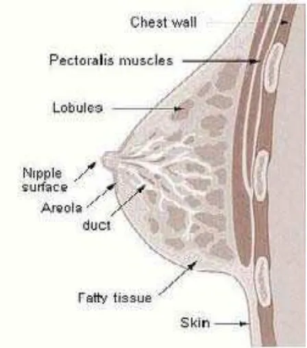
The breast is a modified sweat gland. In 7th week of intrauterinelife, they
are derived from the ectodermal milk lines bilaterally. Breast is an accessory
reproductiveorgan. They are made of glandular tissue containing 15 to
20lobes. Each lobe is made up of group of alveoli and it is drained by
lactiferousduct. Each duct has a dilatation called lactiferous sinusat its
termination beneath thenipple. Group of alveoli draining intoduct is called
Primary secretory unit. Myoepithelium is a single layer of epithelial cells
surrounds only the ducts not lining thelobules. They are contractile and aid the
[image:19.595.206.430.438.692.2]transport of secretions along the duct. Major part of thegland has fatty stroma.
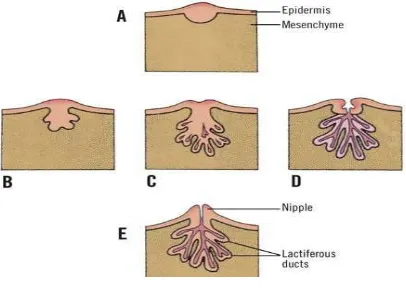
Fig: 2 Development of breast Plane:
The breast lies superficial to the pectoral fascia.
Extent:
Vertically- Second to the sixth rib
Horizontally- the lateral border of the sternum to mid-axillary line lies over
the following muscles:
• Pectoralis major
• Serrartusanterior
Axillary tail of Spence:
Extension of the breast that passes through the foramen of Langer and
therefore this part of breast is deep to the fascia. It has direct contact with
lymph node.
Ligament of cooper:
Anchored to the overlining skin and to the underlying fascia by bands
of these connective tissues.
Nipple:
Nipple is located at the level of fourth intercostal space. It is pierced by
15 to 20 lactiferous ducts which open independently to nipple. The stiffness of
nipple mainly due to circular and longitudinal muscles in it.
Areola:
Areola is a circular pigmented area around nipple. They are rich in
modified sebaceous glands. They secrete oily lubricant.
Blood supply of the breast:
The breast is rich in vascular supply.
• Internal mammary artery- a branch of subclavianartery
• Lateral thoracic
• Superior thoracic Branches of the axillaryartery
• Acromiothoracic
Venous supply:
The veins follow the arteries. Superficial veins drains into the
internal thoracic vein whereas the deep veins drains into the internal thoracic,
axillary and posterior intercostal veins.
The communication of posterior intercostals veins with the Batson’s
vertebral venous plexus is the basis of spread of breast malignancy to
vertebra and skull base.
Nerve supply:
• Anterior cutaneous branches of the 4th to 6th intercostals
nerves
• Lateral cutaneous branches of the 4th to 6th intercostals nerves.
• Sensory fibres to skin
• Autonomic fibres to blood vessels and smooth muscle.
Lymphatic drainage of breast:
Lymph from the breast drains into the following nodes.
Axillary nodes
Level 1:lateral to pectoralis minor
Includes anterior or pectoralgroup, posterior
orsubscapulargroup, lateral or humeralgroup
Level 2: posterior to pectoralis minor - Centralgroup
Internal mammary (parasternal nodes)
- situated along internal thoracic vessels
Interpectoral or rotters nodes: between pectoralis major and minor.
Supraclavicularnodes
Cephalic(deltopectoral)nodes,
Posterior intercostalsnodes
Subdiaphragmatic and subperitoneal lymphplexuses.
Few lymphatic pierce the pectoral fascia & enter thechest
Pattern of drainage:
•75% drains into axillary nodes
•20% drains into internal mammary nodes
•5% drains into posterior intercostalnodes.
• Among the axillary nodes, the anterior group drains mostly followed by
the posterior and apical group.
• The internal mammary nodes drain the lymph from both inner and outer
half of breast.
• A plexus of lymph vessels- sub areolar plexus of Sappey is present deep
to theareola.
• Lymphatics from the deeper surface of the breast pass through the
pectoralis major muscle and the clavipectoral fascia to reach the apical
• Lymphatics from the lower and inner quadrants of thebreast crosses the
costal margin and pierces the anterior abdominal wall through the upper
part of linea Alba and communicate with the subdiaphragmatic and
subperitoneal lymph plexuses.
PHYSIOLOGY OF BREAST: • Estrogen -ductal development
• Progesterone -epithelium and lobular development.
• Prolactin - lactogeneis during late pregnancy and the postpartum
period.
Prolactin results in the upregulation of hormone receptors and aids
in epithelial development.
Positive and negative feedback mechanisms exercise stringent
control of the hormones secreted from the hypothalamus and thepituitary.
Dramatic changes are noted in the female breast changes during
various developmental stages in adolescence, pregnancy, lactation and
senescence. The complex interactions among the various hormones are
responsible for these changes. Imbalances in these hormones result in
Fig 3: Regulation of Breast Development & Function
[image:25.595.169.460.390.611.2]Fig 4: Physiological stages of breast development Fig 3: Regulation of Breast Development & Function
Fig 4: Physiological stages of breast development Fig 3: Regulation of Breast Development & Function
CLINICAL PRESENTATION:
Women with carcinoma breast will present with the following complaints:
• Lump in the breast with or without nipple
retraction– most common
• An axillary nodemass.
• Symptoms due to metastaticdisease
Liver – abdominal pain / mass / obstructive jaundice
Lung – breathlessnes, cough with hemoptysis,
pleural effusion
Bone – pain over long bone site
• Discharge from nipple – rare presentation
• Skin changes:
Paeu d’orange
Is due to cutaneous lymphaticoedema. As the lymphtics
are blocked there is lymphatic oedema of the overlying skin and infiltrated skin
is tethered by the sweat glands.
It can’t swell there by giving an orange peelappearance.
Ulceration
RISK FACTOR FOR BREAST CANCER: Age:
Age is probably the most important risk factor for breast
cancerdevelopment. The incidence of breast cancer continues to increase with
advancing age of the female population. Breast cancer is rare in women
younger than 20 years and constitutes less than 2% of the total cases.
Thereafter, the incidence increases to 1 in 233 from ages 30 to 39 years, 1 in 69
from ages 40 to 49, 1 in 42 from ages 50 to 59, 1 in 29 from ages 60 to 69, and
1 in 8 by age 80 years. The women now have an average risk of 12.2% of being
diagnosed with breastcancer at some time during their lives.
Sex:
Sex is also an important risk factor because most breast cancers occur in
women. Breast cancer also occurs in men. Incidence of breast cancer in men is
less than 1% of the incidence in women.
Personal History of Breast Cancer:
The magnitude of risk depends on the age at diagnosis of the first
primary cancer, estrogen receptor (ER) status of the first primary cancer and
use of adjuvant systemic chemotherapy and endocrine therapy
Histologic Risk Factors:
Histologic abnormalities diagnosed by breast biopsy constitute an
important category of breast cancer risk factors. These abnormalities include
lobular carcinoma in situ (LCIS) and proliferative changes with atypia. LCIS is
premenopausal women. It is typically an incidental finding at biopsy for
another condition and does not manifest as a palpable mass or suspicious
microcalcifications on mammography.
HISTOLOGICAL DIAGNOSIS – RR
Nonproliferative disease - 1.0
Proliferative disease without atypia 1.3-1.9
Proliferative disease with atypia 3.7-4.2
Strong family history 4-9
LCIS >7
Family History of Breast Cancer and Genetic Risk Factors:
The relationship between family history of breast cancer and the risk for
breast cancer is a proven one. First-degree relatives (mothers, sisters, and
daughters) of patients with breast cancer have a twofold to threefold excess risk
for development of the disease. Risk is much higher if affected first-degree
relatives had premenopausal onset and bilateral breast cancer. Risk is not
significantly increased in women with distant relatives (cousins, aunts,
grandmothers) with breast cancer
GENETIC FACTORS:
Genetic factors are estimated to be responsible for 5% to 10% of all
breast cancer cases, but they may account for 25% of cases in women younger
The BRCA1 gene which was identified in chromosome 17(17q21).
Mutations in BRCA1account results for 40% of familial breast cancers. The
BRCA2 which was identified in chromosome 13 account for 30% of familial
carcinoma. In addition to carcinoma breast these patients have additional risk
for other carcinoma.
Reproductive Risk Factors:
Reproductive milestones that increase a woman’s lifetime estrogen
exposure are thought to increase her breast cancer risk. These include
• Onset of menarche before 12 years of age
• First live childbirth after age 30 years
• Nulliparity
• Menopause after age 55 years.
There is a 10% reduction in breast cancer risk for each 2-year delay in
menarche; the risk doubles with menopause after age 55. A first full-term
pregnancy before age 18 years is associated with half the risk for development
of breast cancer of a first full term pregnancy after age 30 years. In contrast to
family history or histologic factors, reproductive risk factors have a large
influence on breast cancer.
Exogenous hormone use:
Therapeutic or supplemental estrogen and progesterone are taken for
various conditions. The two most common reasons are contraception in
for use of exogenous hormones include menstrual irregularities, polycystic
ovaries, fertilitytreatment, and hormone insufficiency states.
Studies have suggested that breast cancer risk is increased in current or
past usersof oral contraceptives but that the risk decreases as the interval
after cessation of use increases.
These data show that women receiving combination HRT with estrogen
and progesterone for 5 years have approximately a 20% increased risk for the
development of breast cancer.
Women who take estrogen-only formulations (because of previous
hysterectomy) do not appear to be at increased risk for breast cancer
Non modifiable risk Factors: • Increasing age
• Female sex
• Menstrual factors
• Early age at menarche (onset of menses before age 12 yr)
• Older age at menopause (onset beyond age 55 yr)
• Nulliparity
• Family history of breast cancer
• Genetic predisposition (BRCA1 and BRCA2 mutation carriers)
• Personal history of breast cancer
• Race, ethnicity (white women have increased risk compared with
Modifiable risk factors: • Reproductive factors
• Age at frst live birth (full-term pregnancy after age 30 yr)
• Parity
• Lack of breastfeeding
• Obesity
• Alcohol consumption
• Tobacco smoking
• Use of hormone replacement therapy
• Decreased physical activity
• Shift work (night shifts)
Histologic Risk Factors:
• Proliferative breast disease
• Atypical ductal hyperplasia
• Atypical lobular hyperplasia
STAGING OF CARCINOMA: Non - invasive Epithelial Cancers
• Lobular carcinoma in situ
• Ductal carcinomain situ or intraductal carcinoma
Papillary
cribriform
solid
comedo
Invasive Epithelial Cancers
Invasive lobular carcinoma (10%)
Invasive ductal carcinoma:
• Invasive ductal carcinoma, not otherwise specifed (50%-70%)
• Tubular carcinoma (2%-3%)
• Mucinous or colloid carcinoma (2%-3%)
• Medullary carcinoma (5%)
• Invasive cribriform carcinoma (1%-3%)
• Invasive papillary carcinoma (1%-2%)
• Adenoid cystic carcinoma (1%)
Mixed Connective and Epithelial Tumors: • Phyllodes tumors, benign and malignant
• Carcinosarcoma
• Angiosarcoma
• Adenocarcinoma
TNM STAGING: Primary tumour (T):
TX - Primary tumor cannot be assessed
T0 - No evidence of primary tumor
Tis - Carcinoma in situ
Tis (DCIS) DCIS
Tis (LCIS) LCIS
Tis (Paget) Paget disease of the nipple not associated with invasive carcinoma
or carcinoma in situ (DCIS and/or LCIS) in underlying breast parenchyma
T1 Tumor ≤ 2 cm in greatest dimension
T1mi Tumor ≤1 mm in greatest dimension
T1a Tumor >1 mm but ≤5 mm in greatest dimension
T1b Tumor >5 mm but ≤10 mm in greatest dimension
T1c Tumor >10 mm but ≤20 mm in greatest dimension
T2 Tumor>2cm but ≤5cm in greatest dimension
T4 Tumor of any size with direct extension to the chest wall and/or to the skin
T4a Extension to the chest wall, not including only pectoralis muscle
adherence or invasion
T4b Ulceration and/or ipsilateral satellite nodules and/or edema of the skin
T4c Both T4a and T4b
T4d Inflammatory carcinoma
Regional Lymph Nodes (N):
Clinical stating
Nx Regional lymph nodes cannot be assessed (e.g., previously removed)
No regional lymph node metastases
N1 Metastases to movable ipsilateral level I, II axillary lymph node(s)
N2 Metastases in ipsilateral level I, II axillary lymph nodes that are clinically
fixed or matted; or in clinically detected* ipsilateral internal mammary nodes in
the absence of clinically evident axillary lymph node metastases
N2a Metastases in ipsilateral level I, II axillary lymph nodes fixed to one
another (matted) or to other structures
N2b Metastases only in clinically detected* ipsilateral internal mammary
nodes and in the absence of clinically evident level I, II axillary lymph node
metastases
N3 Metastasis in ipsilateral infraclavicular (level III axillary) lymph node(s)
with or without level I, II axillary lymph node involvement; or in clinically
level I, II axillary lymph node metastases; or metastases in ipsilateral
supraclavicular lymph node(s) with or without axillary or internal mammary
lymph node involvement
N3a Metastasis in ipsilateral infraclavicular lymph node(s)
N3b Metastasis in ipsilateral internal mammary lymph nodes(s) and axillary
lymph node(s)
N3c Metastasis in ipsilateral supraclavicular lymph node(s)
Pathological classification:
pNX Regional lymph nodes cannot be assessed
pN0 No regional lymph node metastasis
pN0 (i-) No regional lymph node metastasis histologically, negative IHC
pN0 (i+) Malignant cells in regional lymph nodes no greater than 0.2 mm
pN0 (mol-) No regional lymph node metastasis histologically, negative
molecular findings (IHC)
pN0 (mol+) Positive molecular fndings (RT-PCR), but no metastasis detected
by histology or IHC
pN1 Micrometastases; or metastases in 1-3 axillary nodes and/or in internal
mammary nodes with metastases detected by sentinel lymph node biopsy
but not clinically detected
pN1a Metastases in 1-3 axillary nodes; at least one metastasis >2.0 mm
pN1b Metastases in internal mammary nodes with micrometastasis or
macrometastases detected by sentinel lymph node biopsy (not clinically
detected)
pN1c Metastases in 1-3 axillary nodes and in internal mammary nodes with
micrometastases or macrometastases detected by sentinel lymph node biopsy
but not clinically detected
pN2 Metastases in 4-9 axillary nodes or in clinically detected internal
mammary lymph nodes in the absence of axillary lymph node metastases.
pN2a Metastases in 4-9 axillary nodes (at least one tumor deposit >2.0 mm)
pN2b Metastases in clinically detected internal mammary lymph nodes in the
absence of axillary lymph node metastases
pN3 Metastases in ≥10 axillary nodes; or in infraclavicular (level III axillary
nodes) or in clinically detected ipsilateral internal mammary lymph nodes in
the presence of one or more positive level I, II axillary nodes; or in >3 axillary
lymph nodes and internal mammary lymph nodes, with micrometastases or
macrometastases detected by sentinel lymph node biopsy but not clinically
Metastasis:
M0 No clinical or radiographic evidence of distant metastases
cM0(i+) No clinical or radiographic evidence of distant metastases, but
deposits of molecularly or microscopically detected tumor cells in circulating
blood,
bone marrow, or other nonregional nodal tissue that are no larger than 0.2 mm
in a patient without symptoms or signs of metastases
M1 distant detectable metastases as determined by classic clinical and
[image:37.595.191.441.339.720.2]radiographic means and/or histologically proven larger than 0.2 mm
PROGNOSTIC FACTORS: AGE
Age place an important factor in the prognosis of carcinoma breast
.Women diagnosed with breast cancer in their early age have a poorer
prognosis than women diagnosedinmiddle or olderage. There as on for this
unusual pattern is unclear. According to literature breast cancer occurring in
younger women is of more aggressive type.
They are usually,
• large size tumour
• Node positivity
• High grade
• Poorly differentiated
• High tumor invasive potential
• Greater chances of early lymphatic spread
• ER,PR negative, Her2 positive
Thus,thepoorer outcome could at least partly be due to differences in
these important prognostic factors.Recurrence of the tumour is also more
common in younger women than elderly women. In this study we studied the
OBESITY
Obesity adversely affects women’s health.Obesity in women is a
recognized risk factor for metabolic syndrome, type II diabetes mellitus,
cardiovascular disease and several major cancers including breast and
endometrial cancer. The risk of breast cancer increases with obesity and also
associated with poor prognosis of breast cancer. Obesity is measured as weight
gain, body mass index, waist–hip ratio or percent body fat. In our study obesity
is measured in terms of BMI. A one-point gain in BMI is estimated to increase
the risk of postmenopausal breast cancer by 3%.There are several mechanisms
[image:39.595.148.504.401.678.2]by which body weight and obesity affect the prognosis of breast cancer.
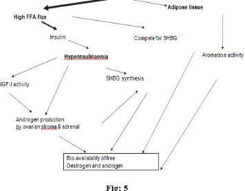
Hormones:
The breast cancer survivors, who are obese, have higher bio available
concentrations of tumour promoting hormones such as estrogen and
testosterone, which may contribute to poor survival. Obese women have 35%
higher concentrations of estrogen and 130% higher concentrations of estradiol
and increased Testosterone concentrations compared with nonobese woman.
Obesity is strongly and inversely associated with sex-hormone-binding
globulin (SHBG)levels,suggestingthatcentralobesitywithassociated
lowplasmalevelofSHBGspromotesbioavailabilityofandro- gens and estrogen.
Obese women may have hyperinsulinemia that promote mammary
carcinogenesisbyincreasingthelevelsofinsulin-likegrowthfactor. (IGF), free
fatty acid and leptin which have a synergistic effect to estrogen on mammary
epithelial cells by promoting angiogenesis and transcriptionalfactors.Leptin, a
type of adipocytokines that are produced by adipocytes have been associated
with carcinogenesis, tumour migration and invasion, enhancement of
angiogenesis and increased aromatase activity.
Nutrition:
The association between breast cancer risk and dietary factors has long
been recognised but the complex relationship between obesity, nutrition and
breast cancer is not fully understood. The regular physical activity after a
breast cancer diagnosis has a strong evidence toreducethe risk of death from
this disease but there is little direct evidence to show that lack ofphysical
TUMOUR SIZE:
Tumour size is one of the strongest prognostic indicators. When tumours
are detected at their early stage we can perform a breast conserving surgery.
When tumour size are larger it implies more number of tumour doubling time
and the chance of distance metastatis is also high.A larger tumour has been
related to more positive lymph nodes , thus their interaction further influences
the survival . Tumour size plays an important role in deciding neo adjuvant
chemotherapy. In this study the mean tumour size in premenopausal women
and post menopausal women and their relation with BMI are studied.
AXILLARY LYMPH NODE:
Axillary lymph node status is one of the most important prognostic
factors for breast carcinoma and a valuable indicator of long-term survival.
Node- positive patients have about a 4–8 times higher mortality than those
without nodal involvement. The more nodes involved the worse the prognosis.
Lymphnode clearance in mastectomy surgery depends on the positivity
of node. Practice has changed from full dissection and/or radiation of the axilla
to the use of sentinel node biopsy (SNB) for many patients. There is still debate
about what constitutes an adequate axillary dissection in terms of the total
number of lymph nodes removed. It is generally accepted that greater than 10
LNs are required .One study found that at least five and 10 nodes are required
for node-negative and node-positive patients respectively. The LN ratio (LNR),
defined as the number of positive LNs over the number of LNs removed. LNR
RECEPTOR STATUS:
The breast cancer is a heterogeneous disease with variable biological
and clinical characteristics because of its different genetic make-up. It is well
known that Proto-oncogenes and tumor suppressor genes are two classes of
genes that play a central role in the regulation of cell growth and
differentiation. So any alterations in one or more of these genes appear to play
an important role in the pathogenesis of most human malignancies.
[image:42.595.142.488.341.694.2]ESTROGEN RECEPTOR:
Estrogen receptors (ERs) are a group of proteins found inside cells.
They are activated by the hormone estrogen (17β-estradiol).Two types (G
protein-coupled receptors)
• 1.ERα
• 2.ERβ
MECHANISM OF ACTION:
ER receptors, once activated by estrogen translocate into nucleus and
binds to the DNA. This regulates the activity of different genes.
Distribution:
The ERα are found in
• Breast cancer cells
• Endometrium
• Ovarian stromal cells
• Hypothalamus.
• Efferent duct epithelium (males).
The ERβ are found in
• ovarian granulosa cells
• heart
• lungs
• prostate
• intestinal mucosa, endothelial cells
The affinity differs with different ligands for binding to alpha and beta
isoforms of the estrogen receptor. The agonistic and antagonistic effects of
Selective estrogen receptor modulators (SERMs) at selective sites are mainly
due to selective binding to either alpha or beta subtype of the receptor.
The ratio of α- to β concentration is important in certain
diseases.Tamoxifen is an antagonist in breast but an ER agonist in bone and is,
therefore, used as a breast cancer treatment and also prevent osteoporosis. But
it increases the risk of endometrial cancer due to its partial agonistic action
atendometrium.In around 70% of breast cancer cases, estrogen receptors are
over- expressed and they are referred to as "ER-positive".
ER POSITIVE TUMOUROGENESIS
TwoHypotheses has been described:
1. When estrogen binds to ER receptors, the proliferation of mammary
cells get stimulated leading to increase in cell division and DNA replication.
This leads to mutation.
2. Estrogen metabolism produces genotoxic waste leading to mutation.
Both these processes leads to Disruption of the cell cycle, apoptosis and DNA
repair Tumor formation
Other cancers associated with estrogen and its receptor is
• ovarian cancer
• endometrial cancer
• colon cancer
The concept of SERMs is based on the ability to initiate the interactions
of estrogen receptors with the proteins such as
• transcriptionalcoactivators
• Transcriptionalcorepressors.
Different tissues has different ratio of coactivator to corepressor
protein. Thus the same ligand may be
• agonist where the coactivatorspredominate
• antagonistic where the corepressors predominate
[image:45.595.161.451.430.707.2]PROGESTERONE RECEPTOR:
The progesterone receptor (PR) also known as NR3C3 is a protein
found inside cells. It is activated by the steroid hormone progesterone.In
humans, PR is encoded by a single PGR gene residing on chromosome 11q22.
Three forms
• PR-A
• PR-B,
• PR-C.
The PR-B - positive regulator
PR-A and PR-C - antagonize the effects of PR-B
• No binding hormone in receptor the carboxyl terminal of the
receptor inhibits transcription.
• Binding in receptor hormone structural change in the receptor
removes the inhibitory action of the carboxyl terminal.
• Progesterone antagonists prevent the structural reconfiguration of the
receptor.
After progesterone interacts with its receptor, dimerization occurs and
the Pg- PgR complex enters into the nucleus and then binds to DNA.It leads
to transcription and then formation of mRNA that is translated by the
Her 2 neu (erbb2) receptors:
HER-2/neu proto-oncogene (also called c-erbB2) is located on chromosome
17q11, which encodes for p185 a transmembrane glycoprotein with
tyrosinekinase activity that belongs to the family of epidermal growth factor
receptors.
HER2 can dimers with their receptors leading to phosphorylation of the
tyrosine residues and initiates a variety of signaling pathways. It promotes
cell proliferation and also opposes apoptosis.
HER-2/neu proto-oncogene amplification or over expression is
one of the most important alterations encountered in breast cancer. The
amplification or over expression of HER-2/neu may lead to following:
• Largetumor size
• high grade
• lack of steroid receptor expression
• axillary lymph nodes metastasis
• advanced stage,early relapse
• Reduced overall survival.
Increased recurrence rateHER2/neu proto-oncogene is amplified and or
over expressed in approximately in 25-30% of invasive primary breast
cancers .An association have become important because only patients
with tumors that over express HER-2/neu benefit from antibody-based
therapy with trastuzumab. For that reason, the determination of
.
Fig: 7
The estrogen, progesterone and the Her2 neu receptors are estimated
from the tissue sample using immunohistochemistry technique. Hormone
receptors were considered negative whenconcentration was below 10%. P53
was consideredpositive when nuclear staining was positive in morethan 10%
of tumoral cells. HER-2/neu overexpressionwas considered positive when
complete and intensemembrane staining was observed in more than 10%of
LYMPHOVASCULAR INVASION:
Lymphovascular invasion (LVI) is defined as tumor emboli present
within a definite endothelial-lined space in the breast surrounding invasive
carcinoma. The existence of LVI may help identify who is at increased risk for
axillary lymph node and metastasis.Lymphovascular invasion (LVI) is
currently considered as an important prognostic factor for primary breast
cancer. Previously it was reported that LVI is an independent prognostic factor
for disease-free survival (DFS) and overall survival (OS) of patients with breast
cancer.
Moreover, LVI was associated with the development of lymph node
metastases and lymphatic micro vessel density. Another clinical study
conducted by Krishnamurti et al showed that LVI may be associated with
peripheral tumor-infiltrating lymphocytes, which have a clinical significance in
breast cancer. A recent study by showed that breast cancer subtypes could
affect the outcomes relatedto LVI. The prognosis of hormone
receptor-negative breast cancers, such as TNBC, is suggested to be highly
influenced by LVI compared to that of hormone receptor-positive breast
INVESTIGATIONS:
NONINVASIVE TESTS MAMMOGRAPHY:
Conventional mammography delivers a radiation dose of 0.1 cGy per
study.
By comparison, chest radiography delivers 25% of this dose.
However, there is no increased breast cancer risk associated with the
radiation dose delivered with screening mammography.
Two types
• Screening.
• Diagnostic.
SCREENING MAMMOGRAPHY:
• Screening mammography is used to detect unexpected breast cancer in
asymptomatic women
• It supplements history taking and physical examination.
• For screening mammography two views of the breast are performed.
Medio lateral oblique (MLO).
Craniocaudal
(CC)
• The MLO view images the greatest volume of breast tissue, including
the upper outer quadrant and the axillary tail of Spence.
• The CC view provides better visualization of the medial aspect of the
• But because of lower specificity of single view screening
most radiologists believe that screening examination
should include both MLO and CC views.
DIAGNOSTIC MAMMOGRAPHY:
• Diagnostic mammography is used to evaluate women with abnormal
findings such as a breast mass or nipple discharge.
• Four views:
Medio lateral oblique (MLO).
Craniocaudal (CC).
90-degree lateral
spot compression
• The 90-degree lateral view is used along with the CC view to triangulate
the exact location of an abnormality.
• Spot compression may be done in any projection by using a small
compression device, which is placed directly over a mammographic
abnormality that is obscured by overlying tissues. Breast compression is a
must in mammography because
Holds breaststill.
Brings objects closer tofilm.
Separates overlapping tissue that might obscure
underlyinglesions.
Disadvantages of mammography:
Pain
Uncomfortable due to compression of breast
NORMAL FINDINGS:
Breast entirely fat.
Scattered fibro glandular densities.
Breast tissue is hetero genetically dense.
Extremely dense.
ABNORMAL FINDINGS:
Micro stippled calcifications
Dense stellate soft tissue mass with irregular margin
and spicky projection
Bilateral asymmetrical distribution of fibro glandular
tissue.
Architectural distortion
Increased thickness of skin due to lymphedema
Nipple retraction.
DIGITAL MAMMOGRAPHY:
This type records the radiography image electronically in a digital
format rather than a film. It is left in computer and is displayed on
fluorescentmonitor.
ULTRASOUND:
• It is useful in evaluation of dense breast
• For screening or diagnostic in women less
than 40 years
• To differentiate b/w cystic and solid masses.
Unfortunately
Masses that are smaller than 5 to 10 mm may not be visualized.
MRI:
• No radiation exposure
• To differentiate scar from recurrence
• Breast with implants
• Screening of high risk woman in younger
age
• Very small lesion can be determined
INVASIVE TESTS:
ASPIRATION CYTOLOGY:
It uses fine needle with a syringe to aspirate cells from a suspicious area
smearing them on a glass slide fixing them immediately (to prevent air
drying) staging for cytology.
22-23gauge needle attached to syringe is used. The air dried preparation
slide is stained with wrights /wrights Giemsa / May Grunnwald Giemsa. If not
air-dried it is immediately fixed with 95% alcohol and staining is done with H
& E stain / with pap stain.
Combination of physical examination, mammography and FNAC will
produce a diagnostic accuracy approaching 100%.FNAC is done in palpable
mass, mass on mammogram. Sensitivity of test is approximately 80%; false
negative varies b/w 2-10 %.
COMPLICATIONS OF FNAC:
• Growing out of tumor along needle tract, which is less in case of
caliber<20 gauze.
• Acutemastitis.
• Pneumothorax.
• Haematomas.
• Interval of 2 weeks required between FNAC and mammography as
they form hematomas and result in false positive mammographic
CORE NEEDLE BIOPSY:
• Standard of care for biopsy of breast lesion
• 11G core needle isused.
• More invasive – more number of wedge of tissues are
taken
• Able to differentiate invasive and in situ lesions
• Receptor status are assessed
• Lymphovascular lesion and grade of tumour are also
assessed
• Betteraccuracy
OPEN BIOPSY: Excision:
• When lump is less than 4 cm
• Removal of whole lump with 1 cm of clearance.
Incisional:
• When core biopsy is inconclusive
• Incision should be within the boundary of
mastectomy scar
• No diathermy should be used
Galactography:
Injection of contrast into one of the ducts through the orifice of the
TREATMENT OF CARCINOMA BREAST:
For treatment purpose carcinoma breast is divided into 3
• Early breast cancer
• Locally advanced carcinoma breast
• Metastatic breast carcinoma
EARLY CARCINOMA BREAST
Includes -
T1N0, T2NO, T1N1, T2N1, T3NO
The main mode of treatment in early breast carcinoma is breast conservative
surgery. It includes
BREAST CONSERVATIVE SURGERY:
• wide local excision of tumour
• axillary sampling
• post op radiotheraphy
• Regular follow up.
The breast specimen that is removed is oriented before sent for
pathology. This allows focal reexcision of involved margins rather than global
reexcision and thus improves the cosmetic result. The surgical defect created
after lumpectomy is closed in cosmetic fashion by using advancement flap
closure method.
Surgical staging of the axilla is performed through a separate incision in
most patients undergoing breast conservation. Anatomic axillary node
FACTORS INFLUENCING BREAST CONSERVATION SURGERY: Tumor Size.
Lumpectomy is considered regardless of size of tumour, if can be
excised with clear margins and an acceptable cosmetic result.
Tumors 5 cm in size, clinically positive nodes and tumors with lobular
and ductal histology were included in therandomized trials of mastectomy
versus breast-conserving therapy
Margins:
The positive margins were associated with a twofold
increase in ipsilateral breast tumor recurrence risk compared with
negativemargins. The clinicopathologic features, biology, endocrine therapy,
administration of radiotherapy have no significant effect
Histology
Invasive lobular cancers and cancers with an extensive intraductal
component can be treated with lumpectomy if clear margins canbe achieved.
Atypical hyperplasia (ductal and lobular) and LCISat resection margins do not
increase local recurrence rates.
Patient Age
Local recurrence rates after breast-conserving surgery are higher
for younger women than for older women. The use of radiation therapy
reduces local recurrence in patients of all ages.
receive radiotheraphy post operatively. Any patients who have
contraindication for RT are not the candidate for breast conservation
theraphy.
CONTRAINDICATION FOR RADIOTHERAPHY
Absolute
• Pregnancy
Relative
• Systemic scleroderma*
• Active systemic lupus erythematosus*
• Prior radiation to breast or chest wall
• Severe pulmonary disease
• Severe cardiac disease (if tumor is left-sided)
• Inability to lie supine
• Inability to abduct arm on affected side
• p53 mutation†
Fig: 8
Tumor that require mastectomy are following
• large tumour breast ratio
• Tumors with extensive calcifications on mammography
• tumors for which clear margins cannot be obtained on wide local
excision
• tumors in patients with contraindications to breast irradiation
• Patient preference for mastectomy or a desire to avoid radiation is also a
MODIFIED RADICAL MASTECTOMY:
Modified radical mastectomy refers to removal of
• The mammary gland
• nipple and areola
• Axillary lymph node dissection (level 1 and 2 sometimes
level 3) (ALND).
An elliptical skin incision is planned to include the nipple and areola and
usually any previous excisional biopsy scars. Skin flaps are raised to separate
the underlying gland from the overlying skin along the sub dermal plexus.
If immediate reconstruction is not planned, sufficient skin is taken to allow
smooth closure of skin flaps without redundant skin folds; this facilitates
comfortable use of breast prosthesis.
If immediate reconstruction is planned, a skin-sparing mastectomy may
be performed in which only the nipple-areola complex is removed and the
[image:60.595.173.473.524.690.2]maximum amount of skin is left for use in the reconstruction.
LOCALLY ADVANCED CARCINOMA BREAST:
METASTATIC CARCINOMA BREAST: • Only palliative care
• Toilet mastectomy
• Palliative chemotheraphy and radiotheraphy
OPERABLE
MRM
ADJUVANT CHEMOTHERATHY
RADIOTHERAPHY
LOCALLY ADVANCED CARCINOMA BREAST:
METASTATIC CARCINOMA BREAST:
Only palliative care
Toilet mastectomy
Palliative chemotheraphy and radiotheraphy
LABC
OPERABLE
MRM
ADJUVANT CHEMOTHERATHY
RADIOTHERAPHY
INOPERABLE
NEOADJUVANT CHEMOTHRAPHY
MRM
ADJUVANT CHREMOTHERAPHY
RADIOTHERAPHY
INOPERABLE
NEOADJUVANT CHEMOTHRAPHY
MRM
ADJUVANT CHREMOTHERAPHY
CHEMOTHERAPHY:
It is give as 3 forms
• Neo adjuvant
• Adjuvant
• Palliative
Despite advances in surgical treatment, most of the woman develops
metastasis with in 5 to 10 years. Hence chemotherapy is a most
important modality of treatment in carcinoma breast.
Most common used are anthracyclins and taxanes.
Most common regimen used is CMF regimen (cyclophosphamide,
Methotrexate, 5- fluorouracil
[image:62.595.110.520.482.663.2]Other regimens are CAF, AC, and CEF.
RADIATION THERAPHY:
Radiotherapy is mainly for locoregional control of disease. It is given as
• Adjuvant therapy
Brest conservation surgery
MRM
• Palliative therapy
Target volume: AFTER BCS • Whole breast RT
• Lumpectomy boost
• Regional nodes
AFTER MASTECTOMY • Chest wall
• Scar
• Regional nodes
DOSE OF RADIATION Conventional:
50 Gy in 25 daily fractions - 5 weeks
Hypo fraction:
BREAST BOOST RADIATION TO TUMIUR BED:
16 Gy in 8 daily fractions- 1.5 weeks
10 Gy in 5 daily fractions- 1 week
LYMPH NODE RADIATION:
50 Gy in 25 daily fractions - 5 weeks
40 Gy in 15 daily fractions - 3 weeks
PALLIATIVE DOSE:
30 Gy / 10#
[image:64.595.123.513.428.612.2]8 Gy / 1#
METHODOLOGY:
Study Design Observational study (Prospective)
Sample Size 160
Inclusion Criteria
All the carcinoma breast patients diagnosed clinically and
confirmed by trucut biopsy undergoing MRM
BODY MASS INDEX – 16 to 40(Normal to severely
obese)
BMI: 16 -25 Normal
BMI: 25 – 40 Overweight and obese
Exclusion Criteria
Advanced carcinoma of breast
Carcinoma breast with metastasis
Undergoing neoadjuvant chemotherephy
Very severely obese patients(BM >.40)
Severely underweight (BMI <16)
Investigations
All Patients who fit the inclusion criteria will be observed and following data collected
• USG breast (age less than 40 years) / mammogram of other breast (age more than 40 years)
• Trucut biopsy
• CT Chest
• Skeletal survey/ Bone scan
• USG abdomen
METHODOLOGY
All patients who fulfill the inclusion criteria will be
enrolled.
Written informed consents will be obtained.
Biopsy proven patients only will be included in the study.
Trucut biopsy will be taken and samples were submitted
for histopathology, determination of estrogen,
progesterone receptor expression and HER-2/neu status.
Associations with other characteristics like age,
menopausal status, body mass index, tumour size, and
node will also be studied.
I will do the investigations mentioned above.
Will stage the carcinoma breast based on TNM
classification Based on staging will proceed with modified
radical mastectomy followed by adjuvant theraphy.
The resected segment will be sent for histopathology to
SAMPLE SIZE CALCULATION:
Based on the study titled “comparison of ER, PR and HER-2/neu
(c-erbB2) reactivity pattern with histologic grade, tumour size and lymph node
status in breast cancer done by Azizun - Nisa, bhurgri Y, Raza F Kayani N
When Z = 1.96 for α 0.05; P= 24.7; q = 75.3 d is fixed as 7%
= 146
Assuming a non response rate of 10%, sample size is fixed at 146+14=160
The collected data were analysed with IBM.SPSS statistics software
23.0 Version. To describe about the data descriptive statistics frequency
analysis, percentage analysis were used for categorical variables and the
mean & S.D were used for continuous variables. To find the significance in
categorical data Chi-Square test was used similarly if the expected cell
frequency is less than 5 in 2×2 tables then the Fisher's Exact was used. The
Odds ratio and relative risk was used to find the effect of cause. In all the
above statistical tools the probability value .05 is considered as significant
level
.
2
2
d
pq
Z
n
=
7
7
3
.
75
7
.
24
96
.
1
96
.
1
×
×
×
×
=
RESULTS
About 160 women who were affected with carcinoma breast were
included in this study based on my inclusion and exclusion criteria. All women
are biopsy proven carcinoma breast and underwent surgery as primary
treatment.
AGE DISTRIBUTION:
Among the 160 women, 40(25%) were under 40 years, 56(35%) were
between 41 to 50 years, 41(25.6%) were between 51 to 60 years, about 20
(12.5%) were between 61 to 70 years and only 3 (1.9%) were above 70 years.
Thus most of the patients in my study were between 41 to 50 years.
AGE Frequency Percent
Valid Up to 40 yrs. 40 25.0
41 - 50 yrs. 56 35.0
51 - 60 yrs. 41 25.6
61 - 70 yrs. 20 12.5
Above 70 yrs. 3 1.9
[image:70.595.107.500.415.637.2]Total 160 100.0
Mean age of women getting carcinoma breast in my study is 48.57 years
of age.
0.0 5.0 10.0 15.0 20.0 25.0 30.0 35.0
Upto 40 yrs 25.0
Graph: 1
Mean age of women getting carcinoma breast in my study is 48.57 years
41 - 50 yrs 51 - 60 yrs 61 - 70 yrs Above 70 yrs
35.0
25.6
12.5
Age distribution%
Mean age of women getting carcinoma breast in my study is 48.57 years
In my study, i took the women having BMI between 16 to 25 as normal
and BMI between 26 to 40 as obese. Among 160 women, 89(55.6%) were
normal BMI whereas
Valid
Table: 5
In my study the mean BMI value of woman getting carcinoma breast
was 25.019 years of age.
BMI:
, i took the women having BMI between 16 to 25 as normal
and BMI between 26 to 40 as obese. Among 160 women, 89(55.6%) were
whereas 71(44.4%) were obese.
BMI
Frequency Percent
Valid Normal 89 55.6
Obese 71 44.4
Total 160 100.0
Graph: 2
In my study the mean BMI value of woman getting carcinoma breast
years of age.
Normal 89 56% Obese
71 44%
BMI
Normal Obese
, i took the women having BMI between 16 to 25 as normal
and BMI between 26 to 40 as obese. Among 160 women, 89(55.6%) were
In my study the mean BMI value of woman getting carcinoma breast Normal
In my study
menopausewhereas
menopause.
Among the 160 carcinoma patients 74(46%) patients have
lymphovascular invasion where remaining 86(54%) has no lymphovascular
invasion.
MENOPAUSAL STATUS:
In my study, 86(54%) women had breast cancer after
74(46%) of women had breast cancer before attaining
Graph: 3
LYMPHOVASCULAR INVASION
Among the 160 carcinoma patients 74(46%) patients have
lymphovascular invasion where remaining 86(54%) has no lymphovascular
Graph: 4
Attained 74 46% Not attained
86 54%
Menopause
Attained Not attained
Positive 74 46% Negative
86 54%
Lymphvascular invasion
Positive Negative
(54%) women had breast cancer after attaining
ast cancer before attaining
Among the 160 carcinoma patients 74(46%) patients have
lymphovascular invasion where remaining 86(54%) has no lymphovascular
ER, PR AND HER 2 NEU RECEPTOR STATUS:
In this study of 160 carcinoma breast women, 54% has ER receptor
positive status, 60 % has PR receptor positive status and 49.4 % has HER 2
NEU receptor positive status. Among them 36.3% have both ER, PR receptor
positive status.
ER Positive 45.6 46% ER NEGATIVE
54.4 54% 0 0%
ER STATUS IN CA BREAST
ER Positive
ER NEGATIVE
PR Positive
40.0 40% PR
Negative 60.0 60%
0 0%
PR STATUS IN CA BREAST
PR Positive
Descriptive Statistics
In this study the following mean values are observed
N
AGE_A 160
HEIGHT 160
WEIGHT 160
BMI 160
Valid N (list wise)
160 HER 2 NEU Negative, 49.4 , 0
HER 2 NEU STATUS IN CA BREAST
Graph: 5
In this study the following mean values are observed
N Minimum Maximum
160 24 76
160 143 171
160 40 86
160 19.0 34.4
[image:75.595.117.513.65.347.2]160
Table: 6
HER 2 NEU Positive, 50.6 HER 2 NEU
Negative, 49.4
HER 2 NEU STATUS IN CA BREAST
HER 2 NEU Positive
HER 2 NEU Negative
Mean
Std.
Deviation
48.74 9.812
155.10 5.786
60.08 9.229
25.019 3.6763
HER 2 NEU STATUS IN CA BREAST
HER 2 NEU Positive
CORRELATION DATAS:
In this study on carcinoma breast the following prognostic factors were
correlated and their relationship was
1. Receptor status with lymphovascular ER receptor status with lymphovascula
In this study among the patient with lymphovascular invasion,
(77%) has negative ER receptor status whereas
invasion majority show
ER -ve
ER +ve 0% 10% 20% 30% 40% 50% 60% 70% 80% 90% 100%
E
R
S
T
A
T
U
S
ER with Lymphovascular invasion
CORRELATION DATAS:In this study on carcinoma breast the following prognostic factors were
relationship was studied.
status with lymphovascular invasion: ER receptor status with lymphovascular invasion:
In this study among the patient with lymphovascular invasion,
77%) has negative ER receptor status whereas those without lymphovascular
invasion majority show positive ER receptor (65.1%) status.
Graph: 6
LVI +ve LVI -ve
77.0% 34.9%
23.0% 65.1%
23.0%
65.1% 77.0%
34.9%
ER with Lymphovascular invasion
In this study on carcinoma breast the following prognostic factors were
In this study among the patient with lymphovascular invasion, majority
those without lymphovascular
PR RECEPTOR STATUS WI
In this study among the patient with lymphovascular invasion,
(86.5%) has negative PR receptor status whereas
invasion majority show
0% 10% 20% 30% 40% 50% 60% 70% 80% 90% 100%
PR -ve
PR +ve
PR with Lymphovascular invasion
PR RECEPTOR STATUS WITH LYMPHOVASCULAR INVASION:
In this study among the patient with lymphovascular invasion,
86.5%) has negative PR receptor status whereas those without lymphovascular
invasion majority show positive PR receptor (62.8%) status.
Graph: 7
LVI +ve LVI -ve
86.5% 37.2%
13.5% 62.8%
13.5%
62.8% 86.5%
37.2%
PR with Lymphovascular invasion
TH LYMPHOVASCULAR INVASION:
In this study among the patient with lymphovascular invasion, majority
those without lymphovascular
HER 2 NEU RECEPTOR STATUS WITH LYMPHOVASCULAR INVASION:
In this study among the patients having lymphovascular invasion,
44.60% has positive HER 2 NEU status whereas 55.4 % has HER 2 NEU
negative status.
GRAPH: 8
HER 2 NEU POSITIVE HER 2 NEU
NEGATIVE
LVI NEGATIVE 55.80% 44.20%
LVI POSITIVE 44.60% 55.40%
44.60% 55.40%
55.80% 44.20%
0.00% 20.00% 40.00% 60.00% 80.00% 100.00% 120.00%
BOTH ER, PR RECEPTOR STATUS WIT
Among the patients affected with lymphovascular invasion only 13.5 %
shows positive ER, PR
receptors status has no lymphovascular invasion.
0% 10% 20% 30% 40% 50% 60% 70% 80% 90% 100%
Any 1+ve/Both -ve
ER & PR +ve
Both ER & PR +ve with LVI
RECEPTOR STATUS WITH LYMPHOVASCULAR INVASION:
Among the patients affected with lymphovascular invasion only 13.5 %
positive ER, PR receptor status whereas 55.8% of positive ER
receptors status has no lymphovascular invasion.
Graph: 9
LVI +ve LVI -ve
ve 86.5% 44.2%
13.5% 55.8%
13.5%
55.8% 86.5%
44.2%
Both ER & PR +ve with LVI
H LYMPHOVASCULAR
Among the patients affected with lymphovascular invasion only 13.5 %
2. Receptor status with
ER RECETOR STATUS VS BMI:
In this study
positive status has normal BMI where
negative ER receptor status
0% 10% 20% 30% 40% 50% 60% 70% 80% 90% 100%
Normal
Obese
with BMI:
ER RECETOR STATUS VS BMI:
In this study 75.3 %( majority) of carcinoma breast patients
positive status has normal BMI whereas only 24.7 % are obese
negative ER receptor status majority (60.9%) were obese.
Graph: 10
ER Positive ER Negative
75.3% 39.1%
24.7% 60.9%
24.7%
60.9% 75.3%
39.1%
ER STATUS VS BMI
) of carcinoma breast patients with ER
PR RECETOR STATUS VS BMI:
In this study 79.63 %( majority) of carcinoma breast patients with PR
positive status has normal BMI whereas 23.4 % are obese
negative PR receptor status 58.3% were obese.
0.0% 20.0% 40.0% 60.0% 80.0% 100.0%
Normal
Obese
TOR STATUS VS BMI:
In this study 79.63 %( majority) of carcinoma breast patients with PR
positive status has normal BMI whereas 23.4 % are obese
negative PR receptor status 58.3% were obese.
GRAPH: 11
PR Positive PR Negative
76.6% 41.7%
23.4% 58.3%
23.4%
58.3% 76.6%
41.7%
PR STATUS WITH BMI
In this study 79.63 %( majority) of carcinoma breast patients with PR
HER 2 NEU RECETOR STATUS VS BMI
In this study among the patients having POSITIVE HER 2 NEU,
%( majority) has normal BMI whereas 38.3% are obese.
0.00% 20.00% 40.00% 60.00% 80.00% 100.00%
HER2 NEU POSITIVE OBESE
NORMAL
HER 2 NEU RECETOR STATUS VS BMI:
In this study among the patients having POSITIVE HER 2 NEU,
) has normal BMI whereas 38.3% are obese.
Graph: 12
HER2 NEU POSITIVE HER 2 NEU NEGATIVE