THESES, SIS/LIBRARY R.G. MENZIES BUILDING N0.2 Australian National University Canberra ACT 0200 Australia
USE OF THESES
This copy is supplied for purposes
of private study and research only.
Passages from the thesis may not be
copied or closely paraphrased without the
written consent of the author.
Telephone: -+61 2 6125 4631 Facsimile: -+61 2 6125 4063
' 1 - i
By
Johannes Marra
A Thesis submitted for the degree of Doctor of Philosophy
at the Australian National University
PREFACE
This dissertation is an account of research carried out at the Department of Ap-plied Mathematics, Research School of Physical Sciences, Australian National Univer-sity, between September 1982 and December 1985.
Except where otherwise stated, the work is my own. The material in Chapter 4 is the result of research carried out in collaboration with Dr J.N. Israelachvili.
None of the work reported here has been submitted to any other institution of learning for any degree.
PUB LI CA TIO NS
1. J. Marra, H.A. van der Schee, G.J. Fleer, and J. Lyklema. "Polyelectrolyte Ad-sorption from Saline Solutions", in Adsorption from Solution, Ottewill, R.H., Rochester, C.H., and Smith, A.L. (eds.) (Academic Press, London, 1983), 245.
2. J. Marra. "Controlled Deposition of Lipid Monolayers and Bilayers onto Mica and Direct Force Measurements between Galactolipid Bilayers in Aqueous Solutions", J. Colloid Interj. Sci. 107 (1985) 446.
3. J. Marra. "Direct Measurements of Attractive Van der Waals and Adhesion Forces between Uncharged Lipid Bilayers in Aqueous Solutions", J. Colloid Inter
f.
Sci. (1986) in press.4. J. Marra and .1. N. TsraP.]achvili. "Direct Measurements of Forces bet.ween
Phos-phatidylcholine and Phosphatidylethanolamine Bilayers in Aqueous Electrolyte Solutions", Biochemistry 24 (1985) 4608.
5. J.N. Israelachvili and J. Marra. "Direct Methods for Measuring Conformational Water Forces (Hydration Forces) bet\veen Mernbrane and Other Surfaces",
Methods in Enzymology (1986) in press.
6. J. Marra. "Direct Measurement of the Interaction between Phosphatidylglycerol Bilayers in Aqueous Electrolyte Solutions", Biophys. J. (1986) (in press).
7. J. Marra. "Effects of Counterion Specificity on the Interaction between Quater-nary Ammoniurn Surfactants in Monolayers and 11ilayers", J. F'hys. (Jhem.
ACKNOWLEDGEMENTS
I would like to thank the members of the Department of Applied Mathematics, Research School of Physical Sciences, Australian National University, for kindly receiv-ing me and improvreceiv-ing my physical insight and scientific knowledge.
Let me first of all acknowledge my supervisors Drs J.N. lsraelachvili and R.G. Horn who initiated me into membrane biophysics and the direct force measurement tech-nique. Without their guidance and advice, this work would not have been possible. Our senior technical officer, Mr A.R. Reading, deserves praise for his professional and always
friendly technical assistance in the laboratory.
During the past three years, I have greatly benefitted from many stimulating dis-cussions, not only on matters scientific, with Professor Ninharn and Drs R.M. Pashley,
S. Marcelja, and H.K. Christenson. Dr Pashley generously provided me with the com-puter program for computing the theoretical double-layer forces.
From outside the department, I thank Professor S.G.A. McLaughlin and Dr D. Sornette for their inspiring discussions and suggestions, the latter also for playing many enjoyable. tennis matches with me. Many thanks go to Norma Chin for her profes-sional typing of this thesis.
ABSTRACT
Direct measurements are reported of the full interbilayer force laws (force vs. distance) between bilayers of zwitterionic phosphatidylcholines (PC) and phosphatidyl-ethanolamines (PE), uncharged galactolipids, anionic phosphatidylglycerols (PG), and cationic dioctadecyldimethylammonium surfactants (DOA) in aqueous electrolyte solu-tions. Bilayers were in each case deposited on molecularly smooth mica surfaces and the interbilayer forces then measured at a distance resolution of 1-2
A.
Three types of forces were identified: attractive van der \Vaals forces, repulsive electrostatic double-layerforces, and (at short range) repulsive steric-hydration forces.
Accurate measurements have been made of the van der \Vaals forces between
un-charged bilayers. In high salt, the van der Waals force between galactolipid bilayers is screened to about half its strength in pure water, which agrees with the theoretical prediction. On the other hand, the van der Waals force between uncharged PE or PC bilayers is already quite weak in pure water. It is proposed that the high concentration of ionic groups between the bilayer surfaces may already screen the van der Waals force in pure water.
Double-layer forces between bilayers arise when the bilayers carry a net surface charge. This occurs when the arnphiphile headgroups are ionized or, in case they are zwitterionic, when ion binding takes place. Fron1 the rneasured double-layer forces as a
function of the bilayer separation and electrolyte concentration, the binding of various
cations to PB, PC and PG, and the binding of various anions to DOA surfactants is investigated. Excellent agreement of the measured double-layer forces with theory is ob-tained. Slight deviations only occur at surface separations less than 25
A,
which might be related to discrete charge effects and/or ion-ion correlation effects.A short-range steric-hydration repulsion was observed only bet\veen uncharged
which balances the van der Waals force at separations of 10-30
A,
is apparently due to a co1nbination of hydration and steric repulsion, the latter arising frorn thcrrnal motions ofheadgroups and thickness fluctuations of fluid bilayers. No repulsive hydration forces were measured between the ionic PG and DOA bilayers.
It is sho\vn that for two bilayers in "contact" at. their equilibrium separation, their
adhesion energies vary on addition of salt due to changes in the repulsive double-layer and hydration forces rather than to a change in the attractive van der Waals force.
Also, it is concluded that bilayer fusion is not simply related to the interbilayer force-law, but must be related to a structural instability of the membrane (see also Horn
(1984)).
An idea about the relative intrabilayer interactions and the equilibrium headgroup area of the amphiphiles in bilayers is obtained by comparing the monolayer compression isotherms for different electrolyte solutions. It emerged that a correlation between inter-bilayer and intrainter-bilayer interactions can only be drawn to a limited extend, which is
mainly due to the complexity of the short-range forces and counterion specificities
Table of Contents
PREFACE ii
PUBLICATIONS iii
ACKNOWLEDGEMENTS iv
ABSTRACT v
CHAPTER 1. THE INTERACTIONS BETWEEN AMPHIPHILIC 1
SURF ACES: GENERAL OVERVIEW
1.1 INTRODUCTION 1
1.2 DOUBLE-LA YER FORCES 4
1.3 THE ELECTRICAL DOUBLE-LA YER FREE ENERGY OF A 10
CHARGED AMPHIPHILIC MONOLA YER AT THE
AIR/WATER INTERFACE
1.4 VAN DER WAALS FORCES AND HYDROPHOBIC FORCES 11
1.5 HYDRATION FORCES 14
1.6 AIMS AND OUTLINE OF THIS WORK 15
CHAPTER 2. MATERIALS AND EXPERIMENTAL METHODS 18
2.1 MATERIALS 18
2.2 SURF ACE TENSION MEASUREMENT TECHNIQUE 19
2.3 LANGMUIR-BLODGETT DEPOSITION TECHNIQUE 21
2.4 THE DIRECT FORCE MEASUREMENT TECHNIQUE 22
CHAPTER 3. FORCES BETWEEN GALACTOLIPID BILAYERS 28
3.1 INTRODUCTION 28
3.2 RESULTS 28
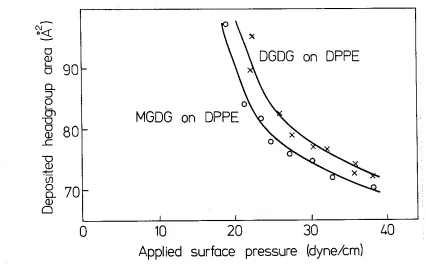
3.2.1 Monolayer Compression Isotherms and Langmuir- 28 Blodgett Deposition
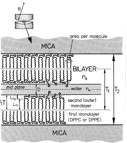
3.2.2 Thickness of the Deposited Bilayers and Operational 29 Definition of the Bilayer-Water Interface
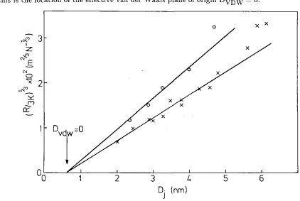
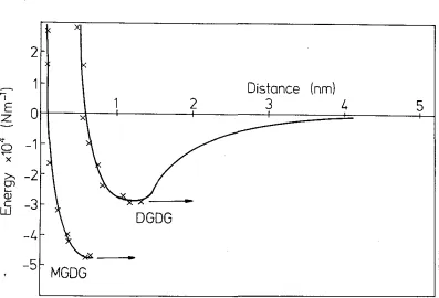
3.2.3 Van der Waals Forces and Hydration Forces between 31 Galactolipid Bilayers
3.3 DISCUSSION 35
CHAPTER 4. FORCES BETWEEN BILAYERS OF THE ZWIT- 39
TERIONIC PHOSPHOLIPIDS PC AND PE IN AQUEOUS SOLUTIONS
4.1 INTRODUCTION 39
4.2 RESULTS 40
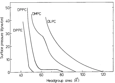
4.2.1 Monolayer Compression Isotherms of PC and PE 40
4.2.2 Deposition of PE and PC Bilayers onto Mica 42
4.2.3 The Bilayer-Water Interface and the Bilayer Thickness 45
4.2.4 Forces in Pure Water 47
4.2.6 Forces in Divalent Electrolyte Solutions 54
4.2. 7 Fusion 61
4.3 DISCUSSION 63
CHAPTER 5. FORCES BETWEEN MOLECULES IN MONOLAYERS 72
AND BETWEEN BILAYERS OF PHOSPHATIDYLGLYCEROL
5.1 INTRODUCTION 72
5.2 RESULTS 74
5.2.1 Monolayer Compression Isothern1s of DMPG and DSPG 74
5.2.2 Langmuir-Blodgett Deposition of PG on Mica 74
5.2.3 Forces between DSPG Bilayers 75
5.2.4 Forces between DMPG Bilayers 81
5.2.5 The Contraction of a DMPG Monolayer at the Air-Water 84 Interface following Addition of CaCI?
5.3 DISCUSSION - 85
CHAPTER 6. EFFECTS OF COUNTERION SPECIFICITY ON THE 89
INTERACTIONS BETWEEN QUATERNARY AMMONIUM SURF ACT ANTS IN MONOLAYERS AND BILAYERS
6.1 INTRODUCTION 89
6.2 RESULTS 92
6.2.1 Monolayer C0111pression Isotherms 92
6.2.2 The Deposited Headgroup Area 93
6.2.3 Forces between DOA Bilayers 94
6.3 DISCUSSION 99
CHAPTER 7. SUMMARY OF THE RESULTS AND POTENTIAL 102
FOR FUTURE STUDIES
7.1 SUMMARY AND CONCLUSIONS 102
7.2 POTENTIAL FOR FUTURE STUDIES 106
CHAPTER 1
THE INTERACTIONS BETWEEN AMPHIPHILIC
SURF ACES: GENERAL OVERVIEW
I.I INTRODUCTION
In the past decade, considerable progress has been achieved in the determination of surface forces, in particular their range and magnitude (lsraelachvili, 1985a). At the current level of understanding, an arbitrary distinction is usually made between long-range forces and short-range forces. Long-range forces cornprise the electrostatic
double-layer force and the electrodynamic van der Waals force. Their properties form the basis of the DLVO theory (Verwcy and Overbeek, 1948) which is regarded as a cornerstone of colloid science. A 1nuch lesser understood long-range force which only
recently has been measured between hydrocarbon surfaces in water (Israelachvili and Pashley, 1982) is the strongly attractive hydrophobic force.
Additional short-range forces arise from the differences rn the organization and structuring of solvent molecules near the surfaces as cornpared to solvent molecules in
the bulk of the solution, and are generally referred to as salvation forces. Of particular interest is the tight association and/or polarization of water molecules near hydrophilic surfaces like phospholipid bilayers (Lis et al., 1982a) and the adsorption of hydrated ions to mineral surfaces (e.g. day) (Van Olphcn, 1977; Pashley, 1981). These phenomena give risr to a strongly repulsive and/or oscillatory short-range force called the hydration
force. \iVhereas continuu1n theory is quite able to explain the general properties of
long-range forces, the rnolccular nature of solvent 1nolecules 1nust be considered to
explain short-range forces.
surfactant monolayers across water is necessary for understanding the properties of
many colloidal and biological systcrns. The interaction between micelles, vesicles and
surfactant coated colloidal particles determines whether they remain dispersed or
whether they aggregate and/or precipitate out of solution. Effective detergent action
and the formation and stability of soap films, foams, and liquid-crystalline surfactant
phases are ultimately dependent on these interactions as well. In the spontaneous
formation process of surfactant aggregates like vesicles and micelles, the shape and the
size of the assemblies in concentrated solutions bears a relationship to the interaction between them (Brady et al., 1985). More generally: the explanation of the phase
behaviour of surfactant/water and surfactant/water/oil systems (e.g. microemulsions) is
only possible from a consideration of the interactions existing in these systems.
ln the biological area: membrane-membrane and vesicle-rnembrane interactions
underlie all phenomena involving intermembrane coupling such as adhesion, membrane
stacking, and fusion. They are relevant to the understanding of biological phenomena
like endocytosis, exocytosis, fertilization, and cell recognition in immunological processes. In the field of neurobiology, the trans1nission of i1npulses across a synapse
seems to be related to the aggregation and fusion of synaptic vesicles along the
presynaptic membrane (Gray, 1973). In the area of photosynthesis, it is believed that
the stacking of thylakoid membranes is involved in the photophosphorylation process
(Punnett, 1970; Chow et al., 1981).
Many biological membranes contain 50% or n1orc protein by weight, and given
that these proteins often protrude beyond the lipid hcadgroups, it is clear that when two
membranes approach each other, it are the proteins that 'see each other first' and so arc
likely to do1ninatc intern1crnbranc interactions. Th(' observations that the aggregation
and fusion of 1nc1nbranes is often protein specific (r~akcr et al., 1980; I-long ct al., 1982)
and that the fusion of rnodel rnernbranes can be triggered by microrr1olar amounts of
Ca2 t only in the presence of Ca-specific proteins further enhance this vie\V. On the other hand, fusion also occurs bet\veen pure phospholipid bilayers (J>apahadjopoulos,
lywlecithin, phosphatidylcthanolarnine, etc.) are effective fusogens. The role of lipids rr1ay therefore not be a purely passive one~ and it is almost certain that the total interaction between any tv..'o real membranes is a cooperative process involving the
concerted action of both the lipids and proteins.
Apart from intermembrane interaction, also intrarr1embrane interactions deserve
attention. One can distinguish between lateral interactions in the hydrocarbon interior
of the bilayer and lateral interactions in the polar headgroup region of the bilayer. The hydrophobic bilayer interior is a relatively homogeneous region of hydrocarbon chains interacting through fairly well-understood van der Waals attractive and steric repulsive forces (Seelig and Seelig, 1974; Marcelja, 1974; Schindler and Seelig, 1975; Gruen, 1980). By contrast, the hydrophilic polar headgroup region containing hydrated and/or ionized charged groups in contact with the aqueous medium is far less understood. Here the interactions are very different, and include steric interactions, dipolar and electrostatic
forces involving specifically bound and free countcrions, strongly associated water and
hydrogen bonds. All these factors contribute to the physical state of the bilayer surface.
I_,ateral interactions between n1olecules determine the effective shape of the
molecules (Israelachvili et al., 1976; Mitchell and Ninham, 1981; Ninham et al., 1983) and their self-assembly into aggregate structures. A detailed knowledge of the effective shape of the molecules allows us to predict size and geometry of the aggregates. In mixtures of amphiphiles, the often observed phase separation of the molecular species is a consequence of differences in the lateral interactions bet\veen the species (Findlay and
Barton, 1978). In a nun1b('r of cases, the fusion bet\veen phospholipid vesicles has been
explained as the result of a difference in lateral interactions between the lipid headgroups on the inner membrane leaflet and between the lipid headgroups on the outer n1err1brane leaflet of the vesicle, leading to a structural instability of the rr1e1nbrane
(Cohen et al.1 1980; I..iiao et al., 1979). Long-range interactions determine \vhcthcr t\vo
vesicles in thernial motion ran approach each other close enough to come into adhesive
intra-bilayer interactions. Because both types of interaction are largely determined by
the chemical nature and properties of the bilayer surface, it can be anticipated that correlations exist between these two (Israclachvili and Sornette, 1985). Together, these forces give bilayers and membranes their highly versatile and complex properties.
1.2 DOUBLE-LA YER FORCES
Many amphiphilic molecules carry an electrical charge due to ionization of or ion
binding to the polar headgroup. When the amphiphiles assemble into aggregates like bilayers, the inter-bilayer interaction is to a large extent determined by the electrostatic forces between the bilayers. In this section, the relevant formulae will be given that describe these forces. The present approach follows the procedure given by Chan, Pashley and White (1980).
In an electrolyte solution, an electrically charged surface gives rise to a diffuse
space charge of opposite sign near the surface. It extends some distance into the solution
and essentially screens the surface charge. The properties of this so-called diffuse double-layer charge arc described by the Gouy-Chaprnan theory (Verwey and Overbeek, 1948).
Now, when two charged planar surfaces approach each other, at son1e distance the
diffuse double-layers of the surfaces will overlap, giving rise to an electrostatic repulsion
between the surfaces. The repulsion is caused by the perturbation of the isolated diffuse double-layers of the surfaces through the presence of the other surface, which increases the free energy and can be treated as an osn1otic pressure bet\veen the L\VO surfaces.
This osmotic pressure is related to the the difference in ion concentration in the region
between the surfaces con1pared with the bulk concentration. \,Vhen the distribution of
ions in this region is known, it becomes possible to calculate the surface rr-pulsion. 'I'his can be done by applying the Gouy-Chapman model of the diffuse double-layer, which uses the Poisson equation 1,o describe the potential gradient bcL\veen the surfaces:
(
(1)
constant t
(t
= 80 for aqueous media).1/'(x)
is the 111can local electrostatic potential. In the special case of two equally charged surfaces, it is obvious that the potential profile and ion distribution bet\veen the surfces must be symmetric about the midplane x=
rnbetween the surfaces. Then, Eq. (I) easily allows us to calculate the charge density u on the surface, since the total accumulated space charge in the x direction per unit area
from x ==- 0 to x
=
m must balance the surface charge per unit area, i.e.<J = -Jx=m p( x )dx =
x=O [
<
d,Pl
x=m t(d,P)
411" dx x=O = - 411" dx x=O·
(2)
Using the Boltzmann distribution for each ionic species ni with a bulk concentration ni(oo) and a valency zi, we have for ni(x):
ni(x) = ni(oo) exp(-zie,P(x)/kT).
(3)
Together with the charge density equation
(4)
we can integrate Eq. (1) to obtain the potential profile and the ion distribution between
the surfaces. Of course, the results must be symmetric about the midplane between the two (equally charged) surfaces.
Now, the interaction pressure bct\veen the charged surfaces is equal to the osmotic
pressure IT at the midplane between the surfaces according to
IT= kTL(ni(m) -ni(oo)), i
(5)
where ni(m) is the concentration of ion 1
i~ at the 1nidplane x = rn \Vhere a potential 1/i(m)
exists. Using Eq. (3) at x
=
m, \Ye obtain that(6) Thus, the problem reduces to that of finding 1/J(m).
Let us now consider a single i:z electrolyte species. The scaled Poisson-J3olLzmann
d2Y
- - = sinhY dX 2
(7)
where Y is the scaled potential ze1)1(x)/kT and X 1s t.}w scaled distance 1<x measured from the midplane between the surfaces with
2 87rn(oo)z2e2
K, = .
tkT '
(8)
(1<-l is the Debye screening length). A first integration of Eq.
(7)
yields(9)
With Q defined as
Q
= [2(coshY- coshYmlJ
11
2. (10)Y m is the scaled mid plane potential. Now,
dQ sinhY
dY
Q
Sgn(Ym)[(Q2 ) 2
]1/2
Q
z
+
coshY m - 1 ( 11)Eq. (9) combined with Eq. (11) gives
dX =
[(Q2 .
coshY)2 -
ll-1/2
dQ 2 T m ( 12)
At the midplane, both Q = 0 and X = 0. ·If we know the value of Q = Q, at the surfaces, then for a given starting value of Y m' we can integrate Eq. (12) from Q = 0 to Q
=
Q, numerically. From the given Q5, the scaled distance X = Xs is obtained and hence the surface separation D = 2X,/1< for the given starting value of Yrn. At that surface separation, the double-layer repulsion is given byIT(D) cc 2n(oo)kT(coshYm - l). ( 13)
1'hus, a set of values (D, \' m) can be generated. The interaction energy per unit surface
area, l~(D), is nun1erically con1puted frorn
In the present study, interactions bct.\veen charged an1phiphilic surfaces have often
been measured in solutions containing a mixture of 1:1 and 2:1 electrolyte, i.e. NaCl and
I
CaCI 2. Suppose we have a total
er
concentrat.ion [Ct] ~ n, then [Ca2+]=
2
n {1 - a)and [Na+] = an, if a denotes the fraction of the c1- concentration which comes from the presence of NaCl electrolyte. From Eq. (6) we now derive that at the midplane
1 1
fl= nkT[(exp<Y m>
+
aexp<-Ym>+
2(1 - a)exp<-2Y m>) -2(3+a)]
= nkTp
{15)
where pis a "dimensionless pressure".
Taking X = KX with 1<2 = 41l'e2n{3 - a)/{kT<), we obtain from Eqs. {1) and (3) that again
{16) with Q now defined as
( 17) Note that
dQ ey- e-Y _ {1 - u)e-2Y
dY
{3 -
a)Q {18)From Eq. {16) and {18) we finally derive
dX Sgn(Y}(3 - a)
dQ (ey - ae-Y -
{I -
a)e_ 2y)· ( 19)In Eq. (19), Eq. {17) must be used to get e yin terms of Q. Once this is done, the values X
5
=
K.D /2 can again be generated as a function of the given Q5 at the surfaces for agiven starting value of Y
01.
It rerr1ains to be indicated ho\v the value Q5 at. the surface 1s detern1ined. This
depends on the type of boundary conditions:
(i) Constant. surface potential
41,.
In this case Q5 is calculat.Pd directly from Eqs. ( 10) or ( 17) taking Y
=
Y5 \vhich is independent of the surf'ace separation.
(ii) Constant surface charge a. Frorn Eq. (2) \\'C s~e that this condition leads to a
constant. value of {dY /dXJx~x, i.e.
(
dY)
4rredX X=X = <kTr;, . "
"
and Q
5 follows from Eq. (9) or Eq. (16).
(20)
(iii) Surface charge regulation. The previous two boundary conditions are extreme cases and often do not describe the boundary conditions of real surfaces adequately. In
recent years, it has become apparent that ions may well adsorb to or desorb from
charged surfaces as they interact (Pashley, 1981). This situation is somewhere in
between the two extremes of constant charge and constant potential.
In the following chapters, we will deal with two types of charged amphiphilic
surfaces: zwitterionic surfaces which acquire a net charge through the binding of
cations; and ionic surfaces whose charge is reduced through the binding of counterions.
Both cases will be discussed here to derive the boundary conditions.
a. Zwitterionic Surfaces
Here, each amphiphile headgroup is intrinsically uncharged. When specific binding
of say Ca2
+
takes place on the headgroups, this binding can as a first approximation bedescribed through the intrinsic binding constant Kea' using a mass action law relating
the ion concentration in the bulk [Ca2+]00 to the number density of bound ions
[SCa2+]0 on the bilayer surface. The intrinsic binding constant Kea for the reaction
where
[Sula
is the surface density of unbound neutral an1phiphiles, is given byK
-Ca -
[S,,Ja[Ca2+Jo
[Sca2+Jexp(2Y,)
l
8ulolCa
2+!00
(21)
(22)
Herc, Eq. (3) has been used to relate [Ca2+j0 to [Ca2 +-]
00• For the charge density we
have
-- 2 "'C 2+]
a - el.::i a. 0
(23)
and for the total a1nphiphilc density [S] 0 \Ve have
{24)
which is the required boundary condition (for any surface separation). Thus, with a
given value for Y , any value of m Q at the surface generates a surface separation D s which is consistent with an existing surface potential and surface charge according to
Eq. (25). Of course, this procedure is only possible when Kea' [Ca2+]
00 and [S] 0 are
known quantities.
b. Ionic Surfaces
Here, the charge on the bilayers originates from the ionized headgroups of the
amphiphiles. An example is phosphatidylglycerol which carries one negatively charged
phosphate group at pH CO'. 7. The bilayer charge can be reduced through specific binding
of say Na+ and Ca 2
+
ions. We then can define intrinsic binding constants KNa andKea for the reactions
(26)
analogous to Eq. (22). Here,
[s-]
0 is the surface density of charged unbound headgroups.1..,he equation for the surface charge <I becoines
(27)
and for the total amphiphile density we now have
(28)
With these conditions, the required boundary condition can easily be derived Lo be
Kc, a. [Ca 2
+J -
co exp(2Y) s o.e:rp(Y8) - - -
-KNa[Na+j
00
+
2exp(Y8)(e[SJo -
o)'(29)
Of course, a sin1ilar equation is valid \vhen the binding of 1-l+ and Ca2
·+·
ions isconsidered or \\'hen the binding of say (~r or so~-ions to a cationic bilayer con1posed of
quaternary arnn1oniun1 surfactants is investigated.
between charged bilayers as a function of their separation. What essentially will be done is that the measured double-layer interactions arc analysed \vith the given formulae in
this section, which allow us to fit a binding constant with which it becomes possible to obtain at each surface separation a surface potential and surface charge consistent with
Eq. (25) or Eq. (29).
At this stage, it is worthwhile to point out the various implicit assumptions made in this section. The Gouy-Chapman theory treats the ions as point charges, assumes a
smeared out surface charge, and takes the dielectric constant < to be independent of the electric field and ion concentration. Also, in writing down Eq. (26), it has been assumed that Ca2
+
does not bridge two adjacent amphiphiles but binds on single headgroups only. We will be able to see from the fit of the experimental results to the theory outlined whether these assumptions are reasonable or not.Finally, the recent theoretical work of Guldbrand et al. (1984) and Kjellander and Marcelja (1985a) indicates that we should expect deviations from the standard Poisson-Boltzmann treatment of the double-layer force1 in particular for divalent
counterions at small surface separations and high charge densities. They attribute these
deviations to correlations bet\veen the counterions in the double-layer and correlated
fluctuations between the ions adsorbed on both surfaces, leading to an extra attractive
interaction (similar to the van der Waals dispersion force type) which has a strength at least comparable to the conventional van der \Vaals attraction. 110\vever, the theory has
not been worked out completely and an unresolved issue is the deconvolution of the ion
binding pheno1ncnon and the ion-ion correlation effect \vhicli both lead to a reduction in
the double-layer force. We will return to this issue in the Chapters ·1, 5 and 6.
1.3 THE ELECTRICAL DOUBLE-LAYER FREE El\ERCY OF A CHARGED AMPHIPHILlC MONO LA YEH
AT THE AIR/WATER INTERFACE
VVhen a monolayer is spread on \vatcr in a Langn1uir trough, the n1011olayer
COITJpreSSJOn isothcrrn Can be !TleCLSllfCd \Vhich gives f.he surface presSUfC fl as a function
of the headgroup area of the a1nphiphilf's. In general, the expansion of tlic n1ouolayf'r
Because the compression isothcrrn provides an insight into the lateral interactions
existing between adjacent amphiphiles, it is of interest to calculate how much of the measured surface pressure is due to diffuse double-layer contributions only.
The theory for a single charged surface gives the diffuse double-layer contribution
!T'1 to the surface pressure fl of a charged monolayer as
l
1,Ps
IT' = 0
a .d,P s'
(30)where 1/>
8 is the potential of the outer Helmholtz plane, i.e. the plane from where the diffuse double-layer charge originates. In the presence of a Stern layer, the value for 1/>s will be different from the actual surface potential.
The general expression for a in terms of 1/>s is (at 25 ° C)
Sgn(,Ps)e(
[
(ez;l/>s)
])1/2
a= ~n-exp
- - -
-1273 L , ' kT '
J
(31)
\vhere ni should be expressed in moles/litre, gives a in units C/A2. In the general case of a mixture of 1:1 and 2:1 electrolyte, Eq. (30) has to be integrated numerically, using Eq. (31). When only a 1:1 electrolyte is present or when the 2:1 electrolyte concentration is much smaller than the 1:1 electrolyte concentration, Eq. (30) is integrated readily to
1 4kT(y1n) [
(el/>
3)
l
II" = cash - - 1
273 2kT ' (32)
a result first obtained by Payens (1955). Here JT'1 has dimensions
J/A
2. In Eq. (32) we can substitute the value of 1/>8 which is obtained from the analysis of the measured double-layer forces between bilayers of the same amphiphiles. It should be noted, that here the JT'1 only accounts for contributions from the diffuse double-layer. When a
Stern layer is present, the total "electrostatic surface pressure" will be larger (see
Chapter 6).
1.4 VAN DER WAALS FORCES AND HYDROl'HOI3IC FORCES
Attractive interactions play an irnportant role in the stabilization of amphiphilic
co1nplicated way. ]fow these molecules can form stable assemblies like membranes, cells,
cell organelles, etc., is one issue. The other issue deals with the interactions between the
separate assemblies, which stabilize or destabilize them as building blocks of biological tissues.
The first issue is to a large extent accounted for by the hydrophobic interaction. The hydrophobic interaction (Franks, 1973) is a strong long-range attractive interaction between hydrocarbon molecules in water and has been shown to be stronger (Israelachvili and Pashley, 1982) and therefore more important than the van der Waals attraction alone. It is responsible for the low solubility of hydrocarbon in water and is probably involved in the conformation of proteins (Kauzmann, 1959). Although a clear theoretical description of the hydrophobic interaction is still lacking, it is believed to be caused by the strong, entropically unfavourable orientation of water molecules near
hydrocarbon molecules.
The second issue is accounted for by an interplay of van dcr Waals forces,
electrostatic forces, and hydration and steric forces. The hydrophilic headgroups of the
amphiphiles face the water phase, and determine to a large extent the interaggregate interactions. The hydrophobic interior is shielded by these headgroups from the water phase so that no long-range hydrophobic attraction is expected and only attraction through the van der Waals interaction will remain. Therefore it should be the van der Waals attraction which is either partly or totally responsible for adhesion (Parsegian, 1973), and membrane stacking (Sculley et al., 1980). Because van der Waals interactions arc the result of the attraction bctv~1een the interiors of the aggregates across
water, the interaction is (apart from the influence of the aggregate geometry) largely non-specific. A modification of the non-specificity of the van dcr \\7aals interaction may
be expected to corne from the interfacial region, and an exaruple of that \\'ill be presentPd in this thesis.
A
E(D)
= - ,J2ir
v
2(33)
where A is the non-retarded Hamaker constant.
According to Lifshitz theory (Parsegian and Ninham, 1970; Mahanty and Ninham, 1976) the non-retarded Hamaker constant can be written as
A = A(T)
+
A(disp).(34)
A(T) represents the temperature-dependent part, and A(disp) the dispersion part of the total Hamaker constant. In the presence of electrolyte, the temperature-dependent term will be screened (Davies and Ninham, 1972; Mitchell and Richmond, 1973), the temperature-dependent term now reflecting correlations between the fluctuations in the
Onsager-Samaris profiles of the ionic species set up by image effects. For 1<D
«
1 (pure water), with 1<-l the Debye screening length (Eq. (8)), the form of A(T) for two hydrocarbon slabs interacting across water is given by the limiting expressionA(T) =
~4
kT(cw -chc)
2,lw
+
lhc{35) where tw and the represent the dielectric constants of \\.:at.er and hydrocarbon at zero
frequency, respectively. A(T) can easily be calculated to have a value of about 3 x 10- 21 J. Using refractive index data for water and hydrocarbon (Parsegian and Ninham, 1970), Lifshitz theory also predicts a similar value for .A(disp), so the theoretical value for the net hydrocarbon/water Hamaker constant A is about 6 x 10-21 J.
In the presence of electrolyte, the expression for A(T) becomes quite complicated. However, for 1<D > 2 the expression becomes simplified (Mahanty and Ninham, 1976) and (to about 10~1a accuracy) is given by
A(T) = A(T,1< =
0)·21<De-
2
~D.
{1
+
0 c/:D)
+ ... } ·
(36) In other words, theory predicts that the ternperature-dependent attractive van derEq. (33) is only valid for short surface separations, say less than 40
A.
Beyond that distance, retardation sets in which makes the interaction less than what is predictedby Eq. (33). Under those circumstances, the full Lifshitz theory must be used to describe the van der Waals interaction properly.
1.5 HYDRATION FORCES
Taking into account only attractive van der Waals and repulsive double-layer forces (DLVO theory; Verwey and Overbeek, 1948), we expect that in the absence of any strong double-layer repulsion - as would arise for nonionic and zwitterionic headgroups
or in high salt for charged amphiphiles - all surfaces would come into strong irreversible adhesive contact with no water re1naining between them. That this does not occur is
due to the existence of an additional strongly repulsive short-range(< 3 nm) force, commonly referred to as a salvation force or (in \Vater) a hydration force \vhich has no\v
been found to occur in several systems. In particular, this force occurs between nonionic
surfactant bilayers with polyoxyethylene headgroups (I.G. Lyle and G.J.T. Tiddy, unpublished work), between zwitterionic lipid bilayers (see Chapter 4), and between galactolipid bilayers (see Chapter 3). Their highly hydrated headgroups ensure that their bilayers and vesicles will not adhere strongly, let alone fuse, even under conditions where there is no repulsive double-layer force.
Repulsive hydration forces bet,veen two surfaces arise whenever there are strongly
hydrated surface groups. As the two surfaces approach each other, the water between them must be removed to the bulk solution. For the hydrated surface groups, this is energetically unfavourable, and appears as a repulsive force between them (Parsegian et
al., J979).
lfydration forces can be intrinsic to a surface, or they can be regulated. Thus,
between uncharged zwitterionic bilayers, they are mainly intrinsic since the hydrophilic
headgroups are covalently attached Lo the surfaces. Regulated hydration forces (or "secondary hydration forces") occur between surfaces containing charged or ionic groups
where the cations or anions bound to these surfaces can be regulated (ion exchanged) by
The range of the hydration forces so far rr1casured between amphiphilic surfaces is
2-3.5 nm, below which the force rises steeply, dominating over the van der Waals and double-layer forces.
Concerning theoretical interpretations of hydration forces, the literature is confusing and the matter wide open. Continuum mean-field theories of solvation forces in general and hydration forces in particular predict an exponential force-law (Marcelja and Radie, 1976; Gruen and Marcelja, 1983; Jonsson and Wennerstrom, 1983). Helfrich (1978) and Sornette and Ostrowsky (1984) have however noted that thermal curvature fluctuations of fluid bilayers will give rise to a repulsive 'steric' or undulation force that is also exponentially repulsive, whose strength depends on the fluidity (e.g. the curvature modulus) of the interacting bilayers. Meanwhile, .Jonsson and Wennerstrom (1983) have proffered yet anot.her interpretation of these forces based on the electrostatic interaction between the lecithin headgroup dipoles ( Z\vitterions) on one surface with their images
reflected by the opposing surface. Again, an exponential repulsion is predicted depending now on the positional correlations between headgroups in each bilayer.
Finally, Kjellander and Marcelja (1985b) have investigated the influence of the dipolar nature of zwitterionic surfaces on the water structure near the surfaces.
According to their theory, the electric field between the oppositely charged groups on the bilayer surface strongly orientates the water molecules in the vicinity of the surface. When these bilayers are brought together, the water structure becomes disturbed which is energetically unfavourable, resulting in a repulsion.
\ \7e rnay mention that Monte-Carlo and molecular dynamics studies of solvation
forces \vhere the discrete molecular nature of the solvent is taken into account, predict
an oscillatory force-law with distance. \.\1e return to reassess these differing theoretical
views in later chapters.
l.6 AIMS AND OUTLINE OF THIS WORK
The aim of this \Vork is to acquire an insight into the interactions (>Xisting between
where the pressure across the aqueous layer was measured as a function of the water
layer thickness. In later work, measurements have been carried out of the osmotic or
hydrostatic "swelling" pressures between stacked multi-bilayers of phospholipids, the bilayer spacings being monitored by X-ray diffraction (Le Neveu et al., 1976, 1977; Parsegian et al., 1979). All these methods have in common that they only can measure repulsive forces. In the present work, use is made of the direct force measurement
technique developed by Israelachvili, which has previously been used extensively to study the forces between molecularly smooth mica surfaces. With this technique, also the forces between two CTAB bilayers adsorbed from solution on mica have been measured (Pashley and lsraelachvili, 1981). In the present study, these mica surfaces are coated with a bilayer of amphiphiles using the Langmuir-Blodgett deposition technique. With this technique, the supporting mica surfaces do not interfere with the bilayer interactions, while the surface smoothness is retained. The present experin1ents extend
the work by Horn (1984) on the measurements of repulsive, attractive and adhesive forces between amphiphilic surfaces and should be of interest to biologists, colloid
chemists and physicists.
Jn summary, Chapter 2 discusses the experimental procedures. Chapter 3 deals with the interactions between uncharged galactolipid bilayers which are of particular interest for measuring the van der Waals force and its screening through electrolyte. Chapter 4 describes the measured interactions bet\vecn Z\vitterionic bilayers. Here,
special attention is given to the van der \\7
aals interaction, the steric-hydration force,
and the binding of divalent cations which gives the bilayers a finite surface charge. The results give an insight into the origin of the short-range steric-hydration force and the
molecular nature of the Z\vitt.erionic surfaces. Chapter 5 presents results on the forces
between charged plwsphatidylglycerol bilayers. They permit a detailed investigation of the DLVO theory, since at high pH the surface charge is known a priori. Some evidence is obtained concerning the existence of ion-ion correlation effects. Finally, Chapter ()
describes the forces bet\veen bilayers of double-chained quaternary amn1oniu1n salt
the spontaneous formation process of vesicles. Both Chapters 5 and 6 discuss the
correlations between interbilayer interactions and intrabilayer interactions. The latter
interactions are studied by recording the monolayer compression isotherms on different
CHAPTER2
MATERIALS AND EXPERIMENT AL METHODS
2.1 MATERIALS
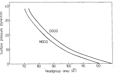
High purity synthetic L-a-dilauroylphosphatidylcholine (DLPC), L-a-dimyristoyl-phosphatidylcholine (DMPC), L-a-dipalmitoy lphosphatidy !choline (DPPC), L-a-distear-oylphosphatidy lcholine (DSPC), L-a-dipalmitL-a-distear-oylphosphatidylethanolamine (DPPE), L-a-dimyristoy lphosphatidy !glycerol (DMPG), and L-a-distearoy I- phosphatidy !glycerol (DSPG) were purchased in lyophylized powder form from Avanti Polar Lipids Inc. (Birmingham, Alabama). Plant monogalactosyldiglyceride lipids (MGDG) and plant digalactosyldiglyceride lipids (DGDG) were purchased in chloroform-methanol solution from Lipid Products (Nutfield Nurseries, Surrey, U.K.). The galactosyl diglycerides were reported to be mostly 18:3 and of purity 993+. All lipids were used without further purification. Dioctodecyldimethylammomium bromide surfactants (DOA) were Eastman (Rochester, New York) and purified by recrystallization from acetone. Fig. 1 gives the headgroup structure of all these amphiphiles.
Hexane-ethanol mixtures or chloroform were found to be suitable spreading solvents for all amphiphiles. All organic liquids were distilled twice before use. Water was purified in the follo\ving consecutive steps: distillation, trcatrnent overnight with
activated charcoal, filtration through a 0.05-µrn-pore size nucleopore filter, and a final
distillation from an all-pyrex still.
Amphiphiles were spread on an all-Teflon Langmuir trough standing in a dust,-frpe larninar flow cabinet. A 9:1 hexane-ethanol spreading solvent was used to spread the
phosphatidylcholincs and the quaternary amrr1onium bromide surfactants. Dl~PE \Vas
spread froin a 4:1 hexane-ethanol rnixturc. Complete dissolution of Dl)f>E in this
PC
PE
DGDG
H
I +HC-0-P-O-CH -CH -N(CH )
I II 2 2 33
R-0-CH 0
I
R-O-CH
2H
3C-CH
2-(CH
2 )16"'-+/CH
3,...-N"'-o-
H
3C-CH
2-(CH
2)16
CH
3H
I +HC-O-P-0-CH -CH -NH
I
II
2 2 3B-0-CH 0
I
R-0-CH
2o-H
IHC-0-P-O-CH -CHOH-CH OH
DOA
I II 2 2
CH:PH
R-0-CH O
PG
O~,
R-0-~H
~0-CH
2
OH
0~0-C(H
2
OH
HC-0-R
I
H
2C-O-R
OH
Fig. 1: Headgroup structure of phosphatidylcholine (PC), phosphat.idylethanolamine (PE), digalactosyldiglyceride (DGDG), phosphatidylglycerol (PG), and dioc-tadecyldimethylammonium surfactant (DOA).
molecule in its headgroup.
MGDG has only one sugar
were dried by evaporation under a strea1n of N2 and redissolved in the spreading solvent
hexane (for MGDG) or a 9:1 mixture of hexane-ethanol (for DGDG). Phos-phatidylglycerols were dissolved and spread from chloroform.
After spreading the amphiphiles on water, the spreading solvent was allowed to evaporate for 5 min. before surface pressure versus headgroup area
(ll-A)
isotherms weremeasured or before the monolayer was compressed to the surface pressure at which
Langmuir-Blodgett deposition of the lipids was done.
2.2 SURFACE TENSION MEASUREMENT TECHNIQUE
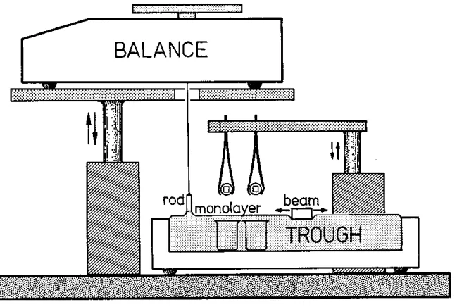
rfhe surface tension of the monolaycr was rneasured \vith the "maximum force on a
[image:28.594.119.540.45.358.2]surface tension is obtained directly frorr1 this rnaximum \veight (after an a<lj11stn1ent for
the weight of the rod). The rod was hung by a fine thread from under a bottom-loading balance of sensitivity 1 mg. This balance rests on a platform that can be raised or lowered smoothly at (variable) speeds down to about 1.0 mm/min and in this way pulls the rod up through the liquid surface at a regulated speed.
BALANCE
H
1.l
iJ'
0 rod
Fig. 2: Schematic drawing of the Langmuir trough. The surface tension 1s measured with a vertical cylindrical rod, attached to a bottom-loading balance. Langmuir-Blodgctt deposition of monolayers or bilayers of lipids is done on two small mica surfaces, glued on glass discs v.rhich are clamped in two tweezers. The t\veezers are
attached to a horizontal steel arm which can be raised or lowered at a speed of 0.2 mm/s. For the calibration depositions, the arm supports a large mica sheet (not shown).
The method does not require any know ledge of the contact angle of the liquid attached to the rod. Use was made of a stainless steel rod of diameter 0.5 cm with sharp circular edges. After consulting the tables given by Padday ct al. (1975) it was found that the surface pressure of a monolayer at the air-water interface at 21 ° C depends on
the maximum weight of water W underneath the rod according to
[image:29.594.190.523.213.431.2]2.3 LANGMUIR-BLODGBTT DEPOSITION TECHNIQUE
Calibration depositions of Langmuir-nlodgett lipid films onto molecularly smooth mica sheets of length 10 cm and width 2 cm were carried out giving a total mica surface area of 40 cm2. Each sheet was attached with two small alligator clips to a horizontal steel arm (see Fig. 2) which could be raised or lowered at a speed of 0.2 rnm/s.
After sweeping the water surface in the trough several times to remove any
surface-active contaminants, the mica was irnrnersed in the water keeping its sides
vertical with respect to the water surface. When the mica was fully immersed in the water, a monolayer was spread on the water surface. After allowing the spreading solvent to evaporate, the monolayer \Vas compressed until some desired surface pressure
was reached at \Vhich deposition \vas carried out. 1~o deposit the lipids, the mica was
raised vertically through the air-water interface keeping the surface pressure constant by slowly moving the barrier over the surface. When the mica was fully removed from the water, it was dried for 20 min. in the air of a larninar flow cabinet. Measurement of the contact angle of a water droplet on a DPPE monolaycr-covcred mica surface showed an
advancing contact angle of 106 °. From this it follows that the surface has become hydrophobic. Clearly, the polar headgroups are directed towards the mica, while the lipid hydrocarbon tails are facing the water phase. Comparing the total mica surface area with the surface area in the trough swept by the barrier during deposition (in order to keep the deposition pressure constant) gives the average headgroup area per
amphiphile on the mica. The headgroup area per amphiphile in the trough at a certain surface pressure is obtained frorr1 the IT-A isoth<~rrri.
A second layer of arnphiphiles can now be deposited by lowering the monolayer-covered surface into the Langrnuir trough at constant surface pressure until it
is fully in1rncrsed. The headgroup area in the second layer is again fou11<l by measuring
the surface area swept by the barrier during dc>position.
Deposition of the first and second layers \Vas attcrnpted at such surface pressures
second layer which corresponds with reported equilibrium headgroup areas in lipid
bilayers or vesicles.
When the forces between two deposited bilayers were to be measured, deposition was done onto two small molecularly smooth mica sheets previously glued on two cylindrically curved glass discs (sec next section). The glass discs with the mica sheets were clamped in two tweezers attached with clips to the movable horizontal steel arm (see Fig. 2) and deposition was done in the same way as with the large mica sheets. It
was assumed that the headgroup area of the deposited amphiphiles was equal to the values obtained from the calibration deposition experiments using the larger mica sheets.
Once the bilayers are deposited, the glass discs can not be retracted from the water as this causes the second layer to desorb. For this reason, the glass discs were put in
5-ml glass beakers standing under water in the Langmuir trough as shown in Fig. 2. The force measurement apparatus was filled with water the previous day and the water was presaturated with amphiphiles from a crystal to prevent desorption of the bilayers from the mica during the subsequent experiment. In this way, thermodynamic
equilibrium was established between amphiphiles in solution and amphiphiles in the bilayers. After removing the front plate from the apparatus the glass discs are transferred under \Vater frorn the trough to the force measurement apparatus where they
are mounted under water. Finally, the front plate is bolted to the front of the apparatus which closes it off so that direct force measurements can now be carried out.
2.4 THE DIRECT FORCE MEASURJC!\1ENT TECHNIQUE
The force measurement device developed by Israelachvili rneasures the force
between t\vo n1ica surfaces in a crossed cylinder configuration as a function of the surface
separation. A detailed account of the technique has been given before (Israelachvili and Adams, 1978) and in this section, only a brief summary will be presented.
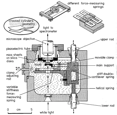
The basic apparatus is shown in Fig. 3. It. has a removable front plate with a nurnber of inlets used for filling and draining the apparatus. rrhe mica surfaces a.re
1nounted on two silica-glass discs with cylindrically polished surfaces facing each other
cleaved frorn 1arge thick mica sheets, so that they have a uniforrn thickness of a few 11m
and a surface area of about 0.5 cm2. Once cleaved, they are immediately placed on another freshly cleaved but thicker larger sheet (the backing sheet) where they are held down firmly by strong adhesive forces. Thus, one side of the mica sheets is protected from contamination. The exposed side of the mica sheets is then silvered by vacuum evaporation with a 480
A
thick highly reflecting layer of pure silver. Then, two silvered mica sheets of equal thickness are picked up from the backing sheet and glued down on the silica discs using a Shell epoxy resin 1004 thermosetting glue (silvered sides down). After the Langmuir-Blodgett deposition of amphiphiles on the mica is carried out, one disc is mounted on a rigid support, the other is positioned, facing the first one, on the end of a spring with a known spring constant {see Fig. 3).The distance between the two surfaces is measured by use of an optical technique using "multiple beam interference fringes" (or fringes of equal chrorriatic order, FECO
(Israelachvili, 1973)). The mica sheets with their outer surfaces silvered, together with the intervening medi1nn form a symrnctrical three layer interferometer. If the two
surfaces are in contact and white light is passed normally through, then the emerging light consists of discrete wavelengths .\~ (n = 1,2,3, ... ) which can be separated and measured as sharp fringes in an ordinary grating spectrometer relative to the wavelength
of the mercury green and yellow lines. If the two mica surfaces are then separated by a distance D, these fringes shift to longer wavelengths .\~given by
(27rµD)
tan
-.\D n
(37)
\Vhere
+
refers to odd order fringes (n odd) and - refers to even order fringes (n even).p.
= µ1nica/ µ, where µmica is the refractive index of mica at..\~,
and µ the refractive index of the rr1ediurn bet\veen the t\vo mica surfaces at ..\~. The fringe order is calculatedusing the formula
_ ;.O
/F
(.\0 _ ;.O)n -· n-1 n n-1 n
Ji' =
n
l 1.024
+ -.
microscope objective
clamp adjusting rod
variable stiffness force-measuring spring
0 cm
5
white light
force-measuring springs
upper rod
movable clamp 1
stiff double-cantilever spring
helical spring
Fig. 3: Schematic drawing of the force measurement apparatus. The force is measured between t\VO molecularly smooth curved surfaces in liquid with a surface
separa-tion resolusepara-tion of~ 1
A
2 using white light and multiple beam interferometry. The mica surfaces are glued onto two optically polished silica-glass discs. The lower disc is mounted on a double-cantilever force-measuring spring whose stiffness canbe varied by .a factor of 1000 by shifting the position of the dove-tail clamps using the positioning rod. This spring can be replaced by any of the other non-variable springs shown above. The spring shown at the right has the advantage of being non-tilting and non-shearing which is important \VhPn large surface forces are
present.
By use of Eq. (37), both the distance D and the medium refractive index 11 can be dclerrnined independently by measuring the shifts in wavelengths of an odd and an even fringe. The accuracy is often as good atl l
A
for I). [image:33.594.137.535.59.451.2]separation of which depends on the relative crystallographic orientation of the two mica
sheets. \Vavelength measurements are perforn1ed on one of these components only and
the refractive index for this component is calculated from the separation of the doublet and a knowledge of the mean refractive index of the mica, which in nature exists as
brown mica or green mica. The mean refractive index of brown and green mica is
reported (Israelachvili and Adams, 1978) as
4.76 x 10⁵
µBrown = 1.5820
+
4.76 x 10⁵ µGreen= 1.5930
+
D. .(-'ni2
I
II
(39)
If the mica sheets were exactly parallel, the fringes would appear as vertical lines in the spectrometer. Because of the crossed cylinder geometry, which to leading order is equivalent to a sphere on a flat, the shape of the fringes is parabolic. The entrance slit of a spectrometer samples a cross-section of the surfaces defined by the positioning of a
two prism system used to direct the light beam into the spectrometer. By using a system of three prisms, a cross-section of the surfaces at right angles to the first can be studied. This allo\vs the measurement of the radii of curvature in two perpendicular
directions from the shapes of the parabolic fringes during an experiment. The mean radius of curvature is calculated by taking the geometric average (R1 R2)1/ 2 of the two radii (typically 1-2 cm). Thus, the fringe profile seen in the spectrometer essentially reflects the shape of a slice through the curved surfaces and gives information on the local radius of curvature as well as on surface deforn1ations occurring under the influence
of strong forces. The lateral resolution is of the order of a few microns and is limited by the magnification of the optical system (about 25x in these experiments). Because the refractive index of the medium bct\veen the surfaces can be measured, it also provides a
method for the determination of the quantity of material deposited or adsorbed on the surfaces \Vhcn its refractive index is kno\vn beforehand. Finally, from the shapes of the
two initially curved surfaces any adhesive deformation and fusion of bilayers can be
The distance bctv,reen the tv.10 surfaces is controlled by use of a three stage
mechanism of increasing sensitivity. The coarse control uses a stepper motor attached
to the upper rod in Fig. 3 which allows a positioning to within 1 µrn; the medium control consists of a synchronous motor connected to the lower rod and allows
positioning to about 10
A.
A piezoelectric crystal (which expands or contracts vertically by ~ 5A
per volt applied axially across the cylindrical wall of the rigid support) is used for the final positioning to about 1A.
Given the facility for moving the surfaces toward or away from each other and, independently, of measuring their separation each with a sensitivity or resolution of
about 1
A,
the force measurements themselves now become straightforward. The force is measured by expanding or contracting the piezoelectric crystal by a known amountand then measuring optically how much the two surfaces have actually moved. Any difference in the two values when multiplied by the stiffness of the measuring spring gives the force difference between the initial and final positions. In this way, both repulsive and attractive forces can be measured \Vith a sensitivity of about 10 mdyne,
and a full force-law can be obtained over any distance.
Jn the present study, an important experimental advance is the introduction of a
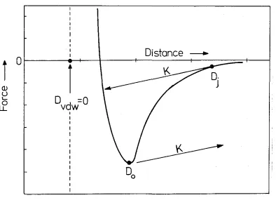
variable spring (see Fig. 3). This was particularly useful in the measurement of van der Waals forces between uncharged amphiphile bilayers (see Chapters 3 and 4). When two surfaces approach each other and the gradient of the surface force equals the spring constant, an instability occurs and the surfaces suddenly jump into contact (see Fig. 4).
By varying the spring constant \Ve obtain the gradient of the attractive force as a function of the surface separation at \Vhich they ju1np into contact. Also, the surfaces will jump apart. from each other at a surface separation where the gradient of the interaction force equals the spring constant. .Frorn Fig. 1 \vhich gives the typical
interact,ion force bet\veen two uncharged bilayer surfaces, it 1s seen that this occurs
almost exactly at the minirnurn locat<~d at D --- 00 in the force versus distance curve.
Distance
-i
0
l
(!)
u
L..
D
=O
0LL
vdw
Fig. 4: Schematic picture of the interaction force between two uncharged lipid bilayers
as a function of the distance D bet\veen them. The anhydrous bilayer /water
inter-face is located at D = 0, and the effective van der Waals plane is at DvDW = 0 (see also Chapter 3). When the bilayers are brought together, they jump into
con-tact from D = Dj as indicated by the arrow where the gradient of the interaction
force equals the spring constant K. Upon separation of the bilayers, they jump
apart from D = 00.
crossed cylindrically curved surfaces of radii Rat a separation D exert a force F(D) on
each other, then according to the Derjaguin approximation, the equivalent interaction
energy E(D) between two flat surfaces per unit area is related to F(D) by
E(D)
F(D)
27rR ( 40)
provided that R
»
D. In the present experiments, the bilayers have a radius of curvatureof about 1.5 cm. Interactions bet\veen bilayers \Vere only n1casurable at surface
[image:36.594.151.545.68.353.2]