R E S E A R C H A R T I C L E
Open Access
Computational analysis of pathogen-borne
metallo
b
-lactamases reveals discriminating
structural features between B1 types
Eithon Cadag
1, Elizabeth Vitalis
2, Kristin P Lennox
3, Carol L Ecale Zhou
1and Adam T Zemla
1*Abstract
Background:Genes conferring antibiotic resistance to groups of bacterial pathogens are cause for considerable concern, as many once-reliable antibiotics continue to see a reduction in efficacy. The recent discovery of the metallob-lactamaseblaNDM-1gene, which appears to grant antibiotic resistance to a variety of Enterobacteriaceae viaa mobile plasmid, is one example of this distressing trend. The following work describes a computational analysis of pathogen-borne MBLs that focuses on the structural aspects of characterized proteins.
Results:Using both sequence and structural analyses, we examine residues and structural features specific to various pathogen-borne MBL types. This analysis identifies a linker region within MBL-like folds that may act as a discriminating structural feature between these proteins, and specifically resistance-associated acquirable MBLs. Recently released crystal structures of the newly emerged NDM-1 protein were aligned against related MBL structures using a variety of global and local structural alignment methods, and the overall fold conformation is examined for structural conservation. Conservation appears to be present in most areas of the protein, yet is strikingly absent within a linker region, making NDM-1 unique with respect to a linker-based classification scheme. Variability analysis of the NDM-1 crystal structure highlights unique residues in key regions as well as identifying several characteristics shared with other transferable MBLs.
Conclusions:A discriminating linker region identified in MBL proteins is highlighted and examined in the context of NDM-1 and primarily three other MBL types: IMP-1, VIM-2 and ccrA. The presence of an unusual linker region variant and uncommon amino acid composition at specific structurally important sites may help to explain the unusually broad kinetic profile of NDM-1 and may aid in directing research attention to areas of this protein, and possibly other MBLs, that may be targeted for inactivation or attenuation of enzymatic activity.
Background
Proteins within theb-lactamase family have long drawn
the attention of researchers and clinicians due to their ability to efficiently hydrolyze many common antibiotics.
Metallob-lactamases (MBLs) in particular are of global
health interest, as many are acquired, capable of travel-ing across species, and are the most commonly encoun-tered transferable carbapenemases [1]. The recently
discovered plasmid-borne New Delhi metallo b
-lacta-mase (NDM-1), capable of hydrolyzing a broad range of antibiotics, is such a metalloenzyme and is noted for its
ability to confer resistance to all but a small handful of
b-lactam antimicrobials. First characterized within a
Swedish patient of Indian origin in 2008 [2], NDM-1 has since been identified in other parts of Asia, North America, Europe, Australia and Africa [3-8].
In addition to its rapid worldwide dissemination, NDM-1 is alarming for its penchant to transfer between speciesviaconjugation. With its initial identification on
a 180-kb Klebsiella pneumoniae plasmid, and
subse-quent re-discovery on aEscherichia coliplasmid isolated
from the same patient, NDM-1 has displayed an ability to spread amongst bacteria [2], and more recent findings have identified it in additional members of the Entero-bacteriaceae family [9,10]. Moreover, the presence of the gene encoding NDM-1 within isolates has been * Correspondence: [email protected]
1
Global Security Computing Applications Division, Lawrence Livermore National Laboratory, Livermore, 94550 CA, USA
Full list of author information is available at the end of the article
associated with the presence of genes and genetic ele-ments which confer additional resistance against other forms of antibiotics, including monobactams, aminogly-cosides, fluoroquinolones and tetracyclines [5,9,11], further reducing treatment options for infected patients.
Taken within this context, NDM-1 has the potential to greatly impact global health, most immediately in hospital settings through nosocomial infections, which appear to be a common mode of infection for NDM-1 carrying bacteria [4]. Further knowledge of the mechan-isms of the encoded protein may help to expedite devel-opment of therapies and countermeasures. Preliminary characterization of NDM-1 conducted by Yong and col-leagues revealed marginal sequence similarity to other members of the MBL family, with the closest sequence homology to VIM-1 and VIM-2 at only 32%; kinetic stu-dies supported this association, although NDM-1 was
noted to possess a superior binding profile for mostb
-lactams compared to VIM-type proteins [2]. They further identified, using sequence alignment, novel fea-tures of the NDM-1 protein not found in other mem-bers of the MBL family, such as uncommon residues around the zinc binding site and a four-residue insertion not observed in other MBLs. These features may help provide NDM-1 with its capability to readily bind to a
very broad range ofb-lactams. Other, more well-known
MBLs such as the VIM-type proteins found in Pseudo-monas, have likewise spread rapidly since their initial discoveries [12-15]. Many infections have been trans-mitted nosocomially, and are often found in developing areas [4,16]. Further knowledge of the mechanisms of these metalloenzymes may help to expedite development of inhibitors with direct clinical significance.
The variety, structure, function and medical signifi-cance of these proteins have been the focus of much research in the past, and they may be classified both molecularly and functionally. Traditionally, MBL
pro-teins are categorized as “class B” b-lactamases, which
can be further divided into subclasses based on the nat-ure of the metal binding site. The presence of specific binding motifs around the active cavity of the proteins, associated with zinc binding and coordination, may be used to classify an MBL as either B1 (zinc binding at H116-H118-H196 and at D120-C221-H263), B2 (N116-H118-H196; D120-C221-H263) or B3 (H/G116-H118-H196; D120-H121-H263) [17]. Notably, four of the six conserved residues are static across all classes, allowing amino acid-based molecular classification at only two positions (H/N/G116 and C221/H121). This classifica-tion scheme, though simple, is thought to be strongly related to the structural plasticity of the enzymes, as the zinc binding sites are critical to the hydrolytic effects of MBLs. Functional groupings have also been used as a means of describing similarities between MBLs.
Inhibition by EDTA, substrate hydrolysis rates and pro-files created by testing against other inhibitors (e.g., cla-vulanic acid) can be used to profile clinically relevant groups of MBL proteins and identify isolates in the lab [18,19].
Prior research on the structure-function relationship of MBL proteins has focused primarily on the region of the active site and mechanism of catalysis. For di-zinc
MBLs, hydrolysis is believed to occur via breaking of
theb-lactam amide bond on the carbonyl by a resident
hydroxide in the active site. This action is zinc-activated, and creates a temporary intermediate tetrahedral carbon, upon which the zinc-bound water donates a proton to the leaving nitrogen of the ligand [20-23]. The steps involved in this action are believed to be ligand-depen-dent, and protonation may or may not coincide with cleavage of theb-lactam ring (e.g., as noted for nitroce-fin bound to ccrA) [24]. For B1 MBLs, binding is thought to be mediated in part by the presence of a large mobile flap that forms a cleft over the active site [21]. Deletion of this flap region in some MBLs has been correlated with weakened affinity for many antibio-tic substrates, with the exception of imipenem [25]. The mobile flap exists in B1 MBL types ccrA and IMP-1 with an aromatic, bulky residue, and has been hypothe-sized to be critically involved in the recruitment, stabili-zation and binding of inhibitors [21,26,27]. This flap is less functionally important in VIM-2, which contains an alanine (A64) in place of an aromatic residue [28], exemplifying the nuanced structural functionality of common B1 MBL components.
The prevalence of methods for classifying MBLs is in large part due to their functional, structural and mole-cular similarities and differences, and our work builds upon the features used for classification currently receiv-ing attention by applyreceiv-ing structure-based analyses of well-characterized MBLs, with the hope of identifying residues and regions that can further aid in functional discrimination. A more detailed picture of residue con-servation and structural uniqueness is assembled for proteins within the B1 MBL subclass, and its constituent types VIM, IMP, ccrA and NDM-1. While the core structure of MBLs is well known to be conserved,
struc-tural alignments revealed a “linker” region with
consid-erable variability among B1 proteins, which we propose as a notable structural classification feature. We apply structural analysis methods to the NDM-1 protein in order to identify significant sequence and structural
dif-ferences from other MBLs that may affect NDM-1’s
analyses and clustering of key features, structurally con-served residues were identified in NDM-1 and compared with the corresponding residues in similar proteins to identify regions of conservation and novelty between the known and new B1 MBLs. Many sites we identify com-putationally as highly conserved correspond to those found to be functionally critical by prior experimental work. Common themes, as well as features unique to NDM-1, are identified. Of particular interest is an
uncharacteristically divergent “linker” region. We find
that while the vast majority of B1 MBLs’ conformation
is well conserved, NDM-1 is marked by both the pre-sence of rare residues in resistance-implicated regions and a linker conformation that is unique among MBL structures.
Methods
MBL-like protein structure library
As one goal of this study was an overall structural char-acterization and comparison of available B1 MBL struc-tures, with emphasis on the recently discovered NDM-1 protein, a library of B1 MBL proteins for which both sequence and structure were available was generated. Protein structures were retrieved from the Protein Data Bank (PDB) [31], and full sequences were taken from UniProt [10]. Special focus was given to three specific B1 types used in comparison to NDM-1 from prior research: IMP-1, VIM-2 and ccrA [2,23,29,30].
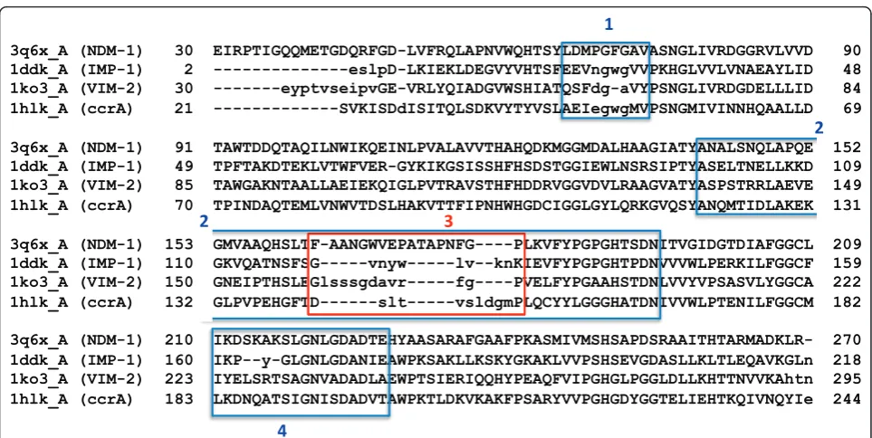
Representatives of IMP-1 (PDB: 1ddk_A), VIM-2 (PDB: 1ko3_A), ccrA (PDB:1hlk_A) and NDM-1 (PDB: 3q6x_A) were selected as seed structures for expanding the number of structures used for variability analysis (see 2.2) to include similar, MBL and MBL-like, proteins; these representatives are used as the reference structures for their respective types throughout the rest of our study, unless otherwise noted. Figure 1 shows structure-based sequence alignment between the selected representatives, showing strong over-all correspondence with relatively few gaps. Structure-based similarity searches were performed for IMP-1, VIM-2, ccrA and NDM-1 against the entire PDB database (release 2011/08/02; 188,448 chains) using the StralSV algorithm [32]. Pruning of the retrieved structures was
per-formed via an LGA_S [33] cutoff value of≥50% structure
similarity to the corresponding reference structures. After removing PDB chains identical in sequence, the result con-sisted of 75 structures. This set of proteins was expanded to include all available NDM-1 crystal structures (nine, as of the writing of this manuscript), which formed the final MBL fold library (83 structures; refer to Additional file 1) used for comparative computational analysis.
Comparative structural analysis
Members of the MBL-like library were subjected to a number of comparative methods in order to determine
distinctive regions of conservation and divergence. Structure-based sequence variability analyses were run for the representative structures of NDM-1, IMP-1, VIM-2 and ccrA, using StralSV [32], which calculates sequence variability from fragment-based local structural alignment. The purpose of this analysis was to identify in analyzed MBL structures local regions where proteins are structurally unique, and regions where they are rela-tively conserved regardless of their sequence similarity, focusing on sequence compositions in such regions.
The StralSV algorithm works, briefly, as follows: a tar-get structure,t, and associated library, L, are specified.
Template structure l Î Lshares structural similarity
withtin at least some structural fragments. Detection
of local similarities and calculations of alignments
between tand all l are performed using the LGA
pro-gram [33], and the specific residue-residue
correspon-dences fortand each member of Lare found. Thus, for
each position in t, a residue“profile” is built using
resi-dues fromLwith which that position structurally aligns.
The output allows one to examine commonalities and eccentricities between a target and any number of tem-plates at the structural level, much like a sequence-based profile would allow one to examine standard posi-tional variability.
To determine structural groupings and gain better insight into both overall and region-specific similarities between MBLs, clustering of the structures was per-formed using StralCP [34] on a whole-chain level and for two specific local substructures selected for their importance or uniqueness: the active site and the linker region, the latter identified from structural variability analysis to be unique in NDM-1 (see 3.1). Clusters were formed hierarchically using Euclidean distance
measure-ments from n-way multiple structural alignments. For
Within the active pocket itself, metal ion distances were measured, and CASTp [36] was used to estimate binding site volumes for B1 MBLs. Because apo and holo forms of IMP-1, VIM-2, ccrA and NDM-1 were available, systematic comparisons of differences in back-bone conformation and ligand binding were made. Changes in small molecule binding within IMP-1, VIM-2 and ccrA at the side chain level were compared to
NDM-1 for the purpose of classifying NDM-1’s
func-tional residue profile using a new pairwise structural comparison service, LGA_pdblist http://proteinmodel. org/AS2TS/LGA_list/.
Comparisons of critical residues based on structural alignments
Pairwise LGA comparisons were used to examine cataly-tic and cricataly-tical residues found in IMP, VIM, ccrA and NDM-1, for the purpose of identifying shared or distinc-tive conformational changes in the immediate vicinity of metal and ligand binding. Using structural alignments as a scaffold, residue-residue correspondences were gener-ated for B1 MBLs using NDM-1 as a reference. Bound representatives were used for this purpose in order to identify ligand-interacting residues (3q6x_A, 1dd6_A, 1a8t_A, 2yz3_A for NDM-1, IMP-1, ccrA and VIM-2,
respectively). Residues specifically examined include those within 4 Å of either the zinc ions or bound ligands. Additionally, residues thought to be critical for other MBL variants based on experimental evidence found in literature, but located outside the active site, were also included and mapped onto NDM-1 for reference.
Results and discussion
Overall MBL structure
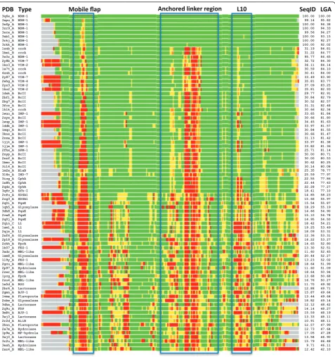
In the present study, focus was placed on B1 MBLs as a way of generalizing toward emerging, transferable anti-biotic resistance genes, such as NDM-1. A global exami-nation of the proteins most closely related to NDM-1 highlighted structural commonalities across B1 MBLs. Comparisons with available NDM-1 structures to the pre-selected MBL-like fold library yielded similar scores, with the closest proteins being representatives from VIM (VIM-2, VIM-4) and ccrA; Figure 2 shows a heat-map of structural alignments of NDM-1 against the pre-selected MBL library, indicating strong (< 2 Å of Ca-Ca deviation; colored in green) structural concordance in the majority of regions for most other MBLs. The
simi-larity of NDM-1’s overall conformation to many other
MBLs despite low sequence identity is unsurprising as
3q6x_A (NDM-1) 30 EIRPTIGQQMETGDQRFGD-LVFRQLAPNVWQHTSYLDMPGFGAVASNGLIVRDGGRVLVVD 90 1ddk_A (IMP-1) 2 ---eslpD-LKIEKLDEGVYVHTSFEEVngwgVVPKHGLVVLVNAEAYLID 48 1ko3_A (VIM-2) 30 ---eyptvseipvGE-VRLYQIADGVWSHIATQSFdg-aVYPSNGLIVRDGDELLLID 84 1hlk_A (ccrA) 21 ---SVKISDdISITQLSDKVYTYVSLAEIegwgMVPSNGMIVINNHQAALLD 69
3q6x_A (NDM-1) 91 TAWTDDQTAQILNWIKQEINLPVALAVVTHAHQDKMGGMDALHAAGIATYANALSNQLAPQE 152 1ddk_A (IMP-1) 49 TPFTAKDTEKLVTWFVER-GYKIKGSISSHFHSDSTGGIEWLNSRSIPTYASELTNELLKKD 109 1ko3_A (VIM-2) 85 TAWGAKNTAALLAEIEKQIGLPVTRAVSTHFHDDRVGGVDVLRAAGVATYASPSTRRLAEVE 149 1hlk_A (ccrA) 70 TPINDAQTEMLVNWVTDSLHAKVTTFIPNHWHGDCIGGLGYLQRKGVQSYANQMTIDLAKEK 131
3q6x_A (NDM-1) 153 GMVAAQHSLTF-AANGWVEPATAPNFG----PLKVFYPGPGHTSDNITVGIDGTDIAFGGCL 209 1ddk_A (IMP-1) 110 GKVQATNSFSG---vnyw---lv--knKIEVFYPGPGHTPDNVVVWLPERKILFGGCF 159 1ko3_A (VIM-2) 150 GNEIPTHSLEGlsssgdavr---fg----PVELFYPGAAHSTDNLVVYVPSASVLYGGCA 222 1hlk_A (ccrA) 132 GLPVPEHGFTD---slt---vsldgmPLQCYYLGGGHATDNIVVWLPTENILFGGCM 182
3q6x_A (NDM-1) 210 IKDSKAKSLGNLGDADTEHYAASARAFGAAFPKASMIVMSHSAPDSRAAITHTARMADKLR- 270 1ddk_A (IMP-1) 160 IKP--y-GLGNLGDANIEAWPKSAKLLKSKYGKAKLVVPSHSEVGDASLLKLTLEQAVKGLn 218 1ko3_A (VIM-2) 223 IYELSRTSAGNVADADLAEWPTSIERIQQHYPEAQFVIPGHGLPGGLDLLKHTTNVVKAhtn 295 1hlk_A (ccrA) 183 LKDNQATSIGNISDADVTAWPKTLDKVKAKFPSARYVVPGHGDYGGTELIEHTKQIVNQYIe 244
YANALSNQLAPQE YASELTNELLKKD YASPSTRRLAEVE YANQMTIDLAKEK
GMVAAQHSLTF-AANGWVEPATAPNFG----PLKVFYPGPGHTSDNI
GKVQATNSFS IEVFYPGPGHTPDNVV
GNEIPTHSLE VELFYPGAAHSTDNLV
GLPVPEHGFT LQCYYLGGGHATDNIV
TF-AANGWVEPATAPNFG----PL G---vnyw---lv--knK Glsssgdavr---fg----PV TD---slt---vsldgmPL
YLDMPGFGAVA FEEVngwgVVP TQSFdg-aVYP LAEIegwgMVP
IKDSKAKSLGNLGDADTEH IKP--y-GLGNLGDANIEA IYELSRTSAGNVADADLAE LKDNQATSIGNISDADVTA
[image:4.595.57.539.86.328.2]
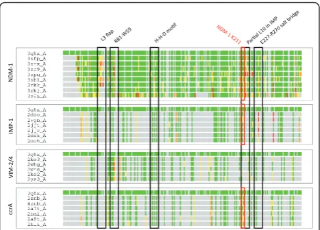
proteins under the B1 MBL grouping are well known to adopt very similar folds and active regions [14]. How-ever, we note that there are significant regions of diver-gence, which include the so-called L3 mobile flap, whose motion is associated with MBL ligand binding,
and a “linker” loop region commonly found in MBLs.
Within VIM-2 this region corresponds to residues 174-186, in IMP it is found between residues 120-129, ccrA residues 142-152 and NDM-1 163-179 (see Figure 1). Structural alignment was generally poor between MBL-types within this region, and it was thus singled out for further analysis.
3q6x_A NDM-1 100.00 100.00
3spu_B NDM-1 99.14 95.82
3sfp_A NDM-1 100.00 94.38
3zr9_A NDM-1 100.00 94.33
3srx_A NDM-1 99.56 94.27
3sbl_A NDM-1 100.00 93.15
3rkj_A NDM-1 100.00 92.27
3rkk_A NDM-1 100.00 92.02
1znb_B ccrA 31.19 84.81
1bmi_B ccrA 31.22 84.77
3s0z_A NDM-1 95.77 84.65
2y8b_A VIM-7 32.72 84.30
1ko3_A VIM-2 36.11 84.14
1a7t_A ccrA 30.59 84.10
4znb_A ccrA 30.41 84.00
2y87_A VIM-7 33.49 83.90
2whg_A VIM-4 35.81 83.77
2yz3_A VIM-2 37.09 83.25
1ko2_A VIM-2 35.81 82.93
1dxk_A BcII 29.77 82.91
2bfl_B BcII 30.84 82.79
2bg7_B BcII 30.52 82.57
3fcz_A BcII 31.31 82.48
1hlk_A ccrA 30.66 82.34
1wuo_A IMP-1 33.65 81.84
2uyx_A BcII 30.66 81.80
1wup_A IMP-1 34.45 81.63
1ddk_A IMP-1 33.97 81.57
1mqo_A BcII 30.84 81.55
3kns_A BcII 30.66 81.47
2nze_B BcII 31.13 81.42
1jjt_A IMP-1 33.97 81.38
1jje_B IMP-1 33.82 81.36
2fhx_A SPM-1 25.71 81.14
2nyp_A BcII 30.00 80.71
2nzf_A BcII 30.00 80.53
1bmc_A BcII 30.62 80.25
3i14_A BcII 30.14 80.08
1m2x_A BlaB 25.35 78.77
3l6n_A IND-7 25.59 77.97
3iog_A CphA 22.55 77.86
1x8h_A CphA 22.44 77.63
2qds_A CphA 22.28 77.27
3q6v_A Sfh-I 18.41 77.10
2cfu_A SDSA1 14.85 58.23
2cg3_A SDSA1 15.66 55.97
2q0i_A PqsE 15.54 55.87
1qh5_A Glyoxylase 18.29 55.19
3dh8_A PqsE 14.66 54.90
2vw8_A PqsE 15.10 54.78
2q0j_A PqsE 14.95 54.50
2qin_C L1 18.42 54.21
1sml_A L1 19.25 53.49
2qjs_A L1 18.09 53.31
2xf4_A Glyoxalase 12.36 53.31
2p18_A Glyoxalase 16.22 53.27
2ohh_A FprA 14.65 52.80
1k07_A FEZ-1 13.30 52.51
3adr_A MBL-like 11.60 52.45
1xm8_A Glyoxalase 20.44 52.27
1l9y_A FEZ-1 13.23 52.02
2gcu_A MBL-like 18.64 51.99
2p97_A Hydrolase 11.60 51.09
2zwr_B MBL-like 18.64 50.94
1ycg_A FprA 13.68 50.68
2zo4_A MBL-like 16.76 50.27
1e5d_A ROO 11.70 49.92
2br6_A Lactonase 12.88 49.71
2gmn_A BJP-1 15.87 49.64
1vme_A Flavoprote 13.44 49.64
2obw_A Glyoxalase 18.82 49.14
3dha_A Lactonase 12.12 48.42
2q9u_A FDP 14.14 48.27
3m8t_A BJP-1 15.59 48.19
3aj3_A Lactonase 13.33 48.11
2r2d_A AiiB 10.91 48.11
3hnn_A Flavoprote 12.57 47.99
2a7m_A Hydrolase 12.73 47.64
1p9e_A MP-hydrola 17.05 47.28
3aj0_A Lactonase 13.58 46.90
3r2u_A MBL-like 15.79 46.48
3esh_A Hydrolase 9.71 46.11
2az4_B MBL-like 12.42 42.10
[image:5.595.60.538.87.595.2]
Beginning at the active site, StralSV analysis of IMP-1, VIM-2, ccrA and NDM-1 type representatives against the preselected MBL library showed well-conserved structural alignment profiles around the di-nuclear zinc binding motif; conservation signals at both the sequence and structure level was strongly evident for the HxHxD zinc binding motif in all four MBLs, even while sur-rounding residues were generally heterogeneous within the MBL library. Chains that matched this structural region, but did not correspond to the B1 HxHxD, motif were generally B2 MBLs (with an NxHxD motif) or
oxi-doreductases (e.g., HxExD), illustrating the overall
shared conformation of the binding pocket despite var-iation between actual residues.
Select regions of the B1 MBLs, and indeed the entire library, are less in consensus. As noted previously, the L3 flap region at the entrance to the binding cavity is observed in different local structural conformations in analyzed MBLs. Analysis of the flap region for VIM, IMP and ccrA highlights the divergence between the structures of this flap across B1 MBLs. While known to be functionally important in IMP and ccrA, sequence conservation is limited and structural profiles are
het-erogeneous due to the region’s mobility. Also, whereas
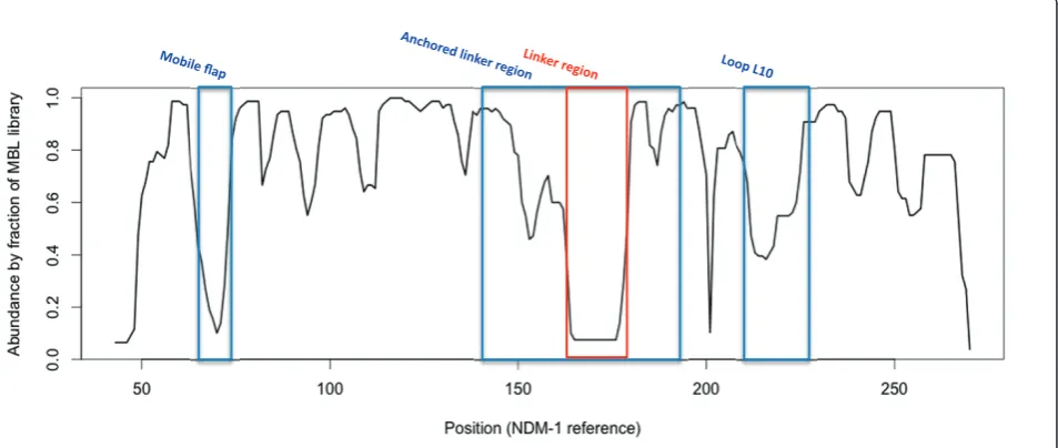
IMP and ccrA possess a large side-chain residue (W28 and W49, respectively), VIM lacks the aromatic side-chain and appears one residue shorter than either IMP or ccrA. Phenylalanine (F70) occupies this position in NDM-1, which may serve a similar purpose to that of its analogues in IMP and ccrA. MBL library alignment to NDM-1 using StralSV is shown in Figure 3 as an abundance plot, and represents the fraction of members of the MBL library with structural alignment to NDM-1 over all positions. Across all members of the library, the L3 flap region shows broad disagreement. Graphical illustration of the structural deviations in this region relative to the NDM-1 is shown in Figure 2. Other regions of dissimilarity include the aforementioned lin-ker region and, interestingly, the L10 loop often asso-ciated with ligand binding. Most regions otherwise show strong structural agreement, including areas of short misalignment between NDM-1 and other MBLs. This was not surprising, given the conservative fold of MBLs, and even with short insertions, the overall conformation of the proteins would not be expected to diverge greatly [37].
Structure-based clustering
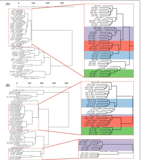
Structure-based clustering of the entire MBL library showed that MBL folds, including those of the B1 MBLs, group together tightly despite distinct sequence and structure variability in various regions. On the whole-chain level within each subclass, structural differ-ences were generally minimal, and groups of structures
cluster cleanly between B1/2/3 MBLs even for the indi-vidual B1 types, where ccrA, BcII, VIM, IMP and NDM-1 form distinct branches (with the exception of NDM-NDM-1 structure 3s0z_A, which appears to cluster closer to VIM; see Additional file 2). This suggests that while MBLs share a very similar structure, evidenced by the small distances between types on the tree, there are suf-ficient and consistent differences at the whole chain level that distinguish IMP, VIM, ccrA and other B1 MBLs.
Further structural comparisons across the B1/2/3 MBLs focused on areas of known importance, including the active cavity where zinc ligation occurs. As we were interested in whether this clustering was also evident around the conserved binding cavity, spherical protein substructures with 7.5 Å and centered at the metal ions were extracted, followed by a second layer of 7.5 Å cen-tered around the residues found in the initial step; this two-step approach at spatially defining the active site provided a substructure centered around the binding region encompassing both direct and secondary interact-ing residues.
Examination of the clusters formed by extracting the active site and its immediate neighborhood paint a much tighter view of NDM-1, with all instances of the protein clustering tightly, showing that structural fea-tures of 3s0z_A outside of the active site are the cause for division (see Figure 4A). VIM- and ccrA-type pro-teins cluster closely with NDM-1 at the active site. As Yong et al. [2] initially proposed that VIM-2 is a close homolog, the small differences between NDM-1 and VIM are not surprising. However, we note that ccrA, despite being a non-transferrable MBL, is strikingly close in the most critical region to NDM-1, surpassing IMP in active site similarity; indeed, IMP is quite distant from the other transferrable MBLs, indicating relatively strong structural differences between IMP and VIM, NDM-1. Much of this change can be explained by
IMP’s L10 loop, which is shorter by three residues than
ccrA, VIM or NDM-1, a factor believed to affect inhibi-tor binding [38].
indicative of an entirely novel conformation within its linker region. This deviation, shown in the alignment of the linker region between NDM-1 and other B1 MBLs on Figure 5, results in a significant portion of the NDM-1 linker being exposed. Further manual inspection of structural alignments of NDM-1 to other proteins in the same dendrogram branch (CphA, SPM-1) reveal that the N- and C- terminals align well, but the majority
of NDM-1’s linker region mismatches.
Within VIM-type proteins, structural alignment reveals that the linker region contains an initial loop approximately five residues longer than the same region in IMP. Incorporating ccrA structures into the pairwise alignment reinforces this five-residue insertion, but also introduces a second insertion in this linker region, pro-ducing a loop approximately four residues longer with respect to VIM-type proteins and two residues longer than IMP. Among these three types, IMP represented the structures with both loops short within the linker
region, an interesting observation given IMP’s
aforemen-tioned shorter L10 loop. NDM-1’s linker region is
extended at the N-terminal, a region where it is most similar to VIM. It then adopts a short helix from posi-tions 170-174, and continues as a loop.
The structural theme found in B1 MBLs is an extended loop pattern, where VIM-type structures pos-sess an initial insertion, followed by a structurally-con-served region approximately five residues in length shared by VIM, IMP and ccrA, and ending with an IMP/ccrA insertion two to four residues in length (see
Figure 1). This is in contrast to earlier sequence-based alignments, where the initial VIM insert differs in loca-tion, and the later insert toward the C-terminal end is entirely absent [2]. The linker region of NDM-1 is a notable departure from this theme, particularly with the presence of a helix and loop extension. The comparative difference of this region across B1 MBL types suggests the possibility that the linker region is an area of flex-ibility within MBLs, and that the unusual length and
conformation of NDM-1’s linker region may confer
higher plasticity. Temperature factors of available NDM-1 crystal structures, while generally higher than other stable regions of the structure, were not abnormally high.
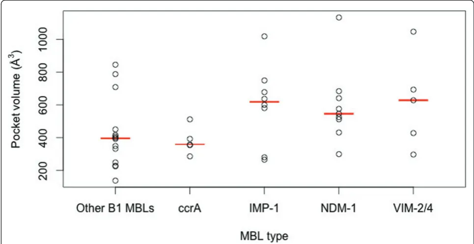
Comparison of MBL pockets and binding changes
Estimated calculations of binding site volumes were higher for the plasmid-borne MBLs versus other MBLs (see Figure 6). A larger binding pocket for the B1 MBLs may aid in accommodating a more diverse set of ligands, and on average IMP-1, VIM-2/4 and NDM-1 have similarly-sized pocket volumes. We also find that ccrA, which has close structural homology to transmissi-ble MBLs, has a notably smaller binding pocket site.
For hydrolytic activity, shallower and tighter zinc ions are associated with more effective catalytic activity [40], and examination of the distances between the MBL zinc ions (for di-zinc species) shows that ccrA ion distances are surprisingly similar to the transferrable MBLs, and IMP-1 and NDM-1 in particular (see Figure 7). Of the
[image:7.595.62.539.92.294.2]
3kns_BcII_ 1dxk_BcII_ 2uyx_BcII_ 2bfl_BcII_ 2bg7_BcII_ 1mqo_BcII_ 2nze_BcII_
3l6n_IND−7_ 3fcz_BcII_ 2nyp_BcII_ 2nzf_BcII_ 3i14_BcII_ 1bmc_BcII_ 3q6x_NDM−1_ 3s0z_NDM−1_ 3rkj_NDM−1_ 3sbl_NDM−1_ 3rkk_NDM−1_ 3zr9_NDM−1_ 3sfp_NDM−1_ 3srx_NDM−1_ 3spu_NDM−1_ 2y87_VIM−7_ 2y8b_VIM−7_ 1ko2_VIM−2_ 2yz3_VIM−2_ 2whg_VIM−4_ 1ko3_VIM−2_ 1bmi_ccrA_ 1hlk_ccrA_ 4znb_ccrA_ 1a7t_ccrA_ 1znb_ccrA_ 3q6v_Sfh−I_ 3iog_CphA_ 1x8h_CphA_ 2qds_CphA_
2fhx_SPM−1_ 1m2x_BlaB_ 1jjt_IMP−1_ 1jje_IMP−1_ 1wup_IMP−1_ 1ddk_IMP−1_ 1wuo_IMP−1_ 2vw8_PqsE_ 2q0j_PqsE_ 3dh8_PqsE_ 2q0i_PqsE_
2zo4_MBL−like_ 3adr_MBL−like_ 2zwr_MBL−like_ 2xf4_Glyoxalase_ 2p18_Glyoxalase_ 1xm8_Glyoxalase_ 1qh5_Glyoxylase_ 2obw_Glyoxalase_
2gcu_MBL−like_ 2p97_Hydrolase_ 2gmn_BJP−1_ 3m8t_BJP−1_ 1k07_FEZ−1_ 1l9y_FEZ−1_
2qjs_L1_ 1sml_L1_ 2qin_L1_ 3hnn_Flavop_ 1vme_Flavoprotein_ 2ohh_FprA_ 2q9u_FDP_ 1ycg_FprA_ 1e5d_ROO_
1p9e_MP−hydrolase_ 3esh_Hydro_ 3aj3_Lactonase_ 3aj0_Lactonase_ 2r2d_AiiB_ 2br6_Lactonase_ 2a7m_Hydrolase_ 3dha_Lacton_ 2cfu_SDSA1_ 2cg3_SDSA1_
3r2u_MBL−like_ 2az4_MBL−like_
0 100 200 300
2cfu_A_SDSA1 2cg3_A_SDSA1 2q0j_A_PqsE 2q0i_A_PqsE 2vw8_A_PqsE 3dh8_A_PqsE
3adr_A_MBL−like 2zo4_A_MBL−like 3aj3_A_Lactonase 3aj0_A_Lactonase
1p9e_A_MP−hydrolase 2r2d_A_AiiB 1vme_A_Flavoprotein 1ycg_A_FprA 2ohh_A_FprA 1e5d_A_ROO 2q9u_A_FDP 3kns_A_BcII 1dxk_A_BcII 2nzf_A_BcII 2nyp_A_BcII 3i14_A_BcII 2nze_B_BcII 1mqo_A_BcII 2uyx_A_BcII 1bmc_A_BcII 3fcz_A_BcII 4znb_A_ccrA 1a7t_A_ccrA 1znb_B_ccrA 1hlk_A_ccrA 1bmi_B_ccrA 1m2x_A_BlaB 3l6n_A_IND−7 2bfl_B_BcII 2bg7_B_BcII
3s0z_A_NDM−1 2y87_A_VIM−7 2y8b_A_VIM−7 2yz3_A_VIM−2 2whg_A_VIM−4 1ko2_A_VIM−2 1ko3_A_VIM−2
1ddk_A_IMP−1 1jje_B_IMP−1 1jjt_A_IMP−1 1wup_A_IMP−1 1wuo_A_IMP−1
2p97_A_Hydrolase 2obw_A_Glyoxalase 1qh5_A_Glyoxylase 2p18_A_Glyoxalase 1xm8_A_Glyoxalase
2gcu_A_MBL−like 2qjs_A_L1 2qin_C_L1 1sml_A_L1 1l9y_A_FEZ−1 1k07_A_FEZ−1 2gmn_A_BJP−1 3m8t_A_BJP−1
3esh_A_Hydrolase 3dha_A_Lactonase 2a7m_A_Hydrolase 2br6_A_Lactonase
3hnn_A_Flavoprotein 3r2u_A_MBL−like 2az4_B_MBL−like 2xf4_A_Glyoxalase
2zwr_B_MBL−like 3sfp_A_NDM−1 3q6x_A_NDM−1 3zr9_A_NDM−1 3spu_B_NDM−1
3rkj_A_NDM−1 3sbl_A_NDM−1 3srx_A_NDM−1 3rkk_A_NDM−1
2fhx_A_SPM−1 1x8h_A_CphA
3q6v_A_Sfh−I 2qds_A_CphA 3iog_A_CphA
0 100 200 300 400
() , ( p ) 3kns_A_BcII 1dxk_A_BcII 2nzf_A_BcII 2nyp_A_BcII 3i14_A_BcII 2nze_B_BcII 1mqo_A_BcII 2uyx_A_BcII 1bmc_A_BcII 3fcz_A_BcII 4znb_A_ccrA 1a7t_A_ccrA 1znb_B_ccrA 1hlk_A_ccrA 1bmi_B_ccrA 1m2x_A_BlaB 3l6n_A_IND−7 2bfl_B_BcII 2bg7_B_BcII
3s0z_A_NDM−1 2y87_A_VIM−7 2y8b_A_VIM−7
2yz3_A_VIM−2 2whg_A_VIM−4 1ko2_A_VIM−2 1ko3_A_VIM−2
1ddk_A_IMP−1 1jje_B_IMP−1
1jjt_A_IMP−1 1wup_A_IMP−1 1wuo_A_IMP−1
3sfp_A_NDM−1 3q6x_A_NDM−1 3zr9_A_NDM−1 3spu_B_NDM−1
3rkj_A_NDM−1 3sbl_A_NDM−1 3srx_A_NDM−1 3rkk_A_NDM−1
2fhx_A_SPM−1 1x8h_A_CphA
3q6v_A_Sfh−I 2qds_A_CphA 3iog_A_CphA 3kns_BcII_ 1dxk_BcII_ 2uyx_BcII_ 2bfl_BcII_ 2bg7_BcII_ 1mqo_BcII_ 2nze_BcII_
3l6n_IND−7_ 3fcz_BcII_ 2nyp_BcII_ 2nzf_BcII_ 3i14_BcII_ 1bmc_BcII_ 3q6x_NDM−1_ 3s0z_NDM−1_ 3rkj_NDM−1_ 3sbl_NDM−1_ 3rkk_NDM−1_ 3zr9_NDM−1_ 3sfp_NDM−1_ 3srx_NDM−1_ 3spu_NDM−1_ 2y87_VIM−7_ 2y8b_VIM−7_ 1ko2_VIM−2_ 2yz3_VIM−2_ 2whg_VIM−4_ 1ko3_VIM−2_ 1bmi_ccrA_ 1hlk_ccrA_ 4znb_ccrA_ 1a7t_ccrA_ 1znb_ccrA_ 3q6v_Sfh−I_ 3iog_CphA_ 1x8h_CphA_ 2qds_CphA_
2fhx_SPM−1_ 1m2x_BlaB_ 1jjt_IMP−1_ 1jje_IMP−1_ 1wup_IMP−1_ 1ddk_IMP−1_ 1wuo_IMP−1_ 2vw8_PqsE_ 2q0j PqsE 2q9u_A_FDP 3kns_A_BcII 1dxk_A_BcII 2nzf_A_BcII 2nyp_A_BcII 3i14_A_BcII 2nze_B_BcII 1mqo_A_BcII 2uyx_A_BcII 1bmc_A_BcII 3fcz_A_BcII 4znb_A_ccrA 1a7t_A_ccrA 1znb_B_ccrA 1hlk_A_ccrA 1bmi_B_ccrA 1m2x_A_BlaB 3l6n_A_IND−7 2bfl_B_BcII 2bg7_B_BcII
3s0z_A_NDM−1 2y87_A_VIM−7 2y8b_A_VIM−7 2yz3_A_VIM−2 2whg_A_VIM−4 1ko2_A_VIM−2 1ko3_A_VIM−2
1ddk_A_IMP−1 1jje_B_IMP−1 1jjt_A_IMP−1 1wup_A_IMP−1 1wuo_A_IMP−1
2p97_A_Hydrolase 2obw A Glyoxalase
2zwr_B_MBL−like 3sfp_A_NDM−1 3q6x_A_NDM−1 3zr9_A_NDM−1 3spu_B_NDM−1
3rkj_A_NDM−1 3sbl_A_NDM−1 3srx_A_NDM−1 3rkk_A_NDM−1
2fhx_A_SPM−1 1x8h_A_CphA
3q6v_A_Sfh−I 2qds_A_CphA 3iog_A_CphA
(A)
(B)
3kns_BcII_ 1dxk_BcII_ 2uyx_BcII_ 2bfl_BcII_ 2bg7_BcII_ 1mqo_BcII_ 2nze_BcII_ 3l6n_IND−7_ 3fcz_BcII_ 2nyp_BcII_2nzf_BcII_ 3i14_BcII_ 1bmc_BcII_
3q6x_NDM−1_ 3s0z_NDM−1_ 3rkj_NDM−1_ 3sbl_NDM−1_ 3rkk_NDM−1_ 3zr9_NDM−1_ 3sfp_NDM−1_ 3srx_NDM−1_ 3spu_NDM−1_ 2y87_VIM−7_ 2y8b_VIM−7_
1ko2_VIM−2_ 2yz3_VIM−2_ 2whg_VIM−4_ 1ko3_VIM−2_
1bmi_ccrA_ 1hlk_ccrA_ 4znb_ccrA_ 1a7t_ccrA_ 1znb_ccrA_ 3q6v_Sfh−I_ 3iog_CphA_ 1x8h_CphA_ 2qds_CphA_
[image:8.595.58.542.88.637.2]2fhx_SPM−1_ 1m2x_BlaB_ 1jjt_IMP−1_ 1jje_IMP−1_ 1wup_IMP−1_ 1ddk_IMP−1_ 1wuo_IMP−1_
plasmid-borne MBLs compared, NDM-1 and IMP appear to have the tightest zinc arrangement, even with the inclusion of a > 4 Å outlier (3q6x_A), whose large inter-zinc distance is likely a result of ampicillin
hydrolysis [23]. NDM-1 accommodates both a relatively large pocket volume, similar to VIM, with slightly tigh-ter zinc conformation; these charactigh-teristics likely influ-ence its broad binding and catalytic capabilities.
[image:9.595.62.539.89.301.2]
Figure 5Visual alignment of the linker region between NDM-1 and other B1 MBLs. NDM-1 is shown in red, against varying shades of gray for IMP-1, ccrA, VIM-2 and BlaB. N- and C-terminal regions align well across all B1 MBLs, but for NDM-1 the center of the linker region is distal, indicated by arrows and labeled by their positioning and residues within NDM-1. The shown structures correspond to 120-166 in ccrA, 98-143 in IMP-1, 138-200 in VIM-2, 138-200 in BcII and 141-193 in NDM-1; these ranges include N- and C-terminal anchor sequences that flank the linker region and are generally well conserved. Graphics were generated using PyMol [39].
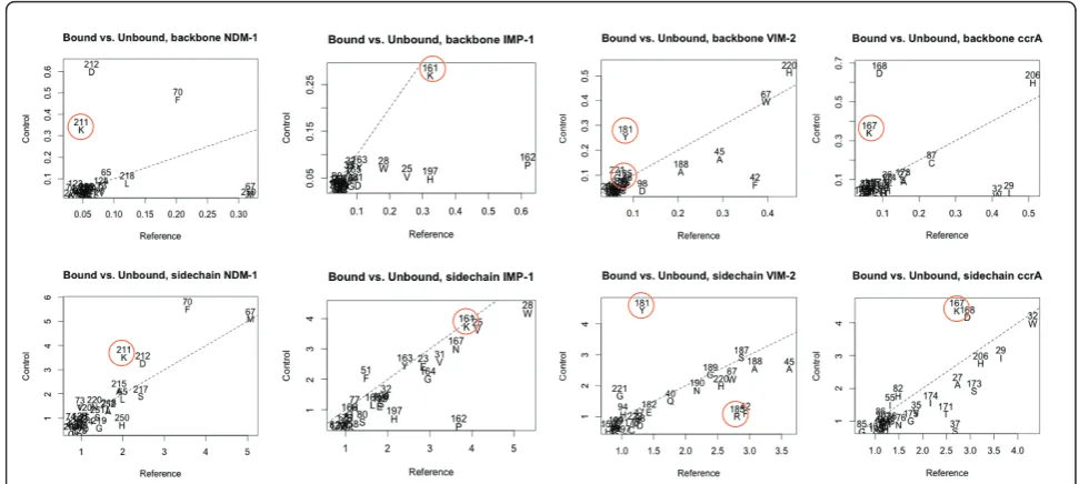
[image:9.595.57.540.452.701.2]Significant backbone and side-chain changes between the bound and unbound states of IMP- and VIM-type proteins, and ccrA and NDM-1 indicated other com-monalities and differences among the MBLs. Figure 8 shows these changes between bound and unbound crys-tal structures around the active site. Specifically, binding across all four MBLs of interest elicits shared, large structural shifts in the flap region: residues M67, F70 in NDM-1 (V25, W28 in IMP-1; I29, W32 in ccrA; F42, A45 in VIM-2) of the mobile flap are shifted during binding via a twist of the loop caused by hydrophobic interactions. Other structural changes are evident in the L10 active site loop, in particular shifts in K211/K161/ K167 in NDM-1, IMP-1, and ccrA, respectively, a posi-tion that has been associated with polar ligand binding activity in B1 MBL members [23,38,41]. In VIM-2, R185 is believed to play a similar role [42], though in strict residue-residue correspondences from structural align-ment, Y181 occupies the position of K211 in VIM-2, and subsequently displays a similar conformational dif-ference between bound and unbound states (Figure 8). Comparison of R185 in VIM-2 shows it undergoes less dramatic a change in conformation, further distinguish-ing it from the other MBLs (in addition to its relatively shorter length and residue composition). D212/D168 in NDM-1/ccrA, undergoes similar changes in conforma-tion as K211, though the analogous residue in IMP-1, P162, does not.
Functional and structural residues of interest in B1 MBLs
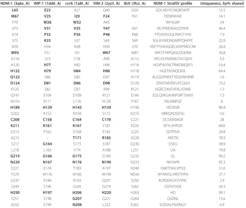
Structural distinctions between VIM, IMP, ccrA, NDM-1 and BcII (with residue positioning per PDB structures 2yz3_A, 1dd6_A, 1a8t_A, 3q6x_A and 3fcz_A, for con-text) are identified for residues within 4 Å of either the zinc or ligand binding regions, are shown in Table 1, such as W67 in VIM-2 (2yz3_A numbering; W93 in NDM-1). This residue was determined to be functionally
important for VIM-2, was shown viamutagenesis to be
[image:10.595.56.539.89.332.2]integral for stability [28] and is bolded in Table 1 due to its colocation (within 4 Å) with the ligand, a mercapto-carboxylate inhibitor [42]. Replacement of this residue in VIM-2 results in decreased ampicillin resistance. 3q6x_A, bound to hydrolyzed ampicillin, indicates the nature of the interaction as a hydrophobic, suggesting a similar antibiotic phenotype [23]. The corresponding residue in ccrA is an isoleucine, which is the second most common residue match using StralSV (see Table 1). IMP-1 possesses a relatively uncommon phenylala-nine, though its effect, if any, on enzymatic function has not yet been experimentally characterized. Given the amino acid and similar proximity to an IMP-1 ligand (also a mercaptocarboxylate inhibitor [38]), an analogous effect with W93 in NDM-1 is plausible. Structural varia-bility analysis was also performed using StralSV to iden-tify additional active site conformations and critical residues based on rarity in NDM-1, and several residues were noted to be unique in various parts of the
structure (see Additional file 4). Notably, some of these unique residues appear within the active cavity of NDM-1, whose structural corollaries in other B1 MBLs are associated with inhibitor or substrate enzymatic activity. This includes the uncommon residue at the L3 loop (positions 68-72), phenylalanine (F70).
NDM-1 also shares functional residues with MBLs outside the IMP, VIM and ccrA types. Earlier directed evolution studies with BcII indicated several residue changes implicated with resistance [40]. Notably, the glycine to serine change at position 262 in BcII maps to S249 within NDM-1, and S196 in IMP-1 (see Table 1). In NDM-1, as in BcII, S249/S262 forms a hydrogen bond with C208 (3.18 Å)/C221 (3.2 Å), directly affecting the second zinc binding site. This change in BcII is noted to result in increased cephalexin turnover [40]. The complementary mutation within BcII, N70S, is not present in NDM-1, though a similar residue, histidine, is found in IMP-1. Cephalosporin profiles for NDM-1 are most similar to IMP-1, though turnover is slightly better for IMP-1 [2,43], and may imply that a mutation of N76 in NDM-1 to H/S76 may result in more efficient cepha-losporin hydrolysis.
Wholesale comparison of these and other possibly cri-tical residues were plotted using LGA_pdblist, permit-ting a view of deviations between functional side chains
of multiple proteins, given a reference. Selection of atom positions for LGA calculation was done using a list of functional ends of protein side-chains, as
described in [44]viaGDC-sc. Use of NDM-1 as a
refer-ence against representatives of VIM, IMP and ccrA highlighted areas of inter- and intra-type functional side-chain difference (Figure 9). Between available NDM-1 structures, side-chain positioning is generally in agreement, with the exception of 3s0z_A, which exhibits notable variation not seen in the other representatives (we note that 3s0z_A, an unbound structure, is missing
part of the linker region–residues 167-170). Within
NDM-1 structures, we observe consistent differences between the bound reference (3q6x_A) and the unbound structures in both R81, the L10 loop and E227 (see annotations on Figure 9). As mentioned in the pre-vious section, IMP, ccrA and NDM-1 also share a lysine (K161 in IMP; K211 in NDM-1) at a residue position associated with mercaptocarboxylate-based inhibition; this residue was highlighted as undergoing conforma-tional changes between bound and unbound states (Fig-ure 8). Side-chain comparisons of this residue using
LGA_pdblist show that NDM-1’s K211 adopts a
func-tional side-chain orientation closer to that of IMP-1 than ccrA, despite the larger differences between
[image:11.595.54.540.89.307.2]NDM-1 and IMP-NDM-1’s overall L10 loop. The presence of these
Figure 8Structural conformation adjustments within the active sites between bound-unbound structures of NDM-1, IMP-1, VIM-2 and ccrA. Charts were generated as follows: in each case, a bound reference from one of the four B1 MBL types was selected for examination. The bound reference was compared to other bound B1 MBL structures using LGA (maximal distance of 4 Å), and similarly to unbound B1 MBL structures. Residue shifts within a 4 Å distance of either the bound ligandorthe dinuclear zincs were drawn on a XY-plot, with the X-axis referring to differences in the bound target and the unbound templates, and Y-axis the bound target against other bound templates,
residues at similar locations, and notably in comparable conformations, for other MBLs within NDM-1 may con-tribute to its broad binding profile, whose characteristics are simultaneously close to other B1 MBLs [2].
Close examination of the NDM-1 structures using side-chain deviations from LGA_pdblist as a guide reveal possible electrostatic interactions between R81 and W59, and E227 may form a transient salt bridge with R270. Notably, E227 is located on the turn imme-diately before the L10 binding loop, and may thus aid
modestly in stabilization. While the same glutamic acid is found in IMP-1, there appears no R270 analogue. Comparison of non-covalent interactions between
3q6x_A and NDM-1’s unbound representatives using
VMD [45] further show that additional salt bridges may form during ligand binding, and that such interactions are more prevalent in NDM-1 than in IMP-1, VIM-2 or ccrA.
[image:12.595.57.542.122.531.2]Emerging research into the mechanisms of MBL pro-teins indicate that variation in resistance profiles can be Table 1 StralSV profiles of B1 MBL active site, functional residues For each five B1 MBL proteins, residues within 4 Å of either the zinc ions or ligand were identified (red denotes metal coordination residues, while bold denotes those in close proximity to the ligand).
NDM-1 (3q6x_A) IMP-1 (1dd6_A) ccrA (1a8t_A) VIM-2 (2yz3_A) BcII (3fcz_A) NDM-1 StralSV profile Uniqueness, by% shared
L65 E23 A27 Q40 G59 GDLAEVYCNIQRSWTF 15.5
M67 V25 I29 F24 F61 FIDMNHAE 14.1
F70 W28 W32 A45 - WFAGKP 24
V73 V31 V35 Y47 V67 VILYPMDKAGQTRHF 46.4
A74 P32 P36 P48 P68 PTDAVISGQLRWCFYHX 7.9
S75 K33 S37 S49 S69 SGLAYVKDNEIMRTQHFPC 22.9
N76 H34 N38 N50 S70 NSFTYVAHGQELWIXPMDCRK 26.8
W93 F51 I55 W67 W87 WIFSTYKPQAVLDGERM 35.8
A116 S73 F78 A90 A112 VIFLASYNWMGTHCQDX 5.5
H120 H77 H82 H94 H116 HGVPAIYNLTRWDMQEFS 53.4
H122 H79 H84 H96 H118 HGETASNQCKVL 64.4
Q123 S80 G85 D97 A119 ALGSDPNFKTYEQVWHIMR 1.6
D124 D81 D86 D98 D120 DSNTAKVEIFLPCQGH 73.2
K125 S82 C87 R99 R121 HGRCSANTVFKLYDIME 1.3
Q147 E104 D109 R121 E144 EDLQVKGAHNPSRFTXMYI 7.5
M154 K111 L116 N128 Y167 YKLNMPGF 6
H189 H139 H145 H159 H196 HEDKVR 96.9
D202 K152 N158 S172 N215 NRKQADSEHG 5.6
C208 C158 C164 C178 C221 DCSXEKMGR 28
K211 K161 K167 Y181 K224 KFYLVHPGR 68.6
D212 P162 D168 E182 S225 SDTEIVA 24.8
A215 - T171 R185 A228 ARSTN 78.9
S217 G164 S173 S187 D230 DSEG 38.9
L218 L165 I174 A188 L231 LIA 74.8
G219 G166 G175 G189 G232 GL 99.2
N220 N167 N176 N190 N233 NKYAPR 85.3
E227 E174 T183 A197 N240 ENKTDIALXPSQ 31.6
Y229 W176 W185 W199 W242 WYMVGLARISTHFN 27.7
G237 K184 K193 Q207 S250 KLRQISEGATVYNC 2.9
S249 S196 G205 G219 S262 GSPAYVDE 24.3
H250 H197 H206 H220 H263 HD 99.1
S251 S198 G207 G221 G264 GSDNL 15.6
A252 E199 N208 L222 E265 EODIALPWVNGY 6.9
These structures were selected because unlike the references structures used to build the MBL library they are ligand bound, with the exception of BcII (3fcz_A). Structural alignments were then used to identify residue-residue correspondences for five B1 MBL proteins for those given residues. For NDM-1, the
associated with residue changes distant from the active site. Studies of VIM variants and residue-specific changes to members of the IMP type [40,46-48] indicate that locations distant from the active site may affect hydrolytic activity. For example, K215 (aligned to a S172 in VIM-2, > 20 Å distant from the active site) in the recently characterized VIM-19 is associated with improved carbapenem resistance when R228 is also pre-sent [48]. The V112A mutation in BcII, similarly distant from the active site, is associated with increased cepha-losporin activity, though the association is unclear. As we noted, mapping of resistance-related BcII regions to NDM-1 shows it possess two of four associated hydroly-tically beneficial residues. The presence of multiple, fit-ness-improving residues within NDM-1 found also in critical structural and functional regions of a myriad number of other MBLs suggests incremental and com-plementary changes in MBL composition, even in regions distant from ligand binding, can have effects on resistance that are difficult to predict.
Conclusions
We have sought to characterize structural features of members of the B1 MBL proteins most closely related to the recently discovered NDM-1 gene using structural conservation and comparisons of sequence conservation. This has included a survey of the structural features of
B1 MBLs from different approaches, including residue variability at specific substructures, clustering varying degrees of structural granularity, and examination of the critical residues of MBLs with an eye toward NDM-1 functionality. While most MBL proteins showed a tightly conserved overall fold structure, structure-based sequence variability methods confirmed the strong structural and sequence conservation at key residues within the active cavity. From this analysis, we find that NDM-1 appears to possess several residues found in variants of IMP, VIM and other MBLs known to confer resistance-like capabilities.
A striking exception to this is the identification of a linker region found within MBLs that appears to vary in structure and length, and is the most divergent and dis-tinguishing structural feature between the IMP, VIM, ccrA and NDM-1 proteins. The identification of this variable linker region within MBLs raised the hypothesis of a distinctive flexible loop; inspection of VIM-2, ccrA and IMP-1 revealed no significant changes in the linker region between apo and holo forms. We identified a marginal difference between the bound and unbound N-terminal ends of the NDM-1 loop on the order of ~1.0-1.5 Å. As the linker region is quite distant from the active site itself, it is unclear if this is a functional shift or an artifact of a possibly more flexible region. Addi-tional study of this region of MBL proteins is necessary to understand how its conformation may affect MBL structure or function.
Deeper knowledge of the structure and mechanism involved in antibiotic resistance in bacteria is highlighted by the continued emergence of transferrable MBLs such as NDM-1. This new enzyme is disturbing for both the speed at which it has spread, its broad capability to bind
many types ofb-lactams uncharacteristic of other MBLs
and its colocation with other resistance-granting genes. Structural alignments of NDM-1 to other B1 MBLs shows that it simultaneously shares critical resistance-associated residues with VIM, IMP, ccrA and even BcII, some of which are distant from the active site. The notion of a structure displaying motifs from multiple protein subclasses is not entirely unknown for B1 MBLs; SPM-1, for example, has structural features found in both B1 and B2 MBL proteins [49]. As others have pos-ited, that this may indicate that while the overall MBL fold structure is critical from a functional standpoint, there is potential for optimization at the residue and
substructure levelviasmall changes in sequence or
con-formation [50]; in this light, NDM-1’s uniqueness in
both composition and structure may serve a multitude of possible function roles, and thus possible targets of further study.
In the future, we hope to expand our computational analysis of these important proteins using ligand
((,$(,&
'& $$
-'$+.
$'
$'
$(#*
)
[image:13.595.57.291.88.256.2]$'(''
screening methods, with the intent to determine resi-dues or structural features that are broadly critical to MBL substrate specificity, thus correlating structure more concretely to phylogenetic profile. The findings described herein provide promising regions for further investigation. Furthermore, experimental follow-up would aid in elucidating the role the linker region may play in MBLs, including NDM-1, with regard to plasti-city, function and binding
Additional material
Additional file 1: MBL_library.csv–(Comma-seprated values file) Enumeration of MBL folds comprising the comparative analysis library. This file contains a list of all structures included in the described MBL library (see 2.1), as well as their type classification.
Additional file 2: Whole_chain_clustering–(Portable document format file) Whole-chain clustering of B1 MBL library using StralCP. This supplemental figure is the whole chain dendrogram for the B1 library, and is depicted in similar form and labeling as Figure 4.
Additional file 3: B1_MBL_active_site_measurements.csv– (Comma-separated values file) Measurements for active sites of selected MBLs. Each row-wise record of this file contains information regarding the active site of the PDB entry noted in the first column. This data includes: the B1 type, the area and volume of the active site (as estimated by CASTp), an indicator variable associated with the presence of a bound ligand in the active site (1 for a present ligand, 0 otherwise), and the measured metal ion distances for di-zinc MBLs (cases where one or less metal ions are present is designated by a dash).
Additional file 4: 3q6x_A_StralSV_w90_5.txt–(Text file) StralSV output for NDM-1 structure 3q6x_A. This file contains the raw output of the StralSV algorithm run on 3q6x_A using the entire PDB (release 2011/08/02). The header of the file contains structural matches (by region) of various PDB templates to NDM-1, followed by the annotations of the templates. The main body of the file consists of the StralSV output profile, where the first data column is the amino acid; the second column is the position of that amino acid in the profile (starting at 1); the third column is the position of the amino acid in the sequence itself; the fourth column denotes the rank of the amino acid present relative to the structural match profile; the fifth column indicates the percentage of matched structures which have an exact residue-residue correspondence to present amino acid; columns six, seven and eight are the percentages of matched structures which contain the most prevalent, second-most prevalent and third-most prevalent residues, respectively; columns nine and ten are the fraction and number of structural hits; the eleventh column is the StralSV profile itself, sorted by the frequency of the amino acid occupying the position to which the present amino acid aligns; the following columns are indicators for various amino acid categories (see header) and unused measures of conservation.
Acknowledgements
This work was conducted at Lawrence Livermore National Laboratory under US DOE Contract DE-AC52-07NA27344. The work was supported by an LLNL-LLNS internally funded grant 09-ERD-054 under Jane Bearinger through the Laboratory Directed Research and Development program, and by a grant from the US DOD Defense Threat Reduction Agency, contract number PE0603384BP. The authors would like to thank the anonymous reviewers for their thoughtful comments and recommendations for improvement.
Author details
1Global Security Computing Applications Division, Lawrence Livermore
National Laboratory, Livermore, 94550 CA, USA.2Biosciences & Biotechnology Division, Lawrence Livermore National Laboratory, Livermore, 94550 CA, USA.
3National Security Engineering Division, Lawrence Livermore National
Laboratory, Livermore, 94550 CA, USA.
Authors’contributions
EV and AZ acquired the data and conceived the project. AZ, KL and EC conducted the analysis and developed related programs. AZ, KL and EC wrote the manuscript. CZ contributed to discussions and ideas for the project. All authors read, edited and approved the final manuscript.
Competing interests
Lawrence Livermore National Laboratory holds the patent for LGA (patent #8024127), and has submitted patents for StralCP and StralSV.
Received: 21 September 2011 Accepted: 14 February 2012 Published: 14 February 2012
References
1. Lee K, Yum JH, Yong D, Lee HM, Kim HD, Docquier J-D, Rossolini GM, Chong Y:Novel acquired metallo-beta-lactamase gene, bla(sim-1), in a class 1 integron from acinetobacter baumannii clinical isolates from Korea.Antimicrob Agents Chemother2005,49(11):4485-4491. 2. Yong D, Toleman MA, Giske CG, Cho HS, Sundman K, Lee K, Walsh TR:
Characterization of a new metallo-beta-lactamase gene, blaNDM-1, and a novel erythromycin esterase gene carried on a unique genetic structure in Klebsiella pneumoniae sequence type 14 from India.
Antimicrob Agents Chemother2009,53(12):5046-5054.
3. Grundmann H, Livermore DM, Giske CG, Canton R, Rossolini GM, Campos J, Vatopoulos A, Gniadkowski M, Toth A, Pfeifer Y, Jarlier V, Carmeli Y, CNSE Working Group:Carbapenem-non-susceptible enterobacteriaceae in Europe: conclusions from a meeting of national experts.Euro Surveill
2010,15(46):1-13.
4. Kumarasamy KK, Toleman MA, Walsh TR, Bagaria J, Butt F, Balakrishnan R, Chaudhary U, Doumith M, Giske CG, Irfan S, Krishnan P, Kumar AV, Maharjan S, Mushtaq S, Noorie T, Paterson DL, Pearson A, Perry C, Pike R, Rao B, Ray U, Sarma JB, Sharma M, Sheridan E, Thirunarayan MA, Turton J, Upadhyay S, Warner M, Welfare W, Livermore DM, Woodford N:Emergence of a new antibiotic resistance mechanism in India, Pakistan, and the UK: a molecular, biological, and epidemiological study.Lancet Infect Dis2010,
10(9):597-602.
5. Mulvey MR, Grant JM, Plewes K, Roscoe D, Boyd DA:New Delhi metallo-Beta-lactamase in Klebsiella pneumoniae and Escherichia coli Canada.
Emerg Infect Dis2011,17(1):103-106.
6. Poirel L, Al~Maskari Z, Al~Rashdi F, Bernabeu S, Nordmann P:NDM-1 producing Klebsiella pneumoniae isolated in the Sultanate of Oman.J Antimicrob Chemother2010,66(2):304-306.
7. Poirel L, Lagrutta E, Taylor P, Pham J, Nordmann P:Emergence of metallo-beta-lactamase NDM-1 producing multidrug-resistant escherichia coli in Australia.Antimicrob Agents Chemother2010,54(11):4914-4916.
8. Poirel L, Revathi G, Bernabeu S, Nordmann P:Detection of ndm-1-producing klebsiella pneumoniae in Kenya.Antimicrob Agents Chemother
2010,55(2):934-936.
9. Detection of Enterobacteriaceae isolates carrying metallo-beta-lactamase–United States, 2010. CDC Morbidity and Mortality Weekly Report
2010,59:750.
10. Jain E, Bairoch A, Duvaud S, Phan I, Redashi N, Suzek BE, Martin MJ, McGarvey P, Gasteiger E:Infrastructure for the life sciences: design and implementation of the UniProt website.BMC Bioinf2009,10:136. 11. Poirel L, Ros A, Carricajo A, Berthelot P, Pozzetto B, Bernabeu S,
Nordmann P:Extremely drug-resistant citrobacter freundii isolate producing NDM-1 and other carbapenemases identified in a patient returning from India.Antimicrob Agents Chemother2011,55(1):447-448. 12. Giakkoupi P, Petrikkos G, Tzouvelekis LS, Tsonas S, Legakis NJ,
Vatopoulos AC, WHONET Greece Study Group:Spread of integron-associated vim-type metallo-beta-lactamase genes among imipenem-nonsusceptible pseudomonas aeruginosa strains in greek hospitals.J Clin Microbiol2003,41(2):822-825.
13. Giske CG, Rylander M, Kronvall G:Vim-4 in a carbapenem-resistant strain of pseudomonas aeruginosa isolated in Sweden.Antimicrob Agents Chemother2003,47(9):3034-3035.
15. Yan JJ, Hsueh PR, Ko WC, Luh KT, Tsai SH, Wu HM, Wu JJ: Metallo-beta-lactamases in clinical pseudomonas isolates in Taiwan and identification of vim-3, a novel variant of the vim-2 enzyme.Antimicrob Agents Chemother2001,45(8):2224-2228.
16. Castanheira M, Deshpande LM, Mathai D, Bell JM, Jones RN, Mendes RE:
Early dissemination of NDM-1 and OXA-181 producing Enterobacteriaceae in Indian hospitals: Report from the SENTRY antimicrobial surveillance program (2006-2007).Antimicrob Agents Chemother2010,55(3):1274-1278.
17. Garau G, Garcia-Saez I, Bebrone C, Anne C, Mercuri P, Galleni M, Frere J-M, Dideberg O:Update of the standard numbering scheme for class b beta-lactamases.Antimicrob Agents Chemother2004,48(7):2347-2349.
18. Bush K, Jacoby GA:Updated functional classification of beta-lactamases.
Antimicrob Agents Chemother2010,54(3):969-976.
19. Bush K, Jacoby GA, Medeiros AA:A functional classification scheme for beta-lactamases and its correlation with molecular structure.Antimicrob Agents Chemother1995,39(6):1211-1233.
20. Badarau A, Page M:The mechanisms of catalysis by metallo beta-lactamases.Bioinorg Chem Appl2008,576297:1-14.
21. Bebrone C:Metallo-beta-lactamases (classification, activity, genetic organization, structure, zinc coordination) and their superfamily.Biochem Pharmacol2007,74(12):1686-1701.
22. Majiduddin F, Materon I, Palzkill T:Molecular analysis of beta-lactamase structure and function.Int J Med Microb2002,292:127-137.
23. Zhang H, Hao Q:Crystal structure of NDM-1 reveals a common beta-lactamase hydrolysis mechanism.FASEB J2011,25(8):2574-2582. 24. Wang Z, Fast W, Benkovic S:On the mechanism of the
metallo-beta-lactamase from Bacteriodes fragilis.Biochemistry1999,38:10013-10023. 25. Moali C, Anne C, Lamotte-Brasseur J, Groslambert S, Devreese B, Van
Beeumen J, Galleni M, Frere JM:Analysis of the importance of the metallo-beta-lactamase active site loop in substrate binding and catalysis.Chem Biol2003,10(4):319-329.
26. Huntley JJA, Fast W, Benkovic SJ, Wright PE, Dyson HJ:Role of a solvent-exposed tryptophan in the recognition and binding of antibiotic substrates for a metallo-beta-lactamase.Protein Sci2003,12(7):1368-1375. 27. Scrofani SD, Chung J, Huntley JJ, Benkovic SJ, Wright PE, Dyson HJ:Nmr
characterization of the metallo-beta-lactamase from bacteroides fragilis and its interaction with a tight-binding inhibitor: role of an active-site loop.Biochemistry1999,38(44):14507-14514.
28. Borgianni L, Vandenameele J, Matagne A, Bini L, Bonomo RA, Frere J-M, Rossolini GM, Docquier J-D:Mutational analysis of vim-2 reveals an essential determinant for metallo-beta-lactamase stability and folding.
Antimicrob Agents Chemother2010,54(8):3197-3204.
29. Guo Y, Niu G, Shui W, Zhou H, Zhang Y, Yang C, Lou Z, Rao Z:A structural view of the antibiotic degradation enzyme NDM-1 from a superbug.
Protein Cell2011,2(5):384-394.
30. King D, Strynadka N:Crystal structure of New Delhi metallo-beta-lactamase (NDM-1) reveals molecular basis for antibiotic resistance.
Protein Sci2011,20(9):1484-1491.
31. Berman HM, Battistuz T, Bhat TN, Bluhm WF, Bourne PE, Burkhardt K, Feng Z, Gilliland GL, Iype L, Jain S, Fagan P, Marvin J, Padilla D, Ravichandran V, Schneider B, Thanki N, Weissig H, Westbrook JD, Zardecki C:The Protein Data Bank.Acta Crystallogr D: Biol Crystallogr2002,
58(Pt 6 No 1):899-907.
32. Zemla A, Lang D, Kostova T, Andino R, Zhou C:StralSV–assessment of sequence variability within similar 3D structures and application to polio RNA-dependent RNA polymerase.BMC Bioinf2011,12:226.
33. Zemla A:LGA–A method for finding 3 d similarities in protein structures.
Nucleic Acids Res2003,31(13):3370-3374.
34. Zemla A, Geisbrecht B, Smith J, Lam M, Kirkpatrick B, Wagner M, Slezak T, Zhou CE:Stralcp–structure alignment-based clustering of proteins.
Nucleic Acids Res2007,35(22):e150.
35. Yoon S, Ebert J, Chung E, Micheli G, Altman R:Clustering protein environments for function prediction: finding PROSITE motifs in 3D.BMC Bioinf2007,8(Suppl 4):S10.
36. Liang J, Edelsbrunner H, Woodward C:Anatomy of protein pockets and cavities: measurement of binding site geometry and implications for ligand design.Protein Sci1998,7(9):1884-1897.
37. Kim R, Guo J-T:Systematic analysis of short internal indels and their impact on protein folding.BMC Struct Biol2010,10:24.
38. Concha NO, Janson CA, Rowling P, Pearson S, Cheever CA, Clarke BP, Lewis C, Galleni M, Frere JM, Payne DJ, Bateson JH, Abdel-Meguid SS:
Crystal structure of the imp-1 metallo beta-lactamase from
pseudomonas aeruginosa and its complex with a mercaptocarboxylate inhibitor: binding determinants of a potent, broad-spectrum inhibitor.
Biochemistry2000,39(15):4288-4298.
39. Schrodinger LLC:The PyMol molecular graphics system August 2010.. 40. Tomatis P, Fabiane S, Simona F, Carloni P, Sutton B, Vila A:Adaptive
protein evolution grants organismal fitness by improving catalysis and flexibility.PNAS2008,105(52):20605-20610.
41. Toney J, Fitzgerald P, Grover-Sharma N, Olson S, May W, Sundelof J, Vanderwall D, Cleary K, Grant S, Wu J, Kozarich J, Pompliano D,
Hammond G:Antibiotic sensitization using biphenyl tetrazoles as potent inhibitors of Bacteroides fragilis metallo-beta-lactamse.Chem Bio1998,
5:185-196.
42. Yamaguchi Y, Jin W, Matsunaga K, Ikemizu S, Yamagata Y, Wachino J, Shibata N, Arakawa Y, Kurosaki H:Crystallographic investigation of the inhibition mode of a VIM-2 metallo-beta-lactamase from Pseudomonas aeruginosa by a mercaptocarboxylate inhibitor.J Med Chem2007,
50:6647-6653.
43. Laraki N, Franceschini N, Rossolini GM, Santucci P, Meunier C, de Pauw E, Amicosante G, Fre’re JM, Galleni M:Biochemical characterization of the Pseudomonas aeruginosa 101/1477 metallo-beta-lactamase IMP-1 produced by Escherichia coli.Antimicrob Agents Chemother1999,
43:902-906.
44. Keedy D, Williams C, Headd J, Arendall W, Chen V, Kapral G, Gillespie R, Block J, Zemla A, Richardson D, Richardson J:The other 90% of the protein: assessment beyond the C-alphas for CASP8 template-based and high-accuracy models.Prot Struct Func Bioinf2009,77:29-49.
45. Humphrey W, Dalke A, Schulten K:VMD–Visual molecular dynamics.J Molec Graphics1996,14:33-38.
46. Marchiaro P, Tomatis P, Mussi M, Pasteran A, Viale A, Limansky A, Vila A:
Biochemical characterization of metallo-beta-lactamse VIM-11 from a Pseudomonas aeruginosclinical strain.Antimicrob Agents Chemother2008,
52:2250-2252.
47. Oelschlaeger P, Mayo S, Pleiss J:Impact of remote mutations on metallo-beta-lactamase substrate specificity: implications for the evolution of antibiotic resistance.Prot Sci2005,14(3):765-774.
48. Rodriguez-Martinez J, Nordmann P, Fortineau N, Poirel L:VIM-19, a metallo-beta-lactamase with increased carbapenemase activity from Escherichia coli andKlebsiella pneumoni.Antimicrob Agents Chemother
2010,54(1):471-476.
49. Murphy TA, Catto LE, Halford SE, Hadfield AT, Minor W, Walsh TR, Spencer J:
Crystal structure of pseudomonas aeruginosa spm-1 provides insights into variable zinc affinity of metallo-beta-lactamases.J Mol Biol2006,
357(3):890-903.
50. Fisher J, Meroueh S, Mobashery S:Bacterial resistance to beta-lactam antibiotics: compelling opportunism, compelling opportunity.Chem Rev
2005,104(2):395-424.
doi:10.1186/1756-0500-5-96
Cite this article as:Cadaget al.:Computational analysis of
pathogen-borne metallob-lactamases reveals discriminating structural features
between B1 types.BMC Research Notes20125:96.
Submit your next manuscript to BioMed Central and take full advantage of:
• Convenient online submission
• Thorough peer review
• No space constraints or color figure charges
• Immediate publication on acceptance
• Inclusion in PubMed, CAS, Scopus and Google Scholar
• Research which is freely available for redistribution