RESEARCH ARTICLE
Normocellular CSF in herpes simplex
encephalitis
Abhinbhen W. Saraya
1*, Supaporn Wacharapluesadee
1, Sininat Petcharat
1, Nuntaporn Sittidetboripat
1,
Siriporn Ghai
1, Henry Wilde
1,2and Thiravat Hemachudha
1Abstract
Background: Herpes simplex virus (HSV) is the most common cause of sporadic encephalitis worldwide. The high mortality rate (70–80 %) of herpes simplex encephalitis (HSE) can be reduced to 20–30 % by antiviral therapy. However, normocellular CSF can lure physicians to look for non-infectious causes, resulting in delayed treatment. This study aimed to investigate, characterize and differentiate HSE patients, with normocellular and pleocytosis CSF, according to neuroimaging patterns, underlying disease, CSF viral load and clinical outcome. Patients with proven (by PCR positive CSF) or presumed viral infections of the CNS admitted to King Chulalongkorn Memorial Hospital between January 2002 and 2011 were analyzed.
Results: HSV was detected in the CSF of 43 patients but only 23 patients had encephalitis. Among these 23 patients, 6 cases (26.1 %) had normal CSF WBC (<5 cells/mm3). One patient in this normocellular CSF group had HIV infection. Although this patient had low CD4 counts (<200 cells/mm3), the peripheral WBC counts showed only mild leuko-penia. The CSF HSV viral load in the pleocytosis group was higher than the normocellular group, with an average of 12,200 vs 3027 copies/ml respectively. There was no correlation between the viral load and the clinical outcome. With respect to neuroimaging, 4 (66.7 %) patients in the normocellular group had unremarkable/non-specific results. Conclusions: Normocellular CSF in HSE is not rare, and can be seen in normal as well as immunocompromised hosts. Clinicians should not exclude CNS infection, especially HSE, merely based on the absence of CSF pleocytosis and/or unremarkable neuroimaging study.
Keywords: Encephalitis, Viral encephalitis, Herpes simplex encephalitis, Herpes simplex virus
© 2016 Saraya et al. This article is distributed under the terms of the Creative Commons Attribution 4.0 International License (http://creativecommons.org/licenses/by/4.0/), which permits unrestricted use, distribution, and reproduction in any medium, provided you give appropriate credit to the original author(s) and the source, provide a link to the Creative Commons license, and indicate if changes were made. The Creative Commons Public Domain Dedication waiver (http://creativecommons.org/ publicdomain/zero/1.0/) applies to the data made available in this article, unless otherwise stated.
Background
Herpes simplex encephalitis (HSE) caused by herpes simplex virus (HSV) is the most common sporadic viral encephalitis among adults worldwide [1, 2]. It has been responsible for up to 20 % of viral encephalitis cases [3]. This disease has a bimodal distribution, where one-third of the patients are under the age of 20, while the remaining are over the age of 50 [4]. Peak incidence is around 60–64 years of age. Neither gender, season, nor immune status of a patient affects its occurrence. The
high mortality rate of HSE (70–80 %) can be reduced to 20–30 % by acyclovir therapy. Acyclovir should be taken within 2 days of neurological symptom’s onset as delayed treatment can result in severe morbidity and mortality in approximately one-third of the patients [5, 6].
HSV-1 is responsible for more than 90 % of HSE in adults [7]. Recent studies have demonstrated many cases of HSE that resulted from HSV-2 [8, 9]. HSV can also be associated with myelitis, radiculitis or both. HSV’s preferred areas are the limbic structures, the medial temporal cortex and the orbito-frontal regions, which are all innervated by branches of olfactory and trigeminal nerves. HSV is contracted via contact with the mucosal surface of mouth, eye or genitalia, and has been speculated to subsequently enter the CNS via any nearby sensory nerve ending into these nerves and the
Open Access
*Correspondence: [email protected]
1 WHO-CC for Research and Training on Viral Zoonoses, Division of Neurology, Department of Medicine, Faculty of Medicine, Neuroscience Centre for Research and Development, Chulalongkorn University and King Chulalongkorn Memorial Hospital, Rama 4 Road, Pathumwan, Bangkok 10330, Thailand
corresponding ganglia. Viral entry into the CNS via olfactory tract has been substantiated by numerous ani-mal experiments [10, 11]. However, the neuropathology of a mouse brain is more diffused compared to the well-defined fronto-temporal distribution in a human brain.
HSV can reside in the nerve and can either replicate or establish a latent infection [1]. Its DNA can be detected in the trigeminal ganglion of previously infected patients. At the time of reactivation, latent HSV in the trigeminal ganglia can travel along the tentorial nerve that inner-vates the meninges of the anterior and the middle cranial fossa. HSV genomes have also been found in olfactory bulb, pons, and the medulla of autopsied brains of nor-mal individuals [12]. Early gross brain pathology of most HSE cases showed hemorrhagic necrosis of the medial or the inferior temporal lobe. These lesions were mainly bilateral and asymmetrical in distribution. Sub-frontal and insular regions were damaged in the latter phase [13]. Brain parenchyma in such regions of HSE patients, who died after 2 weeks of exhibiting HSE, revealed lique-faction necrosis.
The cerebral inflammation from glial cells activation, due to the presence of HSV, includes increase in expres-sion of pattern recognition receptors, especially Toll-like receptors (TLRs) and interferon regulatory factors (IRFs) [4]. Genetic factors, such as deficiency of Toll-like recep-tor-3 (TLR3), can predispose an individual (especially children and young adults) to HSE [10, 11, 14]. Mutation of TLR3 and TLR3 pathway genes such as UNC93B1, TRIF, TRAF3, and TBK1 can result in the defective pro-duction of interferon (IFN). Insufficient amount of IFN-β and IFN-λ leads to an increase in viral replication and enhances cell death. Relapse of HSE tends to occur more frequently in those with abnormal TLR3 immunity. How-ever, these mutations were attributed to only 5 % of HSE in children and young adults [11].
The characteristic symptoms of HSE are abnormal behavior/psychosis (66 %), confusion/disorientation (81 %) and speech disturbance (66 %), which are justifia-ble due to the preferential involvement of the frontal and temporal lobes. Other symptoms of HSE include fever (76–90 %), headache (70–90 %) and seizure (50–55 %) [5, 15]. However, these symptoms are not unique as they can be present in encephalitis caused by other viruses [16, 17]. Moreover, these symptoms can also be found in immune-mediated encephalitis, in particular the anti-NMDA receptor encephalitis, which can mimic or occur as a relapse encephalitis after the first episode of HSE [18].
Hence, diagnosis of HSE requires an integration of clinical presentations, neuroimaging, and laboratory tests, especially molecular diagnostic techniques. The
current gold standard confirmatory test for HSE is poly-merase chain reaction (PCR) to detect HSV DNA in the CSF, whose sensitivity is approximately 96 % with a speci-ficity of 99 % in experienced laboratories [19]. For high accuracy of PCR, specimens should be analyzed within 2–10 days after neurological onset [2].
HSE patients are presumed to have evidence of inflam-matory response in CNS such as CSF pleocytosis or abnormal neuroimaging. CSF pleocytosis in HSE has been found in more than 90 % of the patients, with an average WBC count of 100–200 cells/mm3 [2, 20]. Major-ity of the cases (80 %) had mild to moderately elevated CSF protein levels (~100 mg/dl), with normal glucose. Detection of red blood cells in the CSF was not an indica-tor of HSV associated CNS infection [19], but may repre-sent a poor predictor [21]. CSF examination by real-time PCR to detect HSV DNA was validated as a new gold standard for the diagnosis of HSE in 2004 [2, 19, 22]. However, its value in determining viral amount has been controversial [21]. Further, there have been increasing case reports of normal WBC counts (either normocellu-lar or acellunormocellu-lar) in the CSF of normal and immunocom-promised HSE patients, and some were associated with unremarkable/nonspecific magnetic resonance imaging (MRI) findings [15–17, 23].
Methods
Patients with proven (by PCR positive CSF) or presumed viral infections of the CNS admitted to King Chulalong-korn Memorial Hospital (KCMH), Bangkok, Thailand, between January 2002 and 2011 were analyzed. Encepha-litis was defined as clinical evidence of brain parenchy-mal dysfunction [6], which could not be explained by metabolic, electrolyte imbalance, bleeding disorders or systemic immune disorders such as systemic lupus ery-thematosus (SLE), Behcet’s disease or Sjogren syndrome. Bacterial, Rickettsial, fungal, tuberculosis and parasitic causes were excluded by appropriate clinical and labora-tory examinations. The diagnosis of HSE was based on the clinical features of encephalitis with the exclusion of other mimics as described above, and the presence of HSV DNA in the CSF.
All encephalitis patients with presumed viral causes had their CSF examined by PCR for HSV, Varicella zoster virus (VZV), Epstein-Barr virus (EBV), Cytomegalovirus (CMV) and enteroviruses. Paraneoplastic and autoim-mune panels as previously described [24] were tested in one retrospective patient with normocellular CSF and unremarkable brain MRI examination.
PCR technique for HSV DNA
Qualitative real-time PCR [25] was performed on speci-mens before 2006, where 5 μl of extracted DNA was used for amplification and detection. Specimens received year 2007 onwards, quantitative assay was performed using artus® HSV-1/2 LC PCR kit (Qiagen Inc., Valencia, CA, USA). DNA from 0.1 to 1 mL of CSF was extracted using Nuclisens® extraction kit (bioMérieux, Boxtel, The Netherlands). The amplification was undertaken using a LightCycler® instrument (Roche Diagnostics, Ger-many). HSV-1 and -2 DNA PCR product was differenti-ated by melting curve analysis in the LightCycler® PCR instrument. The standard curve was generated from four quantitation standards of HSV (10, 100, 1000 and 10,000 copies/μl) supplied by the manufacturer. Preparation of PCR assay, PCR profile and data analysis was conducted according to the manufacturer’s protocol. The internal control supplied by the manufacturer was included in all reactions to check for possible PCR inhibition. The analytical detection limit of the PCR Kit was 1 copy/μl (p = 0.05) (Qiagen Inc., CA, USA). For qualitative assay, all steps were the same as quantitative assay, except that the standard curve was not used for calculation, and only one standard positive control was performed along with the assayed sample.
Results Patient data
There were 413 patients with viral neurological infec-tions during the 10-year period. All had lymphocytic pre-dominance with normal glucose CSF profile. Those with normal CSF cell count were considered encephalitic only when their brain dysfunctions could not be explained by compromised cardiopulmonary function or other causes. One hundred and fifty-eight patients (38.3 %) had viral etiologies confirmed by PCR. HSV was detected in the CSF of 43 patients (27.2 %) (23 encephalitis and 20 men-ingitis). Among the 23 HSE patients; there was a slight female predominance (1.3:1), ages ranged between 1 and 85 years [17 (73.9 %) between 16 and 60 (an average of 36)]. Most of HSE patients were previously healthy except 2 (8.7 %) with diabetes, 2 (8.7 %) hematologic malignan-cies, 1 (4.3 %) inactive SLE, 1 (4.3 %) multiple sclerosis (MS) and 1 (4.3 %) seropositive for human immune deficiency virus (HIV). Most of the patients were from
central Thailand. The patient with HIV infection was diagnosed after hospitalization with acute HSE. Prior to hospitalization, the patient had been in good health, never received any highly active antiretroviral therapy (HAART), and did not have any opportunistic infection. The patient’s CD4 count was 200 cells/mm3. The patient with MS was in remission and received 50 mg of azathio-prine daily. Lastly, the patient with inactive SLE had mild thrombocytopenia, but was not on any immunosuppres-sive drugs or corticosteroids.
Clinical findings
Of 23 HSE patients, 16 cases (69.6 %) had fever as a pro-drome. The most common presenting symptoms were seizures (7/23, 30.4 %), behavioral changes (6/23, 26 %), alteration of consciousness (5/23, 21.7 %), focal neuro-logical deficits (3/23, 13 %), and worsening of headaches (2/23, 8.7 %). All patients received intravenous admin-istration of acyclovir. Six patients (26.1 %) recovered completely (The Modified Rankin Scale: mRS 0–1), 14 (60.9 %) partially recovered (mRS 2–3), and 3 patients (13 %) died. Demographic data, underlying diseases, and presenting symptoms of each group, classified according to CSF WBC count, are presented in Table 1.
Laboratory findings
All 23 cases had serum creatinine in the range of 0–3 mg/ dl and normal liver function test. Complete blood count data was available in 20 patients, of whom 14 (70 %) were within the normal limit, 1 (5 %) had pancytopenia (patient no. 17 in pleocytosis group), and 1 (5 %) had leu-copenia (patient no. 6 in normocellular group). Seven-teen of 23 patients (73.9 %) had CSF examination within 2–10 days after neurological symptoms onset, the golden period for CSF PCR examination for HSV [7]. CSF find-ings are detailed in Table 2. CSF protein levels ranged between 14 and 528 mg/dl (mean 108.3 mg/dL), while CSF glucose levels were between 30 and 139 mg/dl (mean 66.2 mg/dl). Seventeen patients (73.9 %) had CSF WBC counts between 6 and 500 cells/mm3, with lymphocytic predominance.
Six cases (26.1 %), with CSF examinations within 10 days after onset of illness (an average of 3 days), had normal CSF WBC (<5 cells/mm3). One patient in this normocellular CSF group (16.7 %) had HIV infection. Although this patient had low CD4 counts (<200 cells/ mm3), the peripheral WBC counts were 3150 and 3570 cells/mm3 in two consecutive tests.
this group was higher than the normocellular group, with an average of 12,200 vs 3027 copies/ml respectively. Of all 14 specimens received after 2007, 10 cases (2 normocel-lular, 8 pleocytosis) were positive for HSV1 and 4 cases (1 normocellular, 3 pleocytosis) were positive for HSV2 encephalitis. Other CSF parameters are described in Table 2.
Imaging findings
Twenty-two patients had complete neuroimaging (either computer tomography (CT) or MRI) records. Eleven (50 %) patients had the predominant temporal or fronto-temporal lobe lesions and 2 (9.1 %) had fronto-fronto-temporal lesion coexisting with thalamic lesions. Another two patients had cortical/subcortical (non fronto-temporal) lesions. Two patients (9.1 %) had bead-like appearance resembling vasculitis on MR angiography, whereas 5 (22.7 %) had normal or non-specific findings. Brain imag-ing data altogether with CSF HSV viral load of normocel-lular and pleocytosis groups are presented in Tables 3
and 4.
Discussion
In this study, 6 of 23 HSE patients (26.1 %) had normal CSF WBC counts as shown in Tables 1 and 2. All nor-mocellular patients had CSF protein levels less than 100 mg/dl, whereas half of the patients in the pleocy-tosis group had CSF protein levels greater than 100 mg/ dl. All brain MRIs of the 15 patients in the pleocytosis group, and 2 of 4 patients in the normocellular group showed abnormal lesions. Two normocellular patients had normal brain MRI and high viral load, 5970 and 3144 copies/ml respectively. One of them had inactive SLE with drowsiness, while the other had MS (in remis-sion) with behavioral change (Table 4). Two patients in the normocellular group had CT scan, which might not have been sensitive enough to detect abnormali-ties, as compared to MRI. However, both did not have any other explainable causes. There was no data on CSF HSV viral load in both cases as only qualitative PCR data was available. One patient was first diagnosed as HIV positive at the time of admission with HSE, with no history of previous opportunistic infections. HIV viral load was not performed in the CSF and data on viral load in the blood was not available. Although HIV encephalitis could not be excluded, such acute presen-tation suggests otherwise. Retrospective autoantibody test was negative. Both patients had partial recovery with acyclovir treatment.
[image:4.595.59.287.131.609.2]Our study showed that HSE with normocellular CSF is more common than what was generally known in the past. The CSF protein levels, number of cases with abnormal neuroimaging, and CSF viral loads in the pleocytosis group were higher when compared to the normocellular group. It was noted that CSF viral load was up to four times higher in CSF pleocytosis group than normocellular group (an average of 12,200 vs 3027 copies/ml), potentially due to increased inflammatory response in pleocytosis group. Viral load alone may not entirely explain the magnitude of CSF cellular response, as patients with nonspecific MRI in the normocellular Table 1 Demographic data and presenting symptoms
of HSV encephalitis in patients with CSF pleocytosis and patients with normocellular CSF
Demographic data CSF pleocy-tosis group (WBC > 5cells/mm3)
Normocellu-lar CSF group (WBC ≤ 5cells/mm3)
Number of patients 17 6
Male:female 9:8 1:5
Median age (years) [range] 35 [1–59] 41 [27–85]
Underlying disease
Normal 13 (76.5 %) 3 (50 %)
HIV infection 0 1 (16.7 %)
Diabetes mellitus 2 (11.8 %) 0
Hematologic malignancy 2 (11.8 %) 0
Multiple Sclerosis (MS) 0 1 (16.7 %)
Systemic Lupus
Erythema-tosus (SLE) 0 1 (16.7 %)
Prodrome
Fever 1 (5.9 %) 1 (16.7 %)
Fever with headache 9 (52.9 %) 1 (16.7 %)
Fever with myalgia 1 (5.9 %) 1 (16.7 %)
Upper respiratory tract
symptoms 1 (5.9 %) 0
Fever with diarrhea 1 (5.9 %) 0
Fever with non-specific
rash 1 (5.9 %) 0
Headache 2 (11.8 %) 0
Non-specific 1 (5.9 %) 3 (50 %)
Presenting symptoms Worsening headache
with/without neck stiffness
2 (11.8 %) 0
Hemiparesis 1 (5.9 %) 0
Facial weakness 1 (5.9 %) 0
Severe spastic body and
limbs 1 (5.9 %) 0
Behavioral change and
confusion 4 (23.5 %) 3 (50 %)
Seizures 5 (29.4 %) 2 (33.3 %)
Alteration of
conscious-ness 3 (17.6 %) 1 (16.7 %)
Outcome
Complete recovery 5 (29.4 %) 1 (16.7 %)
Partial recovery 10 (58.8 %) 4 (66.7 %)
group also had high copies of viral load. When look-ing at individual data in normocellular group (Table 4), the patient with the highest viral load (5970 copies/
ml) had normal MRI and CSF protein level of 14 mg/ dl. Why some HSE patients have normocellular CSF, or normal/nonspecific MRI remains a mystery. Whether Table 2 CSF profiles and imaging findings
a Two of the patients (a non fronto-temporal lesion and one with fronto-temporal and thalamic lesions) also had vasculitis, visualized using magnetic resonance angiography
b Miscellaneous refers to non-specific lesions such as non-specific white matter changes, aging brain etc
CSF Profile CSF pleocytosis group
(WBC > 5cells/mm3) Normocellular CSF group (WBC
≤ 5cells/mm3)
Days of CSF examination after neurological onset Day 0—1: 5 (29.4 %)
Day 2—10: 12 (70.6 %) Day 0—1: 1 (16.7 %)Day 2—10: 5 (83.3 %)
CSF protein 0–60 mg/dl: 1 (6.3 %)
61–100 mg/dl: 7 (43.8 %) >100 mg/dl: 8 (50 %) Not available: 1
0–60 mg/dl: 2 (33.3 %) 61–100 mg/dl: 4 (66.7 %) >100 mg/dl: 0
CSF glucose <30 mg/dl: 1 (6.3 %)
31–60 mg/dl: 7 (43.8 %) >60 mg/dl: 8 (50 %) Not available: 1
<30 mg/dl: 0 31–60 mg/dl 1 (16.7 %) >60 mg/dl: 5 (83.3 %)
Average CSF viral load 12,200 copies/ml (39–11,755,813
copies/ml) (11 cases available: 6 had qualitative test)
3027 copies/ml (59–5970 copies/ml)
(4 cases available: 3 had qualitative test)
Brain imaging findings 16 available records 6 available records
Predominant temporal lobe lesions 5 (29.4 %) 1 (16.7 %)
Fronto-temporal lesions 4 (23.5 %) 1 (16.7 %)
Other cortical and subcortical lesions (non fronto-temporal)a 3 (18.8 %) 0
Fronto-temporal and thalamic lesionsa 3 (18.8 %) 0
Normal or Miscellaneousb 0 2 (33.3 %)
Normal CT (MRI not performed) 1 (5.9 %) 2 (33.3 %)
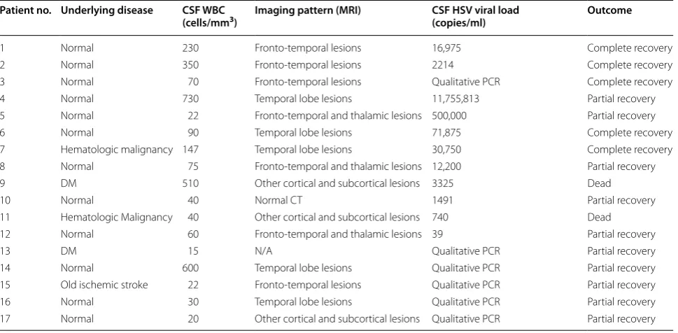
Table 3 Individualized data of the CSF pleocytosis group of HSE patients
Patient no. Underlying disease CSF WBC
(cells/mm3) Imaging pattern (MRI) CSF HSV viral load (copies/ml) Outcome
1 Normal 230 Fronto-temporal lesions 16,975 Complete recovery
2 Normal 350 Fronto-temporal lesions 2214 Complete recovery
3 Normal 70 Fronto-temporal lesions Qualitative PCR Complete recovery
4 Normal 730 Temporal lobe lesions 11,755,813 Partial recovery
5 Normal 22 Fronto-temporal and thalamic lesions 500,000 Partial recovery
6 Normal 90 Temporal lobe lesions 71,875 Complete recovery
7 Hematologic malignancy 147 Temporal lobe lesions 30,750 Complete recovery
8 Normal 75 Fronto-temporal and thalamic lesions 12,200 Partial recovery
9 DM 510 Other cortical and subcortical lesions 3325 Dead
10 Normal 40 Normal CT 1491 Partial recovery
11 Hematologic Malignancy 40 Other cortical and subcortical lesions 740 Dead
12 Normal 60 Fronto-temporal and thalamic lesions 39 Partial recovery
13 DM 15 N/A Qualitative PCR Partial recovery
14 Normal 600 Temporal lobe lesions Qualitative PCR Partial recovery
15 Old ischemic stroke 22 Fronto-temporal lesions Qualitative PCR Partial recovery
16 Normal 30 Temporal lobe lesions Qualitative PCR Partial recovery
[image:5.595.58.536.103.347.2] [image:5.595.57.542.427.665.2]viral load and/or genetic factors influence inflamma-tory responses in the CSF or abnormal MRI findings is not yet known. Moreover, viral load may not affect the outcome, as patients in normocellular group with lower viral load were, in fact, more inclined to be more severely affected by the disease (one died, four par-tially recovered, one complete recovery) compared to the pleocytosis group, which had higher viral loads on average (two died, ten partially recovered, five complete recovery). This conformed to a previous study which reported no correlation between CSF viral load and clinical outcome in HSE patients [21]. Further study in the CSF of HSE patients which compares the inflam-matory responses in both pleocytosis and normocellu-lar group is needed to elucidate the pathophysiology of HSE.
A retrospective study of 35 HSE patients in Spain over a 15 year period revealed that 8 (22.8 %) had normal CSF WBC counts [26]. However, all patients in this Spanish study had neuroimaging abnormalities but only 92 % of patients had PCR positive for HSV. Furthermore, a large encephalitis study in Turkey, which recruited 106 patients between 2001 and 2012, revealed approximately 15 % of HSE patients had acellular and normocellular CSF [5]. The Turkish study also revealed that as many as 5 % had no MRI lesions.
Our study had some limitations, such as small sample size, and lack of complete data in some subjects. How-ever, the study also revealed important data that directly affects the clinical management of HSE. An absence of CSF pleocytosis and/or MRI abnormalities does not exclude infectious encephalitis, in particular HSE. Addi-tionally, examination of HSV DNA by PCR in the CSF is mandatory in the management of encephalitis. Our study indicates that relying on the CSF pleocytosis and/ or presence of classical MRI features of HSE may result in a delay in diagnosis and treatment. A larger study is fur-ther required to confirm results statistically and analyze inflammatory responses in normocellular HSE, when compared to pleocytosis group, to better understand the pathophysiology of HSE.
Conclusion
Absence of CSF pleocytosis in HSE has been described as a rare finding in the past, which can distract physi-cians from the veritable diagnosis. Our study intended to demonstrate the extent to which normal CSF WBC find-ings are found in encephalitis associated with HSV. It is not uncommon, and can be found in previously healthy patients. Therefore, clinicians should not exclude CNS infection (especially HSE) based on the absence of CSF pleocytosis and normal neuroimaging.
Abbreviations
CD4: cluster of differentiation 4; CMV: Cytomegalovirus; CNS: central nervous system; CSF: cerebrospinal fluid; CT: computed tomography; DNA: deoxyribo-nucleic acid; EBV: Epstein–Barr virus; HIV: Human immunodeficiency virus; HSE: Herpes simplex encephalitis; HSV: Herpes simplex virus; IFN: interferon; IRB: Institutional Review Board; IRF: interferon regulatory factor; KCMH: King Chulalongkorn Memorial Hospital; MR: magnetic resonance; MRI: magnetic resonance imaging; MS: multiple sclerosis; NMDA: N-methyl-D-aspartate; PCR: polymerase chain reaction; SLE: systemic lupus erythematosus; TANK: TRAF family member-associated NFKB activator; TBK1: TANK-binding kinase 1; TIR: toll-interleukin 1 receptor; TLR: toll-like receptors; TNF: tumour necrosis factor; TRAF3: TNF receptor associated factors; TRIF: TIR-domain-containing adapter-inducing interferon-β; UNC93B1: Unc-93 homolog B1; VZV: Varicella zoster virus; WBC: white blood cell.
Authors’ contributions
AS and TH participated in patient management. AS carried out data collec-tion, interpretation of the data and prepared draft for the manuscript. TH participated in design, interpretation of the data and reviewed the manuscript to final version. SW and SP participated in PCR diagnosis. NS performed the immunofluorescence laboratory assay. HW and SG contributed to and edited the manuscript. All authors read and approved the final manuscript.
Author details
1 WHO-CC for Research and Training on Viral Zoonoses, Division of Neurol-ogy, Department of Medicine, Faculty of Medicine, Neuroscience Centre for Research and Development, Chulalongkorn University and King Chu-lalongkorn Memorial Hospital, Rama 4 Road, Pathumwan, Bangkok 10330, Thailand. 2 Division of Infectious Disease, Department of Medicine, Faculty of Medicine, Chulalongkorn University and King Chulalongkorn Memorial Hospital, Bangkok, Thailand.
Acknowledgements
This work was part of the encephalitis project in Thailand supported by grants from a Broad Agency Agreement with the Naval Health Research Center (NHRC) under Cooperative Agreement Number W911NF-11-2-0041. It was sponsored by the Research Chair Grant from the National Science and Technology Development Agency (NSTDA) (Thailand). We also thank Michael V. Callahan MD, DTM&H. MSPH. For reviewing this manuscript.
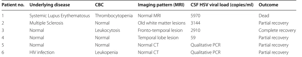
Table 4 Individualized data of the normocellular CSF group of HSE patients
Patient no. Underlying disease CBC Imaging pattern (MRI) CSF HSV viral load (copies/ml) Outcome
1 Systemic Lupus Erythematosus Thrombocytopenia Normal MRI 5970 Dead
2 Multiple Sclerosis Normal Old white matter lesions 3144 Partial recovery
3 Normal Leukocytosis Fronto-temporal lesion 2910 Complete recovery
4 Normal Normal Temporal lobe lesion 59 Partial recovery
5 Normal Normal Normal CT Qualitative PCR Partial recovery
[image:6.595.54.548.103.197.2]• We accept pre-submission inquiries
• Our selector tool helps you to find the most relevant journal
• We provide round the clock customer support
• Convenient online submission
• Thorough peer review
• Inclusion in PubMed and all major indexing services
• Maximum visibility for your research
Submit your manuscript at www.biomedcentral.com/submit
Submit your next manuscript to BioMed Central
and we will help you at every step:
Competing interests
The authors declare that they have no competing interests.
Received: 10 July 2015 Accepted: 8 February 2016
References
1. Kennedy PG, Steiner I. Recent issues in herpes simplex encephalitis. J Neurovirol. 2013;19(4):346–50.
2. Lim HK, et al. TLR3 deficiency in herpes simplex encephalitis: high allelic heterogeneity and recurrence risk. Neurology. 2014;83(21):1888–97. 3. Granerod J, et al. Causes of encephalitis and differences in their clinical
presentations in England: a multicentre, population-based prospective study. Lancet Infect Dis. 2010;10(12):835–44.
4. Koskiniemi M, et al. Herpes encephalitis is a disease of middle aged and elderly people: polymerase chain reaction for detection of herpes sim-plex virus in the CSF of 516 patients with encephalitis. The Study Group. J Neurol Neurosurg Psychiatry. 1996;60(2):174–8.
5. Whitley RJ, et al. Vidarabine versus acyclovir therapy in herpes simplex encephalitis. N Engl J Med. 1986;314(3):144–9.
6. Solomon T, et al. Management of suspected viral encephalitis in adults– Association of British Neurologists and British Infection Association National Guidelines. J Infect. 2012;64(4):347–73.
7. Aurelius E, et al. Encephalitis in immunocompetent patients due to her-pes simplex virus type 1 or 2 as determined by type-specific polymerase chain reaction and antibody assays of cerebrospinal fluid. J Med Virol. 1993;39(3):179–86.
8. Moon SM, et al. Comparison of clinical manifestations, outcomes and cer-ebrospinal fluid findings between herpes simplex type 1 and type 2 cen-tral nervous system infections in adults. J Med Virol. 2014;86(10):1766–71. 9. Mateen FJ, Miller SA, Aksamit AJ Jr. Herpes simplex virus 2 encephalitis in
adults. Mayo Clin Proc. 2014;89(2):274–5.
10. Barnett EM, et al. Herpes simplex encephalitis in the temporal cortex and limbic system after trigeminal nerve inoculation. J Infect Dis. 1994;169(4):782–6.
11. Whitley RJ, et al. Adenine arabinoside therapy of biopsy-proved herpes simplex encephalitis. National Institute of Allergy and Infectious Diseases collaborative antiviral study. N Engl J Med. 1977;297(6):289–94. 12. Tyler KL. Herpes simplex virus infections of the central nervous system:
encephalitis and meningitis, including Mollaret’s. Herpes. 2004;11(Suppl 2):57A–64A.
13. Baringer JR. Herpes simplex infections of the nervous system. Neurol Clin. 2008;26(3):657–74 (viii).
14. Baringer JR, Pisani P. Herpes simplex virus genomes in human nerv-ous system tissue analyzed by polymerase chain reaction. Ann Neurol. 1994;36(6):823–9.
15. Domingues RB, et al. Evaluation of the range of clinical presentations of herpes simplex encephalitis by using polymerase chain reaction assay of cerebrospinal fluid samples. Clin Infect Dis. 1997;25(1):86–91.
16. Kennedy PG, Chaudhuri A. Herpes simplex encephalitis. J Neurol Neuro-surg Psychiatry. 2002;73(3):237–8.
17. Whitley RJ, Gnann JW. Viral encephalitis: familiar infections and emerging pathogens. Lancet. 2002;359(9305):507–13.
18. Steiner I, Tyler KL. The toll (like receptor 3) to the pathogenesis of herpes simplex encephalitis. Neurology. 2014;83(21):1882–3.
19. Steiner I, et al. EFNS-ENS guidelines for the use of PCR technology for the diagnosis of infections of the nervous system. Eur J Neurol. 2012;19(10):1278–91.
20. Lafaille FG, et al. Impaired intrinsic immunity to HSV-1 in human iPSC-derived TLR3-deficient CNS cells. Nature. 2012;491(7426):769–73. 21. Desena A, et al. Herpes simplex encephalitis as a potential cause of
anti-N-methyl-D-aspartate receptor antibody encephalitis: report of 2 cases. JAMA Neurol. 2014;71(3):344–6.
22. Gkrania-Klotsas E, Lever AM. Herpes simplex I encephalitis presenting as a brain haemorrhage with normal cerebrospinal fluid analysis: a case report. J Med Case Rep. 2008;2:387.
23. Mook-Kanamori B, van de Beek D, Wijdicks EF. Herpes simplex encepha-litis with normal initial cerebrospinal fluid examination. J Am Geriatr Soc. 2009;57(8):1514–5.
24. Schoonman GG, et al. Herpes simplex virus encephalitis without cerebro-spinal fluid pleocytosis is not unusual. J Am Geriatr Soc. 2012;60(2):377–8. 25. Kessler HH, et al. Detection of Herpes simplex virus DNA by real-time PCR.
J Clin Microbiol. 2000;38(7):2638–42.