S H O R T R E P O R T
Open Access
Akt and phospholipase C
γ
are involved in the
regulation of growth and migration of
MDA-MB-468 breast cancer and SW480 colon
cancer cells when cultured with diabetogenic
levels of glucose and insulin
Nicola M Tomas
1*, Kai Masur
1,2, Jonas C Piecha
1, Bernd Niggemann
1and Kurt S Zänker
1Abstract
Background:Epidemiological studies revealed a strong correlation between the metabolic syndrome/diabetes
mellitus type 2 (DM2) and higher incidence and faster progression of breast and colon cancer. However, the underlying molecular mechanisms are widely unknown. Akt and phospholipase Cγ(PLCγ) are involved in tyrosine kinase signaling and promote tumor cell growth and migration. Therefore, we examined regulatory functions and expression of Akt and PLCγin a simplified in vitro diabetogenic model.
Findings:Protein expression was determined by western blot analysis in MDA-MB-468 breast cancer and SW480
colon cancer cells previously cultured under physiologic (5.5 mM) and diabetogenic (11 mM) glucose
concentrations (without and with 100 ng/ml insulin). We studied the culture effects on proliferation and migration of these cells, especially after inhibiting Akt and PLCγ. We found that Akt expression was up-regulated with high glucose and insulin in both cell lines, whereas PLCγexpression was enhanced in colon cancer cells only. High levels of glucose and insulin increased cell proliferation and migration in both cell lines in vitro, mediated by Akt and PLCγ, as shown through the specific pharmacological inhibitors A6730 and U73122.
Conclusions:Our molecular data explain glucose- and insulin-induced changes in a cancer cell and help to
understand what might trigger tumor cell proliferation and migration in DM2 patients, too.
Keywords:Breast and colon cancer, Diabetes mellitus type 2, Glucose, Insulin, Akt, PLCγ
Background
Breast cancer is the most common type of cancer in women, and colon cancer is the third most common can-cer in both sexes and the second leading cause of cancan-cer deaths worldwide [1]. The majority of cancer patients, however, will not decease because of their primary tumor, but rather because of the spreading of metastasis, which is responsible for over 90% of cancer deaths [2].
The prevalence of obesity has increased dramatically over the last four decades, and it can be considered the leading health problem of the developed countries in the 21stcentury. Obesity is strongly associated with the devel-opment of diabetes mellitus type 2 (DM2) and co-morbid diseases, such as hypertension and hyperlipidemia, com-monly summarized as the metabolic syndrome [3].
Up-to-date retrospective and prospective epidemiological studies showed strong correlations between the metabolic syndrome and the incidence of malignant neoplasms in different organs [4,5]. Obesity increases the risk of breast and colon cancer [6,7]. DM2 is directly associated with a higher incidence and faster progression of several neoplasms including colon and breast cancer [5,8]. In
* Correspondence:nicola.tomas@uni-wh.de 1
Institute of Immunology and Experimental Oncology, Witten/Herdecke University, Stockumer Str. 10, D-58448 Witten, Germany
Full list of author information is available at the end of the article
particular, increased fasting serum insulin concentrations, commonly found in DM2 patients, seem to increase the risk for breast and colon cancer [9,10]. Besides, tyrosine kinases, namely the insulin receptor (IR) and insulin-like growth factor (IGF) receptors, are over-expressed in several human cancers, including cancer of the breast [11]. Consequently, over-expression of these receptors yields a selective growth advantage to breast cancer cells, especially in the presence of insulin resistance and associated hyperinsulinemia [12]. It has been known for over two decades that glucose is the driving force for tumor cell growth [13] and that high levels of insulin promote metastasis [14].
Insulin operates by binding to its target cell receptor, a heterotetrameric, transmembrane, multisubunit glyco-protein. The insulin signal is propagated through a phos-phorylation network involving intracellular molecules such as phosphoinositid-3-kinase (PI3K), protein kinase B (PKB, Akt) and phospholipase Cγ (PLCγ). The serine/ threonine kinase Akt is a well known regulator of widely divergent cellular processes, and it is implicated in intra-cellular insulin signaling. Akt has been described as an oncogene in several human cancers [15,16], and it is known to be a promoter of tumor cell proliferation [17], prolonged cell survival [18,19], and angiogenesis [20]. The phosphorylation and thus activation of Akt can be specifically inhibited by the trifluoroacetate salt hydrate A6730 [21]. Phospholipases (PLCs) are tyrosine kinase substrates that provide diacylglycerols (DAGs) for intra-cellular signaling in various contexts and its isoform PLCγ is known to be implicated in intracellular insulin signaling [22] and in colon and breast cancer progression [23,24]. U73122 is an aminosteroid that specifically inhi-bits PLCγactivation [25].
Given this knowledge about the gravity of glucose me-tabolism in tumor cells on the one hand and the growing number of studies investigating the epidemiological con-nection between the metabolic syndrome/DM2 and the development and progression of several human cancers on the other hand, surprisingly little is known about the underlying molecular changes and mechanisms that facili-tate tumor development and progression when excessive glucose and insulin are available.
For these reasons, we report about the regulatory func-tions of Akt and PLCγregarding tumor cell proliferation and migration under the specific premise of higher than normal glucose and insulin concentrations.
Methods Cell culture
For all experiments the tumor cell lines SW480 colon adenocarcinoma and MDA-MB-468 mammary gland adenocarcinoma (both American Type Culture Collection, Manassas, VA, USA) were kept in RPMI 1640 media
supplemented with 10% fetal bovine serum (PAA Labora-tories, Pasching, Austria) and 1% penicillin/streptomycin (PAN Biotech GmbH, Aidenbach, Germany). The medium contained either 5.5 mM (99.1 mg/dl) glucose, 11 mM (198.2 mg/dl) glucose, or 11 mM glucose plus 100 ng/ml insulin. All cells were incubated at 37°C humidified atmos-phere and 5% CO2(Binder, Tuttlingen, Germany).
Western blot analysis
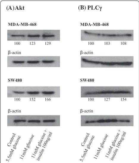
The expression levels of Akt and PLCγ were analyzed by immunoblotting as described previously [26]. Cells (1 × 10⁶) were lysed in Laemmli sample buffer and incubated for 10 min at 95°C. Proteins were separated using SDS-PAGE and transferred to an PVDF-Immobilion-P membrane (Millipore, Schwalbach, Germany), followed by blocking with 10% milk powder (1 h at room temperature (RT)). After incubating the membrane overnight at 4°C with the primary monoclonal antibodies against Akt (Cell Signaling Technology, Boston, USA) and PLCγ (Sigma-Aldrich, Deisenhofen, Germany), membrane were washed vigorously with PBS-Tween. Subsequently, the membrane was incubated with appropriate anti-mouse or anti-goat peroxidase-linked secondary antibodies for 1 h at RT (all Southern Biotech, Birmingham, AL, USA), followed by incubation with the chemiluminescence blotting sub-strate (Roche Diagnostics, Mannheim, Germany) for 1 min at RT for visualization. Bands were detected using the Aequoria Macroscopic Imaging System (Hamatsu, Herrsching am Ammersee, Germany) and quantified using Wasabi 1.4 software (Hamatsu, Herrsching am Ammersee, Germany). The numbers shown are expressed as percen-tages of the 5.5 mM glucose control condition.
Cell proliferation assay
Proliferation analysis was performed in a 96-well-plate with 104 cells per well. Cells were incubated for 48 h at 37°C humidified atmosphere and 5% CO2with and with-out the inhibitors A6730 and U73122 in a concentration of 500nM and 500 mg/dl, respectively (both Sigma-Aldrich, Deisenhofen, Germany). Then, according to the manual, 25μl of MTS color solution (Promega, Madison, WI, USA) was added directly to the culture wells and cells were then incubated under the same conditions for 2 h. Absorbance of the resulting colored formazan prod-uct was recorded at 490 nm in a 96-well-plate-reader. The quantity of formazan product is directly propor-tional to the number of living cells in culture.
Cell migration assay
bicarbonate solution (Sigma, Taufkirchen, Germany), 105 cells to be analyzed, and the inhibitors A6730 (500nM) and U73122 (500 ng/ml). Cell migration within the three-dimensional collagen matrix was recorded for 10 h at 37°C by time-lapse video-microscopy and subsequently ana-lyzed by computer-assisted cell tracking. For analysis of the migratory activity, 30 cells per sample were randomly selected and paths were digitized as x/y coordinates in 15 min intervals. Migratory activity (MA) refers to the percentage of cells moving during a particular 15 min interval, and average migratory activity (AMA) is the average percentage of moving cells, calculated from all 15 min intervals in a 10 h period. Thereby, 100% MA was reached when all 30 cells migrated during the corre-sponding 15 min interval and 100% AMA was reached when all 30 cells migrated constantly over 10 h. The average distance migrated (ADM) inμm was calculated from 10 h of analysis.
Statistical analysis
Data is presented as mean and standard deviation. Statistical significance was determined by Student’st-test, whereby p<0.05 was considered significant. All experiments were performed 3–6 times.
Findings
To analyze Akt/PLCγ expression, proliferation capacity, and cell migration we cultured different tumor cell lines (MDA-MB-468 breast and SW480 colon cancer cells) at physiological (5.5 mM, control) and diabetogenic (11 mM) glucose concentrations without and with 100 ng/ml insulin. Raising the glucose concentration from 5.5 mM to 11 mM increased the expression of Akt by 23% in MDA-MB-468 breast cancer cells (Figure 1A). Addition of 100 ng/ml insulin to the 11 mM culture media increased Akt expression by another 6% when compared to cells cultured with 11 mM glucose alone. An even stronger effect could be seen for SW480 colon cancer cells, which raised Akt expression by 52% when cultured with high glucose concentrations. Here adding 100 ng/ml insulin caused a further increase of 14% (to 66%). The Akt-enhancing effect of insulin can be quantified as approxi-mately one fourth of the glucose effect in both cell lines.
MDA-MB-468 cells did not significantly up-regulate phospholipase Cγ (PLCγ) expression neither when kept under higher than normal glucose concentrations alone, nor when 100 ng/ml insulin was added (Figure 1B). In SW480 cells high glucose and insulin levels showed an increase in PLCγ expression of 27% each, leading to a total increase of 54%.
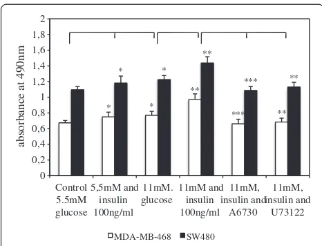
We performed a proliferation assay in order to analyze the influence of both, high glucose and insulin concentrations, as well as inhibition of Akt and PLCγ on the proliferative capacity of the two tumor cell lines
(Figure 2). In this assay, absorbance values at 490 nm are directly proportional to the number of living cells in culture. Both cell lines revealed significantly higher proliferation rates when cultured with additional insulin and hyperglycemic glucose concentrations. Thereby, cells stimulated with glucose (11 mM) had similar proliferation rates to cells stimulated with insulin alone (p= 0,2). Pro-liferation was highest when cells were simultaneously stimulated with glucose and insulin. Furthermore, inhib-ition of Akt and PLCγ by A6730 and U73122, respect-ively, reduced proliferative activity significantly when compared to glucose- and insulin-stimulated cell prolif-eration (Figure 2). On the other hand, no significant effect on tumor cell proliferation was seen after adding A6730 and U73122 to the control media (data not shown). SW480 cells displayed significantly higher pro-liferation rates than MDA-MB-468 cells when kept under normal and increased glucose and insulin concentrations (p<0.001 for all three conditions).
[image:3.595.304.538.89.364.2](A) (B)
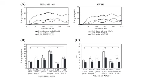
High glucose and insulin concentrations significantly increased migratory activity (MA) in the investigated cell lines. MA refers to the percentage of cells moving during a partiular 15 min interval. Akt-inhibitor A6730 and PLCγ -inhibitor U73122 significantly reduced MA in both cell lines (Figure 3A). This glucose- and insulinduced in-crease in cell migration is also reflected by an augmenta-tion in average migratory activity (AMA), i.e. the average percentage of moving cells, calculated from all 15 min intervals in a 10 h period (Figure 3B). Maximum MA in MDA-MB-468 and SW480 cell lines was seen after con-current stimulation with 11 mM glucose and 100 ng/ml insulin. It could be observed that both cell lines achieved this increase in MA by longer episodes of migration and shortening of pauses. Both inhibitors were able to annihi-late the stimulating effect of insulin. Thereby, addition of U73122 resulted in migratory levels comparable to cells that were stimulated by a high level of glucose alone (11 mM) and thus only abolished the inducing effect of insulin. In comparison, A6730 reduced migration even below the control condition (5.5 mM) in both cell lines (Figure 3B).
The fact that glucose and insulin were strong inducers of tumor cell migration is also reflected by an increase in average distance migrated (ADM, Figure 3C). ADM refers
to the average distance inμm that a cell covered during 10 h of analysis. Both, glucose and insulin, significantly elongated ADM, regardless of the cell type. Cells that were exposed to one of the inhibitors significantly reduced ADM when compared to cells cultured with 11 mM glu-cose and 100 ng/ml insulin. A6730 reduced ADM to a greater extent than U73122 in both cell lines (Figure 3C). Both inhibitors showed no significant effects on migratory parameters when added to the control media (data not shown).
Regarding the distance migrated over 10 h, MDA-MB-468 breast cancer cells appear to be more susceptible to glucose- and insulin-stimulation than SW480 colon cancer cells (p<0.05 for 11 mM glucose, p<0.001 for 11 mM glucose plus insulin). We observed that this effect was due to a greater augmentation in migratory velocity.
Discussion
Tumor staging –including tumor size and invasiveness, lymphatic tissue involvement, and spreading to distant organ sites – is the main predictor of the prognosis for most solid organ tumor patients. Thus, finding bio-chemical signatures that define a tumor’s potential to metastasize can build the basis for new ways of deter-mining a patient’s prognosis and eventually lead to new therapeutic targets. Glucose metabolism and its regula-tion, i.e. transcellular glucose transport and hormonal control via insulin and modification of the following sig-naling pathways, are such possible signatures. The results presented in this work strongly suggest that the glucose-and insulin-induced changes in proliferation glucose-and migra-tion of MDA-MB-468 breast cancer and SW480 colon cancer cells are mediated by changes in Akt and/or PLCγ signaling. The investigated cell lines are widely used in cancer research and are well characterized in the context of their metabolic response to increased availability of glu-cose and insulin [28,29].
Akt is activated in most tumors [30] and various onco-genic functions, such as induction of migration [31] and metastasis [32], proliferation [17], and prolonged cell sur-vival [18], have been described. Interestingly, Akt also enhances cellular glucose uptake by mobilizing glucose transporters to the cell surface [33]. In 2010, Johnson et al. [34] showed that proteins of the phosphoinositid-3-kinase (PI3K)/Akt pathway are significantly over-expressed in colorectal cancer compared to healthy cells, and that ex-pression levels correlated with cancer stage. PI3K is acti-vated by tyrosine kinase receptor stimulation, especially by the binding of insulin [35]. PI3K mediates the generation of phosphatidylinositol (3,4,5)-trisphosphate (PIP3) which allows Akt to be phosphorylated and thus activated [36]. The increased expression of Akt that we found with high glucose and insulin provides augmented substrate for the PI3K-pathway and may explain the increased proliferation
* *
**
*** **
* *
**
*** **
0 0,2 0,4 0,6 0,8 1 1,2 1,4 1,6 1,8 2
Control 5.5mM glucose
5,5mM and insulin 100ng/ml
11mM. glucose
11mM and insulin 100ng/ml
11mM, insulin and
A6730 11mM, insulin and
U73122
absorbance at 490nm
[image:4.595.56.289.87.265.2]MDA-MB-468 SW480
Figure 2Tumor cell proliferation with high glucose and insulin. MDA-MB-468 breast cancer and SW480 colon cancer cells
and migration of the investigated cell lines when stimu-lated by glucose and insulin. The fact that addition of the Akt-specific inhibitor A6730 markedly decreased cell mi-gration and proliferation with high glucose and insulin emphasizes that the up-regulation of Akt plays a crucial role in the processes of proliferation and migration. We could also show that the inhibitory effects of A6730 were specific to the setting with high glucose and insulin as no inhibitory effects of A6730 were evident when used with-out simultaneous stimulation by glucose and insulin.
In breast cancer patients, PLCγ was shown to be up-regulated in distant metastases when compared with the primary tumor, indicating its involvement in metastasis formation [23]. In our experiments, increased glucose and insulin caused an up-regulation of PLCγ in SW480 colon cancer cells, but no up-regulation in MDA-MB-468 breast cancer cells. However, migration and prolif-eration could be reduced significantly by adding U73122 to the diabetic culture conditions. This highlights the regulating capacity of PLCγ in both cell types. Thereby, the inhibitory effect of U73122 was specific to the
condition with increased glucose and insulin as no de-crease in proliferation and migration was seen when U73122 was used with normal glucose and no insulin.
How the treatment of diabetes is linked to the inci-dence of several cancers has gained a lot of interest over the last years and alarming data has recently been pub-lished. Hemkens et. al. [37] found that patients treated with human insulin or insulin analogues had a higher in-cidence of malignant neoplasms than patients who were not treated with insulin. Furthermore, the risk of devel-oping cancer increased with higher dosages of insulin. In 2010, Baur et al. [38] stated that among diabetic patients, those treated with insulin had a four-fold higher risk to die from cancer. However, patients treated with the insulin-sensitizing antidiabetic agent metformin had mortality levels comparable with those of non-dia-betic patients. Our in vitro results support these findings and help to explain how insulin and insulin therapy may increase cancer progression: from our point of view, in-sulin is a strong inducer of tumor cell migration and proliferation and a promoter of oncogenes such as Akt
* * ** *** ** * * ** ** * 0 5 10 15 20 25 30 35 40 45 µ m MDA-MB-468 SW480 * * * *** * * * * ** * 0 2 4 6 8 10 12 14 16 18 20 Control 5.5mM glucose 5,5mM and insulin 100ng/ml 11mM glucose 11mM and insulin 100ng/ml 11mM, insulin and A6730 11mM, insulin and U73122 Control 5.5mM glucose 5,5mM and insulin 100ng/ml 11mM glucose 11mM and insulin 100ng/ml 11mM, insulin and A6730 11mM, insulin and U73122
% migrating cells
MDA-MB-468 SW480
0 10 20 30
0 120 240 360 480 600
% migrating cells
time in minutes
MDA-MB-468
11mM glucose and insulin 100ng/ml 11mM, insulin and A6730 11mM, insulin and U73122
0 10 20 30
0 120 240 360 480 600
% migrating cells
time in minutes
SW480
11mM glucose and insulin 100ng/ml 11mM, insulin and A6740 11mM, insulin and U73122
(A)
[image:5.595.59.535.90.351.2](C)
(B)
and PLCγ. This might indicate that, when patients show a family history for cancer, the use of long-acting insulin in the treatment of DM2 should be reconsidered.
Conclusions
Our data show how glucose and insulin change prolifera-tion and migraprolifera-tion capacity in MDA-MB-468 breast can-cer and SW480 colon cancan-cer cells in vitro. Our results help to understand the diverse roles of glucose and insulin – energy supply and modulation of signaling cascades by modifying Akt and PLCγ expression – and thus explain which changes on a molecular, transcriptional level could be responsible for the epidemiological connection between the metabolic syndrome/DM2 and the progression of vari-ous malignancies. Considering our findings regarding the essential role of Akt in tumor progression, pharmaco-logical research targeting this oncogene might be particu-larly promising.
Abbreviations
ADM: average distance migrated; AMA: average migratory activity; DAG: diacylglycerol; DM2: diabetes mellitus type 2; IR: insulin receptor; IGF: inslin-like growth factor; MA: migratory activity; PI3K: phosphoinositid-3-kinase; PIP3: phosphatidylinositol (3,4,5)-trisphosphate; PKB: protein kinase B; PLC: phospholipase; PLCγ: phospholipase Cγ.
Competing interests
The authors declare that they have no competing interests.
Acknowledgements
This work was supported by the Fritz-Bender-Foundation, Munich. Gratitude goes to Christine Mehner for critically revising the manuscript.
Author details
1Institute of Immunology and Experimental Oncology, Witten/Herdecke
University, Stockumer Str. 10, D-58448 Witten, Germany.2Institute for Plasma
Research and Technology e.V.–INP Greifswald, Felix-Hausdorff-Str. 2, D-17489 Greifswald, Germany.
Authors’contributions
NMT performed the experiments, analyzed and interpreted the results, created the figures, and drafted the manuscript. KM and KSZ designed the study and revised the manuscript. JCP contributed to the experiments and revised the manuscript. BN supported the analysis of the cell migration data. All authors have read and approved the final manuscript.
Received: 5 January 2012 Accepted: 3 May 2012 Published: 3 May 2012
References
1. Ferlay J, Shin HR, Bray F, Forman D, Mathers C, Parkin DM:Estimates of worldwide burden of cancer in 2008: GLOBOCAN 2008.Int J Cancer2010, 127(12):2893–2917.
2. Gupta GP, Massague J:Cancer metastasis: building a framework.Cell2006, 127(4):679–695.
3. Ness-Abramof R, Nabriski D, Apovian CM:Medical therapy for obesity: present and future.Isr Med Assoc J2004,6(12):760–765.
4. Rapp K, Schroeder J, Klenk J, Ulmer H, Concin H, Diem G, Oberaigner W, Weiland SK:Fasting blood glucose and cancer risk in a cohort of more than 140,000 adults in Austria.Diabetologia2006,49(5):945–952. 5. LeRoith D, Novosyadlyy R, Gallagher EJ, Lann D, Vijayakumar A, Yakar S:Obesity
and type 2 diabetes are associated with an increased risk of developing cancer and a worse prognosis; epidemiological and mechanistic evidence.
Exp Clin Endocrinol Diabetes2008,116(Suppl 1):S4–S6.
6. Giovannucci E, Michaud D:The role of obesity and related metabolic disturbances in cancers of the colon, prostate, and pancreas.
Gastroenterology2007,132(6):2208–2225.
7. Pischon T, Nothlings U, Boeing H:Obesity and cancer.Proc Nutr Soc2008, 67(2):128–145.
8. Schiel R, Beltschikow W, Steiner T, Stein G:Diabetes, insulin, and risk of cancer.Methods Find Exp Clin Pharmacol2006,28(3):169–175.
9. Del Giudice ME, Fantus IG, Ezzat S, McKeown-Eyssen G, Page D, Goodwin PJ: Insulin and related factors in premenopausal breast cancer risk.Breast Cancer Res Treat1998,47(2):111–120.
10. Schoen RE, Tangen CM, Kuller LH, Burke GL, Cushman M, Tracy RP, Dobs A, Savage PJ:Increased blood glucose and insulin, body size, and incident colorectal cancer.J Natl Cancer Inst1999,91(13):1147–1154.
11. Frasca F, Pandini G, Sciacca L, Pezzino V, Squatrito S, Belfiore A, Vigneri R: The role of insulin receptors and IGF-I receptors in cancer and other diseases.Arch Physiol Biochem2008,114(1):23–37.
12. Belfiore A, Frasca F:IGF and insulin receptor signaling in breast cancer.J Mammary Gland Biol Neoplasia2008,13(4):381–406.
13. Beckner ME, Stracke ML, Liotta LA, Schiffmann E:Glycolysis as primary energy source in tumor cell chemotaxis.J Natl Cancer Inst1990,82(23):1836–1840. 14. Stracke ML, Kohn EC, Aznavoorian SA, Wilson LL, Salomon D, Krutzsch
HC, Liotta LA, Schiffmann E:Insulin-like growth factors stimulate chemotaxis in human melanoma cells.Biochem Biophys Res Commun 1988,153(3):1076–1083.
15. Cheng JQ, Ruggeri B, Klein WM, Sonoda G, Altomare DA, Watson DK, Testa JR:Amplification of AKT2 in human pancreatic cells and inhibition of AKT2 expression and tumorigenicity by antisense RNA.Proc Natl Acad Sci U S A1996,93(8):3636–3641.
16. Bellacosa A, de Feo D, Godwin AK, Bell DW, Cheng JQ, Altomare DA, Wan M, Dubeau L, Scambia G, Masciullo V, Ferrandina G, Benedetti Panici P, Mancuso S, Neri G, Testa JR:Molecular alterations of the AKT2 oncogene in ovarian and breast carcinomas.Int J Cancer1995,64(4):280–285. 17. Nicholson KM, Anderson NG:The protein kinase B/Akt signalling pathway
in human malignancy.Cell Signal2002,14(5):381–395.
18. Song G, Ouyang G, Bao S:The activation of Akt/PKB signaling pathway and cell survival.J Cell Mol Med2005,9(1):59–71.
19. Dudek H, Datta SR, Franke TF, Birnbaum MJ, Yao R, Cooper GM, Segal RA, Kaplan DR, Greenberg ME:Regulation of neuronal survival by the serine-threonine protein kinase Akt.Science (New York, NY)1997,275(5300):661–665. 20. Chen J, Somanath PR, Razorenova O, Chen WS, Hay N, Bornstein P, Byzova
TV:Akt1 regulates pathological angiogenesis, vascular maturation and permeability in vivo.Nat Med2005,11(11):1188–1196.
21. Zhao Z, Leister WH, Robinson RG, Barnett SF, Defeo-Jones D, Jones RE, Hartman GD, Huff JR, Huber HE, Duggan ME, Lindsley CW:Discovery of 2,3,5-trisubstituted pyridine derivatives as potent Akt1 and Akt2 dual inhibitors.Bioorg Med Chem Lett2005,15(4):905–909.
22. Yamazaki H, Zawalich KC, Zawalich WS:Physiologic implications of phosphoinositides and phospholipase C in the regulation of insulin secretion.J Nutr Sci Vitaminol2010,56(1):1–8.
23. Sala G, Dituri F, Raimondi C, Previdi S, Maffucci T, Mazzoletti M, Rossi C, Iezzi M, Lattanzio R, Piantelli M, Iacobelli S, Broggini M, Falasca M:Phospholipase Cgamma1 is required for metastasis development and progression.
Cancer Res2008,68(24):10187–10196.
24. Noh DY, Lee YH, Kim SS, Kim YI, Ryu SH, Suh PG, Park JG:Elevated content of phospholipase C-gamma 1 in colorectal cancer tissues.Cancer1994, 73(1):36–41.
25. Thompson AK, Mostafapour SP, Denlinger LC, Bleasdale JE, Fisher SK:The aminosteroid U-73122 inhibits muscarinic receptor sequestration and phosphoinositide hydrolysis in SK-N-SH neuroblastoma cells. A role for Gp in receptor compartmentation.J Biol Chem1991,266(35):23856–23862. 26. Entschladen F, Niggemann B, Zanker KS, Friedl P:Differential requirement
of protein tyrosine kinases and protein kinase C in the regulation of T cell locomotion in three-dimensional collagen matrices.J Immunol1997, 159(7):3203–3210.
27. Niggemann B, Drell TLt, Joseph J, Weidt C, Lang K, Zaenker KS, Entschladen F: Tumor cell locomotion: differential dynamics of spontaneous and induced migration in a 3D collagen matrix.Exp Cell Res2004,298(1):178–187. 28. Masur K, Vetter C, Hinz A, Tomas N, Henrich H, Niggemann B, Zanker KS:
29. Masur K, Lang K, Niggemann B, Zanker KS, Entschladen F:High PKC alpha and low E-cadherin expression contribute to high migratory activity of colon carcinoma cells.Mol Biol Cell2001,12(7):1973–1982.
30. Robey RB, Hay N:Is Akt the "Warburg kinase"?-Akt-energy metabolism interactions and oncogenesis.Semin Cancer Biol2009,19(1):25–31. 31. Wang Z, Yang J, Fisher T, Xiao H, Jiang Y, Yang C:Akt Activation is
Responsible for Enhanced Migratory and Invasive Behavior of Arsenic-Transformed Human Bronchial Epithelial Cells.Environ Health Perspect 2011.
32. Saji M, Narahara K, McCarty SK, Vasko VV, La Perle KM, Porter K, Jarjoura D, Lu C, Cheng SY, Ringel MD:Akt1 deficiency delays tumor progression, vascular invasion, and distant metastasis in a murine model of thyroid cancer.Oncogene2011.
33. Elstrom RL, Bauer DE, Buzzai M, Karnauskas R, Harris MH, Plas DR, Zhuang H, Cinalli RM, Alavi A, Rudin CM, Thompson CB:Akt stimulates aerobic glycolysis in cancer cells.Cancer Res2004,64(11):3892–3899.
34. Johnson SM, Gulhati P, Rampy BA, Han Y, Rychahou PG, Doan HQ, Weiss HL, Evers BM:Novel expression patterns of PI3K/Akt/mTOR signaling pathway components in colorectal cancer.J Am Coll Surg2010,210(5):767–776, 776–768.
35. Bertrand L, Horman S, Beauloye C, Vanoverschelde JL:Insulin signalling in the heart.Cardiovasc Res2008,79(2):238–248.
36. Franke TF, Kaplan DR, Cantley LC, Toker A:Direct regulation of the Akt proto-oncogene product by phosphatidylinositol-3,4-bisphosphate.
Science (New York, NY)1997,275(5300):665–668.
37. Hemkens LG, Grouven U, Bender R, Gunster C, Gutschmidt S, Selke GW, Sawicki PT:Risk of malignancies in patients with diabetes treated with human insulin or insulin analogues: a cohort study.Diabetologia2009, 52(9):1732–1744.
38. Baur DM, Klotsche J, Hamnvik OP, Sievers C, Pieper L, Wittchen HU, Stalla GK, Schmid RM, Kales SN, Mantzoros CS:Type 2 diabetes mellitus and medications for type 2 diabetes mellitus are associated with risk for and mortality from cancer in a German primary care cohort.Metabolism2010, 60(10):1363–1371.
doi:10.1186/1756-0500-5-214
Cite this article as:Tomaset al.:Akt and phospholipase Cγare involved
in the regulation of growth and migration of
MDA-MB-468 breast cancer and SW480 colon cancer cells when cultured with diabetogenic levels of glucose and insulin.BMC Research Notes20125:214.
Submit your next manuscript to BioMed Central and take full advantage of:
• Convenient online submission
• Thorough peer review
• No space constraints or color figure charges
• Immediate publication on acceptance
• Inclusion in PubMed, CAS, Scopus and Google Scholar
• Research which is freely available for redistribution
![Poly[hexaaquatri μ malonato didysprosium(III)]](data:image/gif;base64,R0lGODlhAQABAIAAAP///wAAACH5BAEAAAAALAAAAAABAAEAAAICRAEAOw==)