JOURNAL OFVIROLOGY, May1991, p. 2745-2750 0022-538X/91/052745-06$02.00/0
Copyright© 1991, AmericanSociety for Microbiology
Expression
of
Herpes
Simplex Virus
Type
2
Latency-Associated
Transcript
in
Neurons and Nonneurons
RICHARD B. TENSER,l.2* WADE A. EDRIS,2 KATHLEENA. HAY,2 ANDBASTIAAN E. DEGALAN2t Departments ofMedicine (Neurology)' and Microbiology and Immunology,2 The Pennsylvania
State University College of Medicine, Hershey, Pennsylvania 17033 Received 28 September 1990/Accepted 11 February 1991
The presence of herpes simplex virus type 2 (HSV-2) transcription during in vivo latent infection was
investigatedby in situhybridization. Latent infection ofmousedorsalrootganglionwasinvestigatedwith the BamHI p fragment of HSV-2, which resulted in evidence ofganglion hybridization, and other fragments
representing approximately 40% of thegenome, which didnot result in hybridization. Strandspecificity of hybridizationwasinvestigated instudieswithsyntheticoligonucleotides, which supported the conclusion that a latency-associated transcript(s) had been detected. Hybridization was detected with oligonucleotides
complementary to the infected-cell polypeptide 0 (ICPO) template strand but not with oligonucleotides
synthesized from the ICPO template strand. Although mosthybridization occurred over neurons, in some
instances hybridization appearedtooccur overnonneuronal ganglion cells, and thiswas moreevident when
tissue sections were examined by phase contrast microscopy. Although these results supported the usual
neuronalsite of HSV-2 latency, latency innonneuronalcells maybeimportantin consideringthe pathobiology
of HSV-2 infections.
Herpes simplex virus(HSV) latent infection has been well characterized as aninfectionofneurons, usually ofsensory
ganglia (3, 9, 13, 17, 26).Several experimental methods have been used to arrive at this conclusion, which is also in keeping with axonal transport of HSV (4, 8, 15). Recent investigations of the presence of HSV latency-associated
transcript (LAT) inneuronsasdetected by in situ
hybridiza-tion have confirmed the neuronal site of latency (7, 11, 14, 16, 21, 27, 28). Although suggestions have been made that HSV may establish latent infections in vivo in other cell typesandother locations, these results have sometimes been questioned.
In mostinvivo latency studies done with in situ hybrid-ization techniques, HSV type 1 (HSV-1) has been investi-gated, andfewer studies have beenperformedwith HSV-2 (9, 24, 26). Recently, HSV-2 LATwas reported;
hybridiza-tion was detected with one restriction fragment and was
negativewithother fragments representing the remainder of the HSV-2 genome (18). In the present investigation we
extend this report via studies in which hybridization was similarly restricted and hybridization with strand-specific oligonucleotides detectedtranscriptsthatwere
complemen-tary to the strand opposite the infected-cell polypeptide 0 (ICPO) template strand, but did notdetect transcripts from the ICPO template strand. In addition, while HSV-2 LAT
was present over many sensory ganglion neurons,
hybrid-ization also occurred over occasional nonneuronal cells. Although the present results strongly support the usual neuronallocus oflatentHSV-2, latencyinevenarelatively
few nonneuronal cells may alter the way in which the pathobiologyoflong-termHSV infection is considered.
HSV-2(strain 333),originally obtained from theAmerican Type Culture Collection and passed multiple times in cell culture, wasused. Virus stockswere grownand counted in
*Corresponding author.
tPermanent address:FacultyofMedicine, Universityof
Amster-dam, AmsterAmster-dam, TheNetherlands.
primary rabbit kidney cells by standardmeans.HSV-2latent
infection of mouse dorsal root ganglia (DRG) was estab-lished by bilateral rear footpad inoculation of CD-1 mice
(male andfemale, 6to8 weeks old;Charles River Labora-tories, Wilmington, Mass.). Each footpad was inoculated
with 104 PFU by techniques described previously (27). Duringtheperiodoflatency (28to240dayspostinoculation), micewereanesthetized andperfusedwith3% paraformalde-hyde. Thefourth and fifth lumbar DRGwereremoved,and
paraffin sections were collected on 3-aminopropyltriethox-ysilane-treatedslides for in situhybridization. Hybridization
wasperformedwith the3.9-kbBamHIpfragment, obtained from J.Martin(24),oralternativelywithother fragments (D.
0.0 0.1 02 0.3 0.4 0.5 0.6 0.7 0.8 0.9 1.0
..
B.
C. 14 i ,_
c ni y et
D.
LAT,
FIG. 1. Mapof the HSV-2genomeand the DNAprobesused in
in situhybridizationstudies.(A) Mapunits.(B)The HSV-2genome
intheprototypic orientation (20). Repeat regionsarecross-hatched,
and the joint is shown by a vertical dashed line. (C) BamHI
fragments accordingtoWilkieetal.(29).Positivehybridizationwas
achievedonly with theBamHI p fragmentfrom the long internal
repeat.(D)HSV-2 BamHIpfragmentand LAT and ICPOtranscripts accordingto Mitchelletal. (18).The numbers 1, 2,and 3 indicate oligonucleotides complementary to the ICPO template strand (an-tisensetoLAT) (1, LAT1-30; 2, LAT291-320; 3, LAT681-710)and
thenumbers 4 and 5 indicateoligonucleotides synthesizedfrom the
ICPO template strand(antisense to ICPO) (4, ICP03,591-3,220;5, ICP03,111-3,140). Hybridizationwasdetected witholigonucleotides
1 and 2. Oligonucleotide sequences are from the sequence of
BamHI-pfrom D. McGeoch(16a).
2745
Vol. 65,No. 5
on November 10, 2019 by guest
http://jvi.asm.org/
[image:1.612.330.540.485.582.2]2746 NOTES
,>~.s.. ,,Ff X*. * ... .g.. M
.
j....*;.Xw;...ow,-E~~~~~~~~~~~~~~~~~~~~~~
At:
S8
tCt tt
i :^ .!
ie~s
WAIR . : +
f"s!'':>'-':'i tw~j8
... * . kS
.,~~~~~~~~~~~~~~~~~~~~~~~~~~~~~~~~~~~~~~~~~K
s *
._;,
...
t' sE ':
AM
.-MAs,,: .,
..
B
...:>a::
.,'tt...
2.iiA>
:k,s . ¢i . >t.X.fiziJi f. . :;=
's -9X< .. '. ;' .it v i.; " . s
...,A
SlWSz
.tZi.*.
it*m;,
,.,;
e
*'.,.s
<.,'...,.,'''"'.'
R ;92+tteHi-{i...:.:';'':s"'.:..L"
.%+?.. 9 ^,jgp$4, ._s w
eiK # -. :: SsS :; * iF A .^
j Es irffi rF X %
"', ,'< :.'ss<'. ,,
...
o;q@;,,X,.,,wT...
,.A::.-!"'';!''
? 4 . r , .& ., ¢
;- .^ se,i. i///l * s J ,.R :.
s; '; '' !, .. s nF
,tt-.X t - ';i t!<. * a,
D;8
;t
*
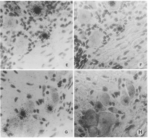
FIG. 2. Insitu hybridization(BamHI-p)ofmouse DRG latentlyinfectedwithHSV-2,4-,um sections. For each pair of panels, standard light microscopy isonthe left (A, C, E, G) and phasecontrastmicroscopy of thesamefields is on the right (B, D, F, H). (A and B) Section showing hybridizationovernonneuronalcell(arrow). (Cand D) Sectionwithhybridizationover a cell likely to be a satellite cell (arrow). (E and F) Sectionwithhybridizationover neuron andnonneuron. (Gand H)Sectionwithhybridization over cells considered possibly nonneuronal whenexamined bystandardlightmicroscopybutprobablyneuronswhenexaminedbyphasecontrast microscopy.
Galloway, Fred Hutchinson Cancer Research Center,
Seat-tle, Wash.) representing approximately 40% of the HSV-2 genome(Fig. 1). HSV DNA fragments werenick translated at 140C with
[35S]dCTP
(1,000Ci/mmol; Amersham Corp., Arlington Heights, Ill.). The specific activities of theprobes were 1 x 10⁸ to 2 x 10⁸ cpml,ug, and 1 to 3 ng of DNA(approximately6 x 105 cpm) was used for eachslide. Tissue preparation and hybridization conditions were asdescribed
previously
(27).Instudiestodetermine the strandspecificityof
hybridiza-tion synthetic oligonucleotides (model 7500 DNA Synthe-sizer; Milligen) were tested by in situhybridization. Oligo-nucleotides were synthesized accordingtothe unpublished
sequence of HSV-2 of D. McGeoch(16a) andkindly made available to us by him. Three 30-mers were used,
corre-spondingtonucleotides 1 to 30, 291to 320, and 681 to 710of the BamHI p fragment, which were fromthestrandopposite tothe ICPOtemplate strand and didnotoverlap ICPO(Fig. J. VIROL.
I1:4
.i.
4-t-I
s;
.i' 4:.f .f
1.1.'W!
It
:.I.:
AA.
on November 10, 2019 by guest
http://jvi.asm.org/
NOTES 2747
*
;Xffl~
I, 2
*.,*,
,Kv
Al
E
.4
F
t-.- ..
...
;. L 40
t~~~~~~~~~~~~~~~~~~'
IE
Opt...!
~~~~~~~~~f~
G
FIG. 2-Continued.
1). The three oligonucleotides will be referredtoasLAT1-30, LAT291-320, and LAT681-710,respectively.Two oligonucle-otideshomologoustothe ICPOtemplatestrand and within the ICPO codingsequence were also tested (ICP03111-3140and ICP03591-3620, of the McGeoch sequence). Oligonucleo-tideswere3' end labeled with[35S]dATP (338 Ci/mmol)and used forin situ hybridization(109cpm/,Lg, approximately 1 ng perslide) ashas been done for HSV-1 (25, 28).
Positive resultswere apparentwithhybridizationof gan-glion sections with the BamHI pfragment (Fig. 2) but not withothercloned fragments (datanotshown). Hybridization signal was present over many neurons, usually over the
nuclei,ashas beenseenforHSV-1(7, 11, 14, 16, 21, 27, 28). Although hybridization signal was usually present in
neu-rons, in some instancessignalwaspresentovercells which didnotappear to be neurons.
To enhance the discrimination of cell types
expressing
LAT,
ganglion
sections of 2 and 4 ,um wereinvestigated.
Given the
relatively large
size ofneurons and theusually
centrallocation of neuronal
nuclei,
itwasthought
thatthinsections would eliminate the
possible
problem
ofoverlap-ping of neurons and nonneurons in a
given
section. Thepossibility of
hybridization
resulting
from neuronaloverlap
ofnonneurons was also minimized
by
thein situhybridiza-tion testingofadjacent serial sections.
Pairs of
photomicrographs
showing
the same fieldsob-tained by standard
light
microscopy(left)
andby
phase
contrast
microscopy
(right)
areshown inFig.
2.Hybridiza-VOL.65, 1991
.ft
.0
1-1,..,:t.1.4
,0
.1.IF)
',1,
IdmF'4s
.I
i
.,
.F;,
z-Alm .,!&
.Fwl
:.;
M. .uni..6...y L,?z.
's. 1,
v
f:.;.
t'.w
t. ala
W,
".; eaqk..
17, -F
't,"'.0 it
,-,t L.;.4
on November 10, 2019 by guest
http://jvi.asm.org/
[image:3.612.56.563.73.552.2]2748 NOTES
jS s ,i
:
.SsX.b...
..WI
A4
.I
.SJE a # sX t t * t 4
t'
t
+'
*."''''
it#,.:s
#
A
seos
''> +' %: :. '.:
Yi # L
{
W
*
: 4 i
:i
st:s
$ A
t
sSit
[image:4.612.62.563.71.552.2]F:2
A
t
t
:*: X
'1||tIIF
:. :: ..
| ;s .$- .S
., r
t ;5>A.
...- .^W *
,: w t:
t :. .:.
.L,....!,s,X
s jvk ws
- rB Br
w...
v;...
F.
t .t4,.
-?
_e0
~~~~~~~~~~~~~~~.
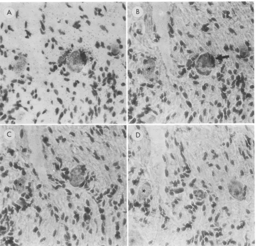
FIG. 3. In situhybridization (BamHI-p) of mouse DRG latently infected with HSV-2,4-pzmsections. (A) Standard light photomicrograph
and (B) phase contrastphotomicrograph of the same section, showing evidence of hybridization over a nonneuronal cell (arrow). (C and D)
Phase contrastphotomicrographs showing adjacent serial sections on either side of the section shown in panels A and B. The lack of label
inpanels C and D supports the nonneuronal site of hybridization seen in panels A and B. For orientation purposes, a star (*) indicates the
sameneuron in each section.
tionovernonneuronalcellswasmostevident when sections wereexamined by phasecontrast microscopy (Fig. 2B and D) andincluded label of probable satellite cells (Fig. 2C and D). Possibly equally important, labeled cells thoughtto be neuronswhenexamined by standard light microscopywere seen to be neurons when examined by phase contrast microscopy (Fig. 2G and H). Testing of serial sections furthersupported the nonneuronal localization of hybridiza-tion(Fig. 3). Itwasestimated that in 10ganglia examined, 5
to 10% of labeled cells were nonneurons.
Hybridization was seen with the BamHI p fragment of HSV-2, which includes the 3' end of the ICPOgene(18), and was not seenwithother HSV-2 fragments, suggesting that
hybridization during latency was similarto thatfor HSV-1 (7, 11, 14, 16, 21, 27, 28). In theirrecentstudy, Mitchelletal.
performed hybridization with fragments representing the entireHSV-2genomeandfoundsimilarly restricted hybrid-ization (18). In the present study, the strand specificity of
A
?.
I
'
V-.
vk
FI
.r
i:
'ft
"I o.
J. VIROL.
*Wf
0
4, Ot.,
i
..%t
-If.z. :.f.,
O'...
.'C'.,..v1.It.:,T,A A
4 fti.,-'tA4. '.
:: ..,
.i, 4
A.A
on November 10, 2019 by guest
http://jvi.asm.org/
NOTES 2749
A
v
S
19
-W ^, t.
is . W
a~~~~~
_10 Am ' b
e.e
o
a~~~~~~~~~~~~~~"llllli,i
DIk&
d~'
"a4.
9
+i --!_
--.
t >
C
':.-**
_4"
a,
W -.10
dft
S.
i-SI -r
K
B Jr
r'tt
Xe* - ne; sli:,-.
-t ¢> + r
~~~
2dta,--;2e'i zr~~~~*'W, S
,:yv
sp
D,
a
-t
-
8tw
r~~~~~
A
~~~*
1 4 ,+ . ,^i i . . *~~~~~~W
.;
t4
4~~~~~~~~~~~Af4
-w
.
:St
4.~~~~~~~4
_11,. },. -v* ; r
404
* X
-<
4 ?siX'ra
i
>
r4es
s
sw
-pA.
IQA
FIG. 4. In situ hybridization (LAT291-320) ofmouse DRG latently infected with HSV-2, 2-p.m serial sections. (A) Standard light photomicrograph and(B)phase contrastphotomicrograph ofthe samesection, showingevidence ofhybridizationover anonneuronal cell (arrow).Furtherevidenceof the nonneuronalnatureof thishybridization-positive cell is provided in panels CandD, whicharephasecontrast photomicrographs showingadjacentserial sectionsoneithersideofthesectionshowninpanelsAand B. Fororientationpurposes, a star (*) indicatesthe same neuronin each section.
hybridizationwasinvestigatedby the useof synthetic
oligo-nucleotides,and the results extend thehybridization results ofMitchell etal. (18), who did notdetermine strand
speci-ficity.
In testing oligonucleotides synthesized from the HSV-2 strand opposite to the ICPO template strand and not
over-lappingICPO(antisensetoLAT),hybridizationwasdetected with LAT1-30 and LAT291-320 but not LAT681-710. Possi-ble reasons for the absence ofhybridization with LAT681-710will beinvestigated further.
Hybridization over a nonneuronal cell after 2-,um serial
sections were tested with LAT291-320 is shown in
Fig.
4. Serialsectiontestingexcluded thepossibility thatlabelwas derived from an overlying neuron, and hybridization de-tectedwithLAT1-30andLAT291-320supportedtheconclu-sion that RNAtranscribed from the strand
opposite
to theICPO template strandwas detected. In
addition,
hybridiza-tionwithBamHIpwaseliminatedbyRNase pretreatmentof tissue sections(26),and
hybridization
was notdetectedwith theICPO-specific oligonucleotides;
as would beexpected,
oligonucleotides
synthesized
from theICPOtemplate
strandhybridizedtoacutelyinfected cells
(data
notshown).
VOL.65, 1991
oi:
*-Wt; 1||"
on November 10, 2019 by guest
http://jvi.asm.org/
2750 NOTES
If it is considered that the presence of HSV LAT denotes latency, it would be concluded that latent infections of nonneuronal cells may be established not infrequently by HSV, at least by HSV-2. It remains to bedemonstrated that latent infection of these nonneuronal cells can be reacti-vated, if it is considered that the definition of latency includes this function (25).
Although HSV latency has beenconvincingly shown to be primarily an infection of sensory ganglion neurons, HSV-1 latency has been suggested to occur in nonneuronal neural cells (7, 23) and HSV-1 and HSV-2 latency to occur in epithelial cells (1, 2, 5, 19). The possibly nonneuronal locus of HSV latency reported here can also be compared with results obtained with varicella-zoster virus (VZV). A neu-ronal site of VZV latencydetermined by in situhybridization was suggested (10, 12), although in more recent studies nonneuronal hybridization was noted (6). The latter led Straus to offer a model which incorporated differences between possible sites of latency of HSV and VZV to explaindifferences in the pathobiology of human infections
by theseviruses (22). Use of phase contrastmicroscopyand the examination of thin tissue sections may resolve some questions of cellidentification. If the occurrence of nonneu-ronal HSV-2 latent infection of the nervous system, as reported in the present study, is confirmed, it may modify some considerations of HSV latency; latent HSV infection ofa celltype(s) which differs functionally and replicatively from neurons may be important in somedisease processes.
This study was supported by Public Health Service award NS20684(R.B.T.).
Helpful discussion with M. Judith Tevethia and the secretarial assistance ofTricia Johnsonare appreciated.
REFERENCES
1. Al-Saadi,S. A., G. B.Clements, and J. H.Subak-Sharpe. 1983. Viral genesmodify herpes simplexviruslatency both inmouse footpad and sensory ganglia. J. Gen.Virol. 64:1175-1179. 2. Clements, G. B., and J. H.Subak-Sharpe.1988. Herpessimplex
virus type 2establishes latency in the mousefootpad. J. Gen. Virol.69:375-383.
3. Cook,M. L., V. B.Bastone, and J. G. Stevens. 1974. Evidence thatneurons harborlatent herpessimplex virus. Infect. Immun. 9:946-951.
4. Cook, M. L., and J.G.Stevens. 1973. Pathogenesis of herpetic neuritis and ganglionitis in mice: evidence for intra-axonal transport ofinfection. Infect. Immun. 7:272-288.
5. Cook,S.D., S. K. Batra, and S. M. Brown. 1987. Recovery of herpes simplex virus from the corneas of experimentally in-fected rabbits. J. Gen. Virol. 68:2013-2017.
6. Croen, K. D., J. M. Ostrove, L. J. Dragovic, and S. E. Straus. 1988. Patternsofgeneexpressionand sites oflatency inhuman nerve ganglia aredifferentforvaricella-zostervirusand herpes simplex viruses. Proc. Natl. Acad. Sci. USA85:9773-9777. 7. Deatly, A. M., J. G. Spivack, E. Lavi, D. R. O'Boyle II, and
N. W. Fraser. 1988. Latent herpes simplex virus type 1 tran-scriptsinperipheral andcentral nervous system tissuesofmice map tosimilar regions of the viral genome. J. Virol. 62:749-756. 8. Dillard,S. H., W. J.Cheatham,and H. L.Moses. 1972.Electron microscopy of zosteriform herpes simplex infection in the mouse. Lab. Invest. 26:391-401.
9. Galloway,D.A., C. Fenoglio, M. Shevchuk, and J. K. McDou-gall. 1979. Detectionofherpes simplexRNA in human sensory ganglia. Virology95:265-268.
10. Gilden, D. H., Y. Rozenman, R. Murray, M. Devlin, and A. Vafai. 1987. Detection ofvaricella-zoster virus nucleic acid in neurons ofnormal human thoracic ganglia. Ann. Neurol. 22: 377-380.
11. Gordon, Y. J., B. Johnson, E. Romanowski, and T. Araullo-Cruz. 1988. RNAcomplementarytoherpes simplexvirus type 1
ICPO genedemonstrated inneurons ofhuman trigeminal gan-glia. J.Virol. 62:1832-1835.
12. Hyman,R. W., J.R.Ecker, and R. B. Tenser. 1983. Varicella-zostervirus RNA in human trigeminal ganglia. Lancet ii:814-816.
13. Kennedy, P. G. E., S. A. Al-Saadi, and G. B. Clements. 1983. Reactivation of latent herpes simplex virus from dissociated identified dorsal root ganglion cells in culture. J. Gen. Virol. 64:1629-1635.
14. Krause, P.R., K. D. Croen, S. E. Straus, andJ. M. Ostrove. 1988. Detection and preliminary characterization of herpes
simplex virus type 1 transcripts in latently infected human trigeminalganglia. J. Virol. 62:4819-4823.
15. Kristensson, K., A. Valne, A. Persson, and E. Lycke. 1978. Neuralspreadofherpessimplexvirustypes1and2inmice after corneal orsubcutaneous(footpad) inoculation. J. Neurol. Sci. 35:331-340.
16. Leib, D. A., C. L. Bogard, M. Kosz-Vnenchak, K. A. Hicks, D. M.Coen,D. M.Knipe, and P. A. Schaffer. 1989. Adeletion
mutant of the latency-associated transcript ofherpes simplex virus type 1 reactivates from the latent state with reduced frequency. J. Virol. 63:2893-2900.
16a.McGeoch,D.(InstituteofVirology, Glasgow,Scotland).Personal communication.
17. McLennan, J. L., and G. Darby. 1980. Herpes simplex virus latency: the cellular location of virusindorsal rootgangliaand the fate ofthe infected cell following virus activation. J. Gen. Virol.51:233-243.
18. Mitchell, W. J., S. L.Deshmane, A. Dolan, D.J. McGeoch, and N. W. Fraser. 1990. Characterization ofherpes simplex virus type 2 transcripts during latent infection ofmouse trigeminal ganglion. J.Virol. 64:5342-5348.
19. O'Brien,W.J.,andJ.L. Taylor. 1989. The isolation ofherpes simplexvirus fromrabbitcorneasduring latency. Invest. Oph-thalmol. Visual Sci. 30:357-364.
20. Roizman, B. 1979. The cellular organization of the herpes simplex virusgenomes.Annu. Rev. Genet. 13:25-57.
21. Stevens, J. G., E. K. Wagner, G. B. Devi-Rao, M. L. Cook, and L. T. Feldman. 1987. RNA complementary to a herpes virus alpha gene mRNA is prominent in latently infected neurons. Science235:1056-1059.
22. Straus, S. E. 1989. Clinicaland biological differences between
recurrent herpessimplex virus andvaricella-zoster virus infec-tions. JAMA262:3455-3458.
23. Stroop, W. G., D. L. Rock, and N. W. Fraser.1984. Localization ofherpessimplex virus inthetrigeminal andolfactorysystems of the mouse central nervous system during acute and latent infection byinsitu hybridization. Lab. Invest. 51:27-38. 24. Suzuki, S., and J. R. Martin. 1989. Herpes simplex virus-type 2
transcripts intrigeminal ganglia duringacuteand latent infection of mice. J. Neurol. Sci. 93:239-251.
25. Tenser, R. B. 1991. The role ofherpes simplex virusthymidine kinaseexpressioninviral pathogenesis andlatency. Intervirol-ogy 32:76-92.
26. Tenser,R.B.,M.Dawson,S.J. Resell, and M. E. Dunstan. 1982. Detection of herpes simplex virus mRNA in latently infected trigeminal ganglion neurons by in situ hybridization. Ann. Neurol. 11:285-291.
27. Tenser, R. B., K. A. Hay, and W. A. Edris. 1989. Latency-associated transcript but not reactivatable virus is present in sensoryganglionneuronsafterinoculationof thymidine kinase-negative mutants of herpes simplex virus type 1. J. Virol. 63:2861-2865.
28. Wechsler, S. L., A. B. Nesburn, R. Watson, S. M. Slanina, and
H. Ghiasi. 1988. Fine mapping ofthe latency-related gene of herpes simplex virus type 1: alternative splicing produces dis-tinct latency-related RNAs containing open reading frames. J. Virol. 62:4051-4058.
29. Wilkie, N. M., A. Davison, P. Chartrand, N. D. Stow, V. G. Preston, and M. C. Timbury. 1978. Recombination of herpes simplex virus: mapping ofmutations andanalysis ofintertypic recombinants. Cold Spring HarborSymp. Quant. Biol. 43:827-840.
J. VIROL.