0022-538X/87/072232-08$02.00/0
Copyright © 1987,American Society forMicrobiology
Polyomavirus Tumor Induction in Mice: Influences of Viral
Coding
and
Noncoding Sequences
on
Tumor Profiles
ROBERT FREUND, GAIL MANDEL,t GORDON G. CARMICHAEL,: JOHN P. BARNCASTLE, CLYDE J. DAWE,
ANDTHOMAS L. BENJAMIN*
Department ofPathology, Harvard MedicalSchool, Boston, Massachusetts02115 Received5January 1987/Accepted 25 March 1987
We determined the DNA sequences of the noncoding regions of two polyomavirus strains that differ
profoundlyintheir abilitiestoinducetumorsinmice. Differences betweenstrainswerefound,bothonthelate sideof thereplication origin in the region containingknown enhancerelementsand ontheearly side of the origin, affecting the number and location of large-T-antigen-binding sites. By constructing and analyzing recombinant viruses betweenthesehigh-and low-tumorstrains, weattemptedtolocalizedeterminantswhich affectthefrequency andhistotype of tumors. Seven recombinantswereconstructedandpropagated invitro,
and the tumorprofileof eachwasestablishedbyinoculation into newbornC3H mice. Recombinantscontaining noncodingsequencesfromthehigh-tumorstrain andcodingsequencesfrom the low-tumorstrainbehavedlike the latter, inducingtumors ata lowfrequency andstrictlyofmesenchymal origin. Reciprocalrecombinants with noncodingsequencesofthe low-tumorstrainlinked tostructural determinants from thehigh-tumorstrain
induced severaltypesofepithelialtumorstypicalofthe high-tumorstrain butatreducedfrequency,inaddition to mesenchymal tumors. A high frequency and full diversity ofepithelial tumors required, in addition to
structural regionsfrom thehigh-tumor strain, noncodingsequencesontheearlysideof theorigin also present
in this strain. A high-tumor profile thus resulted from the combined effects of structural and regulatory determinants inthehigh-tumor strain, with the formeraffecting primarily the tissuetropism andthe latter affectingthefrequencyoftumors.Nodifferential effects of the enhancerregionsfromthe lateside oftheorigin
inthetwo virusstrainswere seen in thisstudy.
Interactions ofpolyomavirus with itsnatural host encom-pass a range of biological processes that remain poorly understood, despitethedetailed knowledge wehave of this virusatthemolecularbiologicallevel. Tumors areknown to
develop frommorethan adozendifferent cell types follow-ing inoculation ofthevirus into newborn mice, and
produc-tiveinfectioncan occurin as many as 30differenttissues (7).
Little isknown at the molecular level about thepathogenesis of tumors and lytic lesions in these laboratory infections. Similarly, muchremains tobelearned aboutvarious
mech-anisms accompanying natural infection in which the virus
persists withoutcausing tumorsandabout the roles that the
transforminggenesofthevirusmight play in such infection. Inthisreportwefollow up on recent observations showing that tumor induction in mice by mouse polyomavirus in-volves viral genetic determinants that are, at least in part,
differentfrom those involved in cell transformation in vitro. Wefoundthat two molecularly cloned andostensibly wild-type virusstrains wereprofoundly different in their abilities
toinduce tumors in mice, despite being equally efficient in the transformation of cells in culture. The PTA strain, referred to here as the high-tumor strain, induced multiple
tumorsin 90 to100% of recipient animals. The tumors were ofepithelial as well as mesenchymal origin and led to a
moribund condition of the host within aperiod of about 12 weeks. The RA, or low-tumor, strain as a rule induced only
single tumors in a small fraction of the animals. These
tumorswereexclusively of mesenchymal origin and led to a
*Correspondingauthor.
tPresent address: Divisionof Molecular Medicine, Tufts New England MedicalCenter, Boston, MA 02111.
tPresent address: Department of Microbiology, University of Connecticut Health Center, Farmington, CT 06032.
moribundconditionafteranaveragesurvivaltime ofover40 weeks. Simultaneous inoculation ofPTA and RA led to a
hightumorprofile, suggestingthat RAdoesnot act toevoke
a dominant systemic resistance, for example, viathe host immune system (7). As a step toward understanding the basis for these differences invivo,weconstructedaseriesof
recombinantviruses between PTA and RA andstudiedthem
in newborn mice. The results reveal two classes ofgenetic
determinants present in PTA, deriving fromnoncodingand
coding regions,thatcontributeto ahigh frequencyandbroad spectrumoftumors.
MATERIALSANDMETHODS
Subcloning and sequencing. Molecularly cloned viral
DNAs from strains PTA and RA (7) were subcloned into
pUC13 byusingstandardrecombinantDNAtechniques (18).
Both the large and smallBamHI-to-EcoRI DNAfragments fromtheviralgenomes weregel purifiedandcloned into the
appropriatelydigestedvector.(Forarestrictionenzyme map
of polyomavirus, see Griffin et al. [15].) The subclones
containing the small BamHI-to-EcoRI viral DNA fragment werefurther subcloned into the BamHI-to-PstI fragment and
PstI-to-EcoRI fragment. TheBamHI-to-PstI fragment,
con-taining the noncoding region of these viral genomes, was subcloned into M13mp8 and M13mp9 for sequencing. The universal M13primer, aswellassynthesizedoligonucleotide
primers throughout the noncoding viral DNA region, was
used to obtain the nucleotide sequence by the
dide-oxynucleotide sequencingmethod (22).
Construction of recombinant viruses.Recombinantviruses
wereconstructed by combiningtheappropriategel-purified
DNA fragments generated from restriction enzyme
diges-tions of the subcloned DNAs. The structures of the viral
recombinantsareshown in Tables 1, 2, and 3. To digest the 2232
on November 10, 2019 by guest
http://jvi.asm.org/
DNAs withBclIorStuIor both, the clones were first grown in thedcmdam Escherichia coli host MM505. Partial diges-tions were done by limiting the enzyme concentration and time of digestion. To generate molecular clones of the recombinant viral genomes, the appropriate gel-purified frag-ments were ligated withEcoRI-digestedpUC13. The cloned DNAs were screened by restriction enzyme digestion and polyacrylamide gel electrophoresis. The cloned viral DNAs were excised from the vector by digestion with EcoRI, ligated under dilute conditions, and transfected into primary baby mouse kidney cells by using the calcium phosphate procedure (7, 14). Virus stocks were grown from these
lysates, and titers were determined by plaque assay on
NIH-3T3 cells.
Mice, virus inoculations, and pathology. All mice were of the inbred strain C3H/BiDa. Litters of newborns were inoc-ulated at less than 18 h of age by subcutaneous injection of 0.05 ml of crude virus suspension. The dose of virus was 5 x
106
to 2 x10⁷
PFU per animal. Mice were necropsied either when moribund or at approximately 1 year of age. Histolog-ical examination of tissues was performed as previously described (7). Tumors scored as overt were seen by gross examination at the time of necropsy and were confirmed by histological examination, except as noted. Tumors scored as occult were revealed by histological examination only. The latter scoring gave a lower limit to the estimate of tumor frequency because of the small amount of tissue that could be examined histologically.RESULTS
Multiple differences between PTA and RA in the noncoding regions. PTA and RA viral DNAs were cloned, and virus stocks were prepared on baby mouse kidney cells from the cloned viral DNAs (see Materials and Methods). Purified viral DNAs were compared by restriction enzyme digestion by using several enzymes singly and incombination. Differ-ences were noted in the region within a few hundred base pairs on either side of the origin of replication. No differ-ences in restriction enzyme digestion patterns were noted in fragments deriving either from the early or late coding regions (data not shown).
On the basis of the above results, the entire noncoding regions of the two viral DNAs were sequenced, going from the ATG for VP2 on the late side through the origin of replication and out to the ATG for the T antigens on the early side. The results are shown in Fig. 1. PTA and RA have nonoverlapping duplications of 33 and 31 base pairs (bp), respectively, on the late side of the origin in the known enhancer region (10, 16, 19, 26). The duplication in RA has been previously reported (21). The duplication in PTA includes sequences containing a core element similar to that found in the simian virus 40 72-bp enhancer (16, 25, 26), as well as a DNase I hypersensitive region (16). This region of polyomavirus DNA (between the PvuII site at 5128 and 5262) has been shown to contain enhancer activity by transfection andtransientexpression with heterologous con-structions (10, 16, 25). Host range variants ofpolyomavirus capable of limited growth on embryonal carcinoma cells have previously been shown to vary in this region (1), although the changes in these variants are different from those seen in either PTA or RA. On the early side of the origin, PTA has three contiguous 11-bp elements containing
large-T-antigen-binding sites (4, 5), while RA has only two. This difference has also been seen between polyomavirus strains A2 and A3, which have three and two
large-T-antigen-binding sites, respectively (9, 13, 24). PTA has an
additional duplication of 40 bp at the BglI site that is not found in RA or any other strain described thus far. Four base-pair substitutions, one on the late side and three onthe early side, were also found (Fig. 1). Anexaminationof PTA and RA sequences in the origin region indicates small changesin preexisting short open reading frames due to the duplications andinsertions; however, in no cases werethese marked by amethionine codon at the start.
PR recombinants in which noncoding sequences of PTA were linked to coding sequences of RA. Recombinant virus strains in this study are given atwo-letterdesignation, PR or RP (PforPTA and R for RA), with the first letterindicating
the strain of origin of most or all of thenoncoding sequences and the second letter indicating the origin of coding se-quences. Results obtained with a series of three PR recom-binant viruses and schematic diagrams of therecombinants
are shown in Table 1. These strains were constructed to
determine the contribution of noncoding sequences of the high-tumor strain on a background of structural
determi-nants from the low-tumor strain. The first ofthese, PR-1,
contained theenhancerregion of PTA onthe late sideofthe origin, while the noncoding sequences on the early side of
the origin along with all early and late coding sequences derived from RA. Only asingle tumor, arenal sarcoma, was
observed. In PR-2, the amount of noncoding sequences derived from PTA was extended in both directions,
incor-porating amino-terminal sequences of VP2 on the late side andadditional noncoding sequences on the early side up to
but not including the 40-bp duplication at the
BgII
site. A third recombinant, PR-3, contained the entire noncodingregion of PTA,including theBglI duplication, with allcoding
sequences comingfrom RA. A low-tumorprofile wasfound
for both of theserecombinants, essentially similartothose of PR-1 and the low-tumor parental strain RA. Neither the enhancer region alone of PTA (PR-1) nor the enhancer region plus the array of additional large-T-antigen-binding
sites on theearly side of the origin (PR-2 andPR-3)was able to direct a high-tumor profile when linked to coding
se-quences from the low-tumor strain. These resultsthus
point
to the existence of one or more structural determinants in PTA essential for a high-tumor profile and
specifically
for induction ofepithelial tumors.RP recombinant containing noncoding sequences of the
low-tumor strain and coding sequences of the high-tumor
strain. The PTA tumorprofile asreported earlier(7)and that of a single RP recombinant, RP-1, which had the entire noncoding region of RA and all coding regions of PTA, is given in Table 2. Schematic diagrams ofPTAand RP-1 are also shown. Most significant in the profile of RP-1 was the appearance of mammary and salivary gland tumors, two of the major epithelial tumor types in the PTA
profile.
These results thus bear out the prediction of at least one PTA coding determinant required for induction ofepithelial
tu-mors. The RP-1 tumorprofile, however, showed lessdiver-sity and lower frequencies ofepithelial tumors than that of PTA; in particular, only a single hair follicle tumor and no thymic tumors were seen in mice inoculated with RP-1.
These animals also lived
significantly
longer
before becom-ing moribund (203 days) compared with those inoculated with PTA (117 days).Another significantfeature of the RP-1
profile
wasthehigh
frequency ofmesenchymal tumors
compared
with the pro-files of the PRrecombinants(Table 1)
orwith that of the RA parental strain itself(7). This suggestsanelement(s)
in PTA coding sequences which favorsdevelopment
ofon November 10, 2019 by guest
http://jvi.asm.org/
A
BcIl
RA
CATTTTGAIAaTTCaCTTaCTTGaTCAGCTTCaGJLAGATGGCGGLGGGCCTCCAACCAGTAATTTTCCTCCCGACTCTTPTA
CATTTTrAaakTTCaCTTaCTTGATCaGCTTCLGLGATGGCGGAGGGCCTCCAAC&CAGTwATTTTCCTCCCGACTCTT
duplication
AAAATAGAAAATGTCAAGTCkGTTAAGCAGGAAGTGACTAkCTGACCGCAGCTGGCCGTGCGACATGACTAACTGACCGC
I
AAAATAGAAAkTGTCAAGTCAGTTAAGCGGkAAGTCACTAACTGACCGCAGCTGGCCGTGCGACAT
AGCTGGCCGTGCGACATCCTCTTTTAATTAGTTGCTAGGCAACTGCCCTCCAGAGGGCAGTGTGGTTTTGCAAGAGGAAG
--- CCTCSTTTTIA TTATTGCTAGGCAACTGCCCTCCAGAGGGCAGTGTGGTTTTGCAAGAGGAAG
Stul
I
CAAAAAGC---CTCTCCACCCAGGCCTAGAATGTTTCCACCCAATCATTA
CAAAAAGCGGCAGTGTGGTTTTGCAAGAGGAAGCAAAAAGCCTCTCCACCCAGGCCTAGAATGTTTCCACCCAATCATTA
duplication
Hpall
CTATGACAACAGCTGTTTTTTTTAGTATTAAGCAGAGGCCGGGG$CTCCTGGCCTCCGCTTACTCTGGAGAAAAAGAAGA
.*
-
14-l
.*
I
I
CTATGACAACAGCTGTTTTTTTTAGTATTAAGCAGAGGCCGGGGGCCCCTGGCCTCCCCTTACTCTGGAGAAAAAGGAGA
deletion
BgIl
I I I
GAGGC----TTCCAGAGGCAACTTGTCALAACAGGACTGGCGCCTTGGAGGCGCTGTGGGG
--- - * * _ .ill...
GAGGCATTGTAGAGGCTTCCAGAGGCAACTTGTCAAAACAGGACTGGCGCCTTGGAGGCGCTGTGGGGCTTGTCAAAACA
duplication
---Cs---CCACCCAAATTGATATATTAAGCCCCAACCGCCTCTTCCCGCCTCATTTCA
GGACTGGCGCCTTGGAGGCGCTGTGGGGCCACCCAkLATTGATATATTAAGCCCCAACCGCCTCTTCCCGCCTCATTTCA
GCCTCACCACCATCATG GCCTCACCACCATCATG
B
BcII
StuI
HpaII
BgI
RA
G
MAPTA
33
11
40
Origin
Late
4-
-*
Early
on November 10, 2019 by guest
http://jvi.asm.org/
TABLE 1. Schematic diagramsand tumor profiles of PR recombinant viruses"
TABLE 2. Schematicdiagrams and tumor profiles of PTA and RP-1"
orl
v' --Jyl
ori
1i1 v L --"-E
p
Lri
IV II
Fraction of mice with
tumors
Meanage at necropsy
(days)
Age range at necropsy
(days)
Epithelialtumors
Hairfollicle Thymus Mammary gland Salivary gland
Mesenchymal tumors
Subcutaneous
connec-tive tissue
Vascular endothelium
Renal medulla Bone
1/37 3/26
292
0 0 0 0
0
0
1
0
403
393-408
No.of mice withgrosstun 0
0 0 0
3
0 0
1
'The PR recombinant viruses contain thecoding regionfromRA.Thediagrams show
thenoncoding region expanded. Black segments are fromRA.and white segmentsarefrom
PITA.Vertical lines above thenoncoding regionarerestriction enzyme sitesBcIl.Stil,
Hpall,andBglIl,from left toright.Triangles indicate insertionsorduplications
characteris-tic of the parentalstrain,asshowninFig.1.PR-1contained thenoncoding sequences from
BelltoStulofPTAand therestof the viral genome fromRA.PR-2contained all of the
sequencesfrom BarmHI toBgll, encompassingthe replication originfrom PTA. This
includes sequencesencoding the N-terminal end ofVP2aswell asall thenoncoding
sequences around theorigin from PTA,notincluding the 40-bp insertionattheBgllsite.
PR-3contained all of thenoncoding sequences of PTA from BclltoBgll. includingthe
33-bp duplication in the enhancer, the 11-33-bp insertioncontainingthethirdT-antigen-binding
site, and the 40-bp duplication around theBgilsite. Allcoding sequenceswerederived
fromRA.LandEindicate lateandearly sides of thereplicationorigin, respectively.
mal tumors, particularly renal sarcomas and bone tumors which were not part of the RA tumor profile and occurred
only rarely withPRrecombinants (Table 1).
Requirement for noncoding sequences from the early side of the origin of the high-tumor strain. Three additional RP recombinants wereconstructed to analyze the contributions ofnoncoding sequences present in thehigh-tumor strain to
thetumorprofileandparticularlytothefrequency and types of epithelial tumors (Table 3). The noncoding regions in
these three recombinants all contained the late-side
en-hancer elements from RA, together with various elements from the early side ofPTA, all on a PTA structural
back-ground. RP-2 contained origin-proximal PTA sequences,
fromtheStuI siteclosetotheoriginout totheBgIIsite. RP-3
Fractionof mice with tumors 5/34 Meanage at necropsy (days)
Agerangeat necropsy (days) 422
310-550
nors Epithelial tumors __________ Hair follicle
Thymus
0 Mammary gland
0 Salivary gland 0
0 Mesenchymaltumors
Subcutaneousconnective tissue
Vascularendothelium 4 Renalmedulla
Bone
37/37
117
45-225
22/22
203 79-390
No. ofmice withtumorsh
OvertOccultTotal
(%)IOvert
Occult Total(9)33 1 34(92) 30 1 31(84) 9 1 10(27)
6 9 15 (41)
7 1 8(22)
0 1 1(3)
4 9 13(35)
10 0 10(27)
1 0 1(5)
0 0 0
6 3 9 (41)
3 6 9(41)
10 3 6 12
3 13 (59) 0 3(14)
1 7(32) 0 12 (55) 'aTherecombinant virus RP-1 contained noncoding sequences from RA and coding
sequences fromPTA. The diagrams show the noncoding regions expanded. Abbreviations
andsymbols are as described in the footnote to Table 1.
bThe percentage represents the number of mice with a particular type of tumor divided
by the total number of inoculated mice.
contained the origin-distal PTA element, consisting of the 40-bp duplication at the BglI site in an otherwise RA noncoding region. RP-4 contained both segments of PTA sequence from the early side of the origin out to and including the duplication around BglI.
RP-2 induced a high frequency of both mammary and
salivarygland tumors,asdid PTA, but only four hair follicle
tumors were confirmed histologically, along with a single thymic tumor, whichwas occult. RP-3 had aprofile similar to those of RP-1 and RP-2, with the notable addition ofa
substantial number ofthymic tumors, both overt andoccult.
When the two early noncoding segments of PTA were
combined, as in RP-4, a high-tumor profile was found,
essentially similar to the PTA profile with respect to fre-quency anddiversity oftumors as well as average latency. These results establish the importance of early-region
noncodingsequencesofPTAinincreasingboth the diversity
and frequency of epithelial tumors. The basic similarity
between PTA and RP-4 reinforces the conclusion that the
enhancerregions ofthe twoparental strains have no differ-ential effect onthetumorprofiles.
A more detailedanalysis of the data for RP recombinants
FIG. 1. Nucleotide sequences of the noncoding regionof PTA and RA. (A) The sequences of strains RA and PTA from the start of translation ofthe lateregions (nucleotide 5000,accordingtothe systemofGriffinetal.[15]) throughtheorigintothestartof translationof theearly region (nucleotide 175). Single-base-pair differencesareindicatedbyaverticalline.RAcontainsaduplicationof 31bp(nucleotides 5114to5144)onthe latesideoftheoriginandan11-bpdeletion(nucleotides44 to54)ontheearlyside of thereplicationorigin.Thedeletion contains the sequenceGAGGC which has been shownto bindlargeTantigen invitro(4, 5, 20). TheseT-antigen-binding-site sequences throughout the noncodingregionareindicatedbydark arrows, andvariants ofthe sequenceareindicatedbyhatchedarrows. PTAcontains
a33-bp duplication(nucleotides5183to5213)onthelate side of theorigin.Theserepeatednucleotides containthesimianvirus40enhancer coresequence(16,25, 26) andaDNase Ihypersensitivesite(16).PTAalsocontainsa40-bpduplication(nucleotides67to106)ontheearly sideofthereplicationorigin.Thisregion containstwolarge-T-antigen-bindingsites(4,5).Restrictionenzyme sites usedin the construction ofrecombinants are indicated above the sequence. TheHpaII site marks the originof DNA replication. (B) Schematicdiagram of the noncoding region ofPTAandRA,indicatingthedifferencesbetween thetwostrains. Duplicationsandinsertionsareindicatedby triangles and the numberofbasepairs. Single-base-pairdifferencesareshownwithverticallines. The restriction enzymesitesBclI(nucleotide5021),
StuI (nucleotide5211),HpaII(nucleotide 5291), andBglI (nucleotide 87)showninpanelAare
designated
forreference.O'i
L E
p .k
PR-3 .11
---%,L..
E
i
Aon November 10, 2019 by guest
http://jvi.asm.org/
TABLE 3. Schematicdiagrams andtumorprofiles of RP recombinant viruses"
L - E l Y
Fraction of mice with
tumors
Meanageatnecropsy
(days)
Agerangeatnecropsy
16/17 120 90-157
OvertOccult TotalCVc)
Epithelialtumors
Hairfollicle Thymus Mammary gland Salivary gland Mesenchymaltumors
Subcutaneousconnective
tissue
Vascular endothelium Renal medulla Bone
4 0 4(24) 0 1 1(6) 5 2 7(41) 9 5 14(82)
1 10 11(65)
2 0 2(12) 12 3 15(88) 9 0 9(53)
L : E
17/17
112
61-178
No. of mice%kithtumorsh
L.
31/31 72 55-93
Over-tOccultTotal OvertOccult Total (I//
t) 0 0
6 4 10(59) 6 0 6(35)
7 5 12(71)
5 2 7(41) 1 0 1(6) 14 1 15(88) 13 0 13(76)
31 0 31 (100) 31 0 31(10() 11 4 15(48)
3 23 26(84) 0 2 2(6)
0 0
21 5 26(84) 7 0 7(23)
[image:5.612.60.300.90.354.2]aAdditional RPrecombinant virusescontainingvariousportionsof thenoncoding regionontheearlyside of theorigin plusallcoding regionsfrom PTA. Abbreviations andsymbolsare asdescribed in the footnoteto Table 1. RP-2contained noncodingsequencesof PTAonthe eairly side oftheorigin fromStiultothe Bell site: noncodingsequences onthe late side of thereplication origin from BclltoSt,ulwerefrom RA. RP-3 contained
thenoncodingsequencesfrom BilltoBgllfrom RA. with the addition of the 40-bp duplication from PTA
centered attheBgllsite. RP-4containedthe enhancersequencesfrom BilltoStlut froni RA.withearly noncodingsequencesfrom PTA. including the 11-bp insertion and the 40-bp duplication.
bThe percentagerepresentsthe number of mice withaparticulartypeoftumordividedby the total number
ofinoculatedmice.
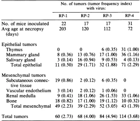
isshown in Table 4. Here, the total number ofgrosstumors ofagiventypein the entire setof animals inoculatedwitha given virus is tabulated ratherthan the number of mice with
particulartumorhistotypes. Thisanalysisbetter reflectsthe roles of different viral sequences in the frequencies of induction ofspecifictumortypes. Atumorfrequency index,
defined as the total number of tumors of a given type or groupdividedbythe number of animalsinoculated, is shown to facilitate comparisonsamong virus strains. Comparisons
of RP-1 with RP-2 and RP-3 indicate that each of the
early-region noncoding segmentsof PTA couldact indepen-dently to increase the frequencies of certain tumors, both
epithelial (mammary and salivary gland) and mesenchymal (renal). RP-1, which contained none of the noncoding
se-quences of PTA, induced fewertotalepithelial tumors than
RP-2, with theorigin-proximal segment ofPTA, RP-3, with the origin-distal segment, or RP-4, containing both PTA
elementsfrom the early side of theorigin.The datainTables 2, 3, and 4 show that the appearance of thymic tumors correlated strongly with the presence of the duplication in PTA around the BglI site. Thus, RP-3 and RP-4, which
contained the duplication, induced gross thymic tumors, while RP-1 and RP-2, which lacked thiselement, did not.
Hairfollicletumorsarenot listed inTable 4 because they couldnotbeaccurately enumeratedatthegrosslevelexcept
in those animals in which they were few. When present in
large numbers, they tended also to be larger in size and confluent or overlapping, making accurate counts virtually impossible. Review of thenecropsyrecords made it evident
thatfor theviruses inthisstudy, therewas awide boundary, quantitatively, between those viruses that induced few hair follicle tumors and the one virus (RP-4) that induced large
numbers of thistumortype. When mice infected withRP-1,
RP-2,orRP-3 bore hairfollicletumors,therewereonly10or
TABLE 4. Total number of tumors and tumorfrequency indices'
No.oftumors(tumorfrequency index) withvirus:
RP-1 RP-2 RP-3 RP-4
No.of mice inoculated 22 17 17 31
Avg age atnecropsy 203 120 112 72
(days) Epithelial tumors
Thymus 0 0 6(0.35) 31(1.00)
Mammary gland 8(0.36) 13(0.76) 17(1.00) 36(1.16) Salivary gland 3(0.14) 16(0.94) 9(0.53) 4(0.13) Totalepithelial 11 (0.50) 29(1.71) 32(1.88) 71(2.29) Mesenchymal tumors
Subcutaneousconnec- 19(0.86) 2 (0.12) 6 (0.35) 0 tivetissue
Vascularendothelium 3 (0.14) 2(0.12) 1 (0.06) 0 Renalmedulla 9(0.41) 18 (1.06) 26(1.53) 33(1.06) Bone 18(0.82) 17(1.00) 19(1.12) 10(0.32) Totalmesenchymal 49(2.23) 39(2.29) 52(3.05) 43 (1.39) Total tumors 60(2.73) 68(4.00) 84(4.94) 114 (3.68)
a Total number oftumors wasdetermined bygrossexamination. Tumor frequency indicesarecalculatedasthe totalnumberoftumorsdividedby the number of animalsinoculated.
fewertumors of this type permouse (Table 5). In contrast, all mice that were infected with RP-4 bore hair follicle
tumors andconsistentlyhad 50or moresuchtumors.
Com-bining the scoring methods in columns 2 and 3 of Table 5,
one sees a5-to 10-fold difference between RP-4 and any of the other RP recombinant viruses, in terms of tumor
re-sponse perunit of targettissue mass.
The data on the RP recombinants suggest relationships
betweenincidences ofparticular epithelialandmesenchymal
tumors and overall survival time. For example,
epithelial
tumorsofthethymustendedto occurearlier and become life
threatening more quickly than other tumors did. Thus,
animals injected with RP-4, with the highest frequency of
overtthymic tumors, had the shortest overall survival time and,consequently, hadrelativelyfew mesenchymaltumors
suchasfibrosarcomas and bonetumors. Amongtheanimals which didnotdevelopthymictumors,renal sarcomas tended
to causeearliest morbidity. Thus, the average survivalwas
longest for animals inoculated with RP-1, which induced no
thymic tumors and the lowest incidence of renal tumors, followed by RP-2, which induced no thymic tumors but a
high incidence of renal tumors, and RP-3 and RP-4, which
induced boththymicand renaltumorsthat contributedtothe shorter average survival times.
No histological differences between tumors induced by
recombinant and parental viruses. In addition to the tabu-lated histotypes, most of the less commonepithelial tumor
[image:5.612.319.561.649.711.2]types known to be inducible by polyomavirus were also
TABLE 5. Hairfollicletumorsin RPprofiles0
Virus Fractionofmice with Estimate ofno.of hair follicle
hairfollicletumors tumorsin affected mice
RP-1 3/22 1-10
RP-2 7/17 1-10
RP-3 6/17 1-10
RP-4 31/31 250
aNumber of hair follicletumorswereestimated foreachanimalat thetime ofnecropsybygrossinspection.
on November 10, 2019 by guest
http://jvi.asm.org/
observed. These included tumors of lacrimal glands, thyroid gland, adrenal glands, periurethral glands, bulbourethral glands, prostate gland, and dental epithelium (ameloblas-tomas). These tumor types were found exclusively in mice that had received RP recombinants. It is noteworthy that ameloblastomas were found only in mice infected with RP-4 (9 of 31 recipients). In view of the small numbers observed, the other less common epithelial tumor types could not be correlated with any particular RP recombinant. No unusual features such as differences in invasiveness or metastasizing ability were noted.
A few unusual neoplasms were discovered at necropsy. These included a microscopic neoplasm of the testis (tenta-tively classified as a Leydig cell tumor), two hepatocellular neoplasms, and a squamous papilloma of the prepuce. The testicular tumor and the papilloma of the prepuce both occurred in one 109-day-old male that had received RP-3. One of the hepatocellular neoplasms occurred in a 70-day-old female that had received RP-4. Histologically, the neo-plasm was not typical of hepatic neoneo-plasms that occur spontaneously and mainly in old males in our C3H/BiDa colony. The other hepatocellular neoplasm occurred in a 158-day-old male that had received RP-3 and was less well differentiated than most spontaneous hepatocellular
neo-plasms in C3H/BiDa mice. Whether any of the above four
neoplasms was induced or accelerated by polyomavirus is unclear.
Lytic infection of kidneys. Evidence of persistent lytic infection of the kidneys was noted in most mice that received recombinants of the RP series but not in those receiving PR virus strains. This evidence consisted of lytic changes mainly in renal tubular epithelium, chiefly in the outer renal cortex. Nuclei exhibited ballooning degeneration and disin-tegration, and intranuclear inclusion bodies characteristic of polyomavirus
lytic
infection were often present. Sloughingof tubular epithelium within distal as well as proximal
convoluted tubules and occasionally even in collecting
tu-bules was common. Tubular epithelial regeneration was
sometimes associated with such lesions. Dense infiltrates of lymphocytes and plasma cells accompanied the lytic tubular lesions, often forming periarterial sleeves or cuffs. Similar tubular and interstitial nephritis has been describedin
asso-ciation with infection by a highly virulent strain of
polyomavirus that causes high mortality associated with
uremia (2). In the present study, twomice that received the
RP-3 recombinant died at 30 and 37days of age and had renal
lytic lesions of such severity that the cause of death was
attributed to this renal damage.
Renal lytic lesions werescored on a scale of 0 to +4 in an exploratory attempt to correlate their severity with the tumorigenicity of the various recombinant virusstrains. The average scores for RP-1, RP-2, RP-3,andRP-4were,
respec-tively, 1.1, 1.5, 2.2, and 1.6. These scores appear to haveno consistent relationship to tumorprofiles, but it is interesting
that RP-3, the virus that induced renal sarcomas at the highest frequency, also caused the most severe degree of renal lytic lesions as judged by this somewhat subjective
method. Renal lytic lesions in mice inoculated with recom-binant viruses of the PR series were absent or much less severe than in mice inoculatedwiththe RPseries, but a valid comparison is not possible becausethePR-infected micehad much higher mean ages at necropsy than the RP-infected animals. By the time of sacrifice, PR mice may have sup-pressed persistent infections that had earlier been more severe.
Viral lytic lesions werealsoidentified in other organs and
tissues, exclusively inmice that had received recombinant
viruses oftheRP series. Epithelial lytic lesions werefound
in salivary glands, lacrimal glands, thyroid glands, adrenal
medulla, and testiculartubules. Lytic lesions unassociated
with
inflammatory
reactionswere seen insmooth muscleofthe thoracic aorta and other large arteries. Of particular
interest werearteriallesionstypical of polyarteritisnodosa,
found in 11 of 17mice that had been infectedwiththe RP-3 recombinant virus, the same recombinant that was
associ-ated with themost severepersistent renallytic lesions and thehighest frequency ofrenal sarcoma.
DISCUSSION
Wehavebegun toinvestigatetheinfluences ofregulatory
versus structural determinants ofpolyomavirus in the
fre-quencyand distribution of tumors in mice. The noncoding regionsoftwoclonedvirus strainsthatdiffer profoundlyin theirtumor-inducing abilities have been sequenced.
Multiple
differences (duplications, deletions, and base
substitutions)
werefound, both on the late side of thereplication originin a region containing known enhancer elements and on the early side, affecting the number and position of
large-T-antigen-binding sites (Fig. 1). We constructed a series of
recombinantvirusstrains inwhich all orpartofthe
noncod-ing regions were exchanged between the parental strains.
The tumorprofiles of seven suchrecombinant strains were
established by inoculation into newborn mice. Three main
conclusions canbe drawn fromthe datathusfar.
(i)
Coding
sequences of the high-tumor strain were essential for the appearance of ahigh-tumor profile and
particularly
for thedevelopmentofepithelial tumors. (ii) Noncoding sequences of thehigh-tumor strain acted toincreasetumor
frequencies
anddiversity, but only inconjunction with structural
deter-minants from the same strain and not with those of the
low-tumorstrain. (iii)Twosegmentsof
noncoding
sequencesfrom the early side oftheorigin inthehightumorstrain acted
inconjunction withhomologous
coding
seqences toinduce ahigh-tumor profile; no effects of the late-side enhancer
regions were seen.
Contributions ofcodingsequences.
Although
DNA restric-tion analysis failed to reveal any differences infragments
deriving from thecodingregions ofthetwo
parental
viruses,
it is clear from the biological data thatsuch differences must
exist. The presence of one or more determinants in the
coding regions of PTA essential for induction of
epithelial
tumors isevident from the fact that the fourRPrecombinant viruses, all with PTA coding regions, induced atleast a low level ofepithelial tumors (Table 2 and 3), whereas none of
the three PR recombinants withRA
coding regions
inducedany such tumors(Table 1).
Coding
determinants in PTAmaysimilarly disposetowarda high
frequency
ofcertaintypes ofmesenchymal tumors, particularly renalsarcomas and bone
tumors.
Comparison of thetumor
profiles
ofPR-3 and RP-1clearly
establishes the importance of structural determinants in
tumor tropism. These two strains are
reciprocal
recombi-nants, involving exchanges ofthe entire
noncoding
regions
between PTA and RA. RP-1, with PTA
coding
and RAnoncodingsequences, inducedmammaryand
salivary
gland
tumors in addition to at least four types of
mesenchymal
tumors,allof whichwerefoundin the PTAtumor
profile.
In sharp contrast, PR-3 inducedonly
a fewmesenchymal
tumors. Thus, a recombinant virus
containing
all of thenoncoding sequences of the
high-tumor
strain linked tocoding sequences of the low-tumor strain failed to inducea high-tumor profile.
on November 10, 2019 by guest
http://jvi.asm.org/
The coding determinant(s) in PTA that affected tumor
tropism did not by itself dispose toward a complete PTA
tumorprofile. This is evident from the fact that RP-1did not
show the full diversity and high frequency of epithelial tumors typical ofPTA. In addition, the RP-1 tumor profile
showedanaverage time to necropsy significantly longer than thatforPTA.
Contributions of noncoding sequences on the early side of the origin. Noncoding sequences on the early side of the
origin in PTA, acting in conjunction with PTA coding
regions,disposed toward a higher frequency and diversity of
epithelial tumors than was found when those sequences
derived from the corresponding noncoding region of RA.
This is apparent from comparisons among the four RP
recombinants(Tables 2 to 5). RP-1, deriving all its noncoding sequences from RA, induced the lowest frequency of
epithe-lial tumors. Addition of either the origin-proximal element
from PTA, which includes the 11-bp insertion and three
single-base-pair
differences (RP-2), or the origin-distal ele-ment, consisting of the 40-bp duplication at theBgIl
site(RP-3), resulted in higher frequencies of epithelial tumors.
The combination of these two sequence elements (RP-4)
induced afull PTA-like profile with respect to diversity and
frequencies oftumors as well as average survival time. A particularly strong correlation existed between overt
thymic tumors and the presence of the 40-bp duplication at
theBglI site in PTA. Rare occult thymic tumors were found
by histological examination with RP-2 (Table 2) as well as
withthe A-2 strain reported earlier (7), both of which lacked the BgIl duplication. This element, which may affect both the frequency ofinduction and the rate of growth of thymic tumors,is under further investigation.
Lack ofeffects of thePTAand RA enhancer regions on the
tumorprofile. While the presence of an enhancer region on thelateside of the origin is presumed to be essential for virus
growth in vivo and induction of tumors, the particular array
ofenhancer sequences present in PTA or RA appears not to
affectthe tumorprofile in any clearly discernible way. Two
comparisons in the data establish this point convincingly.
The first is between the high-tumor strains PTA and RP-4.
Withstructural regionsand early noncoding sequences
com-ingfrom PTA in both of these viruses, a high-tumor profile is
seenregardless of whether the enhancer region derives from PTA or RA(RP-4). Inparticular,the results with RP-4 show that the RA enhancer region did not act negatively in
suppressingtheappearanceofepithelial tumors. The second
comparison is between the low-tumor strains RA and
PR-1,
both with RA coding regions. Low-tumor profiles with no
epithelial tumors were seen, regardless of whether the
en-hanceris from RA or PTA(PR-1). The PTA enhancer region
thus does not appear to constitute a positive cis-acting
element recognized byeither viral or cellular factors in any mannerdifferent from the RA enhancer.
Possible mechanisms. Whilecurrent data are not sufficient
to reveal precise mechanisms, they do suggest likely
inter-pretations as to the kinds of elements that exist and some
general features concerning how they might exert their
effects. It is clear that an important determinant(s) of tissue
tropisms is contained within viral coding sequences.
Whether this determinant(s) involves T antigens or viral structural proteins is not presently known. These possibili-ties imply different mechanisms, involving interactions of viral proteins with eitherintracellularorcell surface compo-nents, and with specificities ofthe interactions determined
by both the virus strain and the type of tissue. Analysis of
additional virus recombinantsaimed at dissecting early and
late coding regions should help to identify and clarify the nature of these determinants.
Noncoding sequences presumably play a regulatory role. Segments ofthe early noncoding region ofPTA influenced overall tumorfrequencies as wellas
frequencies
ofindivid-ual tumors within the profile. Interactions of both viral and cellulartrans-acting factorswith these sequencesare
implied
in the data. Large-T-antigen protein-origin interactions are
well known; specific pentanucleotide sequences have been established as high-affinity large-T-antigen-binding sites
by
in vitro methods (4, 5, 20; Fig. 1), and genetic and
physio-logical experiments carried out in cell culture systems have shownthese interactions to beimportantininitiation of viral
DNAreplication (6, 12), repression of early
transcription
(3,
6, 11, 19), and stimulation of late transcription (17). No direct evidence of in vivo binding to these sequences has been reported, however. The data on recombinant viruses suggest that these interactions may bevirusstrain-specificto
some extent. The early-region noncoding sequences ofPTA acted to boost tumor frequencies only when present along
with the early coding region of PTA and had no effectwhen linked to the RA structural background. Thus, one
possibil-ity is that large-T-antigen and origin sequences in these two strains have coevolved and differentiated along
strain-specific lines, with the consequence that the heterologous
combinations function poorly. The coevolution and tight
linkage ofinteracting cis-trans elements isfamiliar and well documented in both bacterial and animal virus systems, including simian virus 40 (23).
Interactions of a cellular factor(s) with noncoding viral sequences can also be inferred from the data. Forexample, the 40-bp duplication at the BgII site in PTA comprised an element necessary for induction of thymic epitheliomas
(RP-3), although it was not required for high frequencies of other
epithelial
tumors (RP-2). Thymic epithelium musttherefore express a factor(s) capable of recognizing this duplication. These viral sequences did not actautonomously with respect to the presumptive thymic cell factors, but needed a trans-acting viral factor(s) as well. This is indicated by the lack of effect of this duplication when present in an RA structural background, i.e., PR-3failed to inducethymic tumors. This duplication adds two potential large-T-antigen-binding sites (Fig. 1) and could therefore affect large-T-antigen-binding in a strain-specific manner as suggested above. Similar mecha-nisms involving recognition of noncoding viral sequences by factors in mammary gland, salivary gland, kidney, and bone may be involved in tumor formation at these sites, although the quantitative effects were less dramatic than they were for the thymus.
The interpretations given above focus on direct interac-tions between the virus and the target cell for tumor induc-tion and ignore the role of virus replicainduc-tion in the animal and possible variations in effective virus dose reaching the target site. We have begun to examine relationships between replication and tumor induction by determining directly how well and at what sites different virus strains replicate in vivo. The data thus far indicate that replication to high levels in the kidney, the major site of virus amplification, while impor-tant, does not necessarily lead to high levels of tumor induction (T. Dubensky, R. Freund, J. Barncastle, C.
Dawe,
and T. Benjamin, unpublished observations). A separate approach to this question involves the use of preinfected transplants, in which fragments of target tissue are directly exposed to virus in vitro and then transplanted back to the host (8). Current experiments with salivary gland tissue show that following exposure to PTA or RA at equivalent
on November 10, 2019 by guest
http://jvi.asm.org/
high doses, only thePTA-infected tissue fragments giverise to tumors (J. Barncastle, R. Freund, C. Dawe, and T.
Benjamin, manuscript in preparation). Thus, the results of twolines of ongoing experiments point to the importance of
viral genotype in cell interactions at target sites for tumor formation. Additional data from these as well as other approaches will be needed to fully assess the roles of
different viral genetic elements in infections of the natural
host.
ACKNOWLEDGMENTS
This work was supported by Public Health Service grants RO1 CA38722 and P01 CA14723 from the National Cancer Institute. R.F. is the recipient of a National Research Service Award (F32 CA07502)from the National Cancer Institute. G.M. was supported by a Cancer Research Scholar Award from the Massachusetts Division of the American Cancer Society. G.C. is an Established Investigatorof the American Heart Institution.
Wegratefully acknowledge the assistance ofI. Lane and M. J. Cochran in theexperiments and in preparation of the manuscript.
LITERATURE CITED
1. Amati, P. 1985.Polyoma regulatoryregion: a potential probe for mousecell differentiation. Cell 43:561-562.
2. Bolen, J. B., S. E. Fisher, K. Chowdhury, T.-C. Shan, J. E. Willison, C. J. Dawe, andM. A. Israel. 1985. Adeterminantof polyomavirus virulenceenhances virusgrowth in cellsof renal origin. J. Virol.53:335-339.
3. Cogen, B. 1978. Virus-specific earlyRNAin 3T6cells infected byatsAmutant ofpolyomavirus. Virology85:222-230. 4. Cowie, A., and R. Kamen. 1984. Multiple binding sites for
polyomavirus large T antigen within regulatory sequences of polyomavirus DNA. J. Virol. 52:750-760.
5. Cowie, A., and R. Kamen. 1986. Guanine nucleotide contacts within viral DNA sequences bound by polyomavirus large T antigen. J. Virol. 57:505-514.
6. Daily, L., and C. Basilico. 1985. Sequencesin thepolyomavirus DNAregulatory region involved in viral DNAreplication and earlygeneexpression. J. Virol.54:739-749.
7. Dawe, C. J., R. Freund, G. Mandel, K. Ballmer-Hofer, D. A. Talmage, and T. L.Benjamin. 1987.Variationsinpolyomavirus genotype in relation totumorinduction in mice: characteriza-tion of wild type strains with widely differing tumor profiles. Am.J. Pathol. 127:243-261.
8. Dawe, C. J.,W. D.Morgan,and M. S. Slatick. 1966. Influence ofepithelio-mesenchymal interactions on tumor induction by polyomavirus. Int. J. Cancer1:419-450.
9. Deininger, P., A. Esty, P. LaPorte, and T. Friedmann. 1979. Nucleotide sequence andgenetic organization of the polyoma late region: featurescommon tothe polyomaearly region and SV40. Cell 18:771-779.
10. DeVilliers, J., and W. Schaffner. 1981. A small segment of
polyoma virus DNA enhances the expression of a cloned 3-globingeneover a distance of1,400basepairs. Nucleic Acids Res. 9:6251-6264.
11. Farmerie, W. G., and W. R. Folk. 1984. Regulation of polyomavirus transcription by large T antigen. Proc. Natl. Acad. Sci. USA 81:6919-6923.
12. Francke, B., and W. Eckhart. 1973. Polyoma gene function requiredfor viral DNA synthesis. Virology 55:127-135. 13. Friedmann, T., A. Esty, P.LaPorte, and P. Deininger. 1979. The
nucleotide sequence and genome organization of the polyoma early region: extensive nucleotide and amino acid homology withSV40. Cell 17:715-724.
14. Graham, F. L., and A. J. Van der Eb. 1973. A newtechnique for the assayof infectivity of human adenovirus 5 DNA. Virology 52:456-467.
15. Griffin, B. E., E. Soeda, B. G. Barrell, and R. Staden. 1981. Appendix B: Sequence andanalysisof polyoma virus DNA, p. 831-896. In J. Tooze (ed.), DNA tumor viruses. Cold Spring Harbor Laboratory, Cold SpringHarbor, N.Y.
16. Herbomel, P., B. Bourachot, and M. Yaniv. 1984. Twodistinct enhancers with differentcellspecificities coexist in the regula-toryregionofpolyoma. Cell39:653-662.
17. Kern, F. G., S. Pellegrini, A. Cowie, and C. Basilico. 1986. Regulation of polyomaviruslate promoteractivity byviralearly proteins. J. Virol. 60:275-285.
18. Maniatis, T., E. F. Fritsch, and J. Sambrook. 1982. Molecular cloning: alaboratory manual. ColdSpringHarborLaboratory, Cold Spring Harbor, N.Y.
19. Mueller, C., A. Mes-Masson, M. Bouvier, and J. A. Hassell. 1984. Location of sequences in polyomavirus DNA that are
required for early gene expression in vivo and in vitro. Mol. Cell. Biol. 4:2594-2609.
20. Pomerantz, B. J., and J. A. Hassell. 1984. Polyomavirus and simian virus 40 large T antigens bind to common DNA se-quences. J.Virol.49:925-937.
21. Ruley, H. E., andM. Fried.1983. Sequence repeats ina
poly-omavirus DNAregion importantfor geneexpression.J.Virol. 47:233-237.
22. Sanger, F., S. Nicklen, and R. Coulson. 1977. DNA sequences with chain-terminating inhibitors. Proc. Natl. Acad. Sci. USA 74:5463-5467.
23. Shortle, D. R., R. F. Margolskee,and D. Nathans. 1979. Muta-tionalanalysis ofthesimianvirus 40replicon: pseudorevertants ofmutantswithadefectivereplication origin. Proc.Natl.Acad. Sci. USA76:6128-6131.
24. Soeda, E., J. R. Arrand, N. Smolar, J. E. Walsh, and B. E. Griffin. 1980. Coding potential and regulatory signals of the polyoma virus genome. Nature(London) 283:445-453. 25. Veldman, G. M., S. Lupton,and R. Kamen. 1985.Polyomavirus
enhancer contains multiple redundant sequence elements that activate both DNAreplicationand geneexpression. Mol. Cell. Biol. 5:649-658.
26. Weiher, H., M. Konig, and P. Gruss. 1983. Multiple point mutations affecting the simian virus 40 enhancer. Science 219:626-631.