0022-538X/89/020564-08$02.00/0
Copyright© 1989, American SocietyforMicrobiology
Antigenic Structure of the Flavivirus Envelope Protein E
at
the
Molecular Level, Using Tick-Borne Encephalitis
Virus
as a
Model
CHRISTIAN W. MANDL, FARSHAD GUIRAKHOO, HEIDEMARIE HOLZMANN, FRANZX. HEINZ,* AND CHRISTIAN KUNZ
Institute of Virology, University of Vienna, Kinderspitalgasse 15, A-1095 Vienna, Austria Received 6July 1988/Accepted18 October 1988
Amodelofthetick-borne encephalitisvirus envelopeproteinEispresentedthat containsinformationonthe
structural organization ofthis flavivirus protein and correlates epitopes and antigenic domains to defined
sequenceelements. Itthusrevealsdetails ofthestructuralandfunctionalcharacteristicsof thecorresponding protein domains. The localization ofthree antigenic domains (composed of 16 distinct epitopes) within the
primary structure was performed by (i) amino-terminal sequencing ofthree immunoreactive fragments of
proteinEand (ii)sequencing theprotein E-codingregions ofsevenantigenicvariants oftick-borneencephalitis
virus that had been selected in the presence ofneutralizing monoclonal antibodies directed against the E
protein. Further information aboutvariable and conservedregionswasobtainedbyacomparativecomputer
analysisofflavivirusEproteinamino acidsequences. Thesearch forpotentialT-celldeterminantsrevealed at
leastonesequencecompatiblewithanamphipathic at-helixwhich isconserved inallflavivirusessequencedso
far. By combining these data with those on the locationofdisulfide bridges (T. Nowak and G. Wengler,
Virology 156:127-137, 1987)and thestructuralcharacteristics of epitopes, suchasdependencyon
conforma-tionoronintact disulfide bridgesorboth,amodelwasestablishedthatgoesbeyondthelocation ofepitopesin
theprimarysequenceandreveals featuresofthefoldingofthepolypeptide chain, including thegenerationof
discontinuous proteindomains.
Tick-borne encephalitis (TBE)virus represents one of the
majorhumanpathogenic flaviviruses. Of the three structural
proteins (core [C],membrane [M],envelope [E]), proteinE
plays a central role in the biology offlaviviruses (4). It is
capable ofinducing aprotectiveimmune response invivo. It
is responsible for the binding of the virus to the cellular
receptor, and it mediates hemagglutination of erythrocytes
at acid pH. Furthermore, it may be involved in an
intraen-dosomalacid-catalyzedfusion step. Thus, the structural and
functional organization of proteinE isofcentral interest for the understanding of the biology of flaviviruses and the
mechanisms ofvirus-cellinteractions.
For TBE virus and otherflaviviruses(16), much
informa-tion concerning topological relationships, functional and
structuralproperties, andserologicalspecificities of epitopes
has beengained by monoclonal antibody (MAb) work. These
studies led to the establishment ofan epitope modelof the
TBEvirus Eprotein (17;F.Guirakhoo, F. X. Heinz, and C.
Kunz, Virology, in press). Based on competitive binding
studies, three nonoverlapping antigenic domains (A, B, C)
weredefined, each composed of several epitopes(Alto A5,
Bi
toB5,CltoC6) that exhibit different functional activitiesand serological specificities. Furthermore, three MAbs de-fine isolated epitopes (il to i3). The epitopes within each domain vary with respect to their serological specificities
and functional activities. Domain A not only contains
broadly flavivirus-cross-reactive epitopes (Al, A2) butalso
subtype-specific epitopes (A3, A4). Most of the domain B
epitopes are specific for the tick-borne complex of flavivi-ruses (16), whereas domain C predominantly contains sub-type-specific epitopes. Neutralizing activity has been
dem-onstratedforMAbs A3, A4, A5,
Bi,
B2, B4, B5,Cl,andi2.Characteristically, the epitopes within each antigenic do-main also have similar structural properties (sensitivity to
* Correspondingauthor.
denaturation, low pH, proteolysis, reduction of disulfide
bridges). It can thus be assumed that these antigenic
do-mainsactuallycorrespondto different structural entities on
theprotein.Thisepitopemap, however, hassofarnotbeen
linkedtothe primarysequence data(24a).
For a precise localization of individual epitopes in the
sequenceofproteinE,we nowreportcomparativesequence
analysis data ofseven different antigenic variants of TBE
virus which were selected in the presence of neutralizing
MAbs. Inaddition, thelocationsof severalimmunoreactive
fragments ofprotein Ewere determined by amino-terminal
sequence analysis.
By combining these results with previously determined
structuralcharacteristics, includingtheassignment of
disul-fide bridges in West Nile (WN) virus (29), we created a
structural model ofprotein E that contains information on
the folding of the polypeptide chain into distinct protein domains.
MATERIALSAND METHODS
Preparation ofwild-typeand variantviruses. Theisolation
ofseven
antigenic
variantsby growingTBE virus (westernsubtype, strain Neudoerfl) in the presence of neutralizing
MAbs has beendescribed elsewhere(H. Holzmann, C. W.
Mandl, F. Guirakhoo, F. X. Heinz, and C. Kunz, J. Gen.
Virol., in press). The variantsweredesignatedVA3a,VA3b,
VA4, VA5, Vi2, VB1, and VB4accordingtotherespective
MAbstheywereselected with(A3,A4,A5 ofdomain A;Bi
and B4 of domain B; and i2). Both wild-type and variant
viruses were grownin primary chicken embryo cells,
con-centrated by ultracentrifugation, and purified by sucrose
densitygradient centrifugationasdescribedpreviously (20).
Genome sequence analysis. Genomic RNA was isolated from purified variant virus preparations after proteinase K
digestion byphenol and chloroform extractionsasdescribed
previously(24a). Sequence data were obtained by a
modifi-564
on November 10, 2019 by guest
http://jvi.asm.org/
cation of the dideoxy method, basically as described by
Zimmern and Kaesberg (43), with genomic RNA as a tem-plate and synthetic hexadecamer nucleotides as primers. A
set of 19 primers was used to sequence completely the
1,500-nucleotide-long protein E-coding region. When direct
RNA sequencing yielded ambiguous results, cDNA clones
were prepared by the RNase H-DNA polymerase I method
of Gubler and Hoffman (13) and cloned into the Bluescript
(Stratagene) vector system. cDNA synthesis was primed
withthe synthetic hexadecamerP1 that is complementary to
a sequence at the 3' end of the protein E-coding region.
Single-strandedDNA wassequenced by the dideoxy method
ofSanger et al. (34) with the same synthetic primers as for
RNA sequencing.
Fragmentation of protein E. The digestion of protein E
rosettes (19) with trypsin was performed under conditions
that have been described previously (21). Briefly, 100 ,ug of
rosettesprepared as described previously (19) was incubated
with 1
p.g
of trypsin (210 U/mg; Worthington Diagnostics,Freehold, N.J.) at 37°C for either 30 min or 24 h. The
chemical degradation of protein E with cyanogen bromide
(CNBr)wasperformedasdescribedpreviously(21). For the
digestion ofTBE virus with a-chymotrypsin the following
protocol was used. A 10-,ug sample of viruswas denatured
byboiling for5 minin the presence of0.5% sodium dodecyl
sulfate (SDS). Then 2.5 ,ug of enzyme (50 U/mg; Serva,
Heidelberg, Federal Republic ofGermany) was added and
allowedto react at37°C for 80 min.
SDS-polyacrylamide gel electrophoresis and
immunoblot-ting. The fractionation ofpolypeptides on 15% denaturing
polyacrylamide gels was performed as described by Laemmliand Favre(23). For theimmunochemical analysis, thefragmentsweretransferredfromthepolyacrylamide gels
to nitrocellulose paper at 40 V overnight, reacted with
125I-labeled
MAbs,and visualizedby autoradiographyessen-tiallyaspreviously described (18).
Amino-terminal sequencing of protein fragments.
Amino-terminal sequenceanalysis of fragments blotted from
urea-polyacrylamide gels was performed with a gas-phase
se-quencer (ORPEGEN, Heidelberg, Federal Republic of
Germany).
Computer-assisted sequence analysis. The amino acid
se-quences of 15 flaviviral E proteins were aligned for
maxi-mum homology with the help of the Microgenie Software
package (version4.0;Beckman Instruments, Inc., Fullerton,
Calif.). The sequencesutilized forthisanalysisweredengue virus type 1
(DEN-1)
(27), DEN-2 strain PUO-218 (12),DEN-2strainJamaica(9), DEN-2 strain S1(15),DEN-4(42),
SaintLouis encephalitisvirus(36),MurrayValley
encepha-litisvirus (7), Japanese encephalitis virusstrain JaOArS982
(35), Japaneseencephalitisvirus strainNakayama (28),WN
(38), Kunjin (5), yellow fever virus strain 17D (32), yellow
fever virus strain Asibi (14), TBE virus eastern subtype
strain Sofyn (31), and TBE virus western subtype strain
Neudoerfl(24a).From thealignmentof all 15 sequences, the
number of different amino acid residueswasdeterminedfor
each position. Gaps were counted as mismatches. This
yielded a sequence of numbers ranging from 1 (=100%
conservation) to a theoretical maximum value of 15
(=different amino acid residues in each sequence). The
variabilitywasplottedas amoving average (window of nine
positions) of these values.
Fordetermination ofpotential amphipathicsequences, the
MOMENT plotting program (kindly provided by H. Wolf
and S. Modrow, Pettenkofer Institut, Munich, Federal
[image:2.612.315.552.84.186.2]Re-public of Germany) was utilized, which is part of the
TABLE 1. Antigenic variants of TBE virus
Selecting Antigenic Nucleotide Amino acid
Variant
MAb
domain
change change(position)'
(position)"
VA3a A3 A C-+U (212) Ala--Val (71)
VA3b A3 A A-*G (200) Asp-*Gly (67)
VA4 A4 A G-oA (697) Gln-*Lys (233)
VA5 A5 A A-,G (620) Glu-*Gly (207)
Vi2 i2 A- G (511) Lys-*Glu (171)
VB1 B1 B U-*G (1167) Ser--Arg (389)
VB4 B4 B U- C (1150) Tyr--His (384)
aPosition numbersare counted from the firstnucleotide of the protein
E-coding region and thefirst aminoacid of protein E,respectively.
sequence analysis software package of the University of
Wisconsin Genetics Computer Group, version 4 (copyright
1986by John Devereux and Paul Haeberli). This program
calculates the hydrophobic moments of an amino acid
se-quenceby the method ofEisenberg(10).
RESULTS
Nucleotide sequence analysis of antigenic variants.
Anti-genicvariantsof TBEvirushad been generated by growing
thevirus inchickenembryocellsin the presenceof
neutral-izingMAbs(H. Holzmann et al., in press). The MAbs used
for selection were A3, A4, and A5, Bi and B4, and i2.
Sequence analysis of the entire protein E-coding region
revealed for each variant only single nucleotide exchanges
leading to single amino acid changes of the protein E
sequences. These results are summarized in Table 1.
Vari-antsVA3aandVA3b wereboth selectedby thesameMAb
(A3) in two independent experiments. They exhibited
dis-tinct genotypes, the respective amino acid changes being
four residues apart on the primary sequence. On the other
hand,two variantsindependently derivedby selection with
MAb B4 were found to have identical mutations (data not
shown).
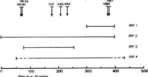
Figure1indicates thepositions ofthemutationspresentin
the sequencesofthesevenantigenicvariantson alinear map
ofproteinE. Thevariants selectedby domainAMAbs(A3,
A4, AS) had mutationsspacedfairly distant fromeach other
VA3a
VA3b Vi2 VA5 VA4
T,T
VB1
VB4
i IRF 1
IRF 2 IRF 3 IRF4
I
-O 100 200 300 400 500
Residue Ntimber
FIG. 1. Location of four IRFs(IRF1toIRF4)and themutations presentinsevenantigenicvariants of TBE virus(VA3a,VA3b,Vi2, VA4,VA5,VB1, VB4)on alinear mapofprotein E. Thelocations of IRF1, IRF2, and IRF3 were determined by amino-terminal
sequencing.Theambiguityof theexactlocation of IRF4 is indicated
bybrokenlinesattheends of thisfragment. Thearrowsdepictthe
positions ofthemutations present in therespective antigenic
vari-ants. Dataonthevariants and IRFsaresummarized in Tables 1to 3.
on November 10, 2019 by guest
http://jvi.asm.org/
[image:2.612.314.553.515.640.2]a
la
l IRF 1
i ...i
b
Om E
IRF2
c
iu:
I
IN IRF3
d
.a...
._0
E
'RF4
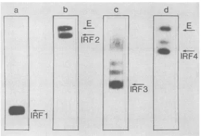
FIG. 2. Immunoblots of fragments of proteinE. ProteinE
frag-ments were separated on SDS gels, blotted onto nitrocellulose paper,andreacted with '25I-labeled MAbsasdescribed inMaterials andMethods. Lanea,24-htryptic digestreacted with MAbBl; only IRF1is detectable.Laneb,30-min tryptic digest reactedwithMAb Al; IRF2 and undigested protein E are visible. Lane c, CNBr fragments reacted with MAb C2; besides IRF3,anumberoflarger
partial degradation products react with the MAb. Lane d,
a-Chymotryptic digest reacted with MAb Al; IRF4, undegraded proteinE,andanotherlargerdegradation productarevisible.
ontheprimarysequence.The MAbs B1 and B4 of domainB,
however, selected for mutations only five amino acidsapart.
Itseemsreasonabletoassumethatthe mutated amino acid is
itselfpartof the epitope recognizedby the selecting MAb,as
hadbeen shown by X-ray crystallography tobe thecasefor
variants of influenza virus (37). However, the possibility
cannot be ruled out that mutations at distant sites cause
conformational changes in the respective epitopes.
Immunoblotting of four IRFs of proteinE.Thereactivity of
fourdifferentfragments ofprotein E(designated
immunore-activefragment 1 [IRF1]toIRF4) with MAbswas analyzed
by immunoblotting. A representative immunoblot of each
fragment is shown in Fig. 2. IRF1wasobtainedby digestion
ofprotein Erosettes(19) with trypsin for 24 h. This fragment
migrates in SDS gelswithan apparent molecularmassof 9
kilodaltons (kDa) and has been characterized previously
(41). IRF2 is observedas anintermediarydegradation
prod-uctoftryptic digestion andmigrates correspondingtoasize
of 47kDa.
IRF3 isa19-kDafragment obtained by chemical
degrada-tion with cyanogen bromide that has been described
previ-ously (18). IRF4wasgeneratedbya-chymotryptic digestion
of SDS-denatured virus and migrated corresponding to a
molecular size of 38 kDa. Each of the four fragments was
tested for its reactivity with the whole panel of MAbs.
Resultsaresummarized inTable 2.IRF1 reactedexclusively
withalldomain B MAbs. IRF2 reacted with all MAbsexcept
A3, A4, A5, il, and i2. The epitopes recognized by MAbs
A3, A4, A5, and i2 are sensitive to denaturation by SDS
(Guirakhoo el al., in press), and therefore these MAbs are
not expected to react under the conditions of
immunoblot-ting. IRF3 was found to contain most of the epitopes of
domain C, and IRF4 reacted with the same MAbsasIRF2.
Localization of IRFs in protein E sequence. The amino
terminiof the fragments IRF1, IRF2, andIRF3 were
deter-mined, permitting us to localize exactly the positions of
thesefragmentsontheprimary sequenceof proteinE.Table
3 lists the amino-terminal sequences and the derived
se-quenceposition numbers of theamino- and carboxy-terminal
ends of these fragments. IRF1 starts atamino acid residue
301.Inagreementwiththe cleavage specificity of trypsin,a
[image:3.612.89.287.67.201.2]Lys residue is found at position 300. Judging from its
TABLE 2. ReactivityofMAbs withfragmentsofproteinE
Molecular Generatedby
Fragment size
clerage
by ReactiveMAbs'(kDa)a cleavage with:
IRF1 9 Trypsin Bl-B5
IRF2 47 Trypsin Al,A2,Bl-B5,
Cl-C6,i3
IRF3 19 CNBr Cl, C3C6
IRF4 38 a-Chymotrypsin Al,A2,Bl-B5,
Cl-C6,i3
a As determined from SDSgels.
b Thereactivity with MAbswasdeterminedbyaWesternblot
(immuno-blot) analysis (see Materials and Methods). MAbs recognizing SDS-sensitive
epitopesdonotreact.
apparentmolecularmassof 9kDa, IRF1isthoughttoextend
approximately
towardresidue 384.ClosesttothispositionisLys-395, whichis assumedto mostlikelyform the
carboxy
terminus ofIRF1 (Table 3). Thus, thecalculatedmolecular
massis 10.5 kDadespitethe apparentsize of 9 kDa derived
from SDS
gels.
The amino terminus of IRF2was determinedto be
iden-tical to that ofprotein E. From its molecular weight, it is
thoughtto extendto approximately residue 400.
The first residue of IRF3wasdeterminedtobeglycine78.
According to thecleavage specificity of CNBr, residue 77 of
TBE virus protein E is a methionine. From its molecular
weight, it is concluded thatIRF3 extends beyond the next
Metresidue(176) untilMet-252, which is assumed tobe its
carboxyterminus (Table3).
The amino terminus of IRF4 was not determined.
How-ever,someinformation about itspositioncanbederivedby
thefollowingconsiderations.Residue 389 is required for the
structural integrity of theepitope ofMAb Bi as shown
by
the mutation present in VB1. MAb B1 reacts with IRF4.
Consequently,IRF4 can be assumed to extend at itscarboxy
terminus at least until residue 389. As deduced from its
molecularweight,thisfragmentisthought to lack at least 50
amino acid residues at the amino terminus of protein E. Figure 1 shows the positions of the IRFs on a linear
representation ofprotein E. Thefollowingconclusions
con-cerningthe positions of antigenic epitopes and domainscan
be drawn. At least the first 50 amino-terminal and the 100
mostcarboxy-terminalaminoacid residuesarenot involved
intheepitopesrecognizedby MAbs Al, A2, B1 toB5, Clto
C6, and i3. The entire domain B is contained between
residues 301 and 395, whereas domain C is located
some-where between residues 78 and 252. Thus, the antigenic
domains B and Ccorrespondtotwodistinctproteindomains
which arebuiltup bynonoverlappingsections of theprimary
sequenceof protein E. Since MAbs Al and A2 do notreact
[image:3.612.323.560.85.184.2]with either IRF1 or IRF3, but react with IRF2 and IRF4,
TABLE 3. Localization ofIRFs of protein E
Amino Carboxy
Fragment Amino-terminalsequencea terminus terminusc
(position)b (position)
IRF1 Gly-Leu-Thr-Tyr-Thr-Met Gly (301) Lys(395)
IRF2 Ser-Arg-?-Thr-His-Leu-Glu Ser(1) Approx. residue 400 IRF3 Gly-Pro-Ala-Thr-Leu-Ala Gly (78) Met(252)
aDeterminedby gas-phasesequencing.
b Position numbersarecounted from thefirst amino acid ofproteinE.
cAs deduced from molecularweight (see text).
on November 10, 2019 by guest
http://jvi.asm.org/
[image:3.612.323.560.625.696.2]2f.V V !'W V'V1
100 200 300 400 500 Residue Number
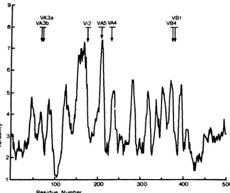
FIG. 3. Variability profile offlavivirusprotein E. Theaminoacid
sequences of 15 flaviviral E proteins were aligned, the number of
different amino acid residueswerecalculated for each position, and
these numberswereplottedas amovingaverageof9.Thus, avalue
of 1 represents 100% conservation, and 15 would be maximum variability. Thearrowsdepictthe positionsofthemutationspresent
in theseven antigenic variants ofTBEvirus.
they most likely involveamino acid residues between posi-tions 50 and 77or 253 and 300(Fig. 1).
The positions ofthe mutations present in antigenic vari-ants ofTBE virus are also indicated in Fig. 1 (see above).
The mutations of variants VB1 and VB4 map within the rangeofIRF1,which carries allepitopes ofdomainB.IRF3,
which carries domain C, also contains amino acid residues involved in the epitopes defined by MAbs A4, A5, and i2. The epitope of MAb A3 is located within the range of IRF2
andperhaps IRF4. MAbs A3, A4, A5, and i2 did not react
withanyof these fragmentsowingtothe SDS sensitivity of the corresponding epitopes (Guirakhoo etal., in press).
Sequence comparisons.Theaminoacid sequencesof theE
proteins of15 different flaviviruses were aligned for
maxi-mum homology, and a variability profile of the flavivirus
proteinE was calculated and plotted as described in
Mate-rials and Methods (Fig. 3). Consistent with previous obser-vations (33), it is obvious from Fig. 3 that the most
con-served region of protein E is located around residue 100. In fact, 12 of14amino acids from positions 98to111 are 100%
conserved amongallflavivirussequences, andateach ofthe othertwopositions twodifferentaminoacidresidues canbe
found. Two further regions that are significantly conserved
inprotein Eare located attheamino terminus (residues1 to
42)and close tothecarboxy terminus (residues 410to445). A hypervariable region extends from residues 145 to 170, and a second one is found from residues 200 to 215. Also included in Fig. 3are thepositions ofthe mutations present
in the antigenic variants of TBE virus, which are generally
located within variable regions.
T-celldeterminant. Peptides thatarecapable offormingan
amphipathic
ox-helix
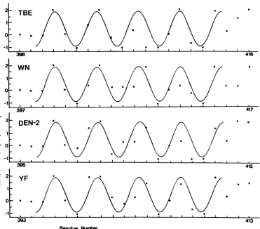
represent potential T-cell antigenic de-terminants (3). A computer scanning of the primaryse-quence of TBE virus protein E indicated only one region
longer than six amino acids with the characteristics of a
potential amphipathic a-helix. This sequence, extending
from residues 398 to 413, was further analyzed (Fig. 4).
Hydrophobicityvalues were plotted for each residue
accord-ing to the scale ofFauchdre andPligka (11), which had been
foundto performbestforthe identification of
immunodom-inant T-cell sites (6). Figure 4 shows that between residues
398 and 413, these hydrophobicity values are well
approxi-mated by a sinusoid with a period of 3.6 residues per cycle. Other flavivirus sequences exhibited a similar potential
T-cell determinant at exactly the same positions when
se-quenceswerealigned for maximumhomology(Fig. 4). Data
from the analyses of the protein E sequences of the viruses
DEN-1 and DEN-4, and Murray Valley encephalitis virus
and Japanese encephalitis virus, are not shown in Fig. 4,
since these results were very similar to those shown for
DEN-2 and WN virus, respectively.
DISCUSSION
The envelope (E) proteins of all flaviviruses sequenced so
far show perfect conservation of 12 cysteine residues which
form six disulfide bridges. In addition, they exhibit highly
homologous hydrophilicity profiles,suggesting that all
mem-bers of the flavivirus family share a fairly common
architec-ture of their E proteins. The assignment ofdisulfide bridges
for WN virus by Nowak and Wengler (29) was a major
breakthrough toward the elucidation of the protein E
struc-ture. Based on these analyses, it was proposed that protein
E was made up of three major regions (Ri to R3) separated
by two intervening sections (Li and L2), as well as two
transmembrane segments at the carboxy terminus. The
model of Nowak and Wengler (29) also included the
loca-tions of potential antigenic sites as deduced from the
hydro-philicity profile of the protein. By using a direct approach,
including the analysis of antigenic variants and IRFs, we
were able to determine the location of structurally defined
antigenic determinants. The compilation of these data with
those on the location of disulfide bridges and the structural
and functionalcharacteristics of epitopes (Guirakhoo et al.,
in press) led to a model (Fig. 5) showing adifferent
arrange-ment of the polypeptide chain into distinct protein domains
that also correspond to the antigenic domains A, B, and C.
Domain A is shown as a discontinuous structural entity
composed of distant regions of the primary sequence,
spe-cifically amino acids 50 to 125 (containing three disulfide
bridges) and 200 to 250. This arrangement is based on a
number of considerations. The antigenic domain Ais defined
by five different MAbs (Ai to A5). Ofthese, MAbs A3 to A5
neutralize TBE virus and were used to select antigenic
variants. The mutations present in these variants mapped on
distant regions of the primary sequence. However, there is
strong evidence that theepitopesrecognized by these MAbs
are located on the same structural entity, since they share
the same structural properties (sensitivity to SDS and acid
pH) and since A3 and A4 as well as A4 and A5 mutually
block each other'sbinding (Guirakhoo et al., in press). The
followingconsiderations provide evidence that the epitopes
recognized by MAbs Al and A2 involve amino acidresidues
between positions 50 and 77. Ai and A2 are located on IRF2
and IRF4, but not on IRF1 and IRF3 (compare Fig. 1 and
Table 2), indicating that they involve any of either amino
acids 50 to 77 or 252 to 300. MAbs A2and A3mutually block
each other's binding, and the mutation present in variants
VA3a and VA3b maps to positions 67 and 71. The epitopes
recognized by Ai and A2 are not destroyed by SDS
treat-ment but are sensitive to reduction by 2-mercaptoethanol,
suggesting that these epitopes are part ofa protein domain
stabilized by disulfide bridges. Such a disulfide-stabilized
on November 10, 2019 by guest
http://jvi.asm.org/
[image:4.612.63.295.72.267.2]0 0
395 415
YF
393 413
Residue Nuber
FIG. 4. Flavivirus protein E sequences that potentially form amphipathic a-helices. The amino acid sequences of four flaviviruses (belongingtofour differentserologicalsubgroups)arealigned for maximumhomology.Thehydrophobicityvalueof each amino acid residue between theindicated borders is plotted accordingtothescale ofFauchdre andPligka(11).Asinusoidwith aperiodof 3.6residues percycle approximates thehydrophobicity plots,indicatingthepotential of theseregionstoformanamphipathica-helicalstructure.
region oftheprotein islocated betweenresidues60 and121,
whichincludes as many asthreedisulfidebridges.
This disulfide-specified region also contains the most
conserved sequence (residues 98to 111) ofprotein E
(indi-catedby trianglesinFig.5), suggestingacrucially important
function ofthis sequence element. It represents a
moder-ately hydrophobic sequence surrounded by a hydrophilic and variable sequence environment. This isreminiscent of
thereceptor-binding areaof influenzavirusthat wasshown
by X-ray crystallography to be a hydrophobic pocket
sur-rounded by variable surface-exposed sequence elements which are recognized by neutralizing antibodies (40). One maytherefore hypothesize that the conservedpeptide98 to
111is part of the flavivirus receptor-binding site.
Domain B is shown as a distinct protein region located
betweenresidues 301 and 395. Its bordersaredefinedby the
end of thetrypticfragmentIRF1 (indicatedby tryp in Fig. 5).
This protease-resistant fragment contains all epitopes of
domainB, suggesting that no other parts of the polypeptide
chain are involved in this structural domain. Domain B
contains one disulfide bridge and apparently represents an
independentlyfolding protein domain with a strong tendency
for renaturation (41). As shown previously, its structural
integrity can be destroyed by opening the disulfide bridge,
butitfullyregains its antigenic reactivity when renaturation
of the disulfide bridge is allowed to occur (41). Domain B
seems to be strongly folded and stabilized by its single
disulfide bond andprobably additional intramolecular forces
as suggested by the presence of several potential tryptic
cleavage sites (small arrows in Fig. 5) which are apparently inaccessible in the native form of this domain. The
involve-ment ofthecarboxy-terminal part of IRF1 in the antigenic
domain B is demonstrated by the mutations present in the
antigenic variants VB1 and VB4.
Antigenic domain C is shown as a single highly variable
loop which does not contain any disulfide bridges, but
carries thesingle carbohydrate side chain of the TBE virus E
protein (solid diamond inFig. 5), sinceIRF1,containingthe
second potentialN-glycosylation site, was showntobe not
glycosylated (41). The location of domain C, which is
contained within the CNBrfragment IRF3 (bordersdepicted
by CNBr in Fig. 5),was deduced asfollows. The mutation
present in variants VA4, VA5, and Vi2 defines areas of
fragment IRF3 that do not belong to domain C, since the
epitopes A4, A5, and i2 do notoverlap with any domain C
epitope as demonstrated by competitive binding assays.
Second, reduction andcarboxymethylationdonotaffect the
structuralintegrity of domain C epitopes,indicating thatno
disulfide bond is involved in the integrity of this structural
entity. Third, domain C is resistant to SDS denaturation,
suggesting that it is notcomposed of discontinuous regions
of the protein like those of domain A. However, this
denaturation resistance is lost when the carbohydrate side
chain is removed(Guirakhooetal., in press),suggestingthat
on November 10, 2019 by guest
http://jvi.asm.org/
[image:5.612.131.502.75.401.2]A
*A4
SLE? >YF?
T8o<\i~~~~~~~~2
tryp-B 3BBTBE B
MVE SLE JE DEN
COOH
450
-FIG. 5. Model of the TBE virusproteinE.Opencirclesrepresenthydrophilicamino acid residues (Arg, Lys, Asn,tisp, Gln, Glu, His), dotted circles showintermediate amino acid residues(Pro, Tyr, Ser, Trp, Thr, Gly), and solidcirclesshow hydrophobicaminoacidresidues (Ile, Val, Leu, Phe, Cys, Met, Ala). Amino acidswereclassifiedby thescaleof KyteandDoolittle (22). Positionnumbers are shown every 50amino acids.Cysteine residues forming disulfide bridgesareconnected by solid lines.Arrowsdepict cleavage sitesthatliberateIRF1 (tryp) and IRF3(CNBr), respectively. Smallarrowsindicate potential cleavage sites within these fragments that are not utilized. Two solid lines stand for thelipid membrane thatisspanned by two transmembrane regions of protein E. Thepolypeptide chain is folded to indicate the antigenic domainsA, B,andC, which are designated by large capital letters. Arrows together with the names of neutralizing MAbsdepict thelocations ofthe mutations identified in the respectiveantigenic variants ofTBEvirus by sequenceanalysis. Aline ofsolid triangles indicates the almost perfectly conserved sequence within domain A. A line of open triangles marks the region of a potential T-cell determinant. Asolid diamondrepresents thecarbohydrate side chain of TBE virus. The MurrayValley (MVE), St.Louis(SLE)andJapanese (JE)encephalitisviruses and DEN viruses havepotentialN-glycosylationsites at thehomologousposition. Yellow fever(YF)and St. Louis encephalitis viruseshave such a site within domain B, DEN viruses within domain A. Thehomologouspositionsof TBE virus are shownby open diamonds.
on November 10, 2019 by guest
http://jvi.asm.org/
[image:6.612.157.463.82.594.2]glycosylation
stabilizes domainC,
although
domain Cepitopes
donotdirectly depend
onthe presence of the sugarresidues.
Last,
MAbs directedagainst
domain Cepitopes
exhibit very little
cross-reactivity (MAbs
C3to C6are evensubtype
specific),
suggesting
the involvement ofhypervari-able amino acid
residues,
such as those betweenpositions
145 and 170
(Fig. 3).
Three MAbs
(il
toi3)
recognize
epitopes
that do notoverlap
with anyofthe threeantigenic
domainsA,
B,
orC.Epitope
i2is definedby
themutationpresentinvariant Vi2. Intheprotein
Emodel,
i2 isplaced
between the threeprotein
domains
A, B,
and C. This intermediate location of i2 iscompatible
with thefindings
that(i)
i2 shares a numberofstructural characteristics with domain A
epitopes,
such assensitivity
toSDSand acidpH,
and(ii)
binding
ofMAbi2isunidirectionally
blockedby
MAbsB4,
B5,
and Cl.Thefirst50aminoacidscouldnotbe
assigned
toanyofthethree
antigenic domains,
butpreliminary
evidence fromprotein expression
studies(W.
Tuma etal., manuscript
inpreparation)
locatesepitope
il within this amino-terminalregion.
The modelalsoreflectsthe
finding
that mostof the B-celldeterminants
described,
especially
thoseof domains A andB,
are conformationdependent
andprobably
evendiscon-tinuous. This is consistentwiththe viewderivedfrom
X-ray
crystallographic
studies that themajority
of B-celldetermi-nants
depend
onthetertiary folding
ofthepolypeptide
chain(1,
2).
In a recent
study, Wengler
et al.(39)
identified twosurface-exposed
regions
of WN virusprotein
Eby
theiraccessibility
toproteolytic cleavage which,
ingood
agree-ment with our
model, correspond
to thecarboxy-terminal
endof domain B andasection of domain
C,
respectively.
Bysequence
analysis
of neutralization escapemutants,Lobigs
et al.
(24)
localized a neutralizationepitope
on the yellow fever virusprotein
E sequence. The mutations present intheir
antigenic
variantsmapped
atamino acidposition
71 or72,
whichperfectly
corresponds
to the mutatedposition
ofthe TBE virus variantVA3a.
By
adifferentapproach
involv-ing
bacterialexpression,
Masonetal. (25,26) identifiedtwoantigenic
domains ofprotein
E. One of these domains iscontained between residues 280 and 414 and was found to
depend structurally
on adisulfide bond(P. Mason,
personalcommunication).
This result agrees very well with thede-scription
of domainBin the TBE virusprotein
Emodel. Theotherdomain maps between residues 76 and93,
correspond-ing
towhatwe identifiedto be part of domain A.Besides these
B-cell-specified epitopes,
apotentialamphy-pathic
ot-helixwhichmight
serve as aT-celldeterminantwasidentified
by primary
sequence analysis and is depicted inthe model in
Fig.
5by
aline of opentriangles.
It is locatedadjacent
tothecarboxy
terminus offragment
IRF1 and doesnot
overlap
with any of the three B-cellantigenic
domains.The model furtherindicates theintramembranouspartsof
the
protein,
i.e.,
thehydrophobic
anchor ofprotein Eand thesignal
sequenceofprotein NS1,
whichareinterrupted byashort
hydrophilic
protein
section. The first transmembranesegment
(residues
448 to 469) includes a very hydrophilicamino acid
(Lys,
Arg,orGln)
at apositionconserved amongallflaviviruses
(number
457 in theTBEprotein Esequence),causing
controversialpredictions
of the actuallengthof thisanchor sequence
(5, 7,
9, 12,27, 28,
32, 36, 38, 42). If thehydrophilic
residuewasnotplaced
within thelipid envelope,thetransmembrane segment would be only 12 amino acids
long,
although
such elements usually consist of 20 to 25hydrophobic
amino acidswhich,
folded as anac-helix,
arerequiredtospan the membrane and provideastable
associ-ation (8). If it is a part of the transmembrane region, the
hydrophilic residue might be involved in some kind of
protein aggregation andthus be protected from the
hydro-phobicenvironment. Alternatively,the amino acidmightbemodifiedby the addition ofafatty acid side chain(30).
The carboxy terminus of WN virusprotein E was
deter-minedby Nowaket al. (T. Nowak,P. Farber, G.
Wengler,
and G. Wengler, Virology, in press) to be the residue
precedingthecleavagesite thatliberates the amino terminus
of protein NS1. This cleavage is probably catalyzed by a
signalase in the lumen of the endoplasmic reticulum.
There-fore, the carboxy terminus of protein E is shown to be
exposedatthe outersurface of the viral membrane.
Owingtotheexpectedoverall structuralconservation,the
model presented here should be useful as a basis for the
further analysis of the antigenic structure and functional
activity of flavivirusprotein E.
ACKNOWLEDGMENTS
Wethank Hans Wolf and Susanne Modrow forallowingustouse their computerfacilitiesatthePettenkofer Institut(Munich,Federal Republic of Germany) and for providing programs for theprediction ofantigenic determinants. We further acknowledge Melby Wilfin-ger, Angela Dohnal, and Walter Holzer for their expert technical assistance,aswellasSusanne Pfauser fortyping themanuscript.
LITERATURE CITED
1. Barlow, D. J., M. S. Edwards, and J. M. Thornton. 1986. Continuous and dicontinuous protein antigenic determinants. Nature(London)322:747-748.
2. Berzofsky,J.A. 1985. Intrinsic and extrinsic factors inprotein antigenic structure. Science 229:932-940.
3. Berzofsky,J. A., K. B. Cease, J. L. Cornette, J. L. Spouge, H. Margalit, I. J. Berkower, M. F. Good, L. H. Miller, and C. DeLisi.1987. Proteinantigenicstructuresrecognized byT-cells: potential applications to vaccine design. Immunol. Rev. 98: 9-52.
4. Brinton, A. B. 1986. Replicationofflaviviruses, p.327-374.In S. Schlesinger and M. J. Schlesinger (ed.), The Togaviridae and Flaviviridae. PlenumPublishing Corp.,New York.
5. Coia, G., M. D. Parker, G. Speight, M. E. Byrne, and E. G. Westaway. 1988. Nucleotide and complete amino acid se-quences ofKunjin virus: definitive gene order and characteris-tics of thevirus-specified proteins. J. Gen. Virol. 96:1-21. 6. Cornette, J. L., K. B. Cease, H. Margalit, J. L. Spouge, J. A.
Berzofsky, and C. DeLisi. 1987. Hydrophobicity scales and computational techniquesfordetecting amphipathic structures
inproteins.J. Mol. Biol. 195:659-685.
7. Dalgarno,L.,D. W. Trent,J. H.Strauss, andC. M. Rice.1986. Partial nucleotide sequenceofthe Murray Valley encephalitis virus genome. J. Mol. Biol. 187:309-323.
8. Davis, N.G.,and P. Model. 1985. Anartificialanchor domain: hydrophobicity suffices to stop transfer. Cell41:607-614. 9. Deube, V., R. M. Kinney, and D. W. Trent. 1986. Nucleotide
sequence and deduced amino acid sequence of the structural proteins ofdengue type 2 virus, Jamaica genotype. Virology 155:365-377.
10. Eisenberg, D., R. M.Weiss, andT. C. Terwillinger. 1984. The hydrophobic momentdetects periodicity in protein hydropho-bicity.Proc.Natl. Acad. Sci. USA81:140-144.
11. Fauchere, J. L.,and V.Pligka.1983.Hydrophobic parameters pi ofamino-acid side chains from the partitioning of N-acetyl-amino-acid amides. Eur. J. Med.Chem. 18:369-375.
12. Gruenberg, A., W. S. Woo, A. Biedrycka, and P. J. Wright. 1988. Partial nucleotide sequence and deduced amino acid sequence of the structuralproteinsofdengue virus type 2, New Guinea C and PUO-218 strains.J. Gen. Virol. 69:1391-1398. 13. Gubler, U.,and B.J. Hoffman. 1983. A simple and very efficient
method forgeneratingcDNAlibraries. Gene25:263-269.
on November 10, 2019 by guest
http://jvi.asm.org/
14. Hahn, C. S., J. M. Dalrymple, J. H. Strauss, and C. M. Rice. 1987. Comparison of thevirulent Asibi strainofyellow fever virus withthe 17D vaccinestrain derived from it. Proc. Natl. Acad. Sci. USA 84:2019-2023.
15. Hahn, C. S., R. Galler,T. Hunkapiller, J. M. Dalrymple, J. H. Strauss,andE. Strauss. 1988.Nucleotide sequenceofdengue 2 RNA and comparison ofthe encoded proteins with those of otherflaviviruses. Virology 161:167-180.
16. Heinz, F. X.1986.Epitope mapping of flavivirus glycoproteins. Adv.VirusRes. 31:103-186.
17. Heinz, F. X., R. Berger, W. Tuma, and C. Kunz. 1983. A topological and functional model of epitopesonthe structural glycoprotein oftick-borne encephalitis virus defined by mono-clonal antibodies.Virology126:525-537.
18. Heinz, F. X., R. Berger,W. Tuma, and C. Kunz. 1983. Location of immunodominant antigenic determinantsonfragments of the tick-borne encephalitis virus glycoprotein: evidence for two different mechanisms by which antibodies mediate neutraliza-tion and hemagglutinaneutraliza-tion inhibineutraliza-tion. Virology130:485-501. 19. Heinz, F. X., and C. Kunz. 1980. Formation of polymeric
glycoprotein complexes fromaflavivirus: tick-borne encephali-tisvirus. J.Gen. Virol.49:125-132.
20. Heinz, F. X., and C. Kunz.1981.Homogeneity of the structural glycoprotein fromEuropeanisolates oftick-borne encephalitis virus: comparison with other flaviviruses. J. Gen. Virol. 57: 263-274.
21. Heinz, F. X., W. Tuma,F.Guirakhoo,R. Berger, and C. Kunz. 1984.Immunogenicity of tick-borne encephalitis virus glycopro-tein fragments: epitope-specific analysis of the antibody re-sponse. Virology 65:1921-1929.
22. Kyte, J., and R. F. Doolittle. 1982. A simple method for displayingthehydropathic character ofaprotein.J. Mol.Biol. 157:105-132.
23. Laemmli, U. K., and M.Favre.1973. Maturation of the head of bacteriophage T4. I. DNA packing events. J. Mol. Biol. 80: 575-599.
24. Lobigs, M., L.Dalgarno,J. J. Schlesinger, and R. C. Weir. 1987. Location ofa neutralization determinant in the E protein of yellow fever virus(17Dvaccine strain). Virology161:474-478. 24a.Mandl,C. W., F. X.Heinz, and C. Kunz. 1988.Sequence of the
structural proteins of tick-borne encephalitis virus (Western subtype)andcomparative analysis with other flaviviruses. Vi-rology 166:197-205.
25. Mason, P.W.,T. L.Mason, and M. J.Fournier. 1988.Current approaches forthedevelopmentofdenguevaccines and related aspects of themolecularbiology of flaviviruses. J. Infect. Dis. 157:1105-1111.
26. Mason, P.W.,P.C. McAda, J. M. Dalrymple, M. J. Fournier, and T. L. Mason. 1987. Expression of Japanese encephalitis virus antigens in Escherichia coli.Virology 158:361-372. 27. Mason, P. W., P. C. McAda, T. L. Mason, and M. J. Fournier.
1987. Sequence of the dengue-1 virus genome in the region encoding the three structural proteins and the major non-structuralprotein NS1.Virology 161:262-267.
28. McAda, P. C., P. W. Mason, C. S.Schmaljohn,J. M. Dalrymple, T. L. Mason, and M. J. Fournier. 1987. Partial nucleotide
sequenceof the Japanese encephalitis virus genome. Virology 158:348-360.
29. Nowak, T., and G. Wengler. 1987.Analysis ofdisulfidespresent inthemembrane proteinsof theWestNile flavivirus.Virology 156:127-137.
30. Pillai, S., and D. Baltimore. 1987. Myristoylation and the post-translational acquisition of hydrophobicity by the mem-brane immunoglobulin heavy-chain polypeptide in B lympho-cytes. Proc. Natl. Acad. Sci. USA84:7654-7658.
31. Pletnev, A. G., V. F. Yamshchikow, and V. M. Blinov. 1986. Tick-borne encephalitis virusgenome.FEBSLett.200:317-321. 32. Rice, C. M., E. M. Lenches, S. R. Eddy, S. J. Shin, R. L.Sheets, andJ. H. Strauss. 1985. Nucleotide sequence ofyellow fever virus: implications for flavivirusgeneexpressionandevolution. Science229:726-733.
33. Rice, C.M.,E.G. Strauss, and J. H. Strauss.1986. Structure of theflavivirusgenome, p. 279-320.InS. Schlesinger andM.J. Schlesinger (ed.), The Togaviridae and Flaviviridae. Plenum PublishingCorp., NewYork.
34. Sanger, F., S. Nicklen, and A. R. Coulson. 1977. DNA sequenc-ing with chain-terminating inhibitors. Proc. Natl. Acad. Sci. USA74:5463-5467.
35. Sumiyoshi,H., K.Morita,C.Mori, I. Fuke, T.Shiba,Y.Sakaki, and A. Igarashi. 1986. Sequence of 3000 nucleotides atthe 5' endof Japaneseencephalitis virusRNA.Gene 48:195-201. 36. Trent, D. W., R. M.Kinney,B.J. B. Johnson, A. V.Vorndam,
J. A.Grant, V. Deubel, C. M. Rice, and C. Hahn. 1987. Partial nucleotide sequence of St. Louis encephalitis virus RNA: structuralproteins, NS1, ns2a, and ns2b. Virology 156:293-304. 37. Webster, R.G., G. M. Air, D. W. Metzger, P. M.Colman, J.N. Varghese, A. T. Baker, and A. G. Laver. 1987. Antigenic structureand variation inaninfluenza virusN9neuraminidase. J. Virol. 61:2910-2916.
38. Wengler,G.,E.Castle, U. Leidner, T. Nowak, andG. Wengler. 1985. Sequence analysis ofthe membrane protein V3 of the flavivirusWestNile virusandofitsgene.Virology 147:264-274. 39. Wengler, G., G. Wengler, T. Nowak, and K. Wahn. 1987. Analysis ofthe influence of proteolytic cleavage onthe struc-turalorganization of the surface of theWestNile flavivirus leads totheisolation ofaprotease-resistantEprotein oligomer from theviral surface. Virology 160:210-219.
40. Wiley,D.C.,andJ. J.Skehel.1987. Thestructureandfunction ofthehemagglutinin membrane glycoprotein ofinfluenza virus. Annu. Rev.Biochem. 56:365-394.
41. Winkler, G., F. X. Heinz, and C. Kunz. 1987.Characterization ofa disulfidebridge-stabilized antigenic domain of tick-borne encephalitis virus structural glycoprotein. J. Gen. Virol. 68: 2239-2244.
42. Zhao, B., E. Mackow, A. Buckler-White, L. Markoff, R. M. Cannock, C.-J. Lai, and Y. Makino. 1986. Cloning full-length denguetype 4 viral DNA sequences: analysis ofgenescoding for structuralproteins. Virology 155:77-88.
43. Zimmern, D., and P. Kaesberg. 1978. 3'-terminal nucleotide sequence ofencephalomyocarditis virus RNA determined by reverse transcriptase and chain-terminating inhibitors. Proc. Natl.Acad. Sci.USA75:4257-4261.