0022-538X/89/114882-08$02.00/0
Copyright X) 1989, American Society for Microbiology
Spatial and Temporal Distribution of Bovine
Herpesvirus
1Transcripts
URS V. WIRTH, KRISTIN GUNKEL,MONIKA ENGELS,AND MARTIN SCHWYZER* Institut fur Virologie der Universitat Zurich, Winterthurerstrasse 266a, 8057 Zurich, Switzerland
Received17 May 1989/Accepted 26 July 1989
Northern (RNA) blot analysis was used to determine the spatial and temporal distribution of bovine herpesvirus 1 (BHV-1) transcripts.Total RNAwasisolatedfromMadin-Darby bovinekidneycells whichhad been infected with BHV-1.2b strain K22or BHV-l.1 strain Jura in the presence or absence ofmetabolic inhibitors. Cloned restriction fragments representingthe entiregenomeof strain K22werelabeled with32Pand hybridizedtoimmobilizedRNA. A total of 54 BHV-1transcriptswerefound, ranginginsize from 0.4 tolarger than 8 kilobases(kb). The inverted repeat regionsand an adjacent segmentof theunique large partofthe BHV-1genomeencodedthree majorimmediate-early (IE) transcripts andoneminorIEtranscriptenriched after cycloheximide treatment of infected cells. Late transcripts were identified by drastically reduced abundanceafter cytosinearabinoside (araC) treatment. Twelve latetranscriptswere encodedmainly by the unique longgenomeregion,withacluster of fourtranscriptslocatedonHindIIIfragmentK(mapunits0.677 to 0.733). The 21 transcripts unaffected by araC treatment were defined as early; they showed dispersed locationsover thewholegenome, withaclusterontheuniqueshortsequence. The 17 remaining transcripts couldnotbe classifiedunambiguouslyasearlyorlatebythesetechniques. The IEtranscriptwithasize of4.2 kb exhibitedhomologywith thesingleIEgeneofpseudorabies virus,and theIEtranscriptwithasize of2.9 kbwasencodedin partby thegenomeregionknown to betranscriptionally activeduringlatency.
Bovine herpesvirus1(BHV-1) isanimportantpathogenof cattle which causes severe respiratory tract infections (in-fectious bovine rhinotracheitis) as well as harmless
infec-tions ofthegenital tract(infectious pustular vulvovaginitis) (forreviews, see references 20 and46).
Theviralgenomeis alinear double-stranded DNA
mole-cule with a length of approximately 140 kilobases (kb) composed of a unique long segment (UL; 105 kb) and a
unique short segment (Us; 11 kb); the latter is flanked by internalrepeat(IR)and terminalrepeat(TR) sequences of 12 kb each. The repeats enable the
Us
component to invert, resulting intwoisomericforms of the BHV-1 genome (23).Figure 1 shows the prototype arrangement ofthe BHV-1 genome(10). On the basis ofits genome structure, BHV-1 hasbeenclassified together withequine herpesvirus 1
(EHV-1), pseudorabies virus (PRV), and varicella-zoster virus (VZV) as group D (36) or class 2 (16). Herpes simplex virus type 1(HSV-1),with two pairs of repeats, has been classified asgroup Eorclass 3.
Like other herpesviruses (37), BHV-1 exhibits regulation
of viralprotein synthesisinatemporal cascade reported first
by Misra etal. (26), who identified 40 to 48
electrophoreti-cally distinct proteins andclassified four as immediate early (IE) and six as late. Similar experiments carried out by Metzleretal. (24, 25) showed 43 and, more recently (A. E.
Metzler, habilitation thesis, University of Zurich, Zurich,
Switzerland), at least 53 electrophoretically distinct pro-teins.Amajor 180-kilodalton (kDa) IE protein, six minor IE
proteins (170, 135, 98, 94, 46, and 35 kDa), and at least six late proteins (260, 180, 87, 68, 22, and 20 kDa) were
identified.
The IE proteins are known as important regulatory
pro-teins forthe productive cycle of herpesvirus replication (11) and mayalso play a role in latent infection (38). Recently, IE
proteinsofHSV-1 were shown to be recognized by cytotoxic
*Correspondingauthor.
Tlymphocytes (22) and may be able to stimulateprotective immunity,asshown earlier for murinecytomegalovirus (31). EHV-1 DNA encodes a single IEtranscript of 6 kb. Surpris-ingly, four electrophoretically distinct, structurally related IEproteinswereidentified(8, 13, 34). Similarly, PRV DNA encodes asingle 5.1-kb IE transcript (7, 42), and in addition tothemajor IE protein (17),twootherIEproteins have been found (12). In contrast, HSV-1 DNA encodes five different IE transcripts and five corresponding IE proteins (43). A transcription map for BHV-1 isnotavailable, and nothing is known about the number, sizes, and locations of the IE transcripts that encode the fourto sevenelectrophoretically distinct IEproteins. Ample information is availableonearly and late BHV-1 gene products (Fig. 1C), inparticular, the
glycoproteins responsible for immune responses during in vivo infections, and therefore they are interesting for vac-cine development (2, 41). Transcription maps have been establishedfor HSV-1 (43), EHV-1(14), and VZV (28, 32). Asabasisforacomparisonofexistingdata about BHV-1 geneproducts, aswellasforacomparison ofthe transcrip-tionally active region during latency (35) with transcripts present during productive infection, we determined the
spatialandtemporal distribution of 54BHV-1transcriptsby Northern (RNA) blot analysis. The transcript sizes ranged
from0.4tolargerthan 8 kb.Ofthese 54transcripts,4 were classified as IE with the protein synthesis inhibitor cyclo-heximide. With the DNA synthesis inhibitor cytosine ara-binoside (araC), 12 transcripts were clearly determined as late and 21wereclassifiedasearly, whereas the 17remaining transcriptscould not be classifiedunambiguously as early or late.
MATERIALSANDMETHODS
Virusand cellculture. Thevirus strains used in this study were BHV-1.1 strain Jura (25) and BHV-1.2b strain K22
(18). Madin-Darby bovine kidney cells were maintained in 4882
on November 10, 2019 by guest
http://jvi.asm.org/
BHV-1 TRANSCRIPTION MAP 4883
mapunits 0;1 0.2 0.3 0.4 0.5 0.6 0.7 0.8 0.9 I
I -.
A genome (1 40kb)
B clned N N I
framonts |I I I
UL
0 G
II:0 A I b
I-I
Iml,j.s
K LIII
I#HindlIl *E.oRl
C genes gill PolMDB 9I TKghVP4 gegi gx
glV
FIG. 1. Map of the BHV-1 genome. (A) UL and Us, and IR and TR short repeat regions are indicated. (B) Cloned Hindlll or
HindIII-EcoRI restriction endonuclease fragments of BHV-1 strain K22orstrain Jura for fragment C used in this study. (C) Genomic locations ofmapped BHV-1 proteins: Pol,homolog of DNA polymerase of HSV-1 (29);gI,homolog of gB of HSV-1 (19, 45); TK, thymidinekinase
of BHV-1(3);MDB, majorDNA-binding protein of BHV-1 (S. K. Bandyopadhyay, S. K. Mittal, and H. J. Field, personal communication); gh,homologtogHofHSV-1 (E. A. Petrovskis and E. L. Post, personal communication); glll, homolog of gC ofHSV-1;VP4,homologof VP4ofHSV-1;ge,homolog of gE ofHSV-1; gi, homolog ofgIof HSV-1;gx,homologof gX of HSV-1(T. Zamb, personal communication);
gIV, homologof gD ofHSV-1(G. Keil, personalcommunication). Eagle minimum essential medium supplemented with 10%
heat-inactivated fetal bovine serum.
Infection scheme. Subconfluent cell monolayers were in-fected with BHV-1 atamultiplicity of infection of10to 20 50%tissue culture infective dosespercellormockinfected. After viral adsorption for 1 h at 37°C, Eagle minimum essential medium with2% fetal bovineserum wasadded and the cells were incubated at 37°C until harvesting ofRNA within 2 to 8 h postinoculation (p.i.). Metabolic inhibitors
wereusedessentially asalready described (26). Toidentify
IE transcripts, cycloheximide (Sigma Chemical Co., St. Louis, Mo.) ata concentration of50 ,ug/ml for strain K22 wasadded with the inoculum and again after the adsorption period withthe medium; RNAwasisolatedat6 hp.i. Strain Jurarequired 100 ,ug/ml for complete inhibition of early and latetranscription. To identify late transcripts dependenton
DNAsynthesis, araC (Sigma)wasadded after adsorptionat aconcentration of50,ug/ml,asdescribed for protein studies of BHV-1 (26), or 100 ,ug/ml, and RNAwas isolated at8 h p.i. At both concentrations, [3H]thymidine (5.0 Ci/mmol; Amersham Corp., Amersham, Buckinghamshire, England) incorporation into acid-insoluble material between6and 8 h p.i. was reduced about 85% by araC treatment compared with untreated cells, and similar Northern blot resultswere obtained; thus, only results of experiments with the higher concentrationare shown.
Isolation of DNA and RNA. Cloned HindIII or
HindIII-EcoRIrestrictionfragments ofBHV-1.2b strain K22 (Fig. 1) describedpreviously (10)wereusedashybridization probes. Plasmids were isolated with Triton lysis buffer and CsCl gradientscontaining ethidium bromideasdescribed by Davis et al. (9). Total RNA was isolated by lysis of cells with guanidine isothiocyanate and centrifugation througha CsCl cushion as already described (9, 30). RNA concentrations
were determined spectrophotometrically; the integrity and
concentration of isolated RNAwereverifiedby agarosegel
electrophoresis. RNA preparations were stored as ethanol precipitates at-70°C.
Northern blot analysis. Northern blot analysis was
per-formed essentially aspreviously described (21), with minor modifications recommended by the manufacturer of the nylon membranes used (Biodyne A BNNG 3R; Pall Ultrafine Filtration Corp., Glen Cove, N.Y.). Portions (450 ,xg) of isolated RNApreparationswereheatedat55°C for 15 minin electrophoresis buffer (20 mM morpholinepropanesulfonic acid [MOPS], 5 mM sodiumacetate, 1 mM EDTA[pH7.0])
containing 50% (vol/vol) formamide and 6%(vol/vol)
form-aldehyde. After quick cooling on ice and addition of 5x loading buffer (15% Ficoll type400, 0.1%bromophenol blue
in 0.1 MEDTA[pH 7.5]),the RNAwasloaded intoa
single
slot(18cm) spanningthe whole width ofa1.2% agarosegel
in electrophoresis buffer containing 6% (vol/vol)
formalde-hyde. Insomeexperiments,agarosegelswith 22 slotswere used andSto 10,ugof total RNAwasloadedperslot.After
electrophoresis, the RNAwastransferredtoa nylon mem-branepresoaked in waterbycapillary action with 20x SSC (lx SSC is 0.15 M sodium chloride plus 0.015 M sodium
citrate [pH 7.0]) as the transfer medium. One edge of the
membranewas alignedwith theslottofacilitate subsequent
identification of the migration origin. After transfer, RNA was UVcross-linked to the membrane by exposition for 2 min on a C-51 mineral light transilluminator (Ultra-Violet
Products, Inc., San Gabriel, Calif.). The membrane was baked for 2 h at 80°C under vacuum and, for experiments
with single slots, cut into 4-mm strips which were marked with apencilat thebottom. Mobility of 28S and 18S rRNA was determined for each gel runby staining flanking strips with methylene blue. Sizes of bovine rRNAweredetermined as4.6 kbfor 28S and 2.0 kb for18Sby comparison with the mobility ofratliverrRNA with a known size (27). Sizes of
transcripts and rRNAs were additionally verified by using
RNA molecular weight markers with a ladder between 0.3 and 7.4 kb (Boehringer Mannheim Biochemicals, Mann-heim, Federal Republic of Germany). Plasmid DNA was labeled with [a-32P]dCTP (3,000 Ci/mmol; Amersham) by
nick translation to a specific activity ofapproximately 108
cpm/,Ig asdescribed by Rigbyetal. (33). Membrane strips
carrying different RNA preparations were prehybridized, hybridized, and washed in 50-ml Falcon tubes in a water
bath with constant agitation at 250 rpm. Prehybridization
was performed at42°C for 6 h with prehybridization buffer containing 5x SSPE (lx SSPE is 0.18 M NaCl, 10 mM sodiumphosphate, and 1 mM EDTA [pH 7.4]), 5x Denhardt solution(1x Denhardt solution is 0.2% bovine serum
albu-min, 0.02% Ficoll, and 0.02% polyvinylpyrrolidone), 50% formamide, 0.1% sodium dodecyl sulfate, and 0.2 mg of sonicatedandheat-denaturedcalfthymusDNAperml. For
hybridization,heat-denatured32P-labeledplasmid DNA(5x 10⁵cpm/cm2ofnylon membrane)wasaddedtofresh prehy-bridization buffer (0.1 ml/cm2) and incubated for 40 h at 42°C. After hybridization, strips were washed with 0.2% sodium dodecyl sulfate-5 mM sodium phosphate-1 mM EDTA(pH7.0)onceatroomtemperatureand twiceat42°C for1 h each time(1 ml/cm2). Wetstrips werearrangedina definedorderbetween sheetsof Saran Wrapand autoradio-graphedon FujiRX filmsatroomtemperature.
IRs TR&
, Us ;; ==: 1_
VOL.63, 1989
on November 10, 2019 by guest
http://jvi.asm.org/
[image:2.612.129.491.77.164.2]I2I 14 5 31 i~81 9I 11011I11121I13V, 1 1 21 21
14
1- 71I81I 9I1j
s11 121 131141 ,r:..- .i;:.1.
:. :N. ::.
40
0
I 40-4.2
4-2.9
41.6
hPi: 6 0 23 5 B 8 02 8
[image:3.612.318.563.82.268.2]cc ~~~~~aa1
FIG. 2. Northernblotexample showinghybridizationof labeled
fragment J (Fig. 1) to RNA isolated under different conditions.
Madin-Darby bovinekidneycellswereeithermock infected(-) or infected with BHV-1.2b strain K22(K)orBHV-1.1strain Jura(J). Total RNAwas isolatedatthe indicatedtimes(hoursp.i. [hpi])and blotted(10
p.g
perlane)asdescribed in Materials and Methods. As inhibitors(inh),cycloheximide (c)wasaddedat50p.g/ml
for strain K22orat 100p.g/ml
for strain Jura from the time of infection and araC (a) was added after the adsorption period at 100 p.g/ml. Autoradiographexposuretimewas1dayfor lanes1 to9and4days for lanes 10to14. RNAsize markers(M)areindicated ontheleft, and calculatedtranscript sizesare ontheright.RESULTS
Hybridization probes
derived from BHV-1.Figure
1 shows amap of the cloned Hindlllfragments (capital letters)
andHindlll-EcoRI
fragments (lowercase
letters) of the BHV-1.2b strain K22 used in thisstudy (10).
Recombinantplas-midswerelabeled and used for
hybridization
withoutremov-ing
the vectors. Controlexperiments
with vectors aloneproduced
nosignals
after 6days
ofautoradiographic
expo-sure with any of the RNApreparations
used(data
notshown).
The sameprobes
from strain K22 were used forhybridization
to BHV-1.1 strain Juratranscripts.
This was consideredlegitimate
because of thehigh degree
of homol-ogy(95%)
between the BHV-1.1 and BHV-1.2 strains (39).Identical results were obtained when the cloned HindIII
fragment
C of BHV-1.1 strain Jura was substituted for thecorresponding fragments
c' andj'
of strain K22 as aprobe.
EcoRIfragment
Fontheright-hand
endof thegenome(10)was notavailable as
cloned,
but this partof thesequence is coveredby
the IRpart ofHindlll-EcoRlfragment
c'.Strategy
used to establish the BHV-1transcription
map. Total RNA was isolated from cells infected with BHV-1(strain
K22 orJura) orfrom mock-infected cellsat different times between 2 and 8 hp.i.;
inparallel experiments,
metabolic inhibitors were used to
distinguish
the differenttemporal
classes oftranscripts.
Theprotein biosynthesis
inhibitor
cycloheximide
causes accumulation of IEtran-scripts,
and as a consequence,synthesis
ofearly
and latetranscripts
whichareexpressed only
in the presence ofIEproteins
isnotturnedon. The DNAsynthesis
inhibitoraraC should abolish latetranscripts
whichdepend
on viral DNAsynthesis
for efficient initiation. Afterelectrophoresis
in agarosegels containing formaldehyde,
RNAsamples
wereC c c c
probo:1 I' QP I
FIG. 3. BHV-1 IE transcriptsenriched bycycloheximide. Indi-cated labeled BHV-1fragments (c',j', L,QP, and1')were hybrid-izedto5p.gof total RNAperlanewhichwasisolatedat6hp.i.from cells infected with strain Jura(J)ormock-infected cells(-)treated with (c) or without (-) cycloheximide. Autoradiograph exposure timewas 10h. RNA size markers(M)areindicatedontheleft,and calculatedsizes of IE transcriptsare onthe right.
blotted on
nylon
membranes,hybridized
with the DNAprobes
described above, and detectedby
autoradiography.
As an
example, Fig.
2 showshybridization
offragment
J(mapunits 0.017to0.083)toselectedRNA
preparations
run on asingle
gel. LaneswithRNAofmock-infected cells with or without inhibitors, included in eachhybridization,
were blank like lanes 1,3, 9, and 10(oneexception
willbe shownbelow).Fordetermination of
transcripts
encodedby
BHV-1,only
bandsyielding
distinct andreproducible signals
in severalexperiments
withboth virus strainswereconsidered.Thus, six viral
transcripts
(1.2, 1.5, 1.8, 3.0, 3.9, and 6 kb) wereassigned
tofragment
J.Additionalbands,e.g.,those in lane 7, did not meet these criteria and were not included,although
somemayrepresent minor BHV-1transcripts.
The 1.2- and 1.8-kbtranscripts
encodedby fragment
Jappeared
very
early
in infection, at about 2 hp.i.
(lane 4), butthey
were notIE
transcripts,
because lane2 had nocorrespond-ing
signals
that arosefromtranscripts
whichwere enrichedby
cycloheximide
treatment (seeFig.
3). The othertran-scripts appeared
later ininfection, atabout 5 hp.i.
(lanes5 to 7); two of thesetranscripts,
with sizes of 3.0 and 6 kb,showed
drastically
reducedsignal intensity
after araC treat-ment (lane 8)compared
with untreated cells (lane 7) and were therefore defined as late.Transcripts
of strain Juragenerally appeared
much earlier and withhigher
abundance at agiven
time than thecorresponding
transcripts
of strain K22. Forexample,
the twotranscripts
with sizes of 1.2 and 1.8 kb gaveaclearsignal
at3hp.i.
for strain Jura(lane5)but were barely detectable at 3 hp.i.
for strain K22 (lane 12)despite
afourfold-increased exposure time.Apart
from thisgeneral delay
in the appearance oftranscripts, equivalent
results were obtained with both strains. Therefore,
only
Northern blots derived from strainJuraare shown below. IE transcripts of BHV-1.
Figure
3 shows RNA fromcycloheximide-treated
BHV-1-infected cellstogether
with control RNApreparations probed
withfragments
from theright-hand end of thegenome.
Cycloheximide
inhibited thesynthesis
ofearly and latetranscripts,
as demonstrated, forexample, by fragment
L, aregion encoding
abundanttran-40- 6
41-3. 9
* .4-12~~~~~~~~~~~'. k
:33.
on November 10, 2019 by guest
http://jvi.asm.org/
[image:3.612.63.302.83.276.2]BHV-1 TRANSCRIPTION MAP 4885 TABLE 1. Spatialandtemporal distribution ofBHV-1 transcripts 123 4 59 7 1 9
Transcript sizes (kb)'
N...j1.1.b1.7,c(30),bg (-7)dh J... 1.2,(1.5),c 1.8, (3.0),bg(3.9),(6)b
[image:4.612.337.549.80.269.2] [image:4.612.68.304.90.279.2]M... 2.7,' 4.5bj
...1.4,2.7,ik3.7
(4.5)b
O ... 1.7,cl3.7ck
k ... 1.7,cl(3.9), 8bm
g.(4.0),~~cn 8bm
g...(4.0), 8
G... (0.4),b1.7c 4O,cn(4.3),C(7.5)b
A... (1.4),c3.4,C 3.9,C(5)o
b'... (1.3),c(1.8), (3.8),c(5)O
ml ...(l.S),c(3.5), (4.0), (6-7)bP
K...o.5,b(1.1)9b(1.7), 2.5,c3.2,c4.1,b67bp
c...2.6,2.9,d4.2d(-7)qdh(>8)bq
...1*6,dr(2.9),s3.0,c'3.6,u 4.0,v(4.7)cw
(-7),dh (>8)bq
L...(1.0),cx(1.5), 1.8,c (2.1),cv 3.0,ct3.5,c
4.0,V(5.6)
QPf...1.0,cx2.1,CY(4.7)CW
1i...1.6,dr(2.9),s (3.6),u(4.7),Cw(-7),dh(>8)bq
aBold letters indicate transcripts of highestabundance, and parentheses
indicate those oflowest abundance. Fortranscripts with superscriptsgtoy,
those with thesamesuperscriptmaybe presumedtocrossfragment
bound-ariesortoarise from repeatedsequences.
bLatetranscriptdrastically reduced by araC.
Early transcriptnotsignificantly reduced by araCtreatment.
dImmediate-early transcript enriched by cycloheximidetreatment.
e1.8 kb forstrainK22.
fIdentical results forboth BHV-1fragments.
scripts in theabsence of cycloheximide (lane 7) but lacking virtually anydetectable signals in its presence (lane 9). All transcripts enriched by cycloheximide can therefore be interpreted as IE transcripts whose expression is indepen-dent ofprotein biosynthesis.
Fragment c' encodedtwomajor IEtranscripts of 4.2 and 2.9 kb (Fig. 3, lane 3). Fragment j' encoded one major IE transcript which exhibited a size of 1.6 kb for strain Jura (lane 6) and 1.8kb for strain K22 (datanotshown). Thiswas the only significant transcript size difference observed be-tween the two strains. Fragments j' and 1' gave rise to identical signals(lanes 6 and 15), indicating the main location of the 1.6-kb transcript on the repeats. RNA from mock-infected cells showed aweak broad signal coinciding with bovine28SrRNA,particularly when fragments
j',
l', and,tosomeextent,c'wereusedasprobes. This background signal
may be due to cross-hybridization of these regions of the
genomewithrRNAashas beenobserved for human
herpes-viruses (40). Therefore, the band that appearedataround 4.6 kb inlanes6 and 15wasnotinterpretedasanIEtranscript. In addition to the three major IE transcripts, a minor, sometimes diffuse bandwasregularly detectedatabout 7 kb with all three probes (c', j', and 1'; see also Fig. 6). All IE transcriptsexceptthe 7-kbspecies weredetected at2 hp.i. in cells not treated with cycloheximide (data not shown). Their signals were faint, and cycloheximide caused an estimated 10- to 30-fold enrichment. At 6 h p.i. in the absence ofcycloheximide, only a minor 2.6-kb transcript wasdetected withfragment c' (lane 1), whereasfragments j'
and 1'encodedseveralmajor transcriptsnotbelongingtothe IE class(lanes 4 and 13).
Other BHV-1 DNAfragments did notgive a signal with cycloheximide treatment, except for fragment N, which showed a 7-kb transcript asjust described. Complete data concerning IE transcripts are summarized in Table 1.
Latetranscriptsof BHV-1. For classification aslate tran-scripts, drastic reductionorabolishmentof the
correspond-'27~~~~~~~~~~~~~~~~~~~~~ ~6-_
401
*w --
w
~~~~1.7
1.
0.4 05
BHV-1: J J J J J J
1',. a a - a a - a a
probe: M G K
e.XL': A 4d 4 d
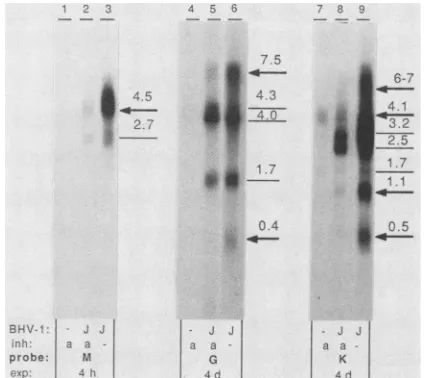
FIG. 4. ExamplesofBHV-1 late transcripts. Indicated labeled BHV-1fragments (M, G,andK)werehybridizedto 10,ugof total RNA perlane fromaraC-treated(a)oruntreated(-)cells infected with strain Jura (J) or mock-infected cells isolated at 8 h p.i. Identical strips were usedfor each condition. Sizes of transcripts
classifiedaslateareindicated byarrows,and othersareindicatedby
thinlines. Autoradiographexposuretime(exp)isindicated foreach probe in hours (h)ordays (d).
ing transcript by araC treatment was demanded. Figure 4 shows latetranscripts encoded by fragments M, G, and Kas
examples. Fragment M encoded twotranscripts with appar-ent sizes of 2.7 and 4.5 kb. Only the latter was reduced sufficiently by araC treatment to be interpreted as a late transcript. Of the five transcripts encoded by fragment G, the7.5-kbtranscriptwasdrastically reduced and the 0.4-kb transcriptwasabsent after araC treatment,justifying classi-ficationaslatetranscripts. Of theseventranscripts encoded by fragment K,twoweredrastically reduced (4.1 and 1.1 kb) and two were completely abolished (0.5 and 6to 7 kb) by araC treatment and therefore classified as late transcripts. Twelvetranscriptswereconsideredtofallinto thiscategory (Table 1).
Early transcriptsof BHV-1. Alltranscripts which did not meet the criteria discussed above for IE orlate transcripts were considered to be potential early transcripts. Tran-scripts whichwereonly slightly reduced by araCtreatment (e.g., the 1.7-kb transcript encoded byfragment G[Fig. 4])
or not reduced at all (e.g., the 4.0- and 4.3-kb transcripts encoded by fragment G [Fig. 4]) were classified as early. Twenty-one transcripts (Table 1) were placedin this
cate-gory.Theremaining17transcripts (Table 1)exhibited
inter-mediate sensitivity to araC and could not be classified unambiguouslyasearlyorlate.The 1.5-kbtranscriptinFig. 2, for example,wasclassifiedasearlyandthe 6-kbtranscript was definedaslate, whereas the 1.2- and 1.8-kbspeciesdid notfiteither category.
Figure 5 shows a particular RNA preparation obtained under conditions of early transcription (araC treatment), electrophoresed in a single gel, blotted, cut into identical strips, and hybridized with the entire set of BHV-1 DNA fragments. This approach is optimal for identifying tran-scripts which cross fragment boundaries. For reasons dis-cussedabove,IEtranscriptswithcontinuedexpressionuntil late times of infection(e.g.,the1.6-kbtranscriptencodedby fragments j' and 1') and late transcripts not completely abolished by araC treatment (e.g., the 4.5-kb transcript Probe
VOL.63, 1989
on November 10, 2019 by guest
http://jvi.asm.org/
N J M 0 k'g G A bmm K c J L P
I
5.3
-
28-^9
-r *-0
i,, r1 :^
I ..
[image:5.612.56.298.79.250.2]*
1
FIG. 5. Northern blot analysis of the indicated labeledBHV-1 fragments (N to
1')
hybridized to identical strips withRNA from cells infected with strain Jura and treated with araC(100 ,ug/ml). RNAsize markers are indicatedontheleft,and adeduced scaleis onthe right. Autoradiograph exposure time was4days forallstrips.encoded by fragment M) also appear in Fig. 5. Figure 5 conveys an overview ofthe great diversity ofBHV-1
tran-scripts with regardtosize,intensity, andspatial distribution.
Theintensities of transcript signals encoded by fragments J,
M, and I (mapunits 0.017 to0.188) andfragments j' and L (mapunits0.811 to 0.904)indicate thehighest abundances of transcripts encoded by these regions.
Examination ofthe RNAstrips from BHV-1-infectedcells
between2and8 hp.i. probed withallofthe cloned BHV-1
fragmentsrevealed that almost everytranscript followed its individual kinetics. These results are not presentedbut are
illustrated by Fig. 2.
Spatial and temporal distribution of BHV-1 transcripts. Table 1 shows a listof all transcriptsdeterminedinthisstudy andexpressed duringlytic infection of Madin-Darbybovine
kidneycells. Dataabout the number, sizesandlocations of
transcriptswerecollected by comparing andsummarizingall
oftheresultsrelating to temporal appearance(exemplifiedin
Fig. 2) and spatial distribution (exemplified in Fig. 5).
Transcripts with similar sizes encoded by the samefragment
atdifferent times(Fig. 2) were presumed to beidentical, and size values wereaveraged. Transcripts with similar sizes and appearances (e.g., diffuse or discrete bands) encoded by
neighboring fragments were presumed to cross fragment
boundaries.
Temporalclassificationof transcripts asIE, early, or late wasdone as long as it was unambiguous with respect to the
criteria discussed above. Three highly abundant transcripts thataccumulated duringcycloheximide treatment were
clas-sified as major IE transcripts. The minor 7-kb signal that arose from fragments c', j', and N may originate from a
single IE transcript detected by partially homologous se-quences within thesefragments (see Discussion). Generally, the IE transcripts were clustered around the IRs and TRs represented by fragments c', j', and
1'
(Fig. 1). Twelve transcripts were classified as late by araC treatment. Latetranscriptswere encoded mainly by the UL sequence of the genome; four ofthem were clustered on fragment K (map
units 0.677 to 0.733). Many of the smallest and largest transcripts belonged to the late class. Among the 38
tran-scripts which could not be classified as IE or late, 21 were considered early transcripts, whereas 17 transcripts
re-1 213141 61 7-181 SI
_
4.2w
j;, to
.4g-
2.92.1.6
-02
K'.--I
Bl -IV-1
pirobI
probe:
Il
IFIJI-C c'(HlSI IE-PRV
FIG. 6. Hybridization of indicated labeled DNAfragments C (c' andj'),c'(HS) (HindIII-SalI subfragmentoffragment c';map units 0.734 to0.748),and IE-PRV(BamHI fragment8ofPRV; 17)to5j±g of totalRNA(perlane) of cells infected with strain Jura(J)ormock infected and treated with (c) or without (-) cycloheximide and isolatedat6 hp.i. Autoradiograph exposure timewas4days. RNA size markers(M) areindicatedontheleft, and calculated sizes of transcriptsare on theright.
mainedunassigned. Theearly transcripts showeddispersed locationsoverthe entire genome, with a cluster on the
Us
genomeregion. Thus,atotalof 54transcripts, rangingin size
from0.4 tolargerthan 8kb, were mapped.
Furthercharacterization and localization oftwoBHV-1 IE
transcripts. A plasmid containingBamHIfragment 8 of the PRV IE protein-coding sequence (17) was labeled and
hy-bridized to blotted RNA from cycloheximide-treated cells.
This probe hybridized specifically with the 4.2-kb IE
tran-script ofBHV-1 encodedby fragmentc'(Fig.6, lanes 9 and 3). Partial sequence homology between IEgenes ofBHV-1 and PRV was also inferred from Southern and dot blots
exhibiting
cross-hybridization
atthe genomelevel(datanotshown). A HindIII-SalI subfragment (map units 0.734 to
0.748)of BHV-1fragmentc' had been shown to be transcrip-tionally active during latency in the BHV-1 rabbitmodelby
in situ nucleic acid hybridization (35). This HindIII-SalI fragmentwas subcloned, labeled, andhybridized toblotted RNAfromcycloheximide-treated cells. Theprobedetected the 2.9-kb IEtranscriptencodedbyfragmentc'(lanes6and 3). In untreatedcells, however, the2.9-kbIEtranscriptwas not detected bythisprobe; insteada2.6-kb early transcript
was revealed (lane 4). The same blot was subsequently
reprobed with the entire HindIll fragment C of strain Jura
(Fig. 1) for comparison (lanes 1 to 3). The 2.9-kbtranscript
detectedby probeC in untreated cells (lane 1) is encodedby
fragment
j'
(Fig.3, lane 4) and does notbelongto the IEclass(Table 1). This transcript is not related to the 2.9-kb IE
transcript revealedin lanes 3 and 6.
DISCUSSION
We mapped 54 transcripts expressed during productive infection of BHV-1 by Northern blot hybridization with
32P-labeled
cloned fragments of the viral genome. On the basis of analyses using cycloheximide as the inhibitor oftranslation, we identified three major transcripts and one
on November 10, 2019 by guest
http://jvi.asm.org/
[image:5.612.338.523.80.276.2]BHV-1 TRANSCRIPTION MAP 4887 minorIE transcript encoded by the repeats and some
adja-cent unique sequences. The IE proteins observed by A. E. Metzler (see above) were not assigned to individual IE transcripts, and the precise locations and orientations of the transcripts remain to be determined. A similar approach has been used to map the transcripts of EHV-1 (8, 13, 14). The researchers found a single 6-kb transcript and four structur-ally related proteins under IE conditions. Similar findings have also been reported for PRV (5, 12), which possesses the same genomic structure as BHV-1 and EHV-1. The IE proteins of BHV-1, however, are encoded by more than one
IEtranscript, like HSV-1 (43), despite the different genomic structures of these viruses. Generally clustering ofIE tran-scripts around repeats can be observed for all alphaherpes-viruses examined so far (7, 13, 14, 17, 28, 32, 43). Prelimi-nary results (data not shown) indicate that the 4.2-kb IE transcript of BHV-1 is encoded by a segment of IRs that borders upon UL (TRs were not tested). An equivalent genomic location has been reported for the 4.2-kb transcript of HSV-1 encodingIEinfected-cell protein 4 (43), as well as for the unique IEtranscripts of EHV-1 (13, 14) and PRV (7, 42). In addition, the coding region of the latter has been found to cross-hybridize with the 4.2-kb IE transcript of BHV-1 (Fig. 6). A similar cross-hybridization between HSV-1 DNA sequences that encode IE infected-cell protein 4 and PRV DNA sequences that encode the 180-kDa IE protein has been described (6). Transfection experiments showed that gene products encoded by the corresponding segments of BHV-1 and PRV were both capable of trans-activating a simian virus 40 promoter; preliminary sequence data re-vealed a well-conserved domain at the amino acid level, indicating the same orientation of the corresponding tran-scripts of BHV-1, PRV, HSV-1, and VZV(0. Menekse and M. Schwyzer, unpublished data). The size of the major BHV-1IE protein (180 kDa) corresponds well to the 180-kDa
IE protein of PRV (5, 17) and the 175-kDa IE infected-cell protein 4 of HSV-1 (43). These data thus suggest that the 4.2-kb IE transcript of BHV-1 codes for a structural and functional analog of the PRV IE protein, which itself has strong amino acid sequence homology with infected-cell protein 4 of HSV-1 and p140of VZV (42).
The UL part of BHV-1 fragment c', which is the region transcribed during latency (35), hybridized only tothe 2.9-kb IE transcript (Fig. 6). T. Mainprize, G. Kutish, and D. L. Rock (personal communication) determined asimilar 2.9-kb polyadenylated transcript inBHV-1.1 strainCooper-infected and cycloheximide-treated bovine lung cells for the same region. Fragments j' and1', which span theborders between the repeats and
Us,
hybridized both to anIE transcript of 1.6 kb for strain Jura and a 1.8-kbIE transcript for strain K22. This strain difference may be related to the observed size heterogeneities of IRs and TRs near the junction withUs
(46). The minor 7-kbIE transcript detected by fragments that also encode the majorIE transcripts (Fig. 3) might represent a precursor to some of the shorterIE transcripts. Surpris-ingly, an apparently identical 7-kb transcriptfrom cyclohex-imide-treated cells was detected with fragment N (2.4 kb) from the left end of the genome (Table 1) but not with the adjacent fragment J (Fig. 2). Homologous sequences be-tween fragments N and c' werefound by sequenceanalysis (15) and might cause cross-hybridization.
Of the 12 defined late transcripts of BHV-1, 4 were clustered on fragment K (map units 0.677 to 0.733), corre-sponding to a map position known formany late transcripts of HSV-1, and as in HSV-1, no latetranscriptswere foundto be encoded by the
Us
genome region (43). Six BHV-1 latetranscripts
appearedinhigh abundance,
and six appeared in low abundance. Late transcripts showed great diversity insize; fourwereveryshort (0.4,0.5, 1.1, and 1.1kb),and six
were very long (>8,8, 7.5, 6 to 7, 6, and 4.5 kb). The six late BHV-1 proteins identified by A. E. Metzler (see
above)
showed similar
characteristics,
with molecularmassesof260kDa for thelargest one and 22 and20 kDa forthe smallest ones. The 0.4- and 0.5-kblatetranscriptsmay have
approx-imatelythecoding capacity ofthe lateproteins of20 and 22 kDaiftheyare assumed tobe glycosylated. The 4.5-kb late
transcriptencoded byfragments Mand I isoneofthe most abundant transcripts of BHV-1. Its characteristics may be
appropriate for the BHV-1gIII protein, amajor late
protein
that has been
mapped
onfragment
I(Fig.
1). The lack ofcomplete reduction ofsignals observed for some late
tran-scripts after araC treatment might be due to incomplete inhibition ofDNAsynthesis (see MaterialandMethods),but thiswouldbe
surprising
inview ofpublisheddataindicating
abolishment ofprogenyvirus ateven loweraraC
concentra-tions (1).
Among the 38 transcripts not classified as IE or late
by
inhibitors, 21 were identified as early. The remaining 17
transcripts exhibited intermediate sensitivities to araC and could not be assigned to the early or late class by the
techniques used. It mustbe kept in mind that Northern blot
analysis provides only a measurement of the relative
abun-dance of transcripts, which is determined in turn by
tran-scription rate and mRNA stability. For HSV-1, late tran-scripts which were detectable before viral DNA synthesis
were defined as a subclass termed "early-late" (43). How-ever, Northern blotanalysis by Weinheimer and McKnight
(44) revealed significant inhibition by phosphonoacetic acid
oftwotranscripts which had been defined as early (43) and
on the other hand, incomplete inhibition of two transcripts
which had been defined as early-late (43). Zhang and Wagner (47) demonstrated that transcription rates ofearly-late and late genes did not decline significantly at late times of
infection, in contrast to those of early genes. Until such
detailedstudies areperformed for BHV-1, finalclassification
of theremaining 17 transcripts or subdivision into more than three temporal classes does not seem to bejustified. In any event, the numbers of 21 early, 12 late, and 17 unassigned transcripts found for BHV-1 fall within the range observed with other alphaherpesviruses, namely, 20 early and 44 late (including early-late) transcripts for HSV-1 (43) or 41 to 45
early and 18 to 20late transcripts for EHV-1 (14).
The total of 54 BHV-1 transcripts described in this study
compares favorably with the number of BHV-1 proteins determined by Misra et al. (26) and A. E. Metzler (see
above), but several factors could have led tooverestimates orunderestimates of the numberoftranscripts. Specifically,
minor RNA species may have escaped detection,
comigrat-ing RNAs with similar sizes cannot be differentiated,
inter-pretation of transcripts that cross fragmentboundaries may be ambiguous, different transcripts may share coding se-quences, different transcript sizes may arise by splicing or from different 5' and 3' ends, and a few weak signals not regularly reproduced or not present for both strains were neglected (see, e.g., Fig. 2). Accordingly,forVZV, 33 minor transcripts were not included in the final transcription map (32), and for HSV-1, eight novel RNA species were found
recently by closer examination of the BamHI B DNA fragment (4).
Figure 1C shows the genomic locations of previously
mapped BHV-1 gene products. Tentative correlations be-tween available data on mapped gene products and tran-VOL.63, 1989
on November 10, 2019 by guest
http://jvi.asm.org/
scripts determined in this work are possible. This is illus-tratedby the fact that the HindlIl Afragmentencoded four transcripts with sizes of 1.4, 3.4, 3.9, and 5 kb. Two subclones of the HindlIl A fragment (map units 0.388 to 0.455) covering the location ofthemappedBHV-1gIprotein hybridized only to the early 3.9-kb transcript (data not shown). For BHV-1 thymidine kinase, an early' protein
encoded by the HindIlI A region, S. Kit(personal
commu-nication) determined a transcript length of 1.3 kb, which would fit well with the 1.4-kb early transcriptdetermined in this work. E. A. Petrovskis and L. E. Post (personal communication) determined the locationand sequence ofa BHV-1 protein homologous to protein gH of HSV-1. De-duced from initiation and termination signals in the
se-quence, thisgeneis probably transcribedfrom a2.7-kblong
sequence which' may correspond to a 3.4-kb [putative
poly(A) tail included] transcriptdetermined in this work. On thejunction 'of the HindIII A and Hindlll-EcoRI b' frag-ments, T. Zamb (personal communication) has localized a region homologous to the VP4 sequence of HSV-1; this protein might be encoded by the 5-kb transcriptdetectedby these fragments.
The transcription map presented provides a basis for further examination ofgenesandgeneproductsthatregulate lytic and latent infections or elicit immune responses in hosts. Selected genes may be mutated in vitro and
recom-bined into thevirus to studytheir functions. Some
recombi-nant viruses may prove useful as vaccine strains with the
advantage that they may be unequivocally distinguished fromchallenge virus inprotection studies.
ACKNOWLEDGMENTS
We thank Robert Wylerfor constantsupport; WalterSchaffner, Alfred Metzler,Peter Wild, and Paul Durieux forcritically reading themanuscript; Bernd Vogt fortechnicalassistance;and AnitaHug
forphotographic assistance.
This work was supported by grant 3.128-0.85 from the Swiss National Science Foundation.
LITERATURE CITED
1. Babiuk,L.A.,S. D.Acres,V.Misra,P. H.G.Stockdale,and E. DeClercq. 1983. Susceptibilityof bovidherpesvirus 1to
antivi-ral drugs: in vitroversus in vivo efficacy of (E)-5-(2-bromovi-nyl)-2'-deoxyuridine. Antimicrob. Agents Chemother.
23:715-720.
2. Babiuk, L. A., J. L'Italien,S. Van Drunen Littel-Van denHurk, T. Zamb, J. P. Lawman, G. Hughes, and G. A. Gifford. 1987. Protection of cattle from bovine herpesvirus type I (BHV-1)
infectionby immunization with individual viral glycoproteins. Virology 159:57-66.
3. Bello, L. J.,J. C. Whitbeck, and W. C. Lawrence. 1987. Map location of thethymidinekinasegeneof bovineherpesvirus1. J.
Virol. 61:4023-4025.
4. Ben-Hur, T., M. Moyal, A. Rosen-Wolff, G. Darai, and Y. Becker.1989. Characterizationof RNAtranscriptsfromherpes simplex virus-1 DNAfragmentBamHI-B. Virology 169:1-8. 5. Ben-Porat, T., and A. S. Kaplan. 1985. Molecular biology of
pseudorabies virus, p. 105-173. In B. Roizman (ed.), The
herpesviruses, vol. 3. Plenum Publishing Corp.,New York. 6. Ben-Porat, T., R. A. Veach, and S. Ihara. 1983. Localization of
theregions of homology between thegenomesofherpes simplex
virus type1 andpseudorabies virus. Virology 127:194-204.
7. Campbell,M. E.M.,andC.M. Preston. 1987. DNAsequences whichregulate the expression of the pseudorabies virusmajor immediateearlygene. Virology 157:307-316.
8. Caughman, G. B.,A.T.Robertson,W. L.Gray,D.C.Sullivan,
and D.J. O'Callaghan.1988. Characterization ofequine
herpes-virus type1 immediateearly proteins. Virology 163:563-571.
9. Davis, L. G., M. D. Dibner, and J. F. Battey. 1986. Basic
methods in molecular
biology,
p. 130-135. Elsevier SciencePublishing,Inc., NewYork.
10. Engels, M.,C.Giuliani,P.Wild,T. M.Beck,E.
Loepfe,
and R. Wyler. 1986. The genome of bovineherpesvirus
1(BHV-1)
strains
exhibiting
aneuropathogenic
potential
compared
to known BHV-1 strains by restriction sitemapping
andcross-hybridization.VirusRes.6:57-73.
11. Everett,R. D.1987.Theregulationof
transcription
of viral and cellular genes byherpesvirus
immediate-early
geneproducts
(review).AnticancerRes. 7:589-604.
12. Fenwick, M. L., and M. McMenamin. 1984.
Synthesis
of a(immediate-early)proteinsin Vero cellsinfected with
pseudora-biesvirus. J.Gen.Virol. 6:1449-1456.
13. Gray,W.L.,R. P.Baumann,A. T.Robertson,G. B.
Caughman,
D. J. O'Callaghan, andJ. Staczek. 1987.
Regulation
ofequine
herpesvirustype 1gene
expression:
characterization of imme-diateearly,early,
and latetranscription. Virology
158:79-87. 14. Gray, W. L., R. P. Baumann,A. T. Robertson, D. J.O'Cal-laghan, andJ.Staczek. 1987. Characterization and mappingof
equine
herpesvirus
type 1 immediateearly,
early,
and latetranscripts. VirusRes. 8:233-244.
15. Hammerschmidt, W.,H. Ludwig,andH.-J.Buhk.1988.
Speci-ficityofcleavagein
replicative-form
DNAof bovineherpesvirus
1. J. Virol. 62:1355-1363.
16. Honess,R. W.,and D. H. Watson. 1977.
Unity
anddiversity
in theherpesviruses. J.Gen. Virol. 37:15-37.17. Ihara, S., L. Feldman, S. Watanabe, and T. Ben-Porat. 1983. Characterization of the
immediate-early
functions ofpseudora-bies virus.Virology 131:437-454.
18. Kendrick, J. W., J.H.Gillespie, and K. McEntee. 1958. Infec-tiouspustular
vulvovaginitis
of cattle. Cornell Vet. 48:458-495. 19. Lawrence,W.C.,R.C.D'Urso,C. A.Kundel,J.C.Whitbeck,and L. J. Bello. 1986.
Map
location of the genefora130,000-dalton
glycoprotein
of bovineherpesvirus
1. J. Virol. 60: 405-414.20. Ludwig, H. 1983. Bovine
herpesviruses,
p. 135-214. In B. Roizman (ed.), Theherpesviruses,
vol. 2. PlenumPublishing
Corp., NewYork.
21. Maniatis,T., E. F. Fritsch, andJ. Sambrook. 1982. Molecular
cloning: alaboratorymanual. Cold
Spring
HarborLaboratory,
ColdSpring Harbor,N.Y.
22. Martin, S., R.J. Courtney, G.Fowler, and B. T. Rouse. 1988. Herpes simplex virus type
1-specific
cytotoxic
Tlymphocytes
recognizevirus nonstructural
proteins.
J.Virol. 62:2265-2273. 23. Mayfield,J.E.,P.J.Good,H.J.VanOort,A. R.Campbell,
andD. E. Reed. 1983.
Cloning
andcleavage
sitemapping
ofDNA from bovineherpesvirus1(Cooper
strain).J.Virol.47:259-264. 24. Metzler, A. E., H. Matile, U. Gassmann, M.Engels,
and R. Wyler. 1985. European isolates of bovineherpesvirus
1: acomparison
ofrestriction endonucleasesites,polypeptides,
andreactivitywithmonoclonal antibodies.Arch. Virol.85:57-69. 25. Metzler, A. E., A. A. Schudel, and M. Engels. 1986. Bovine
herpesvirus1:molecular andantigeniccharacteristics of variant viruses isolated from calves with
neurological
disease. Arch. Virol. 87:205-217.26. Misra, V.,R. M.Blumenthal, and L. A. Babiuk.1981.Proteins
specified by bovine
herpesvirus
1(infectious
bovine rhinotra-cheitisvirus).J. Virol. 40:367-378.27. Noller,H. F. 1984. Structure of ribosomal RNA. Annu. Rev. Biochem. 53:119-162.
28. Ostrove,J. M.,W.Reinhold,C.-M.Fan,S. Zorn, J.Hay,and S. E. Straus. 1985.
Transcription
mapping
ofthevaricella-zoster virusgenome. J.Virol. 56:600-606.29. Owen, L. J., and H. J. Field. 1988. Genomic localization and sequence
analysis
oftheputative
bovineherpesvirus-1
polymer-asegene.Arch. Virol. 98:27-38.
30. Pacha, R. F., and R. C. Condit. 1985. Characterization ofa
temperature-sensitivemutantof vaccinia virusrevealsanovel function that prevents virus-induced breakdown ofRNA. J. Virol. 56:395-403.
31. Reddehase, M. J., W. Mutter,K.
Munch,
H.-J.Buhring,
and U.H.Koszinowski. 1987.CD8-positive
Tlymphocytes
specific
formurine cytomegalovirus
immediate-early antigens
mediateon November 10, 2019 by guest
http://jvi.asm.org/
BHV-1 TRANSCRIPTION MAP 4889
protectiveimmunity. J.Virol. 61:3102-3108.
32. Reinhold, W. C., S. E. Straus, and J. M. Ostrove. 1988. Directionality and further mapping of varicella zoster virus transcripts. Virus Res. 9:249-261.
33. Rigby, P. W. J., M. Dieckmann, C.Rhodes,and P. Berg.1977. Labeling deoxyribonucleic acidtohighspecific activity in vitro by nick translation with DNA-polymerase I. J. Mol. Biol.
113:237-251.
34. Robertson, A. T., G.B.Caughman, W. L. Gray,R. P. Baumann,
J.Staczek, and D. J. O'Callaghan. 1988. Analysis of the in vitro translation products of the equine herpesvirustype1immediate early mRNA. Virology166:451-462.
35. Rock, D. L., S. L. Beam, and J. E. Mayfield. 1987. Mapping bovine herpesvirus type 1 latency-related RNA in trigeminal ganglia of latently infected rabbits. J. Virol. 61:3827-3831. 36. Roizman, B. 1982. The family herpesviridae: general
descrip-tion,taxonomy,andclassification,p.1-23.InB.Roizman (ed.), Theherpesviruses, vol. 1. Plenum Publishing Corp., New York. 37. Roizman, B., and W. Batterson. 1985. Herpesviruses and their replication, p.497-526. In B. N. Fields, D. M. Knipe, R. M.
Chanock, J. Melnick, B. Roizman, and R. Shope (ed.),
Virol-ogy.Raven Press, Inc., New York.
38. Roizman, B., and A. E. Sears. 1987. An inquiry into the mechanisms of herpes simplex virus latency. Annu. Rev. Mi-crobiol.41:543-571.
39. Seal, B. S., S. C. St. Jeor, and R. E. L. Taylor. 1985. Restriction endonucleaseanalysis of bovine herpesvirus 1 DNA and nucleic acidhomology between isolates. J. Gen. Virol. 66:2787-2792. 40. Spector, D. J., T. R. Jones, C. L. Parks, A. M. Deckhut, and
R. W.Hyman. 1987.Hybridizationbetween a repeated region of herpes simplex virus type 1 DNA containing the sequence [GGC]nandheterodispersecellular DNA and RNA. Virus Res. 7:69-82.
41. van Drunen Littel-vandenHurk, S., T. Zamb, and L. A. Babiuk. 1989. Synthesis, cellular location, and immunogenicity of bo-vine herpesvirus 1 glycoproteins gI and glll expressed by recombinant vacciniavirus. J.Virol.63:2159-2168.
42. Vlcek, C., V. Paces, and M. Schwyzer. 1989. Nucleotide se-quenceof the pseudorabies virus immediateearly gene, encod-inga strongtransactivator protein. Virus Genes2:335-346. 43. Wagner, E. K. 1985. IndividualHSVtranscripts:
characteriza-tion of specific genes, p. 45-104. In B. Roizman (ed.), The herpesviruses, vol. 3. PlenumPublishing Corp., New York. 44. Weinheimer, S.P., andS.L. McKnight. 1987. Transcriptional
andpost-transcriptional controls establish the cascade of herpes simplex virusprotein synthesis. J. Mol. Biol. 195:819-833. 45. Whitbeck, J. C., L. J. Beilo, and W. C. Lawrence. 1988.
Comparison of the bovine herpesvirus 1 gIgeneand the herpes simplex virustype1 gBgene.J. Virol. 62:3319-3327.
46. Wyler, R.,M. Engels, and M.Schwyzer. 1989. Infectiousbovine rhinotracheitis/vulvovaginitis (BHV-1),p. 1-72. In G. Wittmann (ed.), Herpesvirus diseases of cattle, horses, and pigs. Devel-opments in veterinary virology. Kluwer Academic Publishers, Boston, Mass.
47. Zhang,Y.-F., and E. K. Wagner. 1987.Thekineticsof expres-sion of individual herpessimplex virustype 1transcripts. Virus Genes 1:49-60.
VOL.63, 1989