Copyright C) 1986,American Society for Microbiology
Organization of Nonstructural Genes of the
Autonomous
Parvovirus
Minute Virus of Mice
SUSAN F. COTMORE ANDPETER TATTERSALL*
Departments of Laboratory Medicine and Human Genetics, Yale University School of Medicine, NewHaven, Connecticut 06510
Received 20 December1985/Accepted 12 February 1986
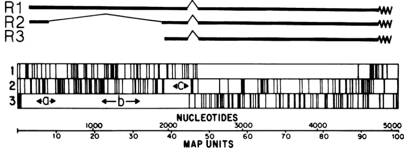
Regionsofopenreadingframe (ORF)from thegenomeoftheautonomousparvovirus minute virus of mice
(MVM) were cloned into a procaryotic expression vector, and bacterial fusion proteins containing MVM-specific amino acid sequences wereisolated. Antibodies raised against these proteins wereused to immuno-precipitate viral proteins synthesized in vitro ina rabbit reticulocytelysatetranslation system programmed with mRNA isolated from cells infected with MVM and a numberof different parvoviruses. These studies
demonstrated that: the 83-kilodalton nonstructural proteinNS-1 and the 25-kilodalton nonstructural protein NS-2 haveacommonamino-terminalsequencewhich is encoded by the singleORF located between nucleotides
225 and 534 inthe viral genome;theORF located between nucleotides 1110 and 1638 is onlyexpressed inthe
NS-1 protein; and the sequence encoded in a small alternative ORF between nucleotides 2075 and 2291 is
expressed exclusively in NS-2. These data confirm that NS-1 is the product of the 4.8-kilobase Rl viral transcript and demonstrate that NS-2 is synthesized from the 3.3-kilobaseR2transcript which arises from the left-hand promoter at map unit 4 on the viral genome. Antibodies against the MVM fusion proteins also
cross-reactedwithsimilarproteins encoded by the viruses H-1 and LulIl, but although antibodies against the carboxy-terminal half of NS-1cross-reactedwithasimilarproteininCPV,wewereunabletodemonstratean
NS-2 proteinencoded by this virus.
Minutevirus of mice (MVM), an autonomous parvovirus,
has alinear, nonpermuted, single-stranded DNAgenome of
some 5 kilobases (kb) contained within an icosahedral
pro-tein capsid approximately 20 nm in diameter (24, 26). Its
coding region is confinedto one DNAstrand(13),and blocks ofopenreading frame (ORF)span mostoftheviralgenome,
with some regions having multiple ORFs (2) (Fig. 1). The
viral genome encodes two overlapping transcription units which produce three major spliced cytoplasmic mRNA
species of4.8 kb(R1), 3.3 kb(R2),and3.0kb(R3)(13)(Fig.
1). TranscriptsRl and R2 are synthesizedfrom a promoter nearthe left-hand end ofthe viral genome at map unit 4,
while the R3 transcript, which is the major virally coded mRNA expressed in infected cells late in infection, arises fromapromoter atmapunit38 (13). All these mRNAspecies coterminateclose to the right-hand endofthegenome(13).
The R3 transcript programs the synthesis of two capsid proteins, VP-1 and VP-2, of 83 and 64 kilodaltons (kDa), respectively(5, 14). The thirdcapsidprotein (VP-3, 62kDa) is not a primary translation product but is derived by proteolytic cleavage which removes the amino-terminal
re-gion ofVP-2 and which occurs only after capsid assembly
andpackaging oftheviralgenome(23).Allthreetranscripts
have a short intron sequence between 46 and 48map units
removed, and the 3.3-kb R2 transcript also has a second
major intronbetweenmap units 10 and 40 which removes a large region of ORF located in the left half of the viral genome. A minor 1.8-kb (R4) transcript has also been
described, but it has not been mappedon the genome (13).
We have previously shown that RNA from MVM-infected cells programs the synthesis in vitro of four viral
polypeptides: the two capsid proteins VP-1 and VP-2 and two nonstructural proteins of 83 kDa (NS-1) and 24 kDa
(NS-2) (5). RNA selected by hybridization to a bacterial
*Correspondingauthor.
plasmid which contained the MVM sequence between nu-cleotides 1084 and 1659yielded onlyNS-1when translated in
vitro, showingthatthisprotein isthe productofa4.8-kb Ri
transcript, but theparticular mRNA species encoding NS-2
was not identified (5). Our original study (5) exploited the
finding that animals infected with a particular parvovirus
make antibodies which, in addition to recognizing the nonstructural proteins ofthat parvovirus, will cross-react
with similar polypeptides synthesized by parvoviruses of
different serotype. The major disadvantages in using such
antibody preparations are theirpolyspecificity with respect to individual polypeptides and the
heterogeneity
of theiraffinities for different domainsofa
single
polypeptide.
In the present paper we used chimeric proteins produced with aprocaryotic expression system toobtain antibodies against
theproteinsequences encodedby particular regions of ORF
in the left half of the MVM genome. These antibodies allowedus todefineblocksofamino acidsequence usedto
specify
the NS-2protein
of MVM and to examine theantigenic relatedness of individual domains of the
nonstructuralproteins ofa numberof differentautonomous
parvoviruses.Theadvantages of this approacharethreefold.
First, the
high-affinity
antibodiessoproduced
are monospe-cific foraparticularproteinsegmentordomain.Second,
by using predeterminedfragmentsof viral DNA andconfirmingthesize andreadingframe of the viral DNA insertsbyDNA
sequencing, the polypeptide sequence against which the antibodies are raised is
unambiguously mapped
within the viral DNA. Finally, the approachprovides invaluable tools for thepreciseanalysis
ofthoseviralgeneproducts
invivo,forexample, in
determining
their intracellularlocation and thekinetics of theirsynthesisand processing.MATERIALS ANDMETHODS
Materials. The procaryotic
expression
vectorspJS413,
pHK412,andpHK4147Xwereobtainedunderlicense from
724
on November 10, 2019 by guest
http://jvi.asm.org/
ORGANIZATION OF MVM NS GENES 725
Ri
AR2
R3
=M'W.V AA%
I
fl
lII1 11
I111
110 u
1E
2
11l
II _rl
cl
1
|
111 1 Diiillll
3
40-4
b
-+
I
II
111
I1
11"1
I
m
1
EI1111
11
NUCLEOTIDES
10,oo
2000 3000 4000sooo
I lO
b
2' 3'0 4'0 5'0 '6'0 70do
90 100MAP
UNITS
FIG. 1. Genetic mapof MVM. The 5149-nucleotide-long viral genome is shown ina3' to5'orientation with a block diagram showing the translationtermination codons in all three ORFs in the complementary strand. The three major cytoplasmic transcripts, Ri (4.8 kb), R2 (3.3 kb), and R3 (3 kb),arerepresented by thick black lines, and the thin lines indicate introns spliced out in theproduction of thematuremessage. Protein-coding regionsarerepresented by open blocks. Sequences labeled a, b, andc onthis diagram represent the nucleotidesequence and ORF expressed in the inserts of theexpression plasmids pYT201, pYT202, and pYT203, respectively,constructedas described in the text. Theboundariesof each MVM insert,determined byDNAsequencing, are nucleotides 225 to 534 for a, 1110 to 1638 for b, and 2075 to 2291 forc.
Molecular Genetics Inc., Minnetonka, Minn. Bacterial strains used were Escherichia coli NF1829 [araD139 A(araABC-leu)7679 galU galK A(lac)X74 rpsL thi (F' laciq lacZ::TnS Y+A+)]and LE392F[supEsupF hsdRgalK trpR metBlacY tonA (F' laciq lacZ::TnS Y+A+)] and were also obtained from Molecular Genetics Inc. Restriction endonucleases and other DNA-modifying enzymes were
obtained fromNew EnglandBioLabs, Inc., Beverly, Mass. ReagentsforDNAsequencingandradiochemicalswerefrom AmershamCorp.,Arlington Heights, Ill. pDR540, aplasmid
containing the trp-lac hybrid or TAC promoter, was
purchased fromP-LBiochemicals, Inc., Milwaukee,Wis.
Construction of MVMexpressionconstructs. (i) Procaryotic
expression vector. pJS413 is one of a series of
I-galactosidase-based expression vectors thathave been
de-scribed in detail elsewhere (21,27-29). Sequences ofinterest
arecloned into sites inapolylinkerin thevector which links twoprocaryoticgene segments outof reading frame.Blocks
of inserted ORF are expressed under control of the lac
promoter asthe middle partofatripartitefusion protein as
described in detail inthe Results section.
(ii) MVM sequences. MVM sequences were obtained by
restriction endonuclease digestion ofafull-lengthinfectious clone of MVM(p)contained in the plasmid pMM984(9). The
following sequenceswerethenpurified fromagarosegels: (i)
theHinfl fragmentbetweennucleotides225and534; (ii) the PvuII toXhoI fragment between nucleotides763 and 2074;
and(iii)theXhoItoNarlfragmentbetweennucleotides2067
and2291. These DNAs were thendigestedwithexonuclease BAL 31for variousperiods,and theextent ofdigestionwas
analyzedby gel electrophoresis. TheKlenow fragment of E.
coli polymerase I was then used in the presence of all four
deoxynucleotides to blunt the ends ofall termini, and the
resulting mixtures were ligated into the SmaI site in the
polylinker
ofpJS413. The position, reading frameassign-ment, and extent of the MVM insert in each construct analyzed in thepresent paper areoutlinedin Fig. 1.
(iii) Screening for expression constructs. Ligationmixtures weretransformed into E.coli NF1829, a strain which lacks a functional
P-galactosidase
gene (29) andcarriestransposon 5 and laciq on an F factor. Bacteria carryingthis Ffactor arekanamycinresistant
(TnS)
andoverproducethelacrepressor(laciq).
Transformants were selected by growth onampicil-lin-kanamycin and replica plated on to lactose-MacConkey
agarindicatorplates.Colonies containinganin-frame fusion protein with
P-galactosidase
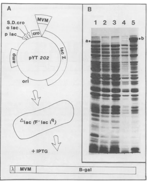
activity are thus induced and can beidentified by theirredcolor. Figure 2Adiagramstheconstructionandidentification ofonesuchexpressionclone,
pYT202, containing a region of the MVM genome derived from the Bfragment. Transformants givingred colonieson
lactose-MacConkey agar wereamplified as 1-ml cultures in yeast-tryptone brothfor severalhours,before being induced
with 1 mM
isopropyl-p-D-thiogalactopyranoside
(IPTG)overnight. Pelletswerethenobtained from100-,u samples of
induced cultures, boiled in sample buffer, analyzed by
sodium dodecyl
sulfate-polyacrylamide
gel electrophoresis (SDS-PAGE) essentially according to the procedure ofLaemmli (7), and stained with Coomassie brilliant blue R.
Owing to theirhigh molecular weight, the tripartite fusion proteins migratedin aregion ofthegel occupiedbyfewother
bacterialproteins(Fig. 2B),andthisprovidesaready means
ofidentifying which bacterial clones accumulatethehighest
levels of fusion protein.
(iv) Introduction of amber termination codons. The con-structpHK4147Xis similartopJS413 but carriesan amber
termination codon(TAG) inthepolylinkerjustdownstream oftheSmaI site (28). Thistermination codoncan bealigned
in-frame with fusion constructs engineered in the pJS413 vector byselectingarestriction endonuclease site(X) which cuts oncein
pJS413
outside ofthepolylinkerand not at all in theinsertandbyusingtheBglIIand BamHIsites whichare at the 5' and 3' borders, respectively, of the polylinker.Thus, ligatingtogether theBglII-Xfragment which contains thepolylinker from pHK4147XandtheBamHI-X fragment
containing the polylinker from the expression construct
produces a competent plasmid which has two in-frame
copies of the polylinkerlinked together by a
BamHI-BglII
fusionwheretheupstreampolylinkerbracketsthesequence to beexpressedand the downstream polylinker containsan in-frame ambertermination codon.
(v) Expressionofamberfragments. Constructs containing
ambertermination codons were transformed separately into NF1829and into LE392Fwhich carries the amber suppres-sor genes supE and supF. Colonies were screened for
--AAA
VOL.58,1986
on November 10, 2019 by guest
http://jvi.asm.org/
[image:2.612.101.512.76.227.2]726 COTMORE AND TATTERSALL
overproduction of fusion proteinsby gel electrophoresis as
described above.
(vi) Replacing the lac promoter with the TAC promoter.
pJS413 was digested with RsaI, and an 864-base-pair
frag-ment containing the Shine-Dalgarno sequence ofcro, the
polylinker, and partof lacZ was gel purified. BamHI linkers were ligated to the termini of this fragment, and it was
cleaved with BamHI to yield two fragments. This mixture
wasthen cloned into theBamHIsiteofpDR540(located just downstream of the TAC promoter), and constructs
contain-ing the pJS413 polylinker in the correct orientation with respect to the TAC promoter were identified. One such plasmid was then digested with PstI and BglII, and the
fragment containingtheTACpromoter was used to replace a similar fragment containing the lac promoter in pJS413 (then called pJS413/TAC). The new promoter was intro-duced intotheexpressionconstructs bysubstitutinga
BglII-X fragment containing the TAC promoter for a similar fragment ineach construct.
DNAsequencing. Constructs were sequenced by
transfer-ring the entire polylinker plus insert (aBglII-BamHI
frag-mentcontainingboth of thejunctions betweentheinsertand the vector) into the BamHI site ofM13mp8 (10), selecting
clones in both orientations, isolating single-stranded DNA,
andusingtheuniversal primertoobtainthesequence by the
dideoxynucleotide chain termination method (19). The MVM nucleotide numbers at both junctions in each con-struct determined in this way are detailed inthe legend to
Fig. 1andinthe Resultssection.
Purification of fusion proteins. Although fusion proteins
are soluble when synthesized at lower levels, induction overnight with IPTG leads to suchmassiveoverproduction
and accumulation ofthe fusion proteins that they become insoluble and effectivelyprecipitateoutin the bacterialcell, making purification exceedingly simple. Cultures (50 ml) of
E. colicarrying eachgene fusionplasmidwere grown to an
optical densityat 600 nmof1.0beforebeinginducedwith 1 mM IPTG overnight. Bacteriawere then collected by
cen-trifugation and incubated in 2 ml of12.5% sucrose-0.15 M
Tris hydrochloride (pH
8.0)-0.005
M EDTA containing 10 mgof lysozymepermlfor30min onice.Sampleswerethenfreeze-thawed twice and mixed with 9 volumes of 0.05 M
Tris hydrochloride (pH
8.0)-0.025
M EDTA-1% Triton X-100-1% sodiumdeoxycholate-1%
Nonidet P-40-0.01% SDS and incubated at roomtemperature for30 min. Aftersonication to reduce sample viscosity, the insoluble fusion proteinswerecollectedbycentrifugationat8,000 x gfor20 min. Pellets were resuspended in SDS-sample buffer and further purified by preparative SDS-PAGE. Gels were
briefly stained with Coomassie brilliant blue R,
protein
bandswereexcised and washed, thegelswerecrushed,and
proteinswere elutedovernight by
agitation
in 5 volumes of0.005 M NaHCO3 (pH 8.5) containing 0.1% SDS. After
centrifugation to remove gel fragments, supernatants were
concentrated with Centricon microconcentrators as de-scribed by the manufacturer (Amicon. Corp., Lexington,
Mass.).
Antisera. Antisera againstall fusion proteinswere raised in MVM-free female BALB/c mice (Jackson Laboratory,
BarHarbor, Maine)byrepeatedintraperitoneal injectionof 100-,ulsamplesof the fusionproteins(20 to 50 ,ug) emulsified with either Freund complete adjuvant (first injection) or Freund incomplete adjuvant (subsequent injections). Ani-mals were immunized over the course of 1 to 2 months
during which time they received three to four injections. Subsequently, antisera were raised against selected fusion
A
S.D.cro
vo lac, plac x
cr0
C.
E
X
0
2
PYT
202 sori
f
Alac
)~~q
L' laciq
.
__...
....
+IPTG
MVM
L
...B..
...- ... ..."I...vB-gal^-.--.-.^^ . . ... ... ..e
FIG. 2. Construction and identification of procaryotic expres-sion clones. (A)Theconstruction andanalysisofpYT202is shown
as anexampleofatripartitegene fusion. Theregion designatedb
(nucleotides1110to1638) inFig. 1, aBAL31digestion productof thePvuIItoXhoIfragmentofMVM(nucleotides763to2074),was
inserted at the SmaI site of the expression vector pJS413. The
resultingfusiongene (cro-MVM-lacZ)is expressedunder the
con-trolof the UV5 lac promoter(plac),thelacoperator(olac),and the
Shine-Dalgarno sequence of the cro protein of phage lambda
(S.D.cro).Thisconstruct wasintroduced intoNF1829,astrain ofE. coli which lacks anactive P-galactosidasegene (Alac) andcarries lacil on an Ffactor. Induction of the lac operon in thisconstruct
with IPTGgivesrisetothesynthesisof thetripartitefusionprotein diagrammed below. The chimeric molecules contain the first 22 amino acids of A cro, 176 amino acids ofMVMbetweennucleotides 1110 and1637in ORF3, and,attheircarboxytermini,almost allof
,-galactosidase (28). Such hybrid molecules have variable, but
significant, ,-galactosidase activity. (B) Coomassie-stained 10%
SDS-polyacrylamide gel showing the proteins synthesized with
(lanes 1, 3, and 5) and without(lanes 2 and 4) IPTG inductionof bacterial clonescarryingthefollowing plasmids:lane1,pHK412(in
whichcroand lacZareinframe andthereisnoinsert);lanes2and 3,pJS413 (in whichcro and lacZare outof frame and there isno
insert);andlanes4and5,pYT202 (inwhich thecroand lacZ genes ofpJS413arejoinedin openreading register byinsert b fromMVM).
Thetripartitefusionprotein (b) producedupon inductionofpYT106
constitutesapproximately 3to5%of thetotalbacterialproteinand has ahigherapparent molecularweightthanthe 116-kDacro-lacZ
fusionsynthesizedfrompHK412 (a).
proteins
in male albino rabbitsby
the initialinjection
ofapproximately
200 ,ug ofprotein
in Freundcomplete
adju-vant at
multiple
intramuscular and subcutaneoussites,
fol-lowed
by repeated
injections
ofsimilar amounts ofprotein
emulsified in Freundincomplete
adjuvant
atmultiple
subcu-taneoussitesduring
thecourseof several months. Serawerecollected 5to6
days
after the lastinjection.
Cellsandviruses.Theprototype strain of MVMwasgrown
IB
a.1
1
2
3
4 5m
~~~~~~~~~~~~~~~~-A
z- Wm. o- ..!.
"b
p;
mk&&AM
M-..AMa
[image:3.612.315.554.70.365.2]J. VIROL.
...
on November 10, 2019 by guest
http://jvi.asm.org/
ORGANIZATION OF MVM NS GENES 727
1 2 3 4 5
-v--:',
F.-sl:.,"..lilm
83kdo
I-T :.Wv-,:,
!"'Ir
-F
:..:.>.
:: ,- 6,
"41.:
'
't.a.
P"
,:
[image:4.612.125.232.71.309.2]64kd
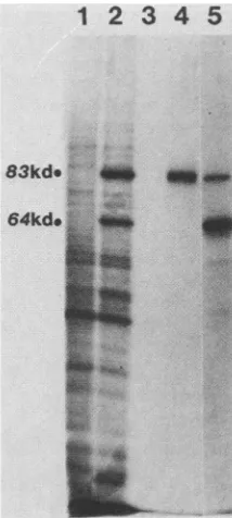
FIG. 3. Immunoprecipitation with antisera to tripartiteand bi-partitefusionpolypeptides.The[35S]methionine-labeledproductsof in vitro translation systems programmed with uninfected A9 cell
RNA(lane 1) and MVM-infected A9 cell RNA(lanes2 through 5)
were prepared and processed as described in the Materials and
Methodssection. Lanes 1 and 2 show total translationproductson a10%SDS-polyacrylamide gel,and lanes 3through5showproteins immunoprecipitated byantisera raised inmiceagainstthetripartite
fusion protein specified by pYT202 (lane 3), the bipartite amber fragment specified bypYT202Am-TAC (lane 4) (see Fig.4 andtext),
andpurifiedemptyMVMcapsids (lane 5). kd, Kilodalton.
in the mouse L-cell derivative A9 ouabrll, and H-1 and
LuIl weregrowninthe human simian virus40-transformed fibroblast line 324K, as previously described (22). Canine
parvovirus (CPV)wasgrownin Crandall felinekidneycells
(CFK), and bovine parvovirus (BPV) was grown in EBTr cells. Cells were parasynchronized by a single thymidine
blockaccordingtothe method of Ward andDadachanji (25),
infected with 30 PFU of virus per cell, washed, and
sus-pended inmedium containing 10-5 M deoxycytidine. Cells
were harvested 22 to 24 hpostinfection.
RNAisolation, cell-free translation,and immunoprecipita-tion. Cytoplasmic RNA was isolated and translated in a
rabbit reticulocyte lysate containing [35S]methionineas
pre-viously described (5). Immunoprecipitation was performed
essentiallyasdescribedbyKessler(6), using Formalin-fixed,
heat-killedStaphylococcusaureus(CowanI strain obtained
from Boehringer Mannheim Biochemicals, Indianapolis,
Ind.) Immunoprecipitation and autoradiography were
per-formedas describedpreviously (5).
RESULTS
Procaryotic expression of MVM sequences. (i) Tripartite fusion proteins. The procaryotic expression vector pJS413 has beendescribed in detail elsewhere (21, 27-29). Briefly,
theconstruct contains asmallpolylinker into whichcoding
sequencesof interestareinserted and thusjoinedinphaseto
a laci-lacZgenefusion (Fig. 2A). Expressionof the fusion
protein is under control of the UV5 mutant lac
promoter-operatorregion, andefficient translation ofhybrid genes is facilitatedbythepresenceofashortpeptideleader(fromthe
croprotein of bacteriophage lambda) which is situated at the correct distance from its own ribosome-binding site and provides a natural initiation codon and the first 22 amino acids at the amino terminus of the fusion. InpJS413 the cro leader is specifically out of translation phase with the lacZ gene owing to the polylinker and thus makes no -galactosidase. However, insertion into the polylinker of DNAfragments 3n + 1 nucleotides long and beginning at the first nucleotide of codon 1 in an ORFwill correctly phase the croleader and the lacZ gene, allowing the translation ofa
tripartiteprotein with demonstrable
P-galactosidase
activity. This enzyme activity is detected inbacteria carrying such recombinantplasmidssincethey grow as red colonies whenreplica plated on to lactose-MacConkey agar indicator plates. When BAL 31-digested fragments A (nucleotides 225 to 534) and C (nucleotides 2067 to 2291) from the MVM genome were inserted into pJS413, between 5 and 10o of the
resultantcolonies had,-galactosidase activity.This approx-imates thefrequency expectedfor the cloning of randomly cutfragmentscontaining one or two ORFs if the presence of theeucaryotic sequences does not substantially impair host
viability. However, withBAL 31 derivatives offragmentB
(nucleotides 763 to 2074) many fewer red colonies were
obtained, and most of these gave low-level expression of fusion proteins when analyzed by SDS-PAGE. The most
likely interpretation of this observation seems to be that,
despite overproduction ofthelacrepressorin thesecells,the
lac promoter is slightly active under these conditions and allows the synthesis of small amounts of a toxic fusion
protein. However, alternative explanations, such as the chimeric RNA or DNA themselves being toxic, have not been excluded. The bacterial clone in group B which pro-ducedbyfar thehighestlevel offusionprotein containedthe
plasmid pYT202, diagrammed in Fig. 2A. DNAsequencing showedthat this construct contained MVM sequences
be-tweennucleotides 1110 and 1638 in the viralgenome,
desig-nated b inFig. 1. Thus,BAL31 removed around 350 bases fromthe left end and 430 basesfrom theright end of the B
fragment. As such, this was certainly one of the shortest sequences presented to the vector and so supports the
suggestion
that longer sequences from this region impairbacterial viability. Interestingly, an overlapping, but much
longer sequence, from this part of the genome (TaqI
frag-ment nucleotides 227 to 2071) was somewhat tolerated by
NF1829.However, prolongedIPTG induction of such clones
didnotleadtothe
production
ofvery largeamountsoftheprotein,
since thecells ceased toreplicate
long before theyreach the normal saturation density of this strain (unpub-lished
observations).
SDS-PAGE showed that many of the clones containing
BAL 31 derivatives of
fragment
A orfragment
Coverpro-ducedfusion
proteins.
Constructs selected for furtherstudyweresubcloned intoM13,andtheinsertsweresequenced to
determineboth theirexact sequenceand the MVMreading
frame
they represented.
The construct pYT201contained afragment
A insert (designatedain Fig. 1) betweennucleo-tides225and 534expressedin ORF 3 oftheMVMgenome, while pYT203 contained nucleotides 2075 to 2291
(desig-natedc)expressedinORF2.Bacteriaexpressingall threeof
the selected clones (pYT201, pYT202, and pYT203) accu-mulatedfusion
proteins
toapproximately 5% ofthe totalcellmasswheninduced withIPTG.
The
tripartite
fusion proteins were then tested for theirability
to elicit antibody responses against the MVMse-quences they contained when injected into mice. In each
casethemicebecameimmunetothe
P-galactosidase
portionVOL. 58,1986
on November 10, 2019 by guest
http://jvi.asm.org/
of the
molecule,
as assessedby
immunoprecipitation
of[35S]methionine-labeled
bacterialproteins
(data
notshown).
However,
none of the seraspecifically immunoprecipitated
any viral
protein
from the in vitro translationproducts
of MVM-infected cellRNA,
as shownin-Fig.
3,
lane3,
for antibodiesagainst
theb-region
tripartite
fusion. Thissug-gested
that the MVM sequenceswere masked insomewayin suchfusion
polypeptides,
andtoovercomethiswefurtherengineered
theseplasmids
toseparate
the MVM sequencesfromthe
P3-galactosidase
sequence,by inserting
anin-frame amber codon between the two.(ii)
Bipartite
fusionproteins-amber fragments.
NF1829bacteria
carrying
constructswith ambertermination codons downstream o'f the MVM sequences did not accumulate eitherbipartite
ortripartite
fusionproteins
after induction withIPTG(Fig.
4B,
lanes9and10).
This resultwasobtained because the UAG codonseffectively
terminatedtranslation,
and the truncated amber
fragments,
whichare detectable in short-term[35S]methionine
labeling experiments,
wererap-idly degraded
inthebacterialcellandfailedtoaccumulatetohigh
levels.However, when
suchconstructsaretransformed into the bacteriumLE392F,
whichcarries thesupE
andsupFgenes, mutant tRNAs may insert
glutamine
ortyrosine,
respectively,
at theposition
ofthe amber codon(28).
supE
and
supF
arereported
togive
14 and 55%suppression
ofUAG
terminators,
respectively
(28), and thus in thisbacte-rium both
tripartite
andbipartite proteins
aresynthesized.
Overpr'oduction
and accumulation ofthefull-length
fusionproteins appeared
toprotect
the truncatedfrom formdegra-dation
(Fig.
4B, lanes 4 and6), and bothproteins
coprecipit-ated
(Fig.
4B,
lane7).
Theamberfragment
wasthenfurtherpurified by preparative gel electrophoresis
toyield
aprotein whichappeared
essentially
free of contaminationasassessedby
analytical
SDS-PAGE(Fig.
4B,
lane8)
and which wasnonpyrogenic
wheninjected
into animals.The 529-base MVM
fragment
inpYT202Am
wasex-pressed
withatotalof92nucleotides from thevector toyield
abipartite
fusionprotein
withanapparent
molecular size ofaround 20 kDa. This
protein
accumulatedappreciably
inLE392F,
but smallerfusions,
such as that derived frompYT2O3Am
(approximately
10kDa),
were less wellpro-tected,
and it wasdifficult,
orimpossible,
to obtain usefulamounts of such
proteins
at the levels ofexpression
ob-tained. TheTACpromoter
(which
contains the -35region
from the trp
promoter
and the -10region
fromthelacUV5promoter)
is muchstronger
thanthelacpromoteralthough
itis still
regulated by
the lacrepressor(18).
Substitution of thispromoter
into the amber constructs allowed considerableaccumulation ofeventhesmaller
bipartite
fusionproteins.
Incontrast tothe resultsobtained with the
tripartite
fusionproteins,
thebipartite
amberfragments
proved highly
immu-nogenic
in both mice andrabbits whenanalyzed
in thesameway, as shown for the
pYT202Am-TAC product
inFig. 3,
lane 4.Nucleotides 1110 to1638inORF 3areexpressedin the NS-1
protein. Antisera raised
against
the amberfragment
ex-pressed by pYT202Am-TAC
specifically' precipitated
theNS-1
polypeptides
from the in vitro translation products ofmRNA isolated from cells infected with
MVM(p), H-i,
LuIII,andCPV
(Fig.
5).For allfourviruses thisprotein
hadanapparent molecular size of around 83kDa,although in the translation
products
of Lulll mRNA NS-1 was sometimes resolved into two bands of similar molecularweig-ht.
Incontrast,no
high-molecular-weight protein
wasprecipitated
from the translation
products
of BPV-infected cell mRNA,but a
low-molecular-weight protein
which comigrates withA
o lac S.D. cro
orIL
+IPTO
UN
mvMMYM-
B-galB
1
2
3
4
5
8 7
8 9
10
=1ppW-40SO=
-NNW a-N
be quo
a-,
FIG. 4. Construction of amberfragmentsandintroduction of the TAC promoter. (A) pYT2O2Am is derived frompTY2O2by intro-duction ofanin-frame amber termination codonjustdownstream of the MVM sequences. In most bacteria proteins synthesized from thisconstruct(representedbyopenblocks)terminateatthis codon.
However,when thisconstructisintroduced intoLE392F,anE. coli strain carrying two amber suppressor genes (supE and supF),
terminationatthisUAG ispartiallysuppressed,andboth truncated and full-lengthfusionproteins aresynthesizeduponinduction with IPTG. (B) An 11% SDS-polyacrylamide gel showing the proteins synthesized bypYT2O2(lanes1and2), pYT202Am (lanes3and4),
andpYT202Am-TAC (lanes 5, 6, 9,and10)before(lanes 1, 3, 5,and
9) andafter (lanes 2, 4, 6, and 10) induction with IPTG. In
Sup-bacteria suchasNF1829which lack ambersuppressortRNAs(lanes 9and10),thefusionproteinsfailtoaccumulatetouseful levels after
induction,whereas in theSup'bacterium LE392F(lanes3through 6), synthesis andaccumulation of thefull-lengthfusion protein(a)
stabilizes andprotects the truncated amberproduct (b), and both accumulate in the cell (lanes4 and 6). Lane 7 shows the proteins
harvested in the 8,000 X g pellet after detergent extraction and sonication of the bacteria. Lane 8 shows the amberfragmentafter
gel purification.
on November 10, 2019 by guest
http://jvi.asm.org/
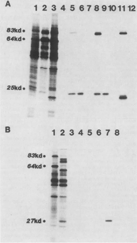
[image:5.612.321.554.66.528.2]ORGANIZATION OF MVM NS GENES 729
A
1
2 3
4 5 6 7
8
910111213
B3kd.
83kd
@
---83kd~
64kd-25kd.
FIG. 5. Protein encoded in the b region of MVM.
Immunopre-cipitationof[35S]methionine-labeledinvitro translationproductsof
MVM,H-i,Lulll,CPV,and BPV withantiseraagainstthebipartite
amberfrag'mentspecifiedbypYT202Am-TAC,encoded in ORF 3 of MVM between nucleotides iii0 and i638. (A) A 10%
SDS-polyacrylamide gel showing the total invitro translationproductsof
mRNA obtained from A9 cells (lane i), A9 cells infected with
MVM(p) (lane 2),324Kcells infected'With H-i (lane 6), and324K
cellsinfected withLulll(lane iO).Lanes4,8,and i2show the NS-i
polypeptides of MVM., H-i, and Lulll, respectively,
im-munoprecipitated withrabbit antiserum raised againstthebipartite
fusionprotein syn'thesizedfrom'pYT202Am-TAC. Lanes3, 7, and
show proteins precipitated from the translation products of
MVM, H-i, and LuIll RNA, respectively, with preimmunization
serum.Forcomparison,inlane 5 the MVMcapsid proteinsVP-i (83
kDa[kd])and VP-2(64kDa)areprecipitatedwith rabbit antiserum
against purifiedMVMcapsids', in lane 9capsidpolypeptidesof H-i
are,precipitated withguineapigantiserumagainst H-i,and in lane
i3 capsid polypeptides of Lulll are precipitated with hamster
antiserumagainst Lulll. (B)A iO%SDS-polyacrylamide gel
show-ingtotalin vitro translation products(lanes and 5)and
immuno-precipitates of the products of mRNA obtained from CFK cells
infected with CPV(lanes through4)and EBTr cellsinfected with
the NP-1 protein of BPV (8) was weakly precipitated byboth
immune and nonimmune sera from this rabbit(Fig. 5B,lanes 6 and7) and is likely to be nonspecific.
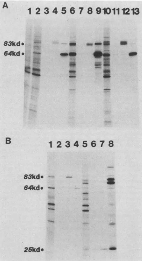
Nucleotides 225 to 534in ORF 3 are expressed in bothNS-1 andNS-2. Antisera raisedagainst the fusion proteinspecified
by pYT201Am-TAC specifically recognized both an NS-1 protein (-83 kDa) and an NS-2 protein (-25 kDa) encoded by MVM, H-1, and LullI (Fig. 6), demonstrating that mRNAs specified by all three of these viruses encode both an NS-1 and an NS-2 polypeptide. Additionally, the data show that in all three viruses this region of the genome (nucleotides 225 to 534 in MVM) must encode an amino-terminalpeptide which is common to both of these proteins and that this amino-terminal region shows strong antigenic
cross-reactions among all three viruses.
Both the NS-1 and NS-2 proteins specified by LuIll mRNA migrated as doublets (Fig. 6A, lane 11), suggesting that the heterogeneity between the two forms of each molecule is likely to reside in the common amino-terminal
residues. We do not know the significance of this doublet, but suspect it may be trivial, for example, the result of transcription from both wild-type virus and a mutant with a relatively small in-frame deletion within the amino-terminal
coding sequence of NS-1.
In contrast to the results obtained with H-1 and LullI,
antiseraagainst the pYT201Am-TACfusiontotally failed to
precipitate translation products specified by CPV-infected cell mRNA(Fig. 6B,lane4). Thus, despite thehighlevel of
antigenic cross-reaction seenbetween the middle region of the NS-1 polypeptides inMVM and CPV demonstrated in Fig. 5, linear antigenic determinants expressed within the amino-terminalregions of these two NS-1 proteins appear to be completelyunrelated. Similarly, this antiserumprovides no evidence for an NS-2 molecule carrying MVM
cross-reactive determinants encodedby CPV.
Mouse antisera against the pYT201Am-TAC fusion did
precipitatea27-kDa protein fromthetranslationproducts of BPV-infected cell mRNA whichwepresume tobethe NP-1
protein described by Lederman et al. (8) (Fig. 6B, lane 7).
Preimmuneserafrom thesamemice failedtoprecipitate this protein,but untilthisresultisconfirmed with specificrabbit
antiserum (not currently available), we remain cautious
about itsinterpretation.
Nucleotides 2075 to2291 in ORF 2 are expressed in the NS-2 protein. Antibodies raised against the fusion protein speci-fied by pYT203Am-TAC, which expresses the MVM sequence in ORF 2 between nucleotides 2075 and 2291,
specifically precipitated
the NS-2 polypeptide from thetranslationproducts ofMVM, H-1, and LuIll (Fig. 6A)but did not react withproteins specified by CPV or BPV (Fig. 6B).
BPV(lanes 5 through 8). An 83-kDaNS-1 polypeptide is precipi-tated with rabbit antiserum against the fusion protein synthesized from pYT202Am-TAC from CPV-specified translation products (lane 3), butnotfrom thoseofBPV(lane7).Proteinsprecipitatedby preimmunization serumfrom this rabbitare seenin lanes2for CPV and 6for BPV. A 25-kDaprotein which comigrates with NP-1 of BPV (lane 8) is weakly precipitated by both the immune and nonimmune serafrom this rabbit(lanes6and 7) and may well be nonspecific. Capsid polypeptidesVP-1 and VP-2of CPVareweakly precipitated inlane 4withamixture ofguinea piganti-H-i capsid and rabbit anti-porcine parvovirus capsid. Capsid polypeptides (VP-1, 80 kDa; VP-2, 72 kDa; and VP-3, 62kDa) and the NP-1 protein (-27 kDa) ofBPV are precipitated with rabbit anti-BPV capsidserum(lane8). Thefigures atthe left of eachpanel indicate apparent molecular sizes.
VOL.58, 1986
on November 10, 2019 by guest
http://jvi.asm.org/
[image:6.612.58.298.72.513.2]A
1 2
3 4 5
6789101112
83kd
64kd.
25kd.o
do
B
1
2345678
83kde
...*
1 1
64kd
v
27kd
o-
-FIG. 6. Proteins encoded in the a and c regions of MVM. Immunoprecipitationof[35S]methionine-labeled in vitro translation
products of MVM, H-1, LuIll, CPV, and BPV mRNAs with antisera raised against the amber fragments specified by pYT201Am-TAC andpYT203Am-TAC (encoded in ORF 3, nucleotides 225 to534, andORF2, nucleotides 2075to2291,ofMVM, respectively). (A)A
10%SDS-polyacrylamide gel showing the total translation products
of mRNA from cells infected with MVM(lane 1), H-1 (lane 2),and
LuIl (lane 3). Lane 5, 8, and 11 show proteins immunoprecipitated
from the total translation products of MVM, H-1, and LullI, respectively, with mouse antiserum against the amber fragment synthesized frompYT201Am-TAC, while lanes 4, 7, and 10 show
thatpreimmunization serafrom these mice didnotprecipitate the nonstructuralproteins from products of MVM,H-1,orLullI RNA, respectively. A rabbit antiserum against the amber fragment
synthe-sized frompYT203Am-TACprecipitates onlythe NS-2polypeptide
ofMVM(lane 6), H-1 (lane 9), and LuIII (lane 12; faint band present
in original autoradiograph not visible infigure). (B) A 10%
SDS-polyacrylamide gel of immunoprecipitates ofin vitro translation
products of RNA from cells infectedwithCPV(lanes 3, 4,and5)
and BPV(lanes 6, 7,and8), using preimmunemouse serum(lanes3
and6), mouse serum against the amber fragment of pYT201Am-TAC(lanes 4 and 7), and rabbitserumagainst the amber fragment
specified by pYT203Am-TAC (lanes5 and8).All of theseserafailto precipitate CPV-specified proteins,whileonlytheserumagainstthe
DISCUSSION
In this study we used procaryotic expression to isolate blocksof protein sequence expressed in particular ORFs of the MVM genome. These sequences, incorporated as part of abipartitebacterial fusion protein, were then relatively easy topurifyfrom the otherbacterial proteinsandallowedus to raise antisera in mice and rabbits which were specific for
particulardomains of the viral nonstructural proteins. Using
the in vitro translation products specified by virus-infected cell mRNA as a source of viralantigen, we were then able to
demonstrate that the NS-1 and NS-2 polypeptides ofMVM,
H-1, and LuIII share a common amino-terminal region which contains the sequence encoded in MVM between nucleotides225and534 inORF3.The carboxy-terminal half of NS-2 does not share protein sequence with NS-1, but rather utilizes a small alternative ORF (ORF 2) located between nucleotides 2075 and 2291, upstream of the minor splice. This suggests that NS-2 is most likely to be the product of a 3.3-kb R2 transcript (Fig. 1) which arises from a promoter at the left-hand (3') end of the genome at map
unit4. Si nucleaseanalysis of the viral transcripts (13) has shown that this size class of message contains exon se-quencesderived frommapunitcoordinates4.0to10.0, 40 to
46,and48to95,withthetwointerveningsequences (10 to40 and 46 to 48) spliced out. Our present observations are
supportedby
unpublished
studies from this laboratory whichshowed that when virus-specific mRNA was purified by
hybridization toindividualplasmid DNAs containingMVM sequence 1 to415, 2067to2204,2290 to2654,2651 to4000,
or 3997 to 5148, the selected RNAs all synthesized NS-2 whentranslated invitro.Conversely,mRNAhybridizedto a plasmid containing the MVM sequence 1084 to 1659 did not translate NS-2, although this latter RNA did program the
synthesisof NS-1. It has been estimated that R2 constitutes between 15 and20% of thevirus-specific mRNA present in
asynchronous cultures of A9 cells infected with MVM(p) (13), although presumablythisvalue mightvary atdifferent times after infection.Wedonotknow howthiscorresponds
to the level of NS-2 synthesized in vitro, since all the
methodswemightuse toevaluaterelative abundance of this moleculeareindirect and involve theuseof [35S]methionine-labeled proteins and specific antibodies
whose
individualaffinities can vary dramatically. However, there is no evi-dence available at present to suggest that NS-2 is the only product
synthesized
from an R2transcript. Recently,
Jongeneel and his colleagues obtained the sequence ofa
cDNA clone derivedfromMVM(i)mRNAwhich carriesthe exon regions characteristic of R2 (C. V. Jongeneel, G.
McMaster, R.
Sahli,
andB.Hirt,
Abstr.Pll,p.64, EMBOWorkshop on
Parvoviruses, Grangeneuve,
Switzerland, September 1985). In this clone, MVM nucleotide 514 lies next to nucleotide 1990,suggesting
asplice
of1,475 bases whichwould transfer anyprotein codinginORF 3 to ORF2.This is clearly
compatible
withthe datapresented
here for NS-2. This clone also contains a minorsplice which juxta-posesnucleotides2280and2377, leaving
theprotein-coding
sequence in ORF 2 for another six amino acids before terminating at an amber codon at nucleotide 2396. In the
commonamino-terminal regionof NS-1 and NS-2(nucleotides225 to534)effectively precipitatesalow-molecular-weightprotein (lane
7) which ispresumedtobe theNP-1protein of BPV.Lanes 1 and 2 show the total translation products of the cytoplasmic poly(A)+ RNAisolatedfromCPV-and BPV-infectedcells,respectively. kd,
Kilodalton.
on November 10, 2019 by guest
http://jvi.asm.org/
[image:7.612.62.299.75.497.2]ORGANIZATION OF MVM NS GENES 731
absence of this second minor splice the protein would still terminate seven residues downstream of nucleotide 2280 at
nucleotide 2299, a fact which is ofinterest because it has
been suggested, although not yet proven, that while the great
majority of R3 transcripts do use this 5' splice site, a
minority, which encode VP-1,do not(1, 4, 15). IftheNS-2
transcriptsusethesplice sites described by Jongeneelet
al.,
aprotein starting at the AUGat position 261 would haveamolecularweight of 25,009, which is in closeagreementwith the apparent molecular weight of NS-2 estimated from SDS-PAGE analysis.
A 76-kDa protein homologous to the NS-1 ofMVM has
been recognized previouslyin the in vitro translation
prod-uctsofmRNAfrom cells infected with H-1(17), but thisis the firstcleardemonstration thatH-1 also encodesanNS-2protein. Although the in vitrotranslation products ofLuIII
have not been examined previously, it is perhaps not
sur-prising that this virus also encodesanNS-1 and NS-2
protein
since heteroduplex mapping studies show that Lulll DNA
sharesconsiderable sequencehomologywith the left half of
thegenomes of both MVM andH-1
(3).
Similarly,
the observation that anantibody
against
themiddle region of the NS-1 molecule of MVM
(nucleotides
1110to 1638) cross-reactswiththe NS-1molecule of CPV isnot surprising since CPV is a host-range mutant of feline panleukopenia virus (FPV), and thenucleotide sequence of FPV(4) couldspecifya
protein
which wouldshare 156of the176 amino acids contained in the MVM insert of pYT202 (i.e., 87% homology). What is
surprising
is that theNS-1 of CPV, although having an apparent molecularweight
verysimilartothat ofMVM
NS-1,
entirely
lackslinearantigenic
determinants which cross-react with the amino terminal of
MVMNS-1.
Unfortunately,
thenucleotide sequenceof this regionremainsto be determined for both CPVand FPV.Nucleotides 1110 to 1638 in the MVM genome cloned in pYT202 specified a protein sequence which is
expressed
exclusively in the NS-1 molecule. The
only
mRNA which incorporates this region of the genome is the 4.8-kb Ri transcript (13) (Fig. 1), and thereforethis observationcon-firms our previous finding that NS-1 is encoded by the
Ri
transcript. Alltheparvoviruses whichhavebeen
sequenced
to date share a region of amino acid homology (MVM nucleotides 1428 to 1832) in the middleoftheNS-1-coding
region (20). The degree of homology through this region varies between viruses, but even such disparate viruses as
MVM(p) and the
dependovirus
AAV-2or MVM(p) and the human serumparvovirusB19sharearound50%homologyatthe amino acidlevel, whilethe more
closely
relatedvirusesMVM and FPV show 96%
homology. Seventy
amino acids fromthe amino-terminal half of this regionarecontained in the fusion protein specified by pYT202Am-TAC, and thefirst55ofthese areknowntoshow over60% homologywith atheoreticalprotein deduced from thenucleotide sequence
of BPV (B. C. Shull, M. Lederman, K. C. Chen, E. S.
Moses, E. R. Stout, and R. C. Bates, Abstr. S1/3, p. 17, EMBOWorkshop on Parvoviruses, Grangeneuve, Switzer-land, September 1985). However, wehave not been ableto
identifyaBPV-specified proteinwhich cross-reacts with this
regionof the MVM NS-1.
Downstream of nucleotide 1832 in MVM the amino acid
homologybetween the NS-1polypeptidesof MVM and CPV
rapidlydiminishes (1, 2, 4, 15), andalthough thereare still
clusters ofsimilar
residues,
it isnecessarytointroducegaps into the nucleotide sequence of CPV to get maximumalignmentof encoded aminoacids.This makes it
essentially
impossible to encode homologous proteins in alternative
ORFs in this
region. Although
there is adual blockof ORF between nucleotides 288 and 566 inCPV,
this sequenceterminates upstream of the 5'
splice
siteproposed
for theminor
splice
(1, 4, 15)(discussed above), and,
whencom-pared
with the amino acid sequenceexpressed
by
pYT202,
the alternative sequence in CPV shows
only
very weakhomology (a maximum
homology
of28%over aregion
of72amino
acids).
The minortranscripts
ofFPV and CPV have not been enumerated ormapped although
themajor (R3
equivalent)
mRNAofFPVis knowntocomprise
a270-baseexon
spliced
to a2,500-base
region
complementary
totheright-hand
half ofthe genome(4).
Thepossibility
thereforeexists that the
transcription
patterns
of CPV and FPV andthe
organization
of the left-hand ends of the genomes ofthesevirusesmay be
significantly
differentfromthose deter-minedfor MVMand H-1.The NS-1
proteins
of MVM and H-1 are nuclearphosphoproteins (11;
S. F.Cotmore andP.Tattersall,
VirusRes., in
press)
which appear to havemultiple
functions invivo, including
transactivation of the middlepromoter(16),
an asyet
unspecified
effectonthelaterstagesof viralDNAreplication (M.
Merchlinsky,
Ph.D.thesis,
YaleUniversity,
New
Haven,
Conn., 1984)
andaninhibitory
effectoncellularDNA
replication
(unpublished
observations).
The NS-2pro-teinhas yettobelocalizedin
vivo,
andatpresentwedonot have either biochemical orgenetic
informationconcerning
its function.
However,
the demonstration that NS-1 andNS-2 share
approximately
84 amino acids at their amino terminalsuggests thatatleastoneofthefunctions of both ofthesemolecules istointeractwith and
coregulate
theactivity
ofacommonelement in the infected cell.
ACKNOWLEDGMENTS
We thank Solon Rhode III for guinea pig anti-H-1 serum and hamster anti-Lulll serum, Tom Molitor for rabbit anti-porcine
parvovirus capsid serum, Jeffry Leary for rabbit anti-BPV capsid
serum, and GunterSieglforsupplyingvirus seed stocks.Wethank Molecular Genetics Inc., Lynn Enquist, Roger Watson, andJohn
Salstromforhelpandadvice with theexpressionvectorsystem. This work was supported by Public Health Service grants CA29303 and CA16038 fromtheNational Cancer Institute.
LITERATURE CITED
1. Astell, C. R., E. M. Gardiner, and P. Tattersall. 1986. DNA sequence of thelymphotropicvariantof minute virus ofmice,
MVM(i), and comparison with the DNA sequence of the
fibrotropicprototype strain. J.Virol. 57:656-669.
2. Astell, C. R., M. Thomas, M. Merchlinsky,and D. C. Ward. 1983. ThecompleteDNA sequenceof minute virus ofmice,an autonomousparvovirus. Nucleic Acids Res. 11:999-1018. 3. Banerjee,P. T.,W. H.Olson,D. P. Allison,R.C.Bates,C. E.
Snyder,andS. Mitra. 1983.Electronmicroscopic comparisonof the sequences of single-stranded genomes of mammalian
parvoviruses by heteroduplex mapping. J. Mol. Biol. 166: 257-272.
4. Carlson, J., K. Rushlow, I. Maxwell, F. Maxwell, S. Winston, and W. Hanh. 1985.CloningandsequencingofDNAencoding
structural proteins of the autonomous parvovirus feline
panleukopeniavirus. J. Virol. 55:574-582.
5. Cotmore,S.F.,L.J.Sturzenbecker,and P. Tattersall.1983.The autonomous parvovirus MVM encodestwo nonstructural pro-teins in addition to its capsid polypeptides. Virology 129: 333-343.
6. Kessler,S. W. 1975. Rapidisolation ofantigensfromcellswith
staphylococcal proteinA-antibodyadsorbent: parameters ofthe interaction ofantibody-antigen complexes with protein A. J. Immunol. 115:1617-1624.
7. Laemmli,U. K. 1970.Cleavageof structuralproteinsduringthe VOL.58, 1986
on November 10, 2019 by guest
http://jvi.asm.org/
assembly of the head of bacteriophage T4. Nature (London) 227:680-685.
8. Lederman, M., R. C. Bates, and E. R. Stout. 1983.Invitro and in vivo studies of bovine parvovirus proteins. J. Virol. 48: 10-17.
9. Merchlinsky, M. J., P. Tattersall, J. J. Leary, S. R. Cotmore, E. M. Gardiner, and D. C. Ward. 1983. Construction ofan infectious molecular clone of the autonomousparvovirusMVM. J.Virol. 47:227-232.
10. Messing, J., and J.Vieira. 1981. A new pair of M13 vectors for selecting either DNA strand of double-digest restriction frag-ments. Gene19:269-276.
11. Paradiso, P. R. 1984. Identification of multiple forms of the noncapsid parvovirus protein NCVP1 in H-1 parvovirus-infected cells. J. Virol. 52:82-87.
12. Paradiso, P. R., K. R. Williams, and R. L. Constantino. 1984. Mapping ofthe amino terminus of the H-1 parvovirus major capsid protein. J. Virol. 52:77-81.
13. Pintel, D., D.Dadachanji, C. R. Astell, and D. C. Ward. 1983. The genome of minute virusof mice,an autonomous parvovi-rus, encodestwooverlappingtranscription units. Nucleic Acids Res. 11:1019-1038.
14. Pintel, D.,M. Merchlinsky, and D. C. Ward. 1984. Expression of minute virus of mice structural proteinsin murine celllines transformed by bovine papillomavirus-minute virus of mice plasmid chimera. J. Virol.52:320-327.
15. Rhode, S. L.,III. 1985. Nucleotide sequence of the coat protein geneof canineparvovirus. J. Virol. 54:630-633.
16. Rhode, S. L., III. 1985. Trans-activation of parvovirus P38
promoterby the76Knoncapsid protein. J. Virol. 55:886-889. 17. Rhode, S.L., III,andP. R. Paradiso. 1983. Parvovirus genome:
nucleotide sequence ofHiandmapping of its genes by hybrid-arrested translation. J. Virol. 45:173-184.
18. Russell, D. R., and G. N. Bennett. 1982. Construction and analysis of in vivo activity of E. coli promoter hybrids and promoter mutants that alter the -35 to -10 spacing. Gene 20:231-243.
19. Sanger, S.,S.Nicklen, andA. R.Coulson.1977. DNA
sequenc-ing with chain terminating inhibitors. Proc. Natl. Acad. Sci. USA74:5463-5467.
20. Shade, R.O.,M.C. Blundell, S. F.Cotmore, P. Tattersall, and C.R. Astell. 1986. Nucleotidesequence and genome organiza-tionofhumanparvovirus B19 isolated from the serum of a child during aplastic crisis. J. Virol. 58:921-926.
21. Silhavy, T. J., M. L. Berman, and L. W. Enquist. 1984. Experiments with gene fusions. Cold Spring Harbor Labora-tory, Cold Spring Harbor,N.Y.
22. Tattersall, P., and J. Bratton. 1983. Reciprocal productive and restrictive virus-cell interactions of immunosuppressive and prototype strains of minute virus of mice. J. Virol. 46:944-955. 23. Tattersall, P., A. J.Shatkin, and D. C. Ward. 1977. Sequence homology between the structural polypeptides of minute virus of mice. J. Mol. Biol. 111:375-394.
24. Tattersall, P., and D. C. Ward. 1978. The parvoviruses-an introduction, p. 3-12. In D.C. Ward and P. Tattersall (ed.), Replication of mammalian parvoviruses. Cold Spring Harbor Laboratory, ColdSpring Harbor, N.Y.
25. Ward,D.C.,and D. K.Dadachanji. 1978.Replication of minute virusof mice DNA, p. 297-313. In D. C. Ward and P.Tattersall (ed.), Replication of mammalian parvoviruses. Cold Spring HarborLaboratory, Cold Spring Harbor,N.Y.
26. Ward, D.C., andP. Tattersall. 1982. Minutevirus of mice, p. 313-334. In H. L. Foster, J.D.Small, and J. G. Fox (ed.), The mouse in biomedical research, vol. 10. Academic Press, Inc., NewYork.
27. Watson, R. J., J. H. Weis, J. S.Salstrom, and L. W. Enquist. 1982.Herpes simplexvirustype-1glycoproteinDgene: nucle-otide sequence and expression in Escherichia coli. Science 218:381-384.
28. Watson, R. J., J. H.Weis, J. S. Salstrom, and L. W.Enquist. 1984. Bacterial synthesis of herpes simplex type 1 and 2 glycoproteinDantigens. J. Invest. Dermatol. 83:102-111. 29. Weis, J. H.,L. W. Enquist, J.S. Salstrom, and R.J. Watson.
1983. An immunologically active chimeric protein containing herpes simplex virus type 1 glycoproteinD. Nature (London)
302:72-74.