OF TWO DIFFERENT FORMS OF IMMEDIATELY LOADED
IMPLANTS SUPPORTING A FIXED FULL ARCH PROSTHESIS.
AN IN-VIVO STUDY
Dissertation Submitted to
THE TAMILNADU Dr. M.G.R. MEDICAL UNIVERSITY
In partial fulfillment for the Degree of
MASTER OF DENTAL SURGERY
BRANCH I
This is to certify that this dissertation entitled “COMPARATIVE EVALUATION
OF STABILITY OF TWO DIFFERENT FORMS OF IMMEDIATELY LOADED IMPLANTS SUPPORTING A FIXED FULL ARCH PROSTHESIS - AN IN-VIVO STUDY” is a bonafide research work done by Dr. P.KAMALASHANKAR under our
guidance and to our satisfaction during his post graduate study period between 2013 – 2016.
This dissertation is submitted to THE TAMILNADU Dr. M.G.R. MEDICAL
UNIVERSITY, in partial fulfillment for the Degree of MASTER OF DENTAL SURGERY in PROSTHODONTICS AND CROWN & BRIDGE - BRANCH I. It has
not been submitted partially or fully for the award of any other degree or diploma.
Head Of The Department Guide
Dr.T.J.Suneetha MDS. Dr. S. Sabarinathan, MDS
Professor and HOD Reader
Department of Prosthodontics and Department of Prosthodontics and Crown & Bridge Crown & Bridge,
HEAD OF THE INSTITUTION
This is to certify that this dissertation entitled “COMPARATIVE EVALUATION
OF STABILITY OF TWO DIFFERENT FORMS OF IMMEDIATELY LOADED IMPLANTS SUPPORTING A FIXED FULL ARCH PROSTHESIS - AN IN-VIVO STUDY” is a bonafide research work done by Dr.P.Kamalashankar under the guidance of Dr.S.Sabarinathan,M.D.S., Reader, Department of Prosthodontics and Crown and Bridge,
Rajas Dental College and Hospital, Kavalkinaru, Tirunelveli-627105.
Date : Dr. MARY KUTTY JOSEPH, M.D.S.,
Place : Kavalkinaru Principal,
Rajas Dental College& Hospital, Kavalkinaru Jn,
This dissertation is the result of work with immense support from many people and it
is a pleasure now that I have the opportunity to express my gratitude to all of them.
I would be failing in my duty if I do not adequately convey my heartfelt gratitude and
my sincere thanks to my Head of the Department, Professor Dr. T.J.Suneetha, M.D.S.,
Department of Prosthodontics and Crown & Bridge, for her exceptional guidance,
tremendous encouragement, well-timed suggestions and heartfelt support throughout my
postgraduate programme, which has never failed to drive the best out of me. I would like to
profoundly thank her for giving an ultimate sculpt to this study. I will remember her help for
life.
I would like to express my real sense of respect, gratitude and thanks to my Guide,
Dr. S.Sabarinathan M.D.S., Reader, for his guidance, constant support, back up and
valuable criticism extended to me during the period of my study. The timely help and
encouragement rendered by him has been enormously helpful throughout the period of my
postgraduate study.His special care in guiding this study is highly inspirational.
I would like to solemnly thank Dr. M.Aarti, M.D.S.,Dr Indumathi M.D.S Reader for
her valuable guidance and encouragement rendered by her. This dissertation has been the
fertile outcome of their massive endurance, support, proficient guidance and counsel.
I would also like to thank Dr.S.I.Joephin Soundar, Dr.Ramesh raja, Dr.Sajna senior
lecturers for their valuable suggestions and timely help given throughout my study.
It is my extreme pleasure to extend my gratitude to my beloved Chairman Dr.Jacob
Raja for his valuable support and constant encouragement throughout the period of my
Principal Dr. Mary kutty Joseph, Vice Principal (Academics) Dr.Cynthia Sathiasekhar,
Vice Principal (Administration) Dr.Johnson Raja, Director of Administration Dr.I.Packiaraj
and Members of the Ethical Committee and Review Board for the permission, help and
guidance throughout the course.
I thank Dr.MATHEW, M.D.S., for helping me with the statistical analysis of this
study.
I am grateful to my colleague, Dr.Vijay K for his patient endurance, co-operation and
whole hearted support he offered me during my postgraduate course. I extend my sincere
thanks to my senior colleagues Dr.Anbu Ila , Dr Shinemanoj , Dr.Aneesh.S, Dr.Jean
Mathew and my junior colleagues Dr. Arul Joshy.A, Dr.Mani Bernard.H, Dr selvin, Dr
shyma for their kind help and support.
Last but not the least, even though words wouldn't do much justice. I would like to
specially thank my parents Mr.S.Palaneeswaran & Mrs.P.Banumathy, my wife
Dr.Vijayabharathi ,my mentors Dr.K.Subramanian MBBS, Dr.T.Asirvatham manoharan
MBBS, Late Mrs.S.Sankara Subbulakshmi for their blessings and unconditional love.
Above all I thank GOD almighty for all the grace endowed upon me.
Place : Kavalkinaru Name : Dr.P Kamalashankar
CONTENTS
S.NO
TITLE
PAGE NO.
1.
INTRODUCTION
01
2.
AIM AND OBJECTIVES 06
3.
REVIEW OF LITERATURE
08
4
MATERIALS AND METHODS
19
5.
RESULTS
38
6.
DISCUSSION
53
7.
SUMMARY
62
8.
CONCLUSION
63
Table no Title Page no
1.
Randomisation schedule for implant placement 242.
Intra oral sites and corresponding implant dimensions 313.
Insertion torque values (ITV ) in NCm 394.
Insertion torque values (ITV ) in NCm for Al oxide blasted / acid 39etched surface
5.
Insertion torque values (ITV ) in NCm for Resorbable blast 40medium treated surface
6.
Insertion torque Values and corresponding Scores 417. Comparison of Insertion torque values (ITV ) of Al Oxide blasted acid etched surface (Group A) and Resorbable blast medium 41
(RBM) treated(GroupB) implants
8.
Site specific Implant Stability Quotient (ISQ) values 429. Implant Stability Quotient (ISQ) values for Al oxide blasted / acid etched surface (group A) 44
10. Implant Stability Quotient (ISQ) values for Resorbable blast 44
medium treated surface (group B) 11. Comparison of mean implant stability quotient (ISQ) values of Al Oxide blasted / acid etched surface (Group A) at different time 45
intervals
12.
Post hoc comparison of implant stability quotient (ISQ) of Al Oxide blasted / acid etched surface (Group A) at different time 46intervals
13.
Comparison of mean implant stability quotient (ISQ) values of 47 Resorbable blast medium (RBM) treated surface(Group B) atdifferent time intervals
15. Comparison of implant stability quotients (ISQ) of Al Oxide 49 blasted / acid etched surface (Group A) and Resorbable blast
medium (RBM) treated surface (Group B) implants at different time intervals.
List of graphs
Graph no Title Page no
1.
Mean ISQ values of Al Oxide blasted / acid etched surface (Group A) 502.
Change in ISQ over 1 year period for Al Oxide blasted / acidetched surface (Group A) 50
3. Mean ISQ values of Resorbable blast medium (RBM) treated
surface(Group B) 51 4. Change in ISQ over 1 year period for Resorbable blast medium
(RBM) treatedsurface(Group B) 51 5. Comparison of ISQ values of Al Oxide blasted / acid etched surface
(Group A) and Resorbable blast medium (RBM) treated surface 52 (Group B) implants at different time intervals.
List of figures
Fig :1 Completely Edentulous Maxillary & Mandibular Arch
Fig :2 Maxillary Arch.
Fig :3 Mandibular Arch.
Fig 6 Face bow Transfer.( lateral view)
Fig 7 Complete denture rehabilitation done in relation to maxillary and
mandibular arch.
Fig 8 Clear Acrylic Duplicated lower denture with radio-opaque
markers.
Fig 9 pre-op CBCT evaluation. A, paraxial sections with markers. B,
Panaromic sections with markers.
Fig 10 Radiographic stent fabrication done.
Fig 11 Accuracy of Radiographic stent evaluation and angulation
conformation by check CBCT. A .panaromic view. B . paraxial
view.
Fig 12 ARMAMENTARIUM.
a, Surgical Instruments. b, Physiodispenser
c, Implants to be used - blinded for six different sites
Fig 13 Muco periosteal flap elevated.
Fig 14 Pilot drill (2 mm ) placed through the stent.
Fig 15 Implant placed in site VI, paralleling pins placed in site IV and V
Fig 16 Insertion torque value evaluated with caliberated Torque wrench.
Fig 19 OSTELL Mentor device.
Fig 20 Implants placed at the bone level- Healing collar placed.
Fig 21 Suturing done with BSS.
Fig 22 Immediate Post OP –OPG with healing collars placed.
Fig 23 Open tray transfer done.
Fig 24 OPG to check the seating of the open tray transfer.
Fig 25 Impression making with custom tray A, Frontal view B, Occlusal view.
Fig 26 PENTAMIX -2 machine.
Fig 27 Impression made with –Monophase( poly ether).
Fig 28 Pick up impression- tissue surface.
Fig 29 1 week post op healing.
Fig 30 Working cast.
Fig 31 Hybrid screw retained mandibular prosthesis with opposing
maxillary complete denture.
Fig 32 1 week healing of soft tissue.
Fig 33 Hybrid Prosthesis placed in the lower jaw .
Fig 34 occlusion of the lower hybrid prosthesis with maxillary complete
denture.
Fig 37 Titanium milled bar ( screw retained)
Fig 38 Immediate Post op OPG after placement of the Hybrid prosthesis.
Fig 39 a ,1 month post op .b, intra oral view. C , intra oral lateral view.
Fig 40 1 month post op healing.
Fig 41 RFA taken a. mesial value .b, buccal value .
Fig 42 1 month post op.
Fig 43 3 month post op.
Fig 44 6 month post op.
Fig 45 1year post op.
Fig 46 case 2 .
Fig 47 case 3 .
Fig 48 Case 4 .
1
Partial or complete edentulism is a major oral health concern in a large part of the adult population and there is a need for treating a sizeable percentage of such population with prosthodontic rehabilitation. Traditional treatments comprising of removable prostheses are often inadequate in restoring full masticatory function and can negatively affect nutrition, physical appearance and self-esteem.14,31Osseointegrated dental implants offer effective and definitive solutions for such scenario. Dental Implants have become a milestone in dentistry and have changed the face of dentistry over the last three decades. Numerous alternative oral therapies and definitive rehabilitation procedures that could not be done with conventional techniques have been realised with the advent of osseointegrated implants. A large body of sound scientific research and decades of clinical use have verified and validated their usefulness in replacing missing teeth.16,28,31 Clinical evidence has shown excellent long term results for osseointegrated implants with success rates above 90%.11,14
2
earlier days. Also, the submerged protocol , two stage surgery and delayed loading were much in vogue as proposed by authors like Branemark and Alberktsson et al.14
Today implants are used to replace either single or multiple missing teeth apart from treating completely edentulous situations. Implants are infact proposed as the first line of treatment for many conditions which earlier were treated with either removable or fixed, tooth or tissue supported prostheses. Innovations in the design , geometry and surface characteristics of dental implants were essentially introduced to widen the scope of implant therapy to a wide range of clinical situations such as complete edentulism , long and short span partial edentulism , missing single tooth, maxillofacial prosthetics, etc. Advancements in the material science, imaging systems and prosthetic technology has also led to a phenomenal change in the way implant treatment planning and execution is carried out.
The successful outcome of any implant procedure needs a series of patient related factors (bone volume and density) and procedure dependent parameters (type of implant and surgical procedure) to be considered . Modern implants come in a variety of shapes and sizes to suit the different edentulous situations they replace and also according to the types of prosthetic teeth or superstructure to be supported by the implants. Their surfaces have been improved to enhance the osseointegration process. Instead of being smooth or machined, they are generally roughened by different manufacturing techniques which dramatically increase the surface area to which bone can attach.22,26,41
3
placement have been developed. Similarly , new implant designs have been proposed to achieve high stability even in poor quality bone. A combination of macro thread design to achieve high initial stability and micro roughened surface to improve bone implant contact is employed in the majority of the commercially available implants today. Modification of screw threads has been shown to increase the pullout strength of implants and influenced the insertion torque values26,41. New implant biomaterials are developed aiming to alter cellular performance at the bone – implant interface. Research has provided strong evidence supporting improved osseointegration , increased bone – implant contact (BIC) , increased mechanical stability and improved soft tissue health with surface treated implants. Schroeder et al observed that a rough and porous surface increases the effective surface about 12 times in relation to a smooth one generating an osteo – inductive effect and increasing the anchoring of the implant in bone.44
4
Attempts to decrease the treatment time with implant therapy have also been made by many authors.30,36,43 Premature loading was considered detrimental to direct bone apposition on the implant surface and was thought to result in fibrous encapsulation. While the initial concept of submerged protocol and delayed loading was based on earlier concepts of osseointegration, continued research has led to an improved understanding of bone biology , osseointegration and host response for immediate and early loading protocols. Immediate loading is no longer considered to be risky and new clinical protocols with shortened healing periods or immediate loading have been proposed with clinical success rates comparable to the original protocol. More recent studies have illustrated that early loading of implants at a low magnitude of force may aid in early peri-implant osteogenesis.37,43As mentioned earlier , the main requirement to allow immediate loading of dental implant is its primary stability when inserted into the osteotomy site. The micromotion of the implant during loading in its early stages should be below 150 µm , above which fibrous encapsulation may occur40.
In addition to the above factors, splinting of multiple implants is also expected to reduce the implant micromotion during healing stage. Immediate prosthetic loading of the implants , either functional or nonfuntional is indicated as part of treatment protocol by many clinicians. The All –on – four technique proposed by Paulo malo is one such protocol with reported success of more than 95%36. Immediate loading of either axially placed or tilted implants in completely edentulous jaws has now become a norm rather than exception.
5
done in artificial bone or cross sectional studies. While the initial mechanical stability of implants can be roughly assessed with synthetic and cadaveric bone models, the influence at cellular level due to the surface characteristics are best researched in an in vivo model which includes animal studies followed by histomorphometric analysis and human clinical trials.
Long term , Prospective human clinical trials are considered to be the gold standard in evidence based dentistry , the results of which can be extrapolated to clinical practice with more accuracy and confidence. Such clinical trials are comparatively few regarding the influence of surface treatments of implants on the primary and long term stability of implants.
Hence this clinical study was undertaken to evaluate the influence of two different surface treatments on the primary and long term clinical stability of endosteal implants supporting an immediately loaded full arch fixed prosthesis.
Aim and Objectives
6
AIM:
The aim of this study was to determine the difference in the primary and progressive clinical stability if any, between two types of root form endosseous implants with two distinct implant surfaces namely Acid etched surface/ Al Oxide blasted surface and Restorable blast medium (RBM) treated surface when loaded immediately with a full arch fixed prosthesis.
OBJECTIVES:
To measure the stability of the above mentioned groups of implants immediately after insertion (Primary stability).
To measure the stability of the above mentioned groups of implants 1 month after insertion of definitive prosthesis.
To measure the stability of the above mentioned groups of implants 3 months after insertion of definitive prosthesis.
To measure the stability of the above mentioned groups of implants 6 months after insertion of definitive prosthesis.
To measure the stability of the above mentioned groups of implants 12 months after insertion of definitive prosthesis.
To compare the difference in stability among the Al Oxide blasted / acid etched surface implants at different time intervals (at the time of insertion, 1 month, 3 months, 6 months & 12 months after insertion).
7
8
Branemark Pl, Zarb GA, Albrektsson T et al (1985)14 In the last three decades, implant dentistry has emerged as a fully accepted discipline in dentistry. During this period of development, its concepts and treatment modalities have undergone tremendous changes. At first, only protocols involving two-stage surgery were recognized as providing reproducible and reliable results .
Antezak-Bouckoms AA, Tulloch JF, Berkey CS (1990)4 Several types of research designs are available including, split-mouth design, whole-mouth design, and cross-over clinical trials. The split-mouth design is a popular design in oral health research. In the most common split-mouth study, each of two treatments are randomly assigned to either the right or left halves of the dentition. The attractiveness of the design is that it removes a lot of inter-individual variability from the estimates of the treatment effect. Split-mouth design seems to be an effective type of clinical trial giving the advantages of reducing bias, obtaining definitive results, and decreasing cost.
Buser D, Weber HP, Bragger U, Balsiger C. et al(1991) 15Proposed a single-stage surgical procedure that became acceptable.
9
Meredith N, Book K, Friberg B, Jemt T, Sennerby L. et al(1997)35 proposed a cross-sectional and longitudinal study of resonance frequency measurements on implants in the edentulous and partially dentate maxilla.
RFA is a test to assess implant stability by measuring the frequency of implant oscillation inside the bone. A transducer connected to the implant is excited by means of an electric or magnetic impulse (depending on the type of transducer used). Thus, the implant is subjected to slight lateral force that causes lateral displacement due to elastic deformation of the bone. The frequency of the registered oscillation depends on the stiffness of bone-implant attachment: the stiffer the system is, the higher the transducer’s oscillation frequency will be.
There are several generations of transducers and assessment instruments. Recent generation instruments (Osstell®; Osstell AB, Gothenburg, Sweden) need no computer to complete analysis, are light, small, quick and easy to use in everyday clinic activity. Unlike previous generations, the transducer demands no calibration in 3G instruments.
Stability values are expressed in ISQ (Implant Stability Quotient) units, which range from 1 (low stability) to 100 (high stability). There is a specific transducer for each type of implant and the obtained values do not depend on the type of transducer (44).
10
Szmukler-moncler s ,salama H,reingewirtz Y et al(1998) 43Opinions vary about the maximal acceptable interval between implant placement and loading. Some researchers use the term immediate loading only when the provisional prosthesis is placed during the same session in which surgery is performed .The delay most often observed for orderly placement of prosthesis directly after the surgical procedure ranges between several hours and 5 days. It would be tempting to use this practical framework in the definition of immediate loading. However, many studies have documented results of treatment that followed an arbitrarily determined delay of 48 to 72 hours. Furthermore, according to numerous authors, patients appear to be increasingly interested in reduced treatment time between tooth removal and delivery of the final implant-supported prosthesis, provided the level of predictability established during the previous two decades is maintained.
Misch et al(1998)16 study has concluded that most of the immediate loaded implants are placed in anatomical sites with dense and good bone quality. The mandible has a better bone quality compared to the maxilla and this is the reason why reports are available regarding immediate loading in mandible than maxilla.
Comparatively, related number of studies has concluded the concept of immediate loading in the mandible for full arch restoration with the survival rates of 80-100% (31-33). Most of the studies published on immediate loading in the mandible have considered mainly in edentulous patients.
11
change in marginal bone level but statistically significant difference in location of marginal bone level in relation to shoulder of the implant was found in favor of self tapping TiO 2 – blasted screw shaped implants made of pure titanium in comparison with self tapping mark two implants machine surface irregularities.
Szmukler-Moncler S, Piattelli A, Favero GA, Dubruille JH et al(2000)42 considered the application of early and immediate loading protocols in dental implantology where the waiting periods for bone healing is shortened; instead of 3 to 8 months, no more than 6 to 8 weeks was deemed necessary.
Aparicio C, Rangert B, Sennerby L., et al(2002)5 Immediate/early loading of dental implants they believe that, to qualify as an immediately loaded implant, the definitive prosthesis must be placed on the same day .
12
Romanos et al,(2003)37 quoted that implant stability in the immediate loading is seems to be increased due to the significant increase of the peri-implant bone density at the implant-bone interface.
Vidyasagar(2004)46 stated that dental implant designs are influenced by overall surface area, length and thread configuration. This can gain initial stability that would reduce the threshold for the tolerated micro motion and minimize the waiting period. The design factors incorporated into the dental implants would decrease the shear forces on the interface. The design features may stimulate the bone formation and in which promotes bone healing and better load distribution . There are different types of dental implants used in dentistry.
Alexandre-Amir Aalam, etal (2005).1 Clinical and radiographic comparison of dental implants with surface roughened by anodic oxidation(tiUnite) dual etched implants (osseotite) and machined implants and concluded that tiUnite, osseotite, and machined implants had similar short term clinical outcomes.
13
the maxilla (23). It is evident that, when compared with the maxilla, the bone surrounding the implant has better volume and quality in the mandible (24).
Barone A, Rispoli L, Vozza I, Quaranta A, Covani U et al(2006)8 In extreme, this involves insertion of an implant immediately after tooth extraction, potentially using simplified procedures such as flapless surgery, and subsequent restoration of the implant in the same session. Ultimately, this combination may not only lead to a reduction in the overall treatment time, but may also substantially decrease the associated costs. Furthermore, it has been claimed that the described approach is clearly associated with reduced surgical procedures and may more efficiently preserve the existing bone and soft tissues at the site of implantation.
Paulo Malo DDS et al (2006)36 In his pilot study indicated that fully edentulous Jaws with various types of bone quality can be treated with high success and good aesthetics using immediately loaded implants featuring a narrow implant apex reduced color height and an anodically oxidized implant surface and that favorable margin bone loss can be maintained.
Vandamme(2007)44 study also showed that threaded implants offer significant bone-to-implant contact during which may also enhance the secondary stability. Hence, cylinder-type implants seem to be contraindicated for immediate loading regimes due to lowering of primary stability and less resistance to vertical movement and shear stress.
14
provided with several advantages such as increased masticatory function, stability to the interim prosthesis, minimizing uncontrolled transmucosal loading, preservation of bone and stimulation of bone remodelling, enhancement of gingival contours and better esthetic. It is also reported with the improvement of psychological impact (34, 35). Despite several advantage of immediate loading, there is no agreement on the technique by which immediate loading can be achieved. The success of immediate loading relies on the technological advances in the texture, shape and material of the implant. In addition to immediate loading the primary stability depends even on the bone quality and density.
Barewal RM(2007) 7 stated the primary stability of implant achieved at the time of implant placement and it is also associated with bone density, length, width and type of implant and drilling technique. The primary stability obtained after implant placement is considered a relevant factor for the prognosis of the implant and has been identified as a prerequisite to achieve osseointegration. This would suggest that high primary stability that makes immediate loading more predictable (20).
15
Romanos GE(2008)37 have stated the varying designs of implants with various degrees of stability and determine their future clinical performance. Hence, he quoted that screw or “threaded” design minimizes the implants micro motion during function thereby maintaining the PS. Furthermore, a threaded design also increases the surface area of the implant thereby offering a higher percentage of bone-to-implant contacts, in comparison to implants with a cylindrical design. Therefore, threaded type implants are generally recommended and particularly for immediate loading.
EspositoM, Grusovin MG, Willings M, Coulthard P, Worthington HV, Esposito M et al(2009)18 proposed different time frames for loading dental implants in replacement of missing tooth.
Fawad javed etal (2010) 20 Assessed the role of primary stability for successful immediate loading of dental implants. And its evident that the degree of achieved primary stability during immediate loading protocols is depend on several factors including bone density and quality, implant shape, design, surface characteristics and surgical technique. Further research is required in situation, such as poor bone quality and quantity and multiple implant or augmentation procedure, which may challenge the attainment of primary stability during immediate loading.
16
studies have demonstrated that the quality of the alveolar bone is the most important factor for achieving good primary stability (21).
Heng-Li Huang (2010).26 Investigated implant stability using resonance frequency measurement of topographically changed and surface chemistry modified implants in rabbit bone. And found that implant surface properties influence RFA measurements of implant stability. surface chemistry modified titanium implants showed higher values than topographically changed implants.
Sun jong kim ,Myung Kim –et al(2010)41 Proposed in the animal study that the bone to implant contact ratio was higher in surfaces with a roughness of 1.02 [73.6%+14.4%] SLA treated and in surfaces with a roughness of 1.76 [ 69.6%+ 12.5%] than in surfaces with roughness of 0.86 m[60.82%+13.11]. There were no significant difference between the 3groups of various surfaces in terms of implant stability.
.
Fung et al et al(2011)22 He concluded that after 36 months of functional loading there was no significant difference in the change in radiographic bone levels between titanium oxide- anodized & machined surface MKIV dental implants. From 12 to 36 months of functional loading ,both anodized machined surfaces implants exhibited gains in RBL(Radiographic Bone Level) which statistically significant for the machined implants.
17
stability showed significant increase in stability over time and increase in stability during healing. In contrast, implant with high primary stability lost some stability over time.
Jan gottlow etal (2012)29. Experimental investigation was done to compare the bone tissue responses and implant stability between two commonly used implants representing different geometries and surface characteristics. Both HSBA and OX implants were well integrated in bone and showed firm and increased stability from placement to after 6 weeks of healing. The HSBA implant showed more BIC after 10 days and OX implant more BIC after 6week of healing. The HSBA implant showed significantly higher shear strength after 3 and 6weeks and higher RTQ values after 3 weeks than OX implant. This result may be due to difference in surface roughness and hydrophilic properties.
.
Gerard torroella saura, Javier mareque etal (2014)24. Evaluated the effect of two different designs, tapered vs cylindrical, on primary stability of implants placed with an immediate loading protocol in edentulous mandible to support fixed prosthesis with in occlusal contacts during first 48hr and concluded that tapered implant achieved greater primary stability values measured with ITVs and less marginal bone loss than the cylindrical implants
18
PS]were absolutely insignificant in well maintained patients & concluded that MBL changes could be affected by the different implant designs A high ISQ values was found for both implants & no statistically significant differences was found for ISQ mean values b/w interventions .
Bilal Al nawas etal (2014)12. Investigated in a split mouth model whether small diameter implants made from titanium-13 zirconium alloy perform at least as well as titanium grade IV implants. This study confirms the TiZr small diameter bone level implants provide at least the same out comes after 12 mounths as grade IV bone level implants. This improved mechanical properties of TiZr may extend implant therapy to more challenging situations.
19
The present in vivo study was conducted for the comparative evaluation of clinical stability of two different types of implants with different surface treatments namely i) Al Oxide blasted / acid etched surface & ii) Resorbable blast medium (RBM) ( Calcium phosphate) treated surface loaded immediately with a fixed full arch prosthesis.
The following materials , instruments and equipment were used for the study.
MATERIALS USED:
1. Root form endosteal implants (Al Oxide blasted / acid etched surface )- Touareg-S (Adin implant system, Adin pvt ltd,Israel)
2. Root form endosteal implants ( Resorbable blast medium (RBM) ( Calcium phosphate) treated surface) - Touareg-OS (Adin implant system, Adin pvt ltd, Israel)
3. Smart peg type 49 (OSSTELL MENTOR, Gothenborg, Sweden).
4. CAD-CAM fabricated Titanium frame work (TDS ME 300 HP,TDS Biotechnologies,Taiwan)
5. Polyether impression material (pentasoft monophase,Pentamix-2,3M ESPE, Seefeld , Germany )
6. Tray adhesive (3M ESPE, Seefeld , Germany )
7. Soft tissue gingival mask (DETAX ,Ettlingen, Germany)
8. Open tray transfers-implant level (Adin implant system, adin pvt ltd, Israel ) 9. Implant replicas. (Adin implant system, adin pvt ltd, Israel)
20
13.Auto polymerizing acrylic resin-pink. (DPI, Mumbai) 14.Auto polymerizing acrylic resin –clear. (DPI, Mumbai)
15.Cross linked acrylic teeth (Gnathostar, Ivoclar Vivadent Inc,NY ,USA ) .
16.Stainless steel sleeves.
17. Light cure Composite resin ( GC CORPORATION,Tokyo, Japan)
18.2% lignocaine with adrenalin (1: 200,000) (Lignox, Indoco remedies pvt ltd .Mumbai) 19.Povidone-Iodine solution 2 % (Betadine ,wockhardt,India ) .
20.Polyethylene glycol suture (vicryl 3-0ETHICON, Johnson & Johnson, Aurangabad)
INSTRUMENTS USED
1)Implant surgical kit (Adin implant system, adin pvt ltd.,Israel).
-Initial drill (lancet drill)
-2.0mm twist drill -.
-2.8 mm twist drill.
-3.2 mm twist drill.
-3.65 mm twist drill.
Paralleling pin.
Depth gauges (short and long)
21
3) Hex driver.
4) Caliberated torque wrench.
5) Bard Parker knife no:3
6) Bard parker blade no :15.
7) Howarth’s periosteal elevator.
8) Austins retractor.
9) Cheek retractor.
10) Vestibular retractor.
11) Tissue forceps.
12)Needle holder.
13) Scissors.
14) Semiadjustable articulator with facebow (Hanau wide vue, whipmix corp, USA)
EQUIPMENTS:
1. Cone beam CT machine (Cone Beam CT CS 9300,Carestream , France) 2. Implant motor with Physio dispenser ( Saeshin, korea )
3. Radio visio graph.(RVG) (Carestream ,France )
22
5. Optical Scanner (Shining 3D, TDS biotechnologies ,Taiwan)
6. TDS ME 300 HP - 5 axis milling machine (TDS biotechnologies ,Taiwan) 7. Pentamix 2 automixing device (3M ESPE ,Seefeld,Germany)
Description of Resonance Frequency Analysis (RFA) & Osstell machine
The RFA technique is essentially a bending test of the bone-implant system in which an extremely small bending force is applied by stimulating a transducer. It is equivalent in terms of direction and type to applying a fixed lateral force to the implant measuring the displacement of the implant. This effectively mimics clinical loading conditions, although on a much reduced scale. The RFA method can potentially provide clinically relevant information about the state of the implant-bone interface at any stage of treatment.
23
Usually the average of the two values are taken to be the ISQ of that particular implant . The implant stability quotient is a nearly linear mapping from resonance frequency measured in kHz to the more clinically useful scale of 1-100 ISQ. The higher the ISQ ,the more stable the implant.
METHODOLOGY
I. Formulation of study design a. Split mouth prospective trial b. Formulation of null hypothesis c. Inclusion and exclusion criteria d. Informed consent
II. Patient selection
III. Fabrication of complete denture IV. Fabrication of radiographic template V. Cone beam CT evaluation
VI. Implant placement
VII. Assessment of primary implant stability VIII. Fabrication of hybrid prosthesis
a.impression making b.framework trial
c.jaw relation and wax trial d.insertion of hybrid prosthesis
IX. Assessment of implant stability – 1 month , 3 months,6 months and 12 months intervals.
24
I. FORMULATION OF STUDY DESIGN :
a. Split mouth prospective trial
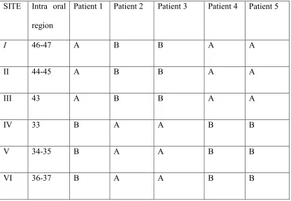
[image:38.612.131.547.410.698.2]The present in-vivo study is designed to be a split mouth prospective clinical trial in which each patient received both the types of implants investigated in either of the two quadrants of the mandibular arch - right or left .Each group of implants was placed on one side of the arch (n-=3) for each patient. A total of 5 patients were included in the study resulting in 15 implants of each group being placed. The Alumina oxide blasted / acid etched surface (TOUAREG-S) is taken as the control (Group A) and the Resorbable blast media (RBM) surface (TOUAREG-OS) is taken as the test group(Group B) . The side selection –right or left for each group is randomized according to the randomization schedule give below.
Table 1 : Randomisation schedule for implant placement SITE Intra oral
region
Patient 1 Patient 2 Patient 3 Patient 4 Patient 5
I 46-47 A B B A A
II 44-45 A B B A A
III 43 A B B A A
IV 33 B A A B B
V 34-35 B A A B B
25
A - Alumina oxide blasted / acid etched surface (TOUAREG-S) B - Resorbable blast media (RBM) surface (TOUAREG-OS)
The study is a triple blind Prospective clinical trial in which the subject, operator and investigator are blinded. An independent research associate prepares the randomization schedule and hands over the implant accordingly to the operator during implant placement. The assessment of the clinical stability is again done by an independent investigator who measures the stability at fixed time intervals namely
a. Immediately after implant placement. b. One month after implant placement. c. Three months after implant placement. d. Six months after implant placement. e. Twelve months after implant placement. b.Formulation of the null hypothesis.
Null hypothesis for this study is that there is no difference in the clinical stability of the two implant surfaces over a period of one year.
c. Inclusion and exclusion criteria.
Inclusion and exclusion criteria for Participation in the study
Inclusion criteria
Voluntary informed consent
Age >45 years and ≤ 65 years
Completely edentulous maxilla and mandible at the time of surgery
26
Available bone height of atleast 10 mm in the molar region, 13 mm in the premolar region and 16 mm in the canine region of the edentulous mandible.
Available bone width of atleast 7mm in molar region, 6 mm in premolar region and 6 mm in canine region of the edentulous mandible.
Commitment to participate in the study for atleast 1 year of follow-up examinations
Exclusion criteria (systemic)
Medical conditions requiring prolonged use of steroids
Severe hemophilia
Bisphosphonate medication
History of leukocyte dysfunction and deficiencies
History of head and neck radiation or chemotherapy
History of renal failure
History of uncontrolled endocrine disorders
Physical handicaps interfering with ability to perform adequate oral hygiene
Use of any investigational drug or device within 30 days prior to implant surgery
Alcoholism or drug abuse
HIV infection
Smoking >10 cigarettes or cigar equivalents per day or chewing tobacco> 10 cigarette equivalents per day .
27
Exclusion criteria (local)
Local inflammation including denture stomatitis.
Mucosal diseases such as erosive lichen planus
History of local irradiation therapy
Presence of osseous lesions
Unhealed extraction sites
History of bone reconstruction and bone grafting techniques at site of intended implant placement.
Severe bruxing or clenching habits
Persistent intraoral infection
Patients with inadequate oral hygiene or unmotivated for adequate home care
Exclusion criteria (secondary)
Need for GBR treatment at implant surgery
Insufficient bone or any other bone abnormality that contraindicated placement
Inappropriate treatment according to study protocol.
Lack of primary implant stability at time of abutment connection (ie, spinning implant at 35 Ncm torque or laterally moving implant)
d.Informed consent. (Annexure 1)
28
were free to ask any questions and were clarified. A written consent was obtained from those subjects who were willing to participate in the study.
II.PATIENT SELECTION :
The completely edentulous patients who reported to the department of prosthodontics of the study center were screened to fulfill the inclusion and exclusion criteria. A detailed general examination and local examination was done for the prospective study population. A panoramic radiograph as a part of the routine investigation for complete denture therapy is also obtained. Based on these clinical and radiographic assessments, the final safety population of five patients (male = 4, female =1) was selected.
III.FABRICATION OF COMPLETE DENTURE :
29
the patient’s mouth and the necessary occlusal adjustments were done clinically. The patients were instructed regarding denture wear and maintenance.
IV.FABRICATION OF RADIOGRAPHIC TEMPLATE
Patients were recalled after one week and the lower denture was alone then duplicated in clear auto polymerizing acrylic resin( DPI, India). The duplication was done by investing the denture in a duplicating flask. Radio-opaque markers (gutta percha) were placed along the long axis of the canine, pre molar and molar teeth extending the whole length from the occlusal surface to the denture base. Thus a radiographic template was obtained to evaluate the bone volume in the area of interest..
V. Cone Beam CT EVALUATION
A cone beam computerized tomographic evaluations (Cone Beam CT CS 9300,Carestream , France) of the mandible was done with the patient wearing the radiographic template. The images were then analyzed for three dimensional evaluation of bone quality in the pre determined site for the implant placement. The available bone height, mesio distal and bucco lingual width at each site was noted down in the clinical case record of the patient. One subject who showed deficient bone volume in the posterior mandible was relieved from the study and one more patient was added who fulfilled the eligibility criteria.
VI.IMPLANT PLACEMENT :
30
were placed earlier were widened to accommodate 2mm wide hollow stainless steel metal sleeves . The sleeves were secured in position by flowing a thin mix of clear auto polymerizing resin into the channels and then inserting the sleeves into the channels. The guiding sleeves were placed only in the areas of proposed implant placement and serve as a guide to position the pilot drill for intial osteotomy. The surgical template thus obtained was cold sterilized by immersion in 2 % gluteraldehyde solution( cidex) for 20 minutes.
31
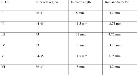
Table 2 : Intra oral sites and corresponding implant dimensions
SITE Intra oral region Implant length Implant diameter
I 46-47 8 mm 4.2 mm
II 44-45 11.5 mm 3.75 mm
III 43 13 mm 3.75 mm
IV 33 13 mm 3.75 mm
V 34-35 11.5 mm 3.75 mm
VI 36-37 8 mm 4.2 mm
The osteotomies were prepared using a contraangle surgical handpiece ( NSK corporation, Japan) mounted on an implant motor (Saeshin, korea) under copious irrigation of cold normal saline( Dexter laboratories , India) under 800 to 1200 rpm speed . All the osteotomies on one side of the arch were completed first and the implants were inserted before the contralateral side osteomies were started.
32
All the implants were placed by the same surgeon for all the study participants inorder to eliminate inter operator bias.
VII . ASSESSMENT OF PRIMARY IMPLANT STABILITY
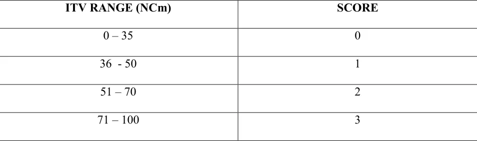
The implants were hand torqued in place and were indexed for favourable emergence. The insertion torque value for each implant was measured in N Cm by means of the calibrated torque wrench and was noted down as assessed by the surgeon. The calibrated torque wrench has readings corresponding to values of 35 Ncm , 50 Ncm,70 Ncm and 100 Ncm. The insertion torque values were taken in reference to the nearest calibration .For example , an ITV between 35 and 50 Ncm is considered to be as >35 Ncm. No definitive numerical value can be obtained but a range is obtained for each implant and scores are assigned based on these ranges. ( table no.6)
33
After assessment of the primary stability of the implants as described above , the implants were covered with a healing collars which were atleast 1 mm above the peri implant soft tissue. Flap closure was obtained by means of multiple interrupted sutures using polyethylene glycol ( vickryl, ethicon, India). The patients were prescribed a course of antibiotics ( amoxycillin ) for 5 days and analgesics ( ketorolac) for 3 days. They were also instructed to use 2 % chlorhexidine mouthwash twice daily for 1 month postoperatively.
VIII.FABRICATION OF HYBRID PROSTHESIS
The implants were subjected to immediate functional loading within 1 week of placement. An implant level screw retained metal – acrylic hybrid prosthesis was fabricated as follows :
A.IMPRESSION MAKING:
34
impression and soft tissue mask was poured. A working model is obtained by pouring type III dental stone (kalabhai, India).
B.FRAMEWORK TRIAL
The model was scanned with a scanner (shining 3D , TDS biotechnologies ,Taiwan) and an implant level titanium milled frame work was obtained.(TDS ME 300 HP - 5 axis milling machine., TDS biotechnologies ,Taiwan).The CAD-CAM fabricated metal framework is tried on the second or third post operative day and a panaromic radiograph is taken to verify the fit of the frame work.
C) JAW RELATION AND WAX TRIAL
A Wax occlusal rim was fabricated on the metal frame work and jaw relation was made opposing the existing maxillary denture. Esthetics , phonetics , occlusal plane and vertical dimension were established. Centric relation record was obtained opposing the existing maxillary denture using Bite Registration paste (exa bite,GC ASIA).The model and the existing maxillary denture were mounted in a semi adjustable articulator using this jaw relation record. Teeth arrangement(Gnathostar,Ivoclar Vivadent Inc,NY ,USA) was done and a wax trial was seen in the patients mouth. A Lingualised occlusal scheme was obtained .
D) INSERTION OF HYBRID PROSTHESIS
35
using calibrated torque wrench. Occlusion was verified and minor occlusal adjustments if any was done in the dentures. The screw access channels in the hybrid denture were closed with a plug of cotton and light cure composite resin . The patient was instructed on oral hygiene maintenance and advised to restrain chewing very hard food items. However the patient was advised to continue his normal food habits. Patients were counseled regarding the recall visits and their continued participation in the study.
IX. Assessment of implant stability – 1 month , 3 months,6 months and 12 months intervals
36
X.Tabulation of data & Statistical analysis
37
METHODOLOGY – OVERVIEW Methodology – Overview
Formulation of Study Design Inclusion & Exclusion Criteria
Case Selection (n = 5) &
Informed Consent
Fabrication of Complete denture
Fabrication of Radiographic template
CBCT Evaluation
Implant Placement (n=30)
Immediate Functional Loading With Hybrid Denture
Aluminium Oxide Blasted / Acid etched Resorbable Blast
Medium Treated (n=15)
Evaluation Of Implant Stability (ISQ) at 1 month, 3 months, 6 months & 12 months
Unblinding of Study & Tabulation of Results
38
The present in vivo study was conducted for the comparative evaluation of clinical stability of two different types of implants with different surface treatments namely i) Al Oxide blasted / acid etched surface & ii) Resorbable blast medium (RBM) (Calcium phosphate) treated surface loaded immediately with a fixed full arch prosthesis.
The test samples (implants) were grouped as follows :
Group A - Al Oxide blasted / acid etched surface (Touareg S) (n=15)
Group B - Resorbable blast medium (RBM) (Calcium phosphate) treated surface (Touareg OS) (n=15).
The samples were placed in 5 patients who received 3 implants of each group in either of the two quadrants of the mandibular edentulous ridge (split mouth design). The initial and progressive stability of these two groups of implants were assessed and the results obtained are tabulated as follows :
39
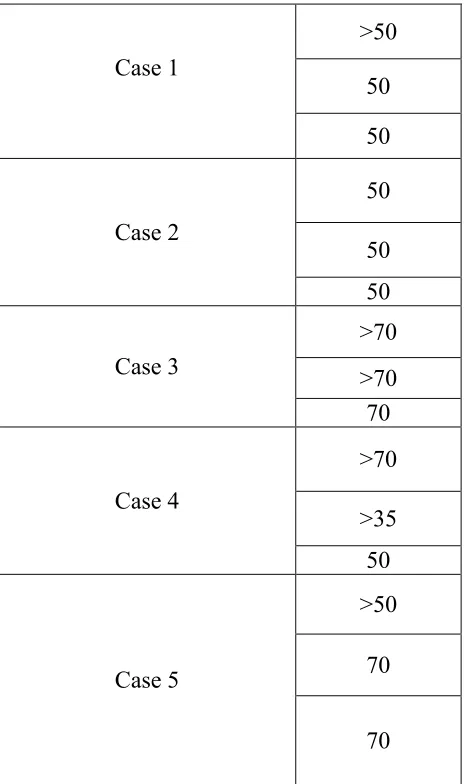
Table 3: Insertion torque values (ITV ) in NCm
Table 4 : Insertion torque values (ITV ) in NCm for Al oxide blasted / acid etched surface Case no Site I Site II Site III Site IV Site V Site VI
Case 1 >50 50 50 70 >50 70
Case 2 >35 70 70 50 50 50
Case3 >70 >70 70 70 50 50
Case 4 >70 >35 70 >70 >35 50
Case 5 >50 70 70 70 >50 >50
Case 1
>50 50 50
Case 2
50 50 50
Case 3
>70 >70 70
Case 4
>70 >35 50
Case 5
>50 70
[image:55.612.191.425.300.692.2]40
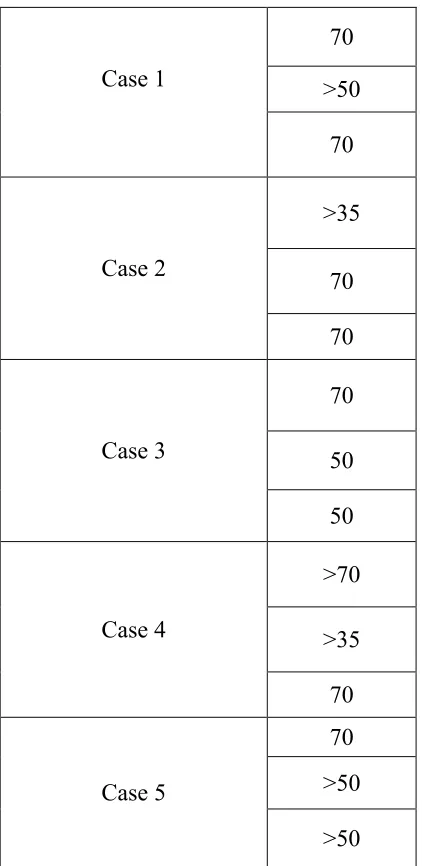
Table 5 : Insertion torque values (ITV ) in NCm for Resorbable blast medium treated surface
Case 1
70 >50
70
Case 2
>35
70 70
Case 3
70
50 50
Case 4
>70
>35 70
Case 5
41
Table 6 : Insertion torque Values and corresponding Scores.
ITV RANGE (NCm) SCORE
0 – 35 0
36 - 50 1
51 – 70 2
71 – 100 3
Table 7 : Comparison of Insertion torque values (ITV ) of Al Oxide blasted / acid etched surface (Group A) and Resorbable blast medium (RBM) treated (Group B)
implants
Group N Mann – whitney U value
P value
A 15
102.500 0.34
[image:57.612.69.541.494.588.2]42
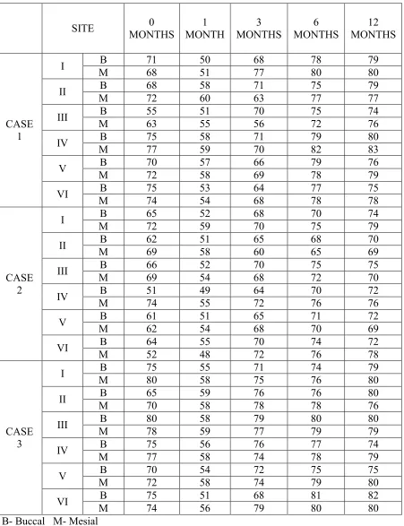
Table 8: Site specific Implant Stability Quotient (ISQ) values
SITE 0
MONTHS 1 MONTH 3 MONTHS 6 MONTHS 12 MONTHS CASE 1
I B 71 50 68 78 79
M 68 51 77 80 80
II B 68 58 71 75 79
M 72 60 63 77 77
III B 55 51 70 75 74
M 63 55 56 72 76
IV B 75 58 71 79 80
M 77 59 70 82 83
V B 70 57 66 79 76
M 72 58 69 78 79
VI B 75 53 64 77 75
M 74 54 68 78 78
CASE 2
I B 65 52 68 70 74
M 72 59 70 75 79
II B 62 51 65 68 70
M 69 58 60 65 69
III B 66 52 70 75 75
M 69 54 68 72 70
IV B 51 49 64 70 72
M 74 55 72 76 76
V B 61 51 65 71 72
M 62 54 68 70 69
VI B 64 55 70 74 72
M 52 48 72 76 78
CASE 3
I B 75 55 71 74 79
M 80 58 75 76 80
II B 65 59 76 76 80
M 70 58 78 78 76
III B 80 58 79 80 80
M 78 59 77 79 79
IV B 75 56 76 77 74
M 77 58 74 78 79
V B 70 54 72 75 75
M 72 58 74 79 80
VI B 75 51 68 81 82
M 74 56 79 80 80
43
Table 8 Continued:
SITE 0
MONTHS 1 MONTH 3 MONTHS 6 MONTHS 12 MONTHS CASE 4
I B 75 58 65 73 75
M 80 59 70 77 76
II B 65 52 66 75 76
M 70 54 72 79 78
III B 80 62 74 80 80
M 78 60 75 79 80
IV B 75 58 71 79 78
M 77 59 70 82 80
V B 70 57 66 79 79
M 72 58 69 78 80
VI B 75 53 64 77 76
M 74 54 68 78 78
CASE 5
I B 68 50 70 72 72
M 66 52 68 70 70
II B 62 53 65 74 72
M 65 51 68 72 70
III B 68 55 70 74 76
M 67 52 72 75 75
IV B 66 54 69 74 78
M 68 54 68 78 78
V B 64 53 68 80 79
M 61 50 69 81 80
VI B 60 51 70 78 78
M 63 52 70 80 81
44
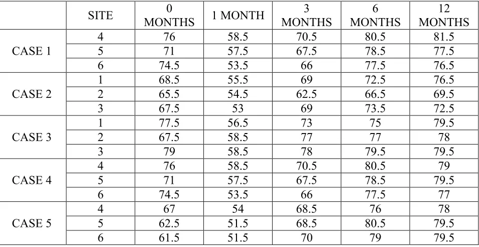
Table 9 : Implant Stability Quotient (ISQ) values for Al oxide blasted / acid etched surface (group A)
SITE 0 MONTH 1 MONTH 3
MONTHS 6 MONTHS 12 MONTHS CASE 1
1 69.5 50.5 72.5 79 79.5
2 70 59 67 76 78
3 59 53 63 73.5 75
CASE 2
4 62.5 52 68 73 74
5 61.5 52.5 66.5 70.5 70.5
6 58 51.5 71 75 75
CASE 3
4 76 57 75 77.5 76.5
5 71 56 73 77 77.5
6 74.5 53.5 73.5 80.5 81
CASE 4
1 77.5 58.5 67.5 75 75.5
2 67.5 53 69 77 77
3 79 61 74.5 79.5 80
CASE 5
1 67 51 69 71 71
2 63.5 52 66.5 73 71
3 67.5 53.5 71 74.5 75.5
Table 10 : Implant Stability Quotient (ISQ) values for Resorbable blast medium treated surface (group B)
SITE 0
MONTHS 1 MONTH
3 MONTHS 6 MONTHS 12 MONTHS CASE 1
4 76 58.5 70.5 80.5 81.5
5 71 57.5 67.5 78.5 77.5
6 74.5 53.5 66 77.5 76.5
CASE 2
1 68.5 55.5 69 72.5 76.5
2 65.5 54.5 62.5 66.5 69.5
3 67.5 53 69 73.5 72.5
CASE 3
1 77.5 56.5 73 75 79.5
2 67.5 58.5 77 77 78
3 79 58.5 78 79.5 79.5
CASE 4
4 76 58.5 70.5 80.5 79
5 71 57.5 67.5 78.5 79.5
6 74.5 53.5 66 77.5 77
CASE 5
4 67 54 68.5 76 78
5 62.5 51.5 68.5 80.5 79.5
[image:60.612.69.546.440.686.2]45
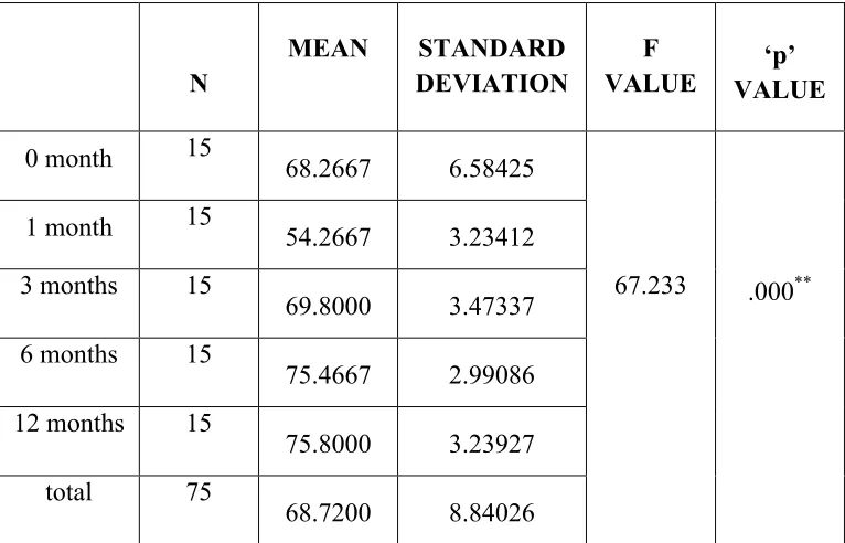
Table 11 : Comparison of mean implant stability quotient (ISQ) values of Al Oxide blasted / acid etched surface (Group A) at different time intervals
N
MEAN STANDARD
DEVIATION
F VALUE
‘p’ VALUE
0 month 15 68.2667 6.58425
67.233 .000**
1 month 15 54.2667 3.23412
3 months 15
69.8000 3.47337 6 months 15
75.4667 2.99086 12 months 15
75.8000 3.23927 total 75
46
Table 12 : post hoc comparison of implant stability quotient (ISQ) of Al Oxide blasted / acid etched surface (Group A) at different time intervals
Tukey’s HSD test
(I) (J)
Mean Difference
(I-J)
Std. Error Sig.
0 month
1 month 14.00000* 1.50833 .000 3month -1.53333 1.50833 .847 6months -7.20000* 1.50833 .000 12months -7.53333* 1.50833 .000
1 month
0 month -14.00000* 1.50833 .000 3month -15.53333* 1.50833 .000 6months -21.20000* 1.50833 .000 12months -21.53333* 1.50833 .000
3month
0 month 1.53333 1.50833 .847 1 month 15.53333* 1.50833 .000 6months -5.66667* 1.50833 .003 12months -6.00000* 1.50833 .002
6months
0 month 7.20000* 1.50833 .000 1 month 21.20000* 1.50833 .000 3month 5.66667* 1.50833 .003 12months -.33333 1.50833 .999
12months
47
Table 13 : Comparison of mean implant stability quotient (ISQ) values of Resorbable blast medium (RBM) treated surface (Group B) at different time intervals
N MEAN STANDARD
DEVIATION
F VALUE
‘p’ VALUE
0 month 15 70.6667 5.44343
77.560 .000** 1 month 15 55.5000 2.59808
3 months 15
69.5667 4.02611 6 months 15
76.8333 3.77334 12 months 15
77.5667 3.04647 total 75
48
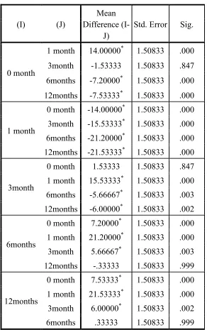
Table 14 : Post hoc comparison of implant stability quotient (ISQ) of Resorbable blast medium (RBM) treated surface (Group B) at different time intervals
Tukey’s HSD test
(I) (J)
Mean Difference
(I-J)
Std. Error Sig.
0 month
1 month 15.16667* 1.42464 .000 3month 1.10000 1.42464 .938 6months -6.16667* 1.42464 .000 12months -6.90000* 1.42464 .000
1 month
0 month -15.16667* 1.42464 .000 3month -14.06667* 1.42464 .000 6months -21.33333* 1.42464 .000 12months -22.06667* 1.42464 .000
3month
0 month -1.10000 1.42464 .938 1 month 14.06667* 1.42464 .000 6months -7.26667* 1.42464 .000 12months -8.00000* 1.42464 .000
6months
0 month 6.16667* 1.42464 .000 1 month 21.33333* 1.42464 .000 3month 7.26667* 1.42464 .000 12months -.73333 1.42464 .986
12months
49
Table 15 : Comparison of implant stability quotients (ISQ) of Al Oxide blasted / acid etched surface (Group A) and Resorbable blast medium (RBM) treated surface
(Group B) implants at different time intervals.
Time
period Group N Mean SD F p
0 month A B 15 15 68.267 70.667 6.5843
5.4434 0.383 0.541
1 month A B 15 15 54.267 55.500 3.2341
2.5981 0.676 0.418
3months A B 15 15 69.800 69.567 3.4734
4.0261 0.002 0.962
6 months A B 15 15 75.47 76.83 2.991
3.773 0.181 0.674
12 months A B 15 15 75.80 77.57 3.239
3.046 0.294 0.592
50
Graph 1 : Mean ISQ values of Al Oxide blasted / acid etched surface (Group A)
Graph 2 : Change in ISQ over 1 year period for Al Oxide blasted / acid etched surface (Group A)
68.26
54.26
69.8
75.46 75.8
1 2 3 4 5
0 Month 1 months 3 Months 6 Months 12 Months
68.26
54.26
69.8
75.46 75.8
1 2 3 4 5
51
Graph 3: Mean ISQ values of Resorbable blast medium (RBM) treated
surface(Group B)
Graph 4 : Change in ISQ over 1 year period for Resorbable blast medium
(RBM) treatedsurface(Group B)
70.66
55.55
69.56
76.83 77.56
1 2 3 4 5
0 Month 1 month 3 Month 6 Month 12 Month
70.66
55.55
69.56
76.83 77.56
1 2 3 4 5
52
Graph 5 : Comparison of ISQ values of Al Oxide blasted / acid etched surface (Group A)
and Resorbable blast medium (RBM) treated surface(Group B) implants at
different time intervals.
0 10 20 30 40 50 60 70 80 90
1 2 3 4 5
Series1 Series2
0 Month 1 month 3 Month 6 Month 12 Month
53
The search for fixed functional and esthetic replacements for
missing natural teeth led to the introduction and subsequent widespread use of dental implants.
Continued research and innovation in implant biomaterials, macro and micro design , surgical
technique and prosthetic options have revolutionalised the field of implant dentistry . Of these ,
surface modification of endosteal implants received great attention among many
researchers1,7,33,44. Attempts to modify implant surfaces were aimed at achieving better
integration at cellular level , obtaining good primary stability , shortening treatment time etc.The
above mentioned aspects have been studied by many authors by employing invitro , histological
and clinical models .Many commercial dental implant manufacturers have developed and
patented unique surface treatments and claim superior results . However the validity of the claim
has to be verified by scientific means . Most of the studies regarding implant surface treatments
and their role in achieving and maintaining implant stability have been carried out in either
animal models or artificial bone models .Prospective controlled human clinical trials on implant
surface treatments are few and limited.
With this background , the present clinical study was done to
evaluate the primary and secondary implant stabilities of two different surface treated implants
and to compare the difference in stabilities of these two groups of implants . The present study
was designed to be a prospective controlled clinical trial employing a split mouth design .Split
mouth designs have many advantages over other clinical study protocols such as elimination of
intra subject bias , standardization of host environment , easy distribution of test samples etc
4
.The completely edentulous mandibular arch was chosen for implant placement in this study.
This is because the bone quality in the mandible is predominantly of D1 and D2 types 4,16. Hence
54
to improve the bone quality by use of condensing osteotomes as is the case with edentulous
maxilla many a times .Also retention can be poor for mandibular complete denture when
compare with maxillary because of the anatomy , pattern of resorption , tongue position and
movement and attachment of muscles. Hence the need to improve the retention of mandibular
complete denture is more pronounced and this can be achieved either by means of removable
overdentures or fully fixed hybrid dentures. Accordingly the subjects in this study were
prescribed a mandibular full arch fixed prosthesis supported by six axially placed root form
endosteal implants opposing a conventional removable maxillary complete denture .
The study was triple blinded – the subject , the surgeon(operator)
and the investigator were blinded so as to eliminate bias at all levels. Since all the implants
placed in this study have the same macro design and there is no visible difference in their
surfaces clinically, the operator cannot identify the group to which the implant belongs to
.Furthermore, the surgical technique is the same for the placement of both the groups of
implants. Investigator bias is eliminated by identifying the implants based on their sites of
placement only. The values are then assigned to the particular sample and group by the
independent research assistant.
Immediate loading of all the implants was done with a metal
reinforced acrylic hybrid prosthesis inserted within a week of implant placement in accordance
to the ITI guide lines .(ITI consenus statement on loading protocols - 2012) . An implant level
screw retained , milled titanium frame work was fabricated since all the implants were axially
55
Primary stability of implants were measured and expressed in two
ways : i) insertion torque value ITV and ii) implant stability quotient(ISQ). The ITVs for
Group A - Al Oxide blasted / acid etched surface (Touareg S) (n=15) and Group B -
Resorbable blast medium (RBM) (Calcium phosphate) treated surface (Touareg OS) (n=15)
were compared using Mann – whitney test for non parametric data and there was no statistically
significant difference between the two groups ( p = 0.34). All the implants achieved an ITV of
>35 N cm , thus fulfilling the inclusion criteria and criteria for immediate functional loading.
The mean ISQ at the time of implant placement for Group A - Al Oxide blasted / acid etched
surface (Touareg S) was found to be 68.267 and that of Group B - Resorbable blast medium
(RBM) (Calcium phosphate) treated surface (Touareg OS) was found to be 70.667. There was
no statistically significant difference between the ISQ s of two groups immediately after
placement ( primary stability) ( p = 0.54). The lack of difference in primary stabilities of the two
groups can be attributed to the fact that both the groups of implants have similar sizes, geometry
and macro design.This finding is in accordance with previous studies such as the one by
Torroella et al where RFA was not able to detect major differences in implant primary stability
between the two implant designs24. Primary implant stability has been proven to be a mechanical
phenomenon .On the other hand , Secondary stability occurs through a cascade of biologic evens
sucha s bone regeneration and remodeling at the bone implant interface.
Another study by Alessandiopozi et al in 2014 affirmed that RFA
measurements present false positive results because it cannot detect the bone – implant contact at
deeper parts2. So it can happen that an implant inserted in a thin cortical bone but with high
density bone at the apical part had acceptable ITV but low ISQ values. However Bischof et al in
56
Primary stability is primarily important for implant
osseointegration. The lack of primary stability has openly been assumed to be the cause factor
for the early implant failure. ( Esposito et al 1998)18. Achieving stability depends on the bone
quality , surgical technique and the micro and macro design of the implant used . Thus implant
stability is the key to clinical success (Gapski et al 2013) 23.Optimal implant stabilization is
especially essential for immediate loading. It is known that implant primary stability depends on
the bone quality , surgical technique and implant design. Implant design plays an important role
in primary intra osseous stabilization. The wide diameter and long implants are recommended in
cases of poor bone density situations.26,40
In the present study , there was a statistically significant difference