1
A RANDOMISED TRIAL TO STUDY THE EFFICACY OF TWO OFFLOADING DEVICES IN THE MANAGEMENT OF PLANTAR
DIABETIC FOOT ULCERS
Dissertation submitted to
THE TAMIL NADU DR. MGR. MEDICAL UNIVERSITY
In partial fulfillment of the degree of
M.S. GENERAL SURGERY Branch – 1
PSG INSTITUTE OF MEDICAL SCIENCE AND RESEARCH DEPARTMENT OF GENERAL SURGERY
2
DECLARATION BY THE CANDIDATE
I hereby declare that this dissertation entitled “A RANDOMISED TRIAL TO
STUDY THE EFFICACY OF TWO OFFLOADING DEVICES IN THE MANAGEMENT OF PLANTAR DIABETIC FOOT ULCERS” is a bonafide and genuine research work carried out by me under the guidance of Dr. VIMAL KUMAR GOVINDAN, M.S., Professor, Department of General Surgery, PSG Institute of Medical Sciences, Coimbatore.
Date: Signature of the Candidate
3
CERTIFICATE BY THE GUIDE
This is to certify that the dissertation entitled “A RANDOMISED TRIAL TO STUDY THE EFFICACY OF TWO OFFLOADING DEVICES IN THE MANAGEMENT OF PLANTAR DIABETIC FOOT ULCERS” is a bonafide work done by Dr. SHEETAL SASIDHARAN in partial fulfillment of the
requirement for the degree of M.S. in General Surgery under my guidance.
Date: Signature of the Guide
Place: Coimbatore Name: Dr. Vimal Kumar Govindan
Professor,
Department of General Surgery,
PSG Institute of Medical Sciences,
4
CERTIFICATE BY THE CO-GUIDE
This is to certify that the dissertation entitled “A RANDOMISED TRIAL TO STUDY THE EFFICACY OF TWO OFFLOADING DEVICES IN THE MANAGEMENT OF PLANTAR DIABETIC FOOT ULCERS” is a bonafide work done by Dr. SHEETAL SASIDHARAN in partial fulfillment of the
requirement for the degree of M.S. in General Surgery under my guidance.
Date: Signature of the Co-Guide
Place: Coimbatore Name: Dr. V. Ramamoorthy
Professor and HOD
Department of PMR,
PSG Institute of Medical Sciences,
5
ENDORSEMENT BY THE HOD/DEAN/HEAD OF THE
INSTITUTION
This is to certify that the dissertation entitled “A RANDOMISED TRIAL TO STUDY THE EFFICACY OF TWO OFFLOADING DEVICES IN THE MANAGEMENT OF PLANTAR DIABETIC FOOT ULCERS” is a bonafide work done by Dr. SHEETAL SASIDHARAN, under the guidance of Dr. VIMAL KUMAR GOVINDAN, Professor, Department of General Surgery, PSG Institute of Medical Science and Research, Coimbatore.
Signature of the HOD Signature of the Principal
Dr.S.Premkumar Dr. Ramalingam. S
Professor and Head, Principal,
Department of General Surgery, Department of General Surgery,
PSG Institute of Medical Sciences, PSG Institute of Medical Sciences,
6
ACKNOWLEDGEMENT
I wish to thank our Principal for having permitted me to conduct this
study in this hospital.
I am ever so grateful to the HOD and the entire faculty of the
Department of Surgery for the kind guidance, valuable advices,
supervision and encouragement while doing this study.
My thanks are also to my colleagues for the considerable help extended
to me.
7
TABLE OF CONTENTS
SI No: PARTICULARS Page No.
1.
ABSTRACT11
2.
INTRODUCTION15
3.
AIMS AND OBJECTIVES18
4.
REVIEW OF LITERATURE20
5.
METHODOLOGY50
6.
RESULTS68
7.
DISCUSSION81
8.
CONCLUSION88
9.
BIBLIOGRAPHY90
10.
ANNEXURES PROFORMA
103
8
LIST OF TABLES
SI No: PARTICULARS Page No.
1.
Age distribution of patients studied60
2.
Gender distribution of patients studied62
3.
Distribution of sites of ulcer64
4.
Adverse events in Total Contact Cast68
5.
Adverse events in Custom made Shoes69
6.
Descriptive characteristics in the two study groups71
7.
Comparative Change in Ulcer Size Between the Two Study Groups73
8.
Percentage Reduction in Wound Size at Each Visit in the Two Study Groups9
LIST OF GRAPHS
SI No: PARTICULARS Page No.
1.
Age wise distribution in Total Contact Cast61
2.
Age wise distribution in Custom made Shoes61
3.
Gender distribution63
4.
Distribution of sites of ulcer65
5.
Adverse events in both study groups69
6.
Average percentage Reduction in Wound Size at Each Visit in the Two Study Groups75
11
ABSTRACT
Introduction
Diabetes related peripheral neuropathy is a major etiological factor in the development of neuropathic foot ulcers. Repeated trauma and pressure on the ulcer bed are the two main reasons for persistence of ulcer.
Offloading allows for pressure relief at areas of high pressure thus facilitating healing process of foot ulcers. It is suggested that “Pressure relief on ulcers commonly referred to as offloading should always be a part of the treatment plan” Cavanagh et al (2005)
Aims and Objectives
The objective of this study is to compare the effectiveness of removable (custom made shoes) and irremovable (total contact cast) devices to offload plantar diabetic ulcers. The following aspects of offloading are looked upon:
1) wound surface areas reduction
12 Materials and Methods
Diabetic foot ulcer patients who have been admitted or have visited PSG hospital on OP basis were included in the study. 32 of these patients, who met inclusion and exclusion criteria of the study, formed the study population. After a detailed history, examination and necessary investigations, ulcers were surgically debrided to remove non viable tissues. Ulcers were photographed and measured and offloading devices were applied. Patients were advised 2 weekly follow-up for the next 3 months. The outcome was studied based on ulcer size reduction and the presence or absence of adverse events.
Results
13
treatment, of which 5 patients healed between 42 to 70days and three out of the thirteen patients (23%) had attained complete healing in custom made shoe group at the end of 12 weeks. The mean number of days for the ulcer to heal in the TCC group was 66.63 days whereas in the custom made shoe group it was 83.23 days which infers that the mean duration of healing time was less with TCC than with custom made shoes.
Conclusion
TCCs are gold standard in offloading diabetic foot ulcers with high proportion of healing rates in lesser amount of time compared to custom made shoes.
15
INTRODUCTION
Diabetic neuropathic ulcers are the most frequent form of ulcers in the foot1. These ulcers are the major determinant of diabetes-related amputations of lower extremity1. About 85% of all the amputation in diabetes is mostly preceded by an ulcer1.
Excessive pressure on sole of these neuropathic feet is the cause for developing these ulcers. The key to effective healing of these ulcers is to provide complete relief of pressure (off-loading) at the site of theses ulcers2. „Offloading is a pivotal but often ignored and neglected aspect of wound care‟- highlighted by Lavery (2003).
Along with pressure relief, surgical debridement of the ulcer and adequate dressing of the wound is essential for the complete healing of these ulcers3. Compliance of patients is generally poor as these patients lack any symptoms due to sensory neuropathy and tend to wear off-loading device very rarely4.
16
debridement, podiatrist in assessing neuro-ischemic status of foot and a technician in off-loading of the ulcer. This team approach helps in effectively treating diabetic foot wounds.
The treatment most commonly employed in pressure reduction at the ulcer site of the foot are either a removable therapeutic shoe or an irremovable total contact cast5.
18
Aims and Objectives
The objective of this study is to compare the effectiveness of removable (custom made shoes) and irremovable (total contact cast) devices to offload plantar diabetic ulcers. Also the following aspects of offloading are looked upon:
1) wound surface areas reduction
20
Review of Literature
History
1.1 History of Diabetes
Diabetes mellitus is one of the many common diseases known to affect the mankind from antiquity6. Its history started approximately in 1550BC7. An ancient literature of Egyptian medical journal has recorded it as a disease causing the patient to lose weight rapidly “Too great emptying of urine” 7
.
Indian physician at the same time identified the sweetness of urine by noting that the urine would attract ants and classified it as madhumeha or “honey urine” 7. It is the Greeks (Apollonius of Memphis) in 230BC who termed it “diabetes” or “to pass through” 7
. The word “mellitus” comes from Latin word meaning sweetened with honey. It was in 1675 when Thomas Willis added mellitus to the word
21
Other historic milestones in diabetes are as follows:
Greek physician Aretaeus of Cappadocia in 2nd century AD noticed the
excess amount of urine being passed through the kidneys also was able to distinguish between Diabetes Melliteus and Diabetes insipidus8..
Aretaeus and Galen Roman physician attributed development of Diabetes to
weakness of kidneys and called it “diarrhea of urine” (diarrhea urinosa) 9.
5th century AD, Sushrutha and Charaka, two Indian physicians
differentiated between two types of diabetes: Type1 diabetes is seen in youths and Type2 diabetes in obese individuals8.
An important milestone in history of diabetes is establishment of role of
liver in glycogen and the fact that diabetes is due to excess glucose production – Claude Bernard in 1857 10.
Avicenna (980-1027)AD gave good description of diabetes and its
22
Mathew Dibson (1713-1784) proved the sweetness of urine is due to sugar
and established sweetness of serum as hyperglycemia. He suggested that diabetes mellitus is a systemic disease9.
Pyrce (1887) described association of foot ulcer, neuropathy and vascular
disease with diabetes mellitus11.
In 1900, the exact link between pancreas and diabetes was established 11.
Charles Best and Fredrick Banting (1922-1936) discovered insulin from
pancreas12.
23 1.2 History of diabetic foot ulcer
Foot ulceration is the most common and disabling complication of diabetes
mellitus. From ancient days, diabetes is a disease characterized by wide range of complications of which foot ulceration can lead to significant disability including lower extremity amputations13.
Between 1850 and 1870, plantar ulcers and gangrene were recognized as
complications of Diabetes11. In 1798, John Rollo noticed that diabetic patients had difficulty in using their limbs due to pain and paraesthesia of lower limbs11.
In earlier days all cases were described as “diabetic gangrene”.
Nitch in 1923 regarded it as senile gangrene due to arteriosclerosis11. Rose and Carless, a decade later recognized the etiology as peripheral
neuritis and endarteritis.
Aird in 1957 identified the importance of infection in young patients. In 1893, a distinction was found between gangrene due to vascular
24
During this time, the only treatment was major amputation of the limb even if the area of gangrene was small 11.
It was regarded that diabetic foot occurred as a result of infection and pressure necrosis of soft tissue which was compressed between callosity of the sole and head of metatarsal bone leading to poor wound healing 14. Also some patients had clawing of foot where the metatarsal heads showed abnormal descent and toes had become hyper extended. 60% of patients with previous ulcer history have chance of developing another ulcer over the same area because the skin over the healed ulcer site will be less resilient to accept repetitive stress. Hence they are more prone to subsequent ulcer15.
1.3 History of treatment of diabetic foot ulcer
In yester years, the final outcome of complications of diabetic foot was invariably amputation16. Even now the cause for inpatient occupancy in diabetic patients is due to foot problems rather than the any other medical complications of diabetes16. Before the Second World War, the gangrenous changes in diabetic foot were
25
gangrene due to vascular insufficiency and gangrene due to infection in a limb with normal blood supply11.
Diabetic foot ulcer healing seems to be arrested at inflammatory or proliferative process, causing infection and inflammation. Many years ago honey was used in dressing of diabetic foot ulcer17. It was found that the anti inflammatory action of honey would decrease the excess activity of collagenase and elastase which are seen in inflammatory condition. It was observed that honey promotes tissue regeneration by stimulating angiogenesis and growth of fibroblasts and epithelial cells17.
For the past 40 years, the concept of moist wound healing of diabetic ulcers has been accepted.
The various types of moist wound dressing are:
26
Collagen dressings: These are used for moderate to heavily draining wounds to enhance healing and tissue repair. These have been found useful on burns, pressure ulcers and dermatologic conditions.
Foam dressings: They are made of hydrophilic polyurethane foam, and offer a moist environment and cushion the wound.
Hydrocolloid dressings: These are soft wafers which become gel like when in contact with wound exudates. They are waterproof and impermeable to bacteria and dust.
27
2. Extent of the problem
2.1 Globally
It has been estimated that, the number of people having diabetes mellitus
worldwide was 131 million in 2000 and it is projected to increase to 366 million by 2030 18. 4%-10% of pts with diabetes have a risk of developing foot ulcer at
28 2.2 India
India has the largest diabetes population in the world. More than 50million people are diagnosed to have diabetes. 85% of amputated cases have found to have
diabetes as the causal factor. The prevalence of amputation is about 3%. In India the prevalence of diabetic foot complications like neuropathy is 15%, peripheral vascular disease is 5% and infections is 7.6%. 55% of foot ulcers are neuropathic, 35% are neuroischemic and 10% are ischemic. Diabetic foot ulcers cause a huge amount of emotional, physical, productivity and financial losses25. .
Pathogenesis of diabetic foot ulcer
29
identifying these high risk patients and educating them will help us in reducing the incidence of foot ulcerations and amputations27.
Diabetes mellitus primarily affects the vessels & nerves causing vasculopathy and neuropathy.
Vasculopathy
Atherosclerotic vascular disease is present in subclinical form in diabetic patients with long duration27. One of the earliest steps in the pathogenesis of
atherosclerosis is the binding of monocytes, leukocytes, and platelets to the
endothelium which is promoted by adhesion molecules. Such adhesion molecules are seen to be elevated in diabetes28. Peripheral vascular disease was found to be 2.5 to 3 times commoner in diabetic than nondiabetic patients29.
Peripheral vascular disease in diabetes mainly effects the vessels between knee and ankle (infra popliteal) leading to poor perfusion of tissues causing friable tissues 27. Thus a mechanical damage to these tissues leads to development of ischemic
30
requirement of blood supply which cannot be met leading to ischemic ulcerations and risk of amputation follow30.
Neuropathy
The incidence of neuropathy is equal to the duration and severity of
hyperglycemia. Patients with neuropathy are at 1.7times greater risk for ulceration compared to patients without neuropathy31.
Causes of neuropathy:
There are 2 theories to causation of diabetic peripheral neuropathy. First, a metabolic factor has been hypothesized as the cause, and the other its association with micro vascular disease. Therefore a nerve biopsy in diabetic neuropathy shows both focal nerve fiber loss along with ischemic injury.
Pathophysiology of diabetic neuropathy32,33
31
2) Hyperosmolality causing edema of nerves.
3) Reduced myoinositol- impairs action of Na- K ATPase and altered myelin synthesis.
4) Occlusive vasanervorum
Effects of neuropathy:
Neuropathy affects the foot both extrinsically and intrinsically.
1. Extrinsic neuropathic foot ulceration
Due to loss of somatic sensation over the plantar aspect of the foot, the patient is unable to perceive the normal painful stimulus. The patient‟s perception of touch, deep pressure, temperature and joint position is impaired. Thus a continuous tissue-damaging excess mechanical load to an insensate foot leads to ulcer formation34,35.
2. Intrinsic neuropathic foot ulceration
32
metabolism. Visceral sensory neuropathy leads to reduced proprioception and the patient continues to walk. The ligaments and joint capsule are further stretched and bony structure of foot is distorted leading to deformities like claw foot with prominent metatarsal heads, or rocker-bottom foot with collapse of longitudinal arch and prominence of tarsal bones. These bony changes produce areas of
localized high pressure in the sole of the foot mainly metatarsal heads, tips of toes and heel35. It initially responds to the high pressure by forming a protective callus and a continued shear force traumatizes the underlying subcutaneous tissue
producing cavities containing blood or serum. Finally the callus breaks down resulting in an ulcer35. The typical feature of neuropathic ulcer is that there will be deep tissue destruction before the epithelial breakdown.
Other risk factors for development of foot ulcers include:
1) Previous foot ulceration: Foot ulcers are more common in those patients with a past history of ulceration or amputation and in patients with a poor social
33
2) Diabetes duration and control: Poor glycemic control as measured by HbAlc, fasting blood glucose, and even single random blood glucose is strongly predictive of subsequent amputation36.
3) Delayed wound healing: In diabetic patients, the process of wound healing is slow and this increases the susceptibility to infection and finally predisposes to amputation37. In diabetic patients, neutrophil function is impaired leading to abnormalities in phagocytosis and killing ability37.
Characteristics of diabetic foot ulcer
Diabetic foot ulcers are most commonly seen over the bony prominences and on the heel. Diabetic foot ulcers are classified into two, ischemic and neurotrophic based on their ulcer characteristics. This classification is important in selecting the appropriate treatment of ulcer.
34
2) Neurotrophic ulcers: These are caused by pressure on the weight bearing areas. The ulcer has granulation surrounded by hyperkeratotic tissue. Good vascular supply is present so the foot is often warm. Callus with bony deformities do occur.
Classification system for Diabetic Foot Ulcers Wagner’s Classification for Diabetic Foot Ulcers
Grade Lesion
1 Superficial diabetic ulcer
2 Ulcer extension involving, ligaments, tendon, joint capsule, or fascia with no abscess or osteomyelitis
3 Deep ulcer with abscess or osteomyelitis
4 Gangrene to portion of fore foot
35
University of Texas Diabetic Wound Classification System
Stage Grade
0 I II III
A (no infection or ischemia)
Pre or post ulcerative lesion completely epithelialised Superficial wound not involving tendon, capsule or bone.
Wound penetrating to tendon, capsule or bone. Wound penetrating to bone or joint.
B Infection Infection Infection Infection
C Ischemia Ischemia Ischemia Ischemia
D Infection and
Ischemia Infection and Ischemia Infection and Ischemia Infection and Ischemia
Treatment of diabetic foot ulcer
36
Strategies for saving the diabetic foot include38:
1) Tight glycemic control
2) Identifying etiological factors 3) Assessment of vascular status 4) Management of infection 5) Offloading strategies
6) Multidisciplinary team approach 7) Patient education
Glycemic control
Adequate glycemic control is the most vital in healing of diabetic foot ulcers. It has been found that leukocyte function is impaired in patients with chronic hyperglycemia leading to delay in wound healing of established foot ulcers38.
Identifying the etiological factors
Foot ulcers are commonly seen in 50% of patients more than 60years39.
37
inflicting trauma while cutting toe nails, history of previous foot ulcers and excessive plantar pressure due to foot deformities39.
Basic examination of foot helps to determine the neuro-vascular status of the foot.
Assessment of vascular status
Palpation of foot pulses (Dorsalis pedis and posterior tibial artery) would be part of the initial examination.
If pulses are feeble or not palpable, vascular investigations are done to evaluate the extent of vaso-occlusion and to assses the healing potential of foot ulcer40.
These include:
1) Doppler study (Duplex scanning with ultrasound analysis) To measure segmental systolic pressure
To provide flow velocity wave form.
38 2) Ankle-Brachial pressure index40
The higher systolic pressure at the ankle is divided by the brachial pressure to give ankle-brachial pressure index (ABPI). Low values are obtained when there is a complete occlusion and high values when there are very minimal atheromatous changes. If there is a constant decrease in ABPI means there is an advancing disease and a constant rise in ABPI indicates development of collaterals.
3) Toe Pressure
Toe pressure measurement is reliable in assessing the healing potential of an ulcer. Transducers are inserted in the sole of footwear to assess the toe pressure. In toe pressure >40mmhg, ulcers heal well. If pressure <20mmhg healing is doubtful41.
4) Transcutaneous oxygen tension (TcPO2)
39 5) Digital subtraction angiography (DSA)43
Vessels are visualized using digital fluorography technique for image enhancement.
Advantage:
DSA accomplishes significantly better contrast resolution.
Highly sensitive screening technique for carotids and lower limb vessels.
When compared to conventional angiography, cost is less. DSA can be performed routinely on OP basis.
DSA may demonstrate small reconstituted vessels distal to an obstruction
not seen on a catheter cut-film study.
An ideal angiography should answer 4 vital questions:
1. The site and extent of the stenosis / occlusion.
2. The „Run-in‟ state of arteries proximal to stenosis is normal or not? 3. The „Run-off‟ Arterial bypass surgery is only feasible if a named distal
40 Neurological assessment of foot44
1. Monofilament test: Using 5.07 Semmes-Weinstein monofilament (10gm) wires for sensory examination.
2. Plantar pressure: Assessed using Harris mat and computer technique. Allows quantitative measurements of plantar foot pressure.
3. Two point discrimination 4. Vibration sense
5. Temperature sensation
Control of wound infection
Early identification of infection and its prompt management is crucial in
41
Sometimes infection of the ulcer can spread into the underlying bone causing osteomyelitis. This is treated by resection of all infected and necrosed bone and antibiotics that penetrate well into the bone46.
Wound care
Initial management includes cleaning of the wound by removing the necrotic and dead tissues and probing the ulcer to check for presence of foreign bodies or to see if the bone is exposed45.
Such sharp debridement of the wound enhances the healing of ulcer which includes removing of the callous using scalpel and forceps. Under aseptic precautions the pus aspirate should be obtained and sent for culture of micro organism and their sensitivity to various antibiotics is tested45. Plain x-ray of foot helps to find out presence of foreign body, gas in tissues or presence of osteomyelitis. Blood
42 Removal of pressure
Elevated plantar pressure is a causative factor in the development of plantar ulcers in diabetic patients with sensory neuropathy45. Repeated trauma and excess
mechanical load over the foot leads to non healing of diabetic foot ulcer. Therefore offloading of the ulcer site helps to prevent repeated trauma and facilitates wound healing. Techniques of removing pressure vary depending on various factors like: patient‟s preference and compliance and severity of ulcer
Patient education
Patent should be educated about good diabetic control, foot care, dangers of smoking etc. All diabetic patients should have an annual foot examination that includes assessing for anatomic deformities, skin breaks, nail disorders, loss of protective sensation, diminished arterial supply, and improper footwear.
Multidisciplinary Team approach
43 Team member‟s include45:
1) Physician to educate about glycemic control. 2) Nurse educator to educate about foot care. 3) Podiatrist to detect and treat foot lesions 4) Surgeon for debriding the ulcer
5) Orthotist for offloading ulcers.
6) Vascular surgeon for revascularization procedures.
Off loading devices
Pressure relief on ulcers and redistributing it to the healthy areas is referred to as offloading47. This reduces the trauma to the ulcer site and allows healing.
Neuropathic ulcers that resisted healing for months to years have healed on use of offloading devices47.
44
person, claw toe deformity and charcot‟s neuroarthropathy are the abnormalities
that can cause disruption of architecture of foot and elevated plantar pressure48.
The use of offloading helps to prevent the repetitive trauma associated with walking which in turn helps in wound healing4.(Amstrong et al 2003)
Offloading of diabetic foot ulcers can be achieved by using various removable and non removable devices49.
Methods of offloading include49:
1. Total non weight bearing- bed rest 2. Total contact cast
3. Foot casts or boots
4. Removable walking braces with rocker bottom sole 5. Half shoes or wedge shoes
6. Accommodative dressing: felt, foam 7. Shoe cutouts
45
Patient compliance is very important when using an offloading device as it is useful only when the patient wears it50. Hence the patient must be educated about the importance of the offloading device in order to improve the patient‟s adherence to offloading. Usually neuropathic patients do not wear their offloading device as they do not feel any pain. That is why total contact cast is considered the golden standard50.
There are various factors that influence a patients desire to wear the device51.
Instability during the gait.
Comfort of the device.
Weight Cosmesis
Ease of application
Removable walker Casts52
Removable walker casts are easy for the patient and the doctor as it can be
46
easy to apply. Removable walker casts are better to use when there is a soft tissue or bone infection because it can be removed to inspect the wound.
Advantages52:
Easily removed for self inspection
Easily removable for local application of therapies.
Easy to daily activities
Can be used for infected wounds
Can be used for superficial wounds
Total Contact Casts (TCC)
This is the most common method of offloading used by doctors53. This method of offloading was first described by Milroy Paul54. TCCs have shown to reduce pressure at ulcer sites by 84-92%. Its healing rates range from 72%-100% over 5-7weeks55. TCC acts by reducing the pressure by transmitting this pressure along the cast wall or to the rear foot. Hence useful in treating fore foot ulcers56.
Advantages of TCCs57:
Protects foot from infection
47 Disadvantages58:
Technically difficult to apply need experienced persons
Time consuming.
Improper application causes skin irritation and ulceration Daily wound assessment cannot be done
Difficulty in daily activities like bathing without wetting the cast
Difficulty in sleeping
Affect gait stability.
Contraindications:
For wounds with ischemia
For infected wounds
48 Half shoes59:
They are designed to decrease the pressure on the fore foot postoperatively.
Advantage:
Removable
Easy to apply – inexpensive
50
MATERIALS AND METHODS
Source of Data:
All patients who have met the inclusion criteria, irrespective of their age or
sex who come to PSG Hospitals, Coimbatore in the department of surgery/ diabetology/medicine with diabetic foot ulcer during the period of February 2012 to December 2013 were included in the study.
Method of Collection of Data:
The data for this study was collected from the 30 subjects fulfilling the
inclusion/exclusion criteria, who came to PSG Hospitals attending Surgery OPD for Diabetic foot ulcer care during the study period February 2012 to December 2013, using a proforma specially designed for the study.
Sample size: 30
51 Inclusion criteria:
Should have Type1 or 2 diabetes mellitus.
Should have peripheral neuropathy60 with a palpable foot pulse.
Should have plantar ulcers for a period of at least 3weeks.
Ulcers should be of Grade 1 according to Wagner‟s Classification61.
Exclusion criteria:
Patients with huge ulcers with size >4cm.
Patients with more than one ulcer in the same foot.
Peripheral vascular disease with ABPI<0.9
Presence of clinical signs of infection- erythema, edema, increase local skin
temp, secretions, fever, gangrene, maggots.
52 Collection of Samples:
30 random cases were selected from the study group either admitted in surgery wards or attending surgery OPD for Diabetic Foot Ulcer management. Cases were selected based on inclusion and exclusion criteria included in our study. Consent was taken from all the subjects.
Patients were randomized through a computerized randomization schedule into two different groups. In which Group A was off-loaded with a non-removable total contact cast, and Group B using Custom Made Shoes. Patients received specific instructions on how to manage the off-loading devices.
A proforma was developed to record the medical history and examination details. Medical history was taken for all the subjects. Details regarding type of diabetes, its duration, treatment, compliance and personal habits were recorded. Patients were assessed for peripheral polyneuropathy which was defined60 as the absence of two of five sensory modalities (vibration sensation using the 128-Hz tuning fork, light touch, blunt-sharp discrimination, Achilles tendon reflex, and 10-g monofilament). Meticulous examination was done including description of the ulcer (site, size, shape and Grade 1 ulcers according to Wagner‟s Classification
53
containing slough were surgically debrided eliminating all non-viable tissues and the entire area of lesion exposed. Ulcers were photographed and traced on to a tracing paper or a graph paper and the surface area of the ulcer was measured and recorded. Following which the ulcers were dressed and covered with sterile dressing before applying the off-loading device.
Patients in Group A were casted with POP (Plaster of Paris) after positioning a layer of protective dressing over the ulcer site to avoid contact with the cast. Attention was given in avoiding friction or trauma with bony prominences by protecting them with extra layers of cotton. Rubber heals were placed to allow the patients to stand and walk.
54
Group A patients treated with Total Contact Cast
55
56
Group B patients treated with Custom made Shoes
Picture showing different types of shoes used to off-load ulcers at
various sites
57
Follow up
58
Outcome
The primary outcome was the rate of wound healing (complete epithelialization or healing at the end of 12weeks). Out of the 33 diabetic patients, 2 failed to complete the course of study. Reasons for this included discomfort, instability (two TCC- cast failures), or failure to return for follow-up appointments (one custom made shoe).
60
Observation and Results
[image:60.612.101.513.342.602.2]Study Design: A randomized control study consisting of 32 patients with diabetic foot ulcers were undertaken to study the effect of removable and non-removable off-loading devices in healing of diabetic foot ulcers and its correlation with various clinical features.
Table 1: Age wise distribution of patients in the two study groups
TCC Shoe
Age in years
No. of
patients Percentage %
No. of
patients Percentage %
30-40 6 37.50 1 7.69
40-50 4 25.00 5 38.46
50-60 2 12.50 4 30.77
60+ 4 25.00 3 23.08
Total 16 13
61
Graph 1: Age wise distribution in TCC
Graph 2: Age wise distribution in Custom-made Shoe
62
[image:62.612.138.475.294.492.2]As shown in Table 1 & Graph 1 and 2, in the present study the age variation was from 34yrs to 68yrs. Majority of the patients were in the age group of 30-40yrs in patients with total contact cast which was 37.5% and age group of 40-50yrs in patients with custom made shoes which was 38.46%.
Table 2: Gender distribution of patients in the two study groups
TCC Shoe
Gender
No. of patients
Percentage %
No. of patients
Percentage %
Male 9 56.25 10 76.92
Female 7 43.75 3 23.08
63
Graph 3 : Gender distribution
64
Table 3: Distribution of sites of ulcers in patients of both study groups
TCC Shoe
Site of Ulcer Number Percentage Number Percentage
Great toe 5 31.2% 4 30.7%
Amputated toe 2 12.5% 1 7.6%
Midfoot 2 12.5% 1 7.6%
1st metatarsal 2 12.5% 3 23%
2nd metatarsal 1 6.2% 1 7.6%
3rd metatarsal - - 1 7.6%
4th metatarsal 3 18.7% - -
5th metatarsal 1 6.2% 1 7.6%
65
Graph 4 : Distribution of sites of ulcer
66
Picture showing various sites of Ulcers
2nd metatarsal head 1st metatarsal head
68
Results
A total of 32 patients were included in the study. Two patients in the TCC group and one patient in the custom made shoe group failed to complete the study. Of these three dropouts, one patient in TCC group developed pre-ulcerative lesion and chafed skin and was not willing for further treatment. The second patient, also in the TCC group, developed discomfort in the cast with instability to walk and breakage of cast, and hence withdrew from the study. One patient in shoe group developed Grade 2 infection which required further debridement and patient lost follow up.
S.No Adverse Events in TCC Treatment
1. Chaffing of skin Adequate padding prior to applying the cast.
2. Breakage of cast Cast was reapplied 3. Discomfort in the cast with
69
S.No Adverse Events in Shoe Treatment
1. Grade 2 infection Minimal debridement and administration of oral antibiotics for 7 days
Graph 5: Adverse events that occurred in both study groups
70
71
[image:71.612.107.499.154.630.2]The Table 4 below shows characteristics of the patients.
Table 4: Descriptive characteristics in the two study groups
TCC Shoe
Variables Mean SD Mean SD
P Value
N 16 13
Age 47.63 10.94 52.00 13.68 0.3466
Male % 56.25 76.92
Duration of Ulcer
(months) 11.06 9.35 5.62 4.37 0.064 Duration of DM
(months) 102.06 70.12 60.54 57.79 0.0982 Ulcer Size at each
visit Surface area of Ulcer (cm2) Initial visit (0
Day) 6.82 4.47 5.86 2.57 0.4961
After 14 Days 5.08 3.52 5.29 2.75 0.8616 After 28 Days 3.60 2.91 4.70 2.96 0.3228 After 42 Days 2.96 2.11 3.86 2.72 0.355 After 56 Days 2.03 1.76 3.50 2.63 0.1128 After 70 Days 1.44 1.52 2.74 2.38 0.1373 After 90 Days 1.34 1.33 2.23 2.10 0.4118 No. of Days for Ulcer
72
There were no significant differences between the two groups in the characteristics evaluated like age, sex and duration of diabetes. The healing time was not influenced by age, sex or duration of diabetes.
Eleven out of the sixteen patients (68.7%) in TCC group had achieved complete healing with 90days of treatment, of which 5 patients healed between 42 to 70days and three out of the thirteen patients (23%) had attained complete healing in custom made shoe group at the end of 12 weeks.
73
Table 5:Comparative Change in Ulcer Size Between the Two Study Groups
No. of
Days TCC Shoe
Ulcer Size(cm2)
P Value1
P Value2
Ulcer Size(cm2)
P Value1
P Value2
0 Day 6.82 5.86
14 Days 5.08 0.2303 0.2303 5.29 0.5932 0.5932 28 Days 3.60 0.0218 0.2036 4.70 0.2991 0.6036 42 Days 2.96 0.008 0.514 3.86 0.0663 0.4578 56 Days 2.03 0.0012 0.2358 3.50 0.033 0.7385 70 Days 1.44 0.0008 0.393 2.74 0.0046 0.4674 90 Days 1.34 0.0154 0.9022 2.23 0.0016 0.5995 P Value1: Comparing each measurement day with the 0 day/baseline
P Value2: Comparing each measurement day with its previous measurement
74
[image:74.612.70.537.293.591.2]The time taken for the ulcer to heal was directly proportional to the area of the ulcer at the time of off-loading. Small ulcers (<3cm) took less than 90days to heal, compared to larger ulcers which took 90days or more for the ulcer to heal.
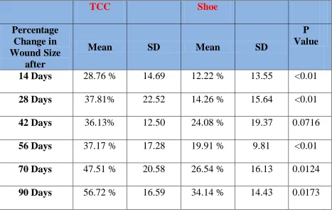
Table 5: Percentage Reduction in Wound Size at Each Visit in the Two Study Groups
TCC Shoe
Percentage Change in Wound Size
after
Mean SD Mean SD
P Value
14 Days 28.76 % 14.69 12.22 % 13.55 <0.01 28 Days 37.81% 22.52 14.26 % 15.64 <0.01 42 Days 36.13% 12.50 24.08 % 19.37 0.0716 56 Days 37.17 % 17.28 19.91 % 9.81 <0.01 70 Days 47.51 % 20.58 26.54 % 16.13 0.0124 90 Days 56.72 % 16.59 34.14 % 14.43 0.0173
75
Graph 6: Comparison of average percentage reduction in ulcer size at each visit in the two study groups
Group A: TCC, Group B: Shoes
76
Graph 7: Comparison of wound healing at end of study
Group A: TCC, Group B: Shoes
77
Pictures Showing Reduction in size of the ulcer over the plantar aspect of great toe during treatment with Total Contact Cast
On Day 0
78
Pictures Showing complete re-epithelialization of the ulcer over the plantar aspect of mid foot after treatment with Total Contact Cast
Picture 1 Picture 2
Picture 1 showing the size of ulcer after 2 weeks of treatment with Total
Contact Cast (14th day)
Picture 2 showing complete re-epithelialization of the size of ulcer after
79
Pictures Showing Reduction in size of the ulcer over the plantar aspect of head of 1st metatarsal during treatment with Custom made Shoes
81
DISCUSSION
The intent of this study was to compare the ability of removable and irremovable devices to effectively offload diabetic neuropathic ulcers. Two aspects of
offloading were looked upon:
1) wound surface areas reduction
2) number and severity of adverse events
The period of this study was from February 2012 to October 2013. Diabetic
82
but only few patients adhered to these instructions. Majority of the patients developed recurrent trauma or infection of the ulcer which required wound debridement, hence leading to further increase in size of these ulcers.
Comparing the acceptability between the two groups, it was found that patients were more easily acceptable with custom made shoes compared to TCC as it had these advantages.
1. Patients were able to enjoy their daily life activities like bathing, sleeping.
2. Shoes could be used for infected wounds and self inspection of wound was possible.
In our study, among the 32 cases of diabetic foot ulcers, who were enrolled,
29cases were studied, which included 16 patients in TCC group and 13 patients in custom made shoe group. Majority of the subjects in the 2 groups were males (56% and 77%) as compared to females (44% and 23%) and were mostly of the age group 30-40 years (37.5%) in TCC group and of 40-50yrs (38.46%) in Custom made shoe group. All the patients included in our study were of Diabetes Mellitus Type II. All the patients included in our study had Wagner‟s Grade I diabetic foot
83
Studies have shown that plantar pressures are highest in the forefoot and less towards the rear and medial arch62. In the present study, majority of the
neuropathic foot ulcers were seen over the plantar aspect more towards the forefoot than in other areas of the foot. Highest percentage of ulcers (31%) were seen over the great toe in both TCC and shoe groups.
Antonella et al (2002) study showed that the forefoot-to-rear foot (F/R) plantar pressure ratio is higher in patients with severe peripheral neuropathy63. This increased F/R plantar pressure ratio causes the forefoot to be more loaded with pressure than rear foot, leading to the development of equinus deformity which is the main causative factor for development of diabetic foot ulcers. His study also showed that highest pressures points were commonly seen along the metatarsal heads.
Increased biomechanical stress is most crucial in leading to ulceration in patients with neuropathic foot ulcers51. In diabetic patients, due to advanced glycation of soft tissues, there occurs a functional shortening of Achilles tendon. This
84
Patients with diabetes also have sensory neuropathy with reduced proprioception35. The ligaments and joint capsule are further stretched and bony structure of foot is distorted leading to deformities like claw foot with prominent metatarsal heads. These bony changes produce areas of localized high pressure in the sole of the foot mainly metatarsal heads, tips of toes and heel35. It would therefore seem
reasonable to assume that reduction of this stress would promote healing. Hence, offloading of diabetic foot ulcer is the treatment for neuropathic diabetic foot ulcers.
Shaw et al in their study noticed that peak plantar pressures seen in the forefoot were remarkably reduced by the use of TCC when compared to Shoes or barefoot walking. TCC achieves forefoot unloading by various mechanisms62, two of which are:
1. Almost 30% of weight from the leg is directly transmitted to the cast wall.
2. TCC removes the load bearing surface from the metatarsal heads by the cavity by the soft foam covering the fore foot.
85
(TCC: 1.34cm2, P value=0.0154, Vs Shoe: 2.23cm2, P value=0.0016). This suggests that the non-removable TCCs heal a higher proportion of wounds by 12 weeks compared to removable Custom made shoes. Hence, we can say that the success of offloading is strictly related to the non removability of the device used. In this present study it was also noticed that there was a faster reduction in size of the ulcer over the first 4weeks (28days) in patients treated with TCC and that after 4 weeks, the ulcer size reduction was almost similar in both the groups.
Hence, it could be suggested that diabetic patients with plantar neuropathic ulcers could start their off-loading with TCC and then switched over to custom made shoes, if they wished.
86
there was an increase in the number of footsteps and activities in patients with removable shoes off than footsteps with removable shoes on50. This revealed that patients were incompliant with removable device. Hence non removable casts are superior over removable devices50 because of the fact that patients cannot remove the TCC and they take less active when using it. Armstrong et al (2003) study also noticed that though the patients with removable shoes were advised to wear the shoes continuously, patients wore them only for 28% of their footsteps52. In our study, most of the patients in the custom made shoes group, admitted to the fact that they wore the shoes only during outdoor activities. It therefore emphasizes the point that effective counseling is necessary prior to starting the treatment with custom made shoes.
It was also seen that patients with TCC had reduced repetitive stress to the ulcer as it was completely covered. This was not the case with custom made shoes,
because the wounds were open. A few patients developed wound infection, which required debridement, thereby further leading to increase in size of ulcer and increased healing time. This in turn explains the success of TCC over Custom made shoes.
87
control edema57. The most important point for TCC is its ability to “force compliance” in patients. Patients using TCC are forced to use the cast all
throughout as the device cannot be removed easily without the clinician‟s orders.
Thus TCC is indeed the better choice to offload neuropathic diabetic plantar ulcers.
88
Conclusion
The main treatment of diabetic neuropathic, non-infected, non-ischemic ulcers is proper debridement and pressure reduction over the ulcer.
In conclusion, this study suggests that based on the different offloading devices selected there is a significant difference in healing of diabetic foot ulcers.
TCCs are gold standard in offloading diabetic foot ulcers with high proportion of healing rates of 68.7%. Eleven out of the sixteen patients achieved complete
90
BIBLIOGRAPHY
1. Reiber GE, Boyko EJ, Smith DG: Lower extremity foot ulcers and amputations in diabetes. Diabetes in America. 2nd ed. National Diabetes Data Group, Eds. Washington, DC, National Institutes of Health, 1995; p. 409–428.
2. Caputo M, Cavanagh PR, Ulbrecht JS, Gibbons GW, Karchmer AW:
Assessment and management of foot disease in patients with diabetes. N Engl J Med , 1994,331:854–860.
3. Miller OF III: Chronic foot wounds in diabetics and total contact casting. Clin Dermatol12:39–45, 1994
91
5. Levin ME: Foot lesions in patients with diabetes mellitus. Endocrinol Metab Clin North Am25:447–462, 1996
6. Ripoll, Brian C. Leutholtz, Ignacio. Exercise and disease management (2nd ed. ed.). Boca Raton: CRC Press. p. 25.
7. Leonid Poretsky, (2009). Principles of diabetes mellitus (2nd ed.). New York: Springer. p. 3.
8. Jacek Zajac, Anil shreshtha, Parini Patel, Leonid Poretsky. Main Events in the History of Diabetes Mellitus. Principles of Diabetes Mellitus 2010, pp 3-16.
9. Dobson, M. (1776). "Nature of the urine in diabetes". Medical Observations and Inquiries 5: 298–310.
92
11. Faris I. A brief history of diabetes and its complications. The management of the diabetic foot.Edinburgh: Churchill Livingstone;1982.p.1-4.
12. Banting FG, Best CH, Collip JB, Campbell WR, Fletcher AA (November 1991). "Pancreatic extracts in the treatment of diabetes mellitus: preliminary report. 1922". CMAJ 145 (10): 1281–6.
13. Boulton AJ, Vileikyte L J Fam Pract. The diabetic foot: the scope of the problem. 2000 Nov; 49(11 Suppl):S3-8.
14. Armstrong DG, Lavery LA, Nixon BP, Boulton .Techniques for debriding and off-loading the diabetic foot wound. AJ Clin Infect Dis. 2004 Aug 1; 39 Suppl 2:S92-9.
15. Helm PA, Walker SC, Pullium GF Arch. Recurrence of neuropathic ulceration following healing in a total contact cast. Phys Med Rehabil. 1991 Nov;
93
16. Dang CN, Boulton . Changing perspectives in diabetic foot ulcer management. AJ Int J Low Extrem Wounds. 2003 Mar; 2(1):4-12.
17. Molan PC. Potential of honey in the treatment of wounds and burns. Am J Clin Dermatology 2001; 2(1):13–19
18. Wrobel JS, Mayfield JA, Reiber GE Geographic variation of lower extremity major amputation in individuals with and without diabetes in the Medicare population. Diabetes care 2001;24:860-864
19. Singh N, Armstrong DG, Lipsky BA . Preventing foot ulcers in patients with diabetes. JAMA. 2005 Jan 12; 293(2):217-28
20. Reiber GE. Epidemiology of foot ulcers and amputations in the diabetic
94
21. Gregg EW, Sorlie P, Paulose-Ram R. et al. Prevalence of lower-extremity disease in the US adult population ≥40 years of age with and without diabetes:
1999-2000 National Health and Nutrition Examination Survey. Diabetes Care. 2004;27:1591-1597
22. National Diabetes DataGroup. Diabetes in America. Vol.2. Bethesda, MD: National Institutes of Health;1995. NIH Pub.No. 95-1468.
23. Ramsey SD, Newton K, Blough D. et al. Incidence, outcomes, and cost of foot ulcers in patients with diabetes. Diabetes Care. 1999;22:382-387
24. Tennvall GR, Apelqvist J, Eneroth M. Costs of deep foot infections in patients with diabetes mellitus. Pharmacoeconomics. 2000;18:225-238
95
26. Reiber GE. The epidemiology of diabetic foot problems. Diabet Med. 1996;13:(suppl 1) S6-S11
27. The Pathogenesis of Diabetic Foot Problems Diabetes 46 (Suppl. 2):S58-S61, 1997
28. Fasching P, Wagner OF. Elevated circulating adhesion molecules in NIDDM: potential mediators in diabetic macroangiopathy.
Diabetology39:1242-1244,1996.
29. Walters DP, Gatling W, Mullee MA, Hill RD. The prevalence, detection and epidemiological correlates of peripheral vascular disease. A comparison of diabetic and non diabetic patients. Diabetic Med9:710-715, 1992.
96
31. Lavery LA, Armstrong DG, Vela SA, Quebedeaux TL, Fleischli. Practical criteria for screening patients at high risk for diabetic foot ulceration. JG Arch Intern Med. 1998 Jan 26; 158(2):157-62.
32. Roy Freeman.MD, Richard S Beaser. Joslin‟s Diabetic text book. 2nd edition. A Guide for Primary Care providers. Chap16. Diabetic Neuropathy;Pg481-518
33. Alvin C Powers. Chap338.Diabetes mellitus. Harrison‟s principles of Internal medicine-Vol II. 17th Edn;Pg2275.
34. Wu SC, Crews RT, Armstrong DG Curr .The pivotal role of offloading in the management of neuropathic foot ulceration. Diab Rep. 2005 Dec; 5(6):423-9.
97
36. Selby JV, Zhang D: Risk factors for lower extremity amputations in persons with diabetes. Diabetes Care 18:509-516,1995
37. Sapico FL, Bessman AN: Diabetic foot infections. In The High Risk Foot in Diabetes Mellitus. Frykeberg RG, Ed. New York, Churchill Livingstone, 1991, p. 197-211
38. Cavanagh PR, Lipsky BA, Bradbury AW et al (2005) Treatment for diabetic foot ulcers. Lancet 366: 1725-35.
39. Young MJ, Boulton AJ, MacLeod AF, Williams DR, Sonksen PH. A multicentre study of the prevalence of diabetic peripheral neuropathy in the United Kingdom hospital clinic population. Diabetologia. 1993;36:150-154.
98
41. Pitei DL, Edmonds ME. Foot pressure measurements. Wounds. 2000;12:(6 suppl B) 19B-29B
42. American Diabetes Association. Peripheral arterial disease in people with diabetes. Diabetes Care. 2003;26:3333-3341
43. Robert B Rutherford. Vascular surgery. 3rd edition.Vol 1, Chap 5; Pg 62
44. Perkins BA, Olaleye D, Zinman B, Bril V. Simple screening tests for
peripheral neuropathy in the diabetes clinic. Diabetes Care. 2001;24:250-256
45. Edmond M (2005) Infection in the neuroischemic foot. Lower extremity wound 4(3):145-53
99
47. Armstrong DG, Lavery LA, Bushman TR Rehabil Res Dev. Peak foot pressures influence the healing time of diabetic foot ulcers treated with total contact casts. 1998 Jan; 35(1):1-5
48. Piaggesi A, Viacava P, Rizzo L, et al. Semiquantative analysis of
histopathological features of the neuropathic foot ulcer: effects of pressure relief. Diabetes Care 2003;26:3123-8.
49. Wu SC, Crews RT, Armstrong DG: The pivotal role of offloading in the management of neuropathic foot ulceration. Curr Diab Rep 5:423–429, 2005
100
51. Armstrong DG, Lavery LA, WU S, Boulton AJM: Evaluation of removable and irremovable cast walkers in the healing of diabetic foot wounds. Diabetes Care 28:551–554, 2005.
52. Lavery LA, Vela Sa, Lavery DC, Quebedeaux TL. Reducing dynamic foot pressures in high-risk diabetic subjects with foot ulcerations. A comparison of treatment. Diabetes care.1996;19(8);818-821.
53. American Diabetes Association consensus development conference on diabetic foot wound care. Diabetes Care 1999; 22(8):1354.
54. Coleman W, Brand PW, Brike JA. The total contact cast: a therapy for planter ulceration on insensitive feet. J Am Podiatr Med Assoc.1984; 74:548 - 552.
101
56. Armstrong DG, Stacpoole-Shea S. Total contact casts and removable cast walkers: mitigation of plantar heel pressure. J Am Podiatr Med
Assoc.1999;89:50-53.
57. Mueller MJ, Diamond JE, Sinacore DR, Delitto A, Blair VPD, Drury DA, Rose SJ:Total contact casting in treatment of diabetic plantar ulcers: controlled
clinical trial. Diabetes Care 12:384 –388, 1989
58. Caravaggi C, Faglia E, De Giglio R, et al: Effectiveness and safety of a nonremovable fiberglass off-bearing cast versus a therapeutic shoe in the treatment of neuropathic foot ulcers: a randomized study. Diabetes
Care 23:1746–1751, 2000
102
60. Schaper NC: Diabetic foot ulcer classification system for research purposes: a progress report on criteria for including patients in research studies. Diabetes Metab Res Rev 20 (Suppl. 1):S90–S95, 2004
61. Frykberg RG, Zgonis T, Armstrong DG et al (2006) Diabetic foot disorders: a clinical practice guideline. J foot ankle surg 45(5) supp: 1-65
62. Shaw JE, Hsi WL, Ulbrecht JS, Norkitis A, Becker MB, Cavanagh PR: The mechanism of plantar unloading in total contact casts: implications for design and clinical use. Foot Ankle Int 18:809–817, 1997
63. Antonella Caselli, John M, et al: The Forefoot-to-Rearfoot Plantar Pressure Ratio Is Increased in Severe Diabetic Neuropathy and Can Predict Foot Ulceration. Diabetes Care 25:1066–1071, 2002
103
Proforma
Patient Identication:
Date: Time: Height: Weight:
1. Describe the reason for the visit?
2. History of present illness / wound status: When the wound was first noticed? How did the wound start?
How do you clean your wound?
What do you use for a dressing change? How often do you change your dressing?
What previous treatments have you had for the wound? Did any previous treatments help your wound?
Does the wound cause you pain?
If so describe the pain on a scale: at its worst______ at its best_______ When you have pain how long does it last?
When does the pain occur most often? What makes the pain worse?
What makes the pain better?
Do you have any other symptom with the pain?
104 2. Past hospitalizations
Date Reason for hospitalization
__________ ____________________________________________ __________ ____________________________________________
3. Current medications:
Medication Dose/How often
__________ ____________________________________________ __________ ____________________________________________ __________ ____________________________________________
4. Allergies Reaction:
__________ ____________________________________________ __________ ____________________________________________ __________ ____________________________________________ __________ ____________________________________________ Allergy to rubber or latex containing products?
5. Habits:
105 6. Diet: Special diet/ diet restrictions:
____________________________________________
7. Family history:
Diabetes Heart problem Stroke Cancer Leg ulcer
8. Social history:
106 EXAMINATION at each visit:
Any new complaints:_________________________________________________ Wound assessment:
Location : Wound etiology:
Size(cm): L____ x B_____ Photo:
Painful: Yes No Wound color:
Fibrous tissue: Granulation tissue Necrotic tissue
Margins: wnl macerated Edema : No Yes
Erythema: No Yes Odour:
107 Pulses:
Dorsalis pedis: R:____, L:______ Popliteal : R:____, L:______ Femoral: R:____, L:______
Deep tendon reflexes:
Sensations: vibratory sensation proprioception Lymphatics:
Labs / Imaging results:
Assessment:
________________________________________________________________
Treatment Plan:
Wound assessed: Yes ( ) No ( ) Wound cleaned: Yes ( ) No ( ) Patient/family education: Yes ( ) No ( ) Sharp debridement: Partial thickness ( ) Full thickness ( )
108 Dressings / cast / device / shoe gear:
_____________________________________________________________
Wound care orders:
_____________________________________________________________
Procedures/ diagnostic tests ordered:
____________________________________________________________
Others:
_____________________________________________________________
Treatment:
_____________________________________________________________
Next visit: 1week 2week 3week 4week
Others: _____________________________________________________
Physician‟s signature and date:
109
110