Copyright © 2001, American Society for Microbiology. All Rights Reserved.
Structural Features of Nectin-2 (HveB) Required for
Herpes Simplex Virus Entry
WANDA M. MARTINEZANDPATRICIA G. SPEAR*
Department of Microbiology-Immunology, Northwestern University Medical School, Chicago, Illinois 60611
Received 14 June 2001/Accepted 8 August 2001
One step in the process of herpes simplex virus (HSV) entry into cells is the binding of viral glycoprotein D (gD) to a cellular receptor. Human nectin-2 (also known as HveB and Prr2), a member of the immunoglobulin (Ig) superfamily, serves as a gD receptor for the entry of HSV-2, variant forms of HSV-1 that have amino acid substitutions at position 25 or 27 of gD (for example, HSV-1/Rid), and porcine pseudorabies virus (PRV). The gD binding region of nectin-2 is believed to be localized to the N-terminal variable-like (V) Ig domain. In order to identify specific amino acid sequences in nectin-2 that are important for HSV entry activity, chimeric mol-ecules were constructed by exchange of sequences between human nectin-2 and its mouse homolog, mouse nectin-2, which mediates entry of PRV but not HSV-1 or HSV-2. The nectin-2 chimeric molecules were ex-pressed in Chinese hamster ovary cells, which normally lack a gD receptor, and tested for cell surface expres-sion and viral entry activity. As expected, chimeric molecules containing the V domain of human nectin-2 exhibited HSV entry activity. Replacement of either of two small regions in the V domain of mouse nectin-2 with amino acids from the equivalent positions in human nectin-2 (amino acids 75 to 81 or 89) transferred HSV-1/ Rid entry activity to mouse nectin-2. The resulting chimeras also exhibited enhanced HSV-2 entry activity and gained the ability to mediate wild-type HSV-1 entry. Replacement of amino acid 89 of human nectin-2 with the corresponding mouse amino acid (M89F) eliminated HSV entry activity. These results identify two different
amino acid sequences, predicted to lie adjacent to the Cⴕand Cⴖbeta-strands of the V domain, that are critical
for HSV entry activity. This region is homologous to the human immunodeficiency virus binding region of CD4 and to the poliovirus binding region of CD155.
The entry of herpes simplex virus (HSV) into cells is a multistep process that requires the interaction of several viral glycoproteins with various cell surface receptors. Initial attach-ment occurs via the binding of viral glycoprotein C (gC) or glycoprotein B (gB) to cell surface heparan sulfate. Subse-quently, glycoprotein D (gD) interacts with one of its several cellular receptors. This somehow triggers fusion of the viral and cellular membranes, a step that requires glycoprotein H (gH), glycoprotein L (gL), gB, gD, a cellular gD receptor, and possibly additional cellular molecules (reviewed in references 40 and 41). Other members of the alphaherpesvirus family, such as porcine pseudorabies virus (PRV) and bovine herpes-virus-1 (BHV-1), encode homologous sets of viral glycopro-teins and enter cells through a very similar mechanism. These animal herpesviruses are able to utilize some of the human receptors for entry, which partly explains their ability to infect cultured human cells (41).
Two of the four gD receptors identified to date are nectin-1 (44), previously named HveC (13), HIgR (8), and Prr1 (22), and nectin-2, previously named HveB (45) and Prr2 (10). The mouse homologs of these human receptors have considerable sequence identity with their human counterparts, 95% for nec-tin-1 and 72% for nectin-2, and also exhibit viral entry activity (25, 38, 39). Both the mouse and human forms of nectin-1 can serve as entry receptors for HSV-1, HSV-2, PRV, and BHV-1
(8, 13, 25, 26, 38). On the other hand, the mouse and human forms of nectin-2 have a more limited entry activity and dif-ferent specificities. Mouse nectin-2 is an entry receptor for PRV but not BHV-1 or HSV strains (39), whereas human nectin-2 can mediate the entry of PRV and variant forms of HSV-1 that have amino acid substitutions at position 25 or 27 of gD (21, 45). HSV-1 strains expressing gDs with the Q27P or Q27R amino acid substitution have been designated Rid vari-ants (9). Human nectin-2 can also serve as a weak receptor for HSV-2 entry but has very little entry activity for wild-type HSV-1 strains (21, 45).
Nectin-1 and nectin-2 belong to a subgroup of the immuno-globulin (Ig) superfamily, based on structural and sequence similarities. Other members of this subgroup include nectin-3 (34, 36) and the poliovirus receptor (CD155) (24). Nectin-3 has no reported viral entry activity, but CD155 is able to me-diate entry of PRV and BHV-1 (13). Each of these proteins can be expressed as multiple isoforms that are secreted or membrane bound due to the use of different C-terminal exons (6, 16, 20, 36, 41). All members of this Ig subfamily contain three homologous Ig domains, an N-terminal variable-like (V) domain and two constant-like (C2) domains. The membrane-bound forms have the most efficient viral entry activity, appar-ently independent of the sequences of the transmembrane region or cytoplasmic tail (8, 13, 21). A secreted form of nec-tin-1 was reported to bind to cells and to have detectable HSV entry activity (20).
The cellular role of the nectins includes participation in cell-to-cell adhesion. These proteins localize to cadherin-based ad-herens junctions in polarized epithelium or to cell junctions in * Corresponding author. Mailing address: Department of
Microbi-ology-Immunology, Northwestern University Medical School, 320 E. Superior St., Mailcode S213, Chicago, IL 60611. Phone: (312) 503-8230. Fax: (312) 503-1339. E-mail: [email protected].
11185
on November 9, 2019 by guest
http://jvi.asm.org/
nonpolarized cells and link neighboring cells through trans -homophilic or heterophilic interactions (36, 43, 44). Certain isoforms of the nectins containing specific carboxy-terminal sequences can associate with the actin cytoskeleton through interactions with afadin, an F-actin binding protein (15, 23, 36). The binding of nectin to afadin is important for localiza-tion of the nectins to cadherin-based junclocaliza-tions (15, 23, 44). Although binding of nectin-1 to afadin is not necessary for HSV entry (8, 13, 35), this interaction does facilitate the effi-cient cell-to-cell spread of HSV infection (35). Since nectin-1 and nectin-2 are probably coexpressed in a variety of cell types, they may serve redundant roles in certain cell types. However, mutations that abolish expression of full-length nectin-1 in humans result in autosomal recessive cleft lip or palate ecto-dermal dysplasia syndrome (42). Knockout of the nectin-2 gene in mice resulted in male sterility through defects in sper-matogenesis, without obvious effects on females (5).
Various lines of evidence suggest that the interaction of HSV with nectin-1 or nectin-2 occurs through the V domain. Soluble nectin-1 consisting of only the V domain blocks the entry of HSV into nectin-1-expressing cells and binds soluble gD (7, 19). Similar results were observed with a truncated nectin-2 protein containing only the V domain when tested with the HSV-1 variant U21 and soluble U21 gD (21). Also, a nectin-1 protein deleted for the two C2-like domains can me-diate HSV entry, albeit inefficiently (7). Epitope mapping of anti-nectin-1 antibodies that can inhibit gD binding and HSV entry suggests that amino acids (a.a.) 80 to 104 of the V domain of nectin-1 are critical for the gD interaction (17).
The aim of this study was to identify specific regions and amino acids within the V domain of nectin-2 that are impor-tant for HSV entry. We chose to study nectin-2 to take advan-tage of the difference in entry specificity of the human and murine homologs. A chimeric approach was used to identify human nectin-2 sequences that are able to confer HSV entry activity on mouse nectin-2 and therefore are critical for viral entry activity. Various sets of nectin-2 chimeras were con-structed by replacing regions of mouse nectin-2 with the cor-responding amino acids of human nectin-2. The chimeric pro-teins were tested for ability to mediate entry of several alphaherpesviruses, including wild-type HSV-1, HSV-1/Rid1, HSV-2, PRV, and BHV-1.
As expected, the V domain of human nectin-2 was shown to contain the critical determinants for HSV entry. Two short regions (a.a. 75 to 81 and a.a. 89) within the V domain of human nectin-2 were identified that could independently con-vert mouse nectin-2 into an entry receptor for HSV-1/Rid1. The resulting chimeras also exhibited HSV-1 and improved HSV-2 entry activity, neither characteristic of the parental molecules. A human nectin-2 molecule containing the substi-tution M89F lost HSV but not PRV entry activity. Thus, we have identified two regions and specific amino acid residues within the V domain of nectin-2 that are critical for HSV entry activity. These amino acid residues map to one side of the V domain within loops adjacent to the C⬘ and C⬙ beta-strands. This region is homologous to that of CD4 and CD155 that is critical for human immunodeficiency virus (HIV) and poliovi-rus entry, respectively. This region is also adjacent to the ho-mologous region in nectin-1 that contains epitopes for mono-clonal antibodies (MAbs) that can inhibit gD binding. In light
of the homology between nectin-1 and -2, these studies will aid in the identification and characterization of specific sequences in nectin-1 that are important for viral entry.
MATERIALS AND METHODS
Cells and viruses.CHO-K1 cells were provided by J. Esko (University of California, San Diego).-Galactosidase (-gal) reporter viruses used were wild-type HSV-1 (strain KOS) and HSV-1/Rid1, described previously as KOS/tk12 and KOS-Rid1/tk12, respectively (45); gH-negative PRV(Kaplan) (3), provided by T. Mettenleiter (Federal Research Center for Virus Diseases of Animals, Insel Reims, Germany); and BHV-1(Cooper)TK-bgal⫹v4a (27), provided by L. Bello (University of Pennsylvania). The reporter virus HSV-2(333)gJ⫺, engi-neered to contain a cytomegalovirus-lacZcassette in place of part of the glyco-protein J (gJ) gene, will be described in detail elsewhere (C. Rowe and P. G. Spear, unpublished results). PRV was propagated and titered on gH-expressing Vero SW78 cells (3), and BHV-1 was propagated and titered on MDBK cells. All other viruses were propagated and titered on Vero cells.
Plasmids. Plasmid pMW20, containing the human nectin-2 open reading frame (ORF) (GenBank AF058448) in pcDNA3, has been described previously (45). This plasmid was used for the expression of human nectin-2 in all assays. Plasmid Mph-pcDNA3, containing the mouse nectin-2 ORF, was provided by D. Shukla (University of Missouri). To generate this plasmid, primers 5⬘-CTGAA GCTTCCCATGGCCCGGGCCGCAGTC and 5⬘-GTCTCTAGAGTAGGGTC ACACGTAAACTGC were used to amplify mouse nectin-2 coding sequences from a 15.5-day-old embryonic mouse (C57BL/6J) cDNA library (Gibco-BRL). The amplified segment was digested withHindIII andXbaI and cloned into pcDNA3 (D. Shukla and P. G. Spear, unpublished results). The coding sequence was identical to that described before (GenBank M12197) (32).
(i) Plasmids expressing chimeric or mutated receptors.All sequences were first cloned into pUC19 (New England Biolabs) for mutagenesis and then cloned into pcDNA3.1 (Invitrogen) for mammalian expression. Site-directed mutagen-esis was performed according to the manufacturer’s instructions using a PCR-based system (QuikChange site-directed mutagenesis kit; Stratagene).
(ii) Plasmids expressing Ch 1 to Ch 5.Plasmid pWM31 contains the mouse nectin-2 ORF altered by the introduction of aSacII restriction site between the V and C2 domains (5⬘-AACGGTACCCGCCGCGGGGTGACCTGG) and an EcoRV site between the C2-C2 domains (5⬘-CCCTCCAGAAGTATCGATAT CCGGCTATGATGAC). Upon sequencing, a nucleotide change was detected in this plasmid which resulted in an amino acid substitution (R463W) in the fifth amino acid from the carboxyl-terminal end of mouse nectin-2. The nectin-2 protein expressed from pWM31 is indistinguishable from that expressed from Mph-pcDNA3 in viral entry assays (data not shown). pWM31 was used in all experiments as the wild-type mouse nectin-2 expression plasmid. pWM33 con-tains the ORF of human nectin-2 (SacI fragment of pMW20) in pUC19 altered by the addition of aSacII site between domains V and C2 (5⬘-GGTCCGTCCG CGGGATGACCTGGCTC) and anEcoRV site between domains C2 and C2 (5⬘-CCTCCTGAAGTGTCGATATCCGGCTATGAT). Plasmid pWM39 (Ch 1) contains the ectodomain of human nectin-2 (SrfI-BlpI fragment of pWM33) fused to the transmembrane (TM) and cytoplasmic domains of mouse nectin-2 (BlpI-SrfI fragment of pWM31). pWM41 (Ch 2) contains the V and first C2 domain of human nectin-2 (SrfI-EcoRV fragment of pWM39) followed by the C2 domain, TM, and cytoplasmic sequences of mouse nectin-2 (EcoRV-SrfI frag-ment of pWM31). pWM38 (Ch 3) contains the V domain of human nectin-2 (SrfI-SacII of pWM33) followed by mouse nectin-2 sequences (SacII-SrfI frag-ment of pWM31). pWM36 (Ch 4) contains the two C2 domains of human nectin-2 (SacII-BlpI fragment of pWM28, a plasmid similar to pWM33) ligated between the V domain and TM sequences of mouse nectin-2 (BlpI-SacII frag-ment of pWM31). pWM20 (Ch 5) contains the ectodomain of mouse nectin-2 (HindIII-BlpI fragment from Mph-pcDNA3) fused to the TM and cytoplasmic domains of human nectin-2 (BlpI-HindIII fragment of pMW20).
(iii) Plasmids expressing Ch 7 to Ch 20, F84 mutants, and M89 mutants.
Plasmids were constructed by performing site-directed mutagenesis on pWM29, which contains the mouse nectin-2 ORF (SacI fragment of pWM33) in pUC19. For the M89 mutants, site-directed mutagenesis was performed on pWM68, which contains the ORF of human nectin-2 (BamHI fragment of pMW20) in pUC19. Mutated plasmids were sequenced to ensure that there were no unin-tended mutations and the mutagenized coding region was cloned into the HindIII site or BamHI site (for human nectin-2 mutants) of pcDNA3.1 for mammalian expression. The nectin-2 molecules are encoded by the following plasmids: Ch 6, pWM61; Ch 7, pWM62; Ch 8, pWM63; Ch 9, pWM64; Ch 10, pWM65; Ch 11, pWM66; Ch 12, pWM75; Ch 13, pWM74; Ch 14, pWM77; Ch 15, pWM78; Ch 16, pWM71; Ch 17, pWM70; Ch 18, pWM72; Ch 19, pWM79;
on November 9, 2019 by guest
http://jvi.asm.org/
Ch 20, pWM76; F84A, pWM87; F84I, pWM89; F84T, pWM88; F84K, pWM85; F84E, pWM86; F84Y, pWM84; M89I, pWM100; and M89F, pWM101.
CELISA for detection of proteins on cell surfaces. A cell enzyme-linked immunosorbent assay (CELISA) was performed as described previously (11, 12). Briefly, transfections were performed on subconfluent CHO-K1 cell monolayers using Lipofectamine (Gibco-BRL). Each well of a six-well plate received 1.5g of plasmid, 5l of Lipofectamine, and Opti-MEM (Gibco-BRL) in 1 ml. Twenty-four hours later, cells were replated into 96-well dishes. The next day, cells were incubated with PBS-BSA (phosphate-buffered saline [PBS] plus 0.5 mM MgCl2, 1 mM CaCl2, and 3% bovine serum albumin [BSA]). This solution was also used for all washes and for antibody dilutions. After 30 min, the appropriate primary antibody was added to cells. Antibodies used included anti-human nectin-2 R146 (45), provided by G. Cohen and R. Eisenberg (University of Pennsylvania), anti-mouse nectin-2 6B3 [␣-mouse nectin-2 (V)], and anti-mouse nectin-2 18C12 [␣-mouse nectin-2 (C2)] (1), provided by A. Nomoto (University of Tokyo). Cells were washed four times and fixed using 2% formaldehyde–0.2% glutaraldehyde in PBS. Then, cells were incubated for 30 min with biotin-conjugated secondary antibody (1:500 dilution) (Sigma), washed as before, and incubated with Amdex streptavidin-conjugated horseradish peroxidase at 1:20,000 dilution (Amersham) for 30 more min. Cells were then washed and reacted with substrate solution containing 3,3⬘,5,5⬘-tetramethylbenzidine (BioFX). At various times after sub-strate addition, plates were read at 370 nm. Alternatively, the reaction was stopped by the addition of stopping solution (BioFX), and plates were read at 410 nm.
Virus entry assays.Virus entry assays were performed as previously described (30). Briefly, CHO-K1 cells were transfected as described above and replated into 96-well plates after 24 h. Cells were then exposed to serial dilutions of -gal-expressing virus diluted in PBS plus 0.1% glucose and 1% heat-inactivated serum. After 6 h, cells were washed, incubated with the-gal substrate ONPG (o-nitrophenyl--D-galactopyranoside) and analyzed as described previously
(30).
RESULTS
Critical determinants for HSV entry map to the V domain of
human nectin-2. To generate the first set of human-mouse
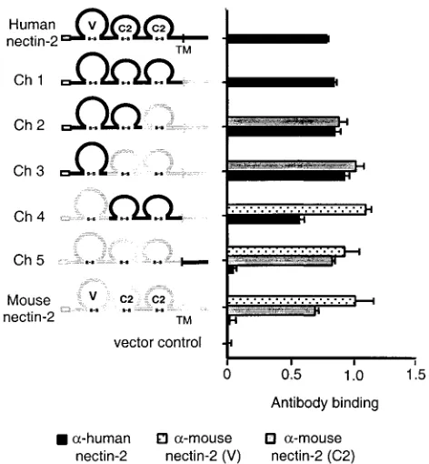
nectin-2 chimeric molecules, identical restriction enzyme sites were engineered between the V-C2 and C2-C2 domains of the cloned human and mouse nectin-2 genes by site-directed mu-tagenesis. These sites were used, together with existing shared enzyme sites, to exchange the V, first C2, or second C2 domain gene segments between the human and mouse nectin-2 genes in different combinations. Five different chimeric molecules were constructed (Fig. 1).
To determine whether the chimeric molecules retained prop-er conformation and wprop-ere expressed on the cell surface, Chi-nese hamster ovary cells (CHO-K1) were transfected with plas-mids expressing the wild-type nectin-2 molecules or chimeras or with control plasmid and tested, by CELISA, for the binding of anti-human or anti-mouse nectin-2 antibodies (Fig. 1). The antibodies used were a polyclonal human nectin-2 anti-body and two monoclonal anti-mouse nectin-2 antibodies that recognize the mouse nectin-2 V domain and second C2 do-main, respectively. Results shown in Fig. 1 demonstrate that the polyclonal anti-human nectin-2 antibodies bound, at near-ly equivalent levels, to all chimeras containing some portion of the ectodomain of human nectin-2. The monoclonal anti-mouse nectin-2 antibodies detected, at similar levels, cell sur-face expression of all chimeric molecules containing the mouse nectin-2 V and C2 domains. These results indicate that all chimeric molecules were expressed on the cell surface as effi-ciently as the parental molecules and retained at least some antigenic determinants.
To determine which chimeric molecules were able to medi-ate HSV entry, viral entry assays were performed. CHO-K1 cells, normally resistant to the entry of HSV, were transfected
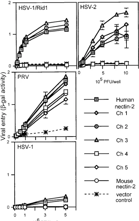
with plasmids expressing wild-type human or mouse nectin-2 or the chimeric molecules or with control plasmid and then exposed to serial dilutions of reporter viruses HSV-1/Rid1 (an HSV-1 variant expressing gD with the amino acid substitution Q27P), HSV-2, and PRV. These reporter viruses contain a
[image:3.587.304.540.69.324.2]lacZcassette in the viral genome and express-gal upon entry into cells.-Gal activity was used as a measure of viral entry. Results from a representative experiment are shown in Fig. 2. Wild-type human and mouse nectin-2 and all the chimeras were functional for PRV entry, which confirms cell surface expression and retention of functional activity by the chimeric molecules. Only chimeric molecules that contained the V do-main of human nectin-2 (Ch 1, Ch 2, and Ch 3) were able to mediate entry of HSV-2 or HSV-1/Rid1 at efficiencies similar to that of wild-type human nectin-2. A very low level of entry activity was also observed with human nectin-2 and Ch 1, Ch 2, and Ch 3 for wild-type HSV-1. Conversely, chimeric molecules containing the V domain of mouse nectin-2 (Ch 4 and Ch 5) FIG. 1. Nectin-2 chimeric molecules and cell surface expression. A schematic representation of human, mouse, and chimeric nectin-2 molecules is shown. Human nectin-2 sequences are represented by black lines, and mouse nectin-2 by gray lines. Empty boxes correspond to the proposed signal peptides, and the transmembrane region (TM) is marked by a vertical line. Ig-like domains (V, C2, and C2) are drawn as loops, connected by the predicted disulfide linkage. The name of each molecule is shown to the left of the drawing. To determine cell surface expression of the various nectin-2 molecules, CHO-K1 cells were transfected with plasmids expressing human or mouse nectin-2 or chimeric molecules or with control plasmid. Forty-eight hours later cells were exposed to the appropriate primary antibody and fixed. Antibody binding was detected with a biotinylated secondary antibody, followed by streptavidin-horseradish peroxidase. Peroxidase activity was used as a measure of antibody binding. Values shown are the means and standard deviations of quadruplicate determinations. Data for each molecule are shown directly to its right. Antibodies used were polyclonal rabbit anti-human nectin-2 antibody (␣-human nectin-2) and monoclonal anti-mouse nectin-2 antibodies recognizing an epitope in the V domain or second C2 domain.
on November 9, 2019 by guest
http://jvi.asm.org/
behaved similarly to wild-type mouse nectin-2 and were not able to mediate HSV entry. Exchange of the other domains (first C2, second C2, or TM-cytoplasmic) did not significantly affect the entry ability of the molecules, which was determined solely by the V domain present. We noted, however, that Ch 3 was reproducibly more efficient than human nectin-2 at medi-ating HSV-2 entry. These experiments indicate that the V domain of human nectin-2 is sufficient to convert mouse nec-tin-2 into a functional HSV entry receptor and support data by others (21) indicating that the V domain of human nectin-2 contains critical determinants for HSV entry into cells.
Identification of amino acids within V domain of human nectin-2 that can confer HSV entry activity on mouse nectin-2.
To identify amino acid residues within the V domain of human nectin-2 required for HSV entry activity, new chimeric mole-cules were constructed in which single or multiple amino acids
in the V domain of mouse nectin-2 were replaced with the corresponding residues from human nectin-2, as explained be-low. HSV entry activity of all chimeras was determined, in viral entry assays, by exposing transfected CHO-K1 cells to serial dilutions of reporter alphaherpesviruses HSV-1, HSV-1/Rid1, HSV-2, PRV, and BHV-1 and measuring -gal activity 6 h after infection. The chimeric molecules were also tested for cell surface expression, in a CELISA, by measuring the binding of anti-mouse nectin-2 (C2) antibody to CHO-K1 cells trans-fected with the various molecules.
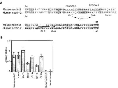
(i) V domain chimeras.Alignment of sequences between the
two cysteines of the V domain of human and mouse nectin-2 identified discrete regions of divergence between these mole-cules (Fig. 3A). Six new chimeric molemole-cules were constructed in which these regions in mouse nectin-2 were replaced by the corresponding sequences from human nectin-2 using site-di-rected mutagenesis. Note that Ch 11 has both of the substitu-tions made individually for Ch 7 and Ch 9. As shown in Fig. 3B, the anti-mouse nectin-2 (C2) antibody bound to CHO-K1 cells expressing mouse nect2 and all the chimeric molecules, in-dicating that the chimeras were expressed on the cell surface. The finding that reduced levels of antibody bound to cells expressing Ch 8 and Ch 10 was reproducible and suggests that these chimeras were not expressed or transported to the cell surface as efficiently as wild-type mouse nectin-2 or that the substitutions in the V domain somehow altered the conforma-tion of the membrane-proximal C2 domain, which seems un-likely.
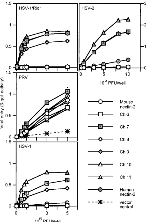
Results of a representative experiment to test the viral entry activity of these chimeric molecules are shown in Fig. 4. All wild-type and chimeric molecules were able to mediate entry of PRV despite the apparently reduced levels of Ch 8 and Ch 10, confirming their cell surface expression and retention of some functional activity. Ch 6, 8, and 10 behaved like mouse nectin-2 and were not able to mediate HSV entry. However, Ch 7, 9, and 11 gained the ability to mediate entry of HSV-1/Rid1 to levels similar to that of human nectin-2. These chimeras were also able to mediate HSV-2 entry but much more efficiently than did human nectin-2. Since the chimeric molecules exhib-ited enhanced HSV-2 entry activity, we tested whether the chimeras could mediate entry of other alphaherpesviruses (HSV-1 and BHV-1). Chimeras 7, 9, and 11 were able to me-diate entry of wild-type HSV-1 into cells much more efficiently than human nectin-2. None of the molecules exhibited BHV-1 entry activity (data not shown). The results from these exper-iments show that two regions from human nectin-2, regions A (a.a. 75 to 81) and B (a.a. 88 to 92) (Fig. 3A), could indepen-dently or jointly transfer HSV-1/Rid1 entry activity to mouse nectin-2. The resulting chimeras also exhibited enhanced HSV-1 and HSV-2 entry activity compared to wild-type human nec-tin-2.
(ii) Single amino acid substitutions.To determine whether
specific amino acids within region A or B of human nectin-2 could transfer HSV entry activity to mouse nectin-2, new chi-meric molecules were constructed in which one or a few amino acids in these regions in mouse nectin-2 were replaced with the corresponding amino acids from human nectin-2. The amino acid sequences of regions A and B of the new chimeras and the wild-type nectin-2 molecules are shown in Fig. 5A.
[image:4.587.49.277.72.434.2]All the region A and B chimeras were expressed on the cell FIG. 2. Alphaherpesvirus entry activities of wild-type and chimeric
nectin-2 molecules. CHO-K1 cells transfected with plasmids express-ing human nectin-2, mouse nectin-2, or chimeric molecules or with control plasmid were replated onto 96 wells and exposed to serial dilutions of reporter viruses (HSV-1/Rid1, PRV, HSV-1, and HSV-2) for 6 h. Infected cells were then washed, and -gal substrate was added.-Gal activity was used as a measure of viral entry. Values shown are the means and standard deviations of triplicate determina-tions. Similar results were obtained in two other experiments.
on November 9, 2019 by guest
http://jvi.asm.org/
surface, as shown by CELISA using the anti-nectin-2 (C2) antibody (Fig. 5B). All the chimeras were also able to mediate entry of PRV, although with different efficiencies, which con-firms cell surface expression and retention of some functional entry activity for the chimeric molecules (Fig. 6 and 7).
A representative experiment for the viral entry assays using region A chimeras is shown in Fig. 6. Ch 7, which contains the entire region A of human nectin-2 (Fig. 3A), was included for comparison. All of the region A chimeric molecules were able to mediate entry of HSV-1/Rid1, but not as efficiently as did human nectin-2 (Fig. 6). These chimeras also gained the ability to mediate entry of HSV-2 to levels similar to, or greater than that of human nectin-2. Only Ch 15 exhibited wild-type HSV-1 entry activity to a level approaching that of Ch 7. These results show that substitutions in region A have different effects on entry of the four alphaherpesviruses tested. None of the sub-stitutions affected PRV entry activity. Any of the subsub-stitutions in region A resulted in acquisition of HSV-1/Rid1 entry activity but not to the levels observed with wild-type human nectin-2. For HSV-2, any of the substitutions in region A is enough for acquisition of entry activity; the more region A amino acids derived from human nectin-2, the more enhanced the entry
activity. For wild-type HSV-1, at least the three amino acids substituted in Ch 15 are required for significant levels of entry activity.
A representative experiment for the viral entry assays using region B chimeras is shown in Fig. 7. Ch 9, which contains the entire region B of human nectin-2 (Fig. 3A), was included for comparison. All chimeric molecules that contained a Met in-stead of Phe at position 84 (Ch 17, Ch 19, and Ch 20) gained the ability to mediate entry of HSV-1/Rid1 and had enhanced entry activity for HSV-1 and HSV-2. Ch 19, containing substi-tution of two amino acids (S83K and F84M) exhibited the highest levels of entry activity for all three HSV strains tested. Other substitutions in this region (Ch 16 and Ch 18) did not confer HSV entry activity on mouse nectin-2. Interestingly, the single amino acid substitution at position 84 (F84M) actually reduced PRV entry activity (Ch 17).
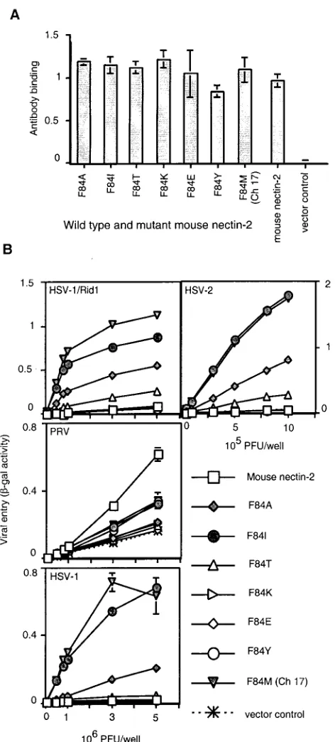
Effect of various substitutions in position 84 of mouse
nec-tin-2 on alphaherpesvirus entry. Ch 17, which contains the
[image:5.587.92.497.73.377.2]F84M mutation, was functional for HSV entry. The Phe at that position in mouse nectin-2 may cause steric hindrance (or a different conformational structure in that area) to prevent HSV entry. Alternatively, a Met, as present in human nectin-2, FIG. 3. Alignment of human and mouse nectin-2 sequences in the V domain and cell surface expression of V domain chimeric molecules. (A) The sequence between the two cysteines of the V domain of mouse nectin-2 is aligned above the corresponding sequence of human nectin-2. Amino acids are numbered from the initial methionine. Gray shading indicates identity between the sequences. New chimeric molecules were constructed by replacing the underlined mouse nectin-2 sequences with the sequences at equivalent positions in human nectin-2 and given the names shown below the human sequence. Ch 11 has both of the substitutions shown for Ch 7 and Ch 9. The amino acids exchanged in Ch 7 and Ch 9 are referred to in the text and later figures as regions A and B, respectively. (B) Cell surface expression of wild-type and V domain chimeric molecules. CHO-K1 cells were transfected with plasmids expressing human nectin-2, mouse nectin-2, or chimeric molecules or with control plasmid and 48 h later tested for the binding of anti-mouse nectin-2 (C2) antibody as explained for Fig. 1. Values shown are the means and standard deviations of triplicate determinations. The same transfected cell populations were used to obtain the results shown in panel B and in Fig. 4.
on November 9, 2019 by guest
http://jvi.asm.org/
may be specifically required for entry activity. To determine the effect on HSV entry of other amino acid substitutions in position 84 of mouse nectin-2, we constructed mouse nectin-2 molecules containing different amino acid residues in that po-sition (Fig. 8). All mouse nectin-2 mutants were expressed on the cell surface at levels similar to mouse nectin-2, as detected by the binding of the anti-mouse nectin-2 (C2) antibody to transfected CHO-K1 cells (Fig. 8A).
The mutant mouse nectin-2 molecules were tested for viral entry activity, and a representative experiment is shown in Fig. 8B. In addition to Ch 17 (F84M), mutants containing either an Ile (F84I) or an Ala (F84A) gained the ability to mediate entry of HSV-1, HSV-1/Rid1, and HSV-2. The activity of F84I was very similar to that of Ch 17 (F84M), indicating that an Ile can substitute very well for Met in that position to allow HSV entry. F84T exhibited a somewhat reduced but detectable entry activity for HSV-1/Rid1 and HSV-2. Mutants having a charged residue (F84K and F84E) or an aromatic amino acid (F84Y) were inactive for viral entry. Some mutants exhibited
signifi-cantly reduced ability to mediate PRV entry (F84E, F84T, F84K, and F84A).
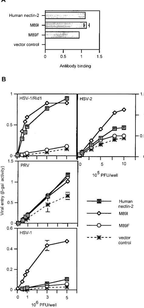
Altering the Met at position 89 of human nectin-2 influences
HSV entry activity.Because changing the Phe at position 84 of
mouse nectin-2 to Met or Ile confers HSV entry activity, we tested the effect of substituting the Met at the equivalent po-sition (a.a. 89) in human nectin-2 with other amino acids. Human nectin-2 mutants were constructed in which the Met at position 89 was replaced by a Phe, as in mouse nectin-2, or by Ile (Fig. 9). We hypothesized that the M89F mutation would eliminate human nectin-2 HSV entry activity. Also, because the F84I substitution in mouse nectin-2 conferred HSV entry activity, the M89I mutation should not affect the ability of human nectin-2 to mediate HSV entry.
[image:6.587.45.280.71.428.2]The human nectin-2 mutants were tested for cell surface expression using the polyclonal anti-human nectin-2 antibody.
[image:6.587.308.534.276.619.2]FIG. 4. Alphaherpesvirus entry activities of V domain nectin-2 chi-meric molecules. CHO-K1 cells transfected with plasmids expressing human nectin-2, mouse nectin-2, or V domain chimeric molecules or with control plasmid were infected with reporter alphaherpesviruses (HSV-1/Rid1, PRV, HSV-1, and HSV-2) as described for Fig. 2. Val-ues shown are the means and standard deviations of triplicate deter-minations. Similar results were obtained in two other experiments.
FIG. 5. Amino acid sequences of regions A and B in nectin-2 chi-meras Ch 12 to Ch 20 and cell surface expression. (A) The amino acid sequences of regions A and B in nectin-2 chimeric molecules are shown between the mouse nectin-2 (lowercase letters) and human nectin-2 (uppercase letters and underlined) sequences. The name given to each chimera is shown to the left of the sequence. (B) Cell surface expression of region A and B nectin-2 chimeric molecules. CHO-K1 cells were transfected with plasmids encoding human nec-tin-2, mouse necnec-tin-2, or region A or B nectin-2 chimeric molecules and tested for the binding of anti-mouse nectin-2 (C2) antibody as described for Fig. 1. Values are the means and standard deviations of quadruplicate determinations.
on November 9, 2019 by guest
http://jvi.asm.org/
As shown in Fig. 9A, the mutants were detected at the cell surface at levels similar to wild-type human nectin-2. When tested for viral entry activity (Fig. 9B), all molecules were able to mediate PRV entry. However, the M89F mutant lost the ability to mediate entry of HSV-1/Rid1, HSV-1, and HSV-2. As hypothesized, substitution of an Ile in this position (M89I) did not affect HSV-1/Rid1 entry. Interestingly, the M89I mu-tant exhibited enhanced HSV-1 and HSV-2 entry activity. Thus, replacing the Met at position 89 with Phe alters the herpesvirus entry activity of human nectin-2, eliminating HSV but not PRV entry activity, yielding an activity profile similar to that of mouse nectin-2. An Ile in the equivalent position is associated with enhanced HSV-1 and HSV-2 entry activity, as when an Ile is present in mouse nectin-2. Clearly the particular amino acid at position 84 in mouse nectin-2 or 89 in human
nectin-2 is crucial for the ability of these cell surface molecules to mediate HSV and PRV entry.
DISCUSSION
[image:7.587.45.279.73.443.2]In this study our goal was to identify structural differences between human and mouse forms of nectin-2 that accounted for ability of the human form but not the mouse form to mediate HSV entry. Our results confirmed the previous finding that determinants for HSV entry reside in the N-terminal V-like Ig domain of human nectin-2 (21). More importantly, they demonstrated that the particular amino acid residues present in two small regions of the V domain determined whether either mouse or human nectin-2 could serve as an HSV entry receptor. Moreover, we found that changes in amino acid se-quence in these regions had different effects on the ability of several alphaherpesviruses tested to utilize these receptors.
[image:7.587.307.536.284.643.2]FIG. 6. Alphaherpesvirus entry activities of region A nectin-2 chi-meric molecules. CHO-K1 cells were transfected with plasmids ex-pressing wild-type and region A chimeric molecules, Ch 7, or control plasmid. Forty-eight hours later, cells were exposed to serial dilutions of reporter alphaherpesviruses (HSV-1/Rid1, PRV, HSV-1, or HSV-2) as described for Fig. 2.-Gal activity was used as a measure of virus entry. The means and standard deviations of triplicate determinations are shown for a representative experiment. The assays were repeated three times with similar results.
FIG. 7. Alphaherpesvirus entry activities of region B nectin-2 chi-meric molecules. CHO-K1 cells were transfected with plasmids ex-pressing wild-type and region B chimeric molecules, Ch 9, or control plasmid. Forty-eight hours later, cells were exposed to serial dilutions of reporter alphaherpesviruses (HSV-1/Rid1, PRV, HSV-1, or HSV-2) as described for Fig. 2.-Gal activity was used as a measure of virus entry. The means and standard deviations of triplicate determinations are shown for a representative experiment. Similar results were ob-tained in two other experiments.
on November 9, 2019 by guest
http://jvi.asm.org/
This indicates that the various forms of alphaherpesvirus gD must differ in their interactions with each of the various wild-type and mutated forms of nectin-2.
The exchange of Ig domains between human and mouse forms of nectin-2 gave straightforward results. All chimeras with the V domain of human nectin-2 had the entry activities characteristic of human nectin-2, and all chimeras with the V domain of mouse nectin-2 had the more restricted entry activ-ity characteristic of mouse nectin-2. This confirms that the V domain contains the critical determinants for HSV entry (21). The only anomaly was the reproducible enhancement of entry activity for HSV-2 when the V domain of human nectin-2 was fused to the remainder of the mouse nectin-2 molecule (Ch 3 in Fig. 2). Possibly, the C2 domains of mouse nectin-2 provide a better scaffold for presentation of the human V domain to HSV-2 than do the C2 domains of human nectin-2.
When short stretches of mouse sequence within the V do-main were replaced with human sequences from equivalent positions, replacements in two of the five regions of divergence tested gave remarkable results. Replacements within regions A or B or both (Fig. 3 to 7) conferred ability to mediate entry of HSV-1/Rid1 and also conferred ability to mediate entry of HSV-1 and HSV-2 much more efficiently than is characteristic of human nectin-2. Further dissection of region A revealed that replacement of mouse amino acids GTV with human amino acids HQN (Ch 15) was almost as effective as substitut-ing with APANHQN (the entire sequence of human nectin-2 present at this position) (Ch 7). Further dissection of region B revealed that all mouse chimeras having Met at position 84 (as in human nectin-2 in the equivalent position) had acquired HSV-1/Rid1 entry and also new entry activities, whereas all those that retained the Phe at position 84 had acquired no new entry activities, irrespective of other changes made in this re-gion. The optimal replacement in region B, even better than replacing the entire region, was the substitution of mouse amino acids SF at positions 83 and 84 with the human amino acids KM from the equivalent positions 88 and 89.
Replacement of amino acids in region A or B of mouse nectin-2 with human nectin-2 sequences conferred normal (for HSV-1/Rid1) or enhanced (HSV-1 and HSV-2) entry activi-ties. However, the replacement of one amino acid in region B of human nectin-2 with the corresponding mouse amino acid (M89F) eliminated all HSV entry activities, even though re-gion A was unchanged. Thus, the presence of a Phe at position 89 in the context of the human V domain or at position 84 in the context of the mouse V domain is associated, in both cases, with absence of any HSV entry activity. The presence of a Phe at position 84 in the context of a mouse V domain containing human sequences in region A is associated with HSV entry activity. Whether the presence of a Phe at position 84 in mouse nectin-2 permits HSV entry activity depends on the sequence in region A. It might be possible to alter human nectin-2 so that presence of a Phe at position 89 would also permit HSV entry activity.
[image:8.587.47.283.67.589.2]The amino acid present at position 84 in the mouse V do-main or at position 89 in the human V dodo-main is clearly critical for entry of all the viruses tested, but the particular amino acid preferred at this position for optimal entry activity is different for PRV and the HSV strains. The only molecules that exhib-ited reduced entry activity for PRV were Ch 9 (which has the FIG. 8. Cell surface expression and alphaherpesvirus entry
activi-ties of mouse nectin-2 mutants containing amino acid substitutions at position 84. (A) Six mouse nectin-2 mutants were constructed in which the amino acid at position 84 was replaced with another amino acid (Ala, Ile, Thr, Lys, Glu, or Tyr) and given the name of the substitution as shown on thexaxis. To assess cell surface expression, CHO-K1 cells transfected with plasmids expressing the mouse nectin-2 position 84 mutants, wild-type mouse nectin-2, or control plasmid were tested for the binding of anti-mouse nectin-2 (C2) antibody as described for Fig. 1. Results shown are the means and standard deviations of triplicate determinations. (B) CHO-K1 cells transfected as above were infected with reporter alphaherpesviruses (HSV-1/Rid1, PRV, HSV-1, and HSV-2) as described for Fig. 2. The means and standard deviations of triplicate determinations are shown for a representative experiment. The assays were repeated twice with similar results.
on November 9, 2019 by guest
http://jvi.asm.org/
entire region B replaced with human sequences) and various mouse nectin-2 mutants containing substitutions in position 84 in region B, including F84M (Ch 7). For PRV entry, the pre-ferred amino acid at position 84 was Phe, at least for mouse nectin-2. The results obtained with the HSV strains provide a clear contrast. Phe at position 84 in mouse (wild-type mouse nectin-2) or 89 in human (M89F) was associated with absence of HSV entry activity, suggesting that PRV and HSV strains respond differently to the presence of Phe at this position. The substitutions F84M and F84I in mouse nectin-2 conferred en-try activity for HSV-1/Rid1 to levels similar to that observed for human nectin-2. The same substitutions conferred en-hanced entry activity for wild-type HSV-1 and HSV-2, high-lighting differences in entry requirements for HSV-1/Rid and wild-type HSV-1 and HSV-2. Also, the M89I substitution in human nectin-2 was without effect on HSV-1/Rid1 entry, whereas the M89F substitution eliminated this entry activity. As expected, the M89F substitution also eliminated the entry activity observed for wild-type HSV-1 and HSV-2, but surpris-ingly, the M89I substitution enhanced entry activity for both wild-type HSV-1 and HSV-2. Assuming that these variations in sequence of the nectin-2 V domain influence gD binding, the gDs encoded by PRV and HSV-1/Rid1 must each differ from those encoded by HSV-1 and HSV-2 in their interactions with each form of nectin-2 tested. The differences observed between HSV-1 and HSV-2 entry seem to be principally quantitative and not qualitative.
Although the direct binding of alphaherpesvirus gDs to nec-tin-1 and other entry receptors has been observed (7, 12, 18, 19, 25, 38, 46), it has been difficult to detect the binding of gD to nectin-2. A previous report demonstrated by ELISA the weak binding of the variant form of gD U21 (amino acid substitution at position 25) to soluble forms of human nectin-2 (21). We were able to detect low levels of binding of a soluble form of HSV-1/Rid1 gD to cells expressing a variant form of mouse nectin-2 with high entry activity (Ch 19), but not to wild-type human nectin-2 or some of the other active chimeras. The failure to detect HSV gD binding to human nectin-2 is prob-ably due to low affinity of the interaction. Alterations made here, resulting in chimeric molecules with enhanced entry ac-tivity, may also enhance the affinity of gD-nectin-2 interactions enough to begin to detect binding of soluble gD to cells or binding of gD to receptor by ELISA. Other methods will have to be devised to obtain quantitative comparisons of the gD-receptor interactions, in order to determine whether the ap-parent affinity of the interaction correlates with entry activity of the parental and chimeric receptors described here.
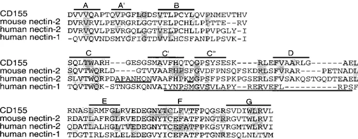
[image:9.587.301.538.80.587.2]Alignment of mouse and human nectin-2 sequences with that of CD155 allowed us to make rough predictions about the location of the amino acids in regions A and B relative to the beta-strands that form the V domain (Fig. 10). Loop regions between the beta-strands, specifically B-C, C-C⬘, C⬘-C⬙, C⬙-D, and D-E loops, showed the most variability in sequence among the molecules. Region A appears to localize to an area encom-passing the C-C⬘ loop and region B to the C⬘-C⬙ loop. The C⬘-C⬙region is predicted to be exposed and available for in-teractions with virus, and in fact, equivalent regions of other members of the Ig superfamily have been shown to be contact points for viruses. Studies on CD155 have identified the C⬘ -C⬙-D region as an important component of the binding site for
FIG. 9. Cell surface expression and alphaherpesvirus entry activi-ties of human nectin-2 mutants containing amino acid substitutions at position 89. (A) Two human nectin-2 mutants were constructed in which the amino acid at position 89 was exchanged for an Ile or a Phe, as shown on theyaxis. To assess cell surface expression, CHO-K1 cells transfected with plasmids expressing wild-type human nectin-2, M89I, M89F, or control plasmid were tested for the binding of anti-human nectin-2 antibody, as described for Fig. 1. Results shown are the means and standard deviations of triplicate determinations. (B) Alphaherpes-virus entry activities of wild-type human nectin-2 and position 89 mu-tants. CHO-K1 cells were transfected as above and 48 h later exposed to serial dilutions of reporter alphaherpesviruses (HSV-1/Rid1, PRV, HSV-1, and HSV-2) as described for Fig. 2. Results shown are the means and standard deviations of triplicate determinations. The assays were repeated three times with similar results.
on November 9, 2019 by guest
http://jvi.asm.org/
poliovirus. Specifically, amino acids localized to the C⬘-C⬙loop are crucial for viral binding to receptor (4, 31). Substitution of a single amino acid to a bulkier residue (Q55F), in the position equivalent to the Met in region B of human nectin-2, abolished poliovirus binding (31). The region in CD4 encompassing the C⬘-C⬙-D beta-strands and intervening loops has been identified as the binding site for HIV (2, 29). Transfer of this region of human CD4 to the rat homolog can confer HIV entry activity on the hybrid molecule (37). In addition, substitutions of ami-no acids in the C⬘-C⬙ loop affect virus binding (33). Thus, it seems that the equivalent region of the V domains of different Ig molecules can be utilized by several viruses for binding to the cell surface and for entry.
Because nectin-2 is believed to be a gD receptor, as are all the other HSV entry receptors, regions A and B in human nectin-2 could form part or all of the contact site with gD or could otherwise influence the conformation of the contact site. In a recent study of human nectin-1, three monoclonal anti-bodies were shown to inhibit the binding of gD to nectin-1. The epitopes of two of these antibodies were mapped using over-lapping peptides (17). These epitopes were localized to se-quences homologous to region B in nectin-2 and extending downstream to the D-E loop (Fig. 10). Taken together, these results suggest that the region in nectin-2 including the C⬘and C⬙beta-strands and surrounding loops is the site to which HSV gD binds.
It has been shown that soluble forms of HSV-1 gD can inhibit cell adhesion mediated by nectin-1 (35), suggesting that the interface fortrans-homophilic interactions between nec-tin-1 ectodomains may overlap with the gD-binding domain. We have preliminary evidence, however, that the M89F muta-tion in human nectin-2 has little or no effect ontrans -homo-philic interactions between nectin-2 ectodomains despite the absence of HSV entry activity. A mutation at position 136 in mouse nectin-2 was reported to abolish thetrans-homophilic interaction (28). Work is in progress to assess the effects of an equivalent mutation in human nectin-2 and other mutations on both alphaherpesvirus entry andtrans-homophilic interactions. Because of the homology between nectin-1 and nectin-2, the information obtained in this study on nectin-2 will facilitate identification of the regions in nectin-1 that are critical for
alphaherpesvirus entry activity. Also, the nectin-2 chimeras and mutants generated in this study will be useful for high-resolution structural studies designed to determine how the variations in primary sequence influence three-dimensional structure, interactions with various forms of alphaherpesvirus gD, and entry activity.
ACKNOWLEDGMENTS
We thank N. Susmarski and M. L. Parish for excellent technical assistance and C. Rowe, G. Cohen, R. Eisenberg, and A. Nomoto for reagents.
This work was supported by Public Health Service grants R37 AI36293 and U19 AI31494 from the National Institute for Allergy and Infectious Diseases. W.M.M. was supported by Public Health Service fellowship F31 GM 19765.
REFERENCES
1.Aoki, J., S. Koike, H. Asou, I. Ise, H. Suwa, T. Tanaka, M. Miyasaka, and A. Nomoto.1997. Mouse homolog of poliovirus receptor-related gene 2 prod-uct, mPRR2, mediates homophilic cell aggregation. Exp. Cell Res.235:374– 384.
2.Arthos, J., K. C. Deen, M. A. Chaikin, J. A. Fornwald, G. Sathe, Q. J. Sattentau, P. R. Clapham, R. A. Weiss, J. S. McDougal, C. Pietropaolo, R. Axel, A. Truneh, P. J. Maddon, and R. W. Sweet.1989. Identification of the residues in human CD4 critical for the binding of HIV. Cell57:469–481. 3.Babic, N., B. G. Klupp, B. Makoschey, A. Karger, A. Flamand, and T. C.
Mettenleiter.1996. Glycoprotein gH of pseudorabies virus is essential for penetration and propagation in cell culture and in the nervous system of mice. J. Gen. Virol.77:2277–2285.
4.Bernhardt, G., J. Harber, A. Zibert, M. de Crombrugghe, and E. Wimmer.
1994. The poliovirus receptor: identification of domains and amino acid residues critical for virus binding. Virology203:344–356.
5.Bouchard, M. J., Y. Dong, B. M. McDermott, Jr., D.-H. Lam, K. R. Brown, M. Shelanski, A. R. Bellve, and V. R. Racaniello.2000. Defects in nuclear and cytoskeletal morphology and mitochondrial localization in spermatozoa of mice lacking nectin-2, a component of cell-cell adherent junctions. Mol. Cell. Biol.20:2865–2873.
6.Campadelli-Fiume, G., F. Cocchi, L. Menotti, and M. Lopez.2000. The novel receptors that mediate the entry of herpes simplex viruses and animal al-phaherpesviruses into cells. Rev. Med. Virol.10:305–319.
7.Cocchi, F., M. Lopez, L. Menotti, M. Aoubala, P. Dubreuil, and G. Cam-padelli-Fiume.1998. The V domain of herpesvirus Ig-like receptor (HIgR) contains a major functional region in herpes simplex virus-1 entry into cells and interacts physically with the viral glycoprotein D. Proc. Natl. Acad. Sci. USA95:15700–15705.
8.Cocchi, F., L. Menotti, P. Mirandola, M. Lopez, and G. Campadelli-Fiume.
1998. The ectodomain of a novel member of the immunoglobulin subfamily related to the poliovirus receptor has the attributes of a bona fide receptor for herpes simplex virus types 1 and 2 in human cells. J. Virol.72:9992– 10002.
[image:10.587.110.478.72.215.2]9.Dean, H. J., S. Terhune, M.-T. Shieh, N. Susmarski, and P. G. Spear.1994. FIG. 10. Alignment of nectin-2, nectin-1, and poliovirus receptor (CD155) V domain sequences. The sequence of the V domain of CD155 (24) was aligned with the corresponding sequences of mouse and human nectin-2 and human nectin-1. The proposed location of the beta-strands of CD155 (14) is indicated above the CD155 sequences. Gray shading represents identity in amino acid of at least three of the molecules. Regions A and B of human nectin-2 are underlined. The proposed epitopes for MAbs that inhibit gD binding to nectin-1 are underlined (17).
on November 9, 2019 by guest
http://jvi.asm.org/
Single amino acid substitutions in gD of herpes simplex virus 1 confer resistance to gD-mediated interference and cause cell type-dependent alter-ations in infectivity. Virology199:67–80.
10.Eberle´, F., P. Dubreuil, M. G. Mattei, E. Devilard, and M. Lopez.1995. The human PRR2 gene, related to the human poliovirus receptor gene (PVR), is the true homolog of the murine MPH gene. Gene159:267–272.
11.Geraghty, R. J., A. Fridberg, C. Krummenacher, G. H. Cohen, R. J. Eisen-berg, and P. G. Spear.2001. Use of chimeric nectin-1 (HveC)-related re-ceptors to demonstrate that ability to bind alphaherpesvirus gD is not nec-essarily sufficient for viral entry. Virology285:366–375.
12.Geraghty, R. J., C. R. Jogger, and P. G. Spear.2000. Cellular expression of alphaherpesvirus gD interferes with entry of homologous and heterologous alphaherpesviruses by blocking access to a shared gD receptor. Virology
268:147–158.
13.Geraghty, R. J., C. Krummenacher, G. H. Cohen, R. J. Eisenberg, and P. G. Spear.1998. Entry of alphaherpesviruses mediated by poliovirus receptor-related protein 1 and poliovirus receptor. Science280:1618–1620. 14.He, Y., V. D. Bowman, S. Mueller, C. M. Bator, J. Bella, X. Peng, T. S. Baker,
E. Wimmer, R. J. Kuhn, and M. G. Rossmann.2000. Interaction of the poliovirus receptor with poliovirus. Proc. Natl. Acad. Sci. USA97:79–84. 15.Ikeda, W., H. Nakanishi, J. Miyoshi, K. Mandai, H. Ishizaki, M. Tanaka, A.
Togawa, K. Takahashi, H. Nishioka, H. Yoshida, A. Mizoguchi, S.-I. Nishi-kawa, and Y. Takai.1999. Afadin: a key molecule essential for structural organization of cell-cell junctions of polarized epithelia during embryogen-esis. J. Cell Biol.146:1117–1131.
16.Koike, S., H. Horie, I. Ise, A. Okitsu, M. Yoshida, N. Iizuka, K. Takeuchi, T. Takegami, and A. Nomoto.1990. The poliovirus receptor protein is produced both as membrane-bound and secreted forms. EMBO J.9:3217–3224. 17.Krummenacher, C., I. Baribaud, M. Ponce de Leon, J. C. Whitbeck, H. Lou,
G. H. Cohen, and R. J. Eisenberg.2000. Localization of a binding site for herpes simplex virus glycoprotein D on herpesvirus entry mediator C by using antireceptor monoclonal antibodies. J. Virol.74:10863–10872. 18.Krummenacher, C., A. V. Nicola, J. C. Whitbeck, H. Lou, W. Hou, J. D.
Lambris, R. J. Geraghty, P. G. Spear, G. H. Cohen, and R. J. Eisenberg.
1998. Herpes simplex virus glycoprotein D can bind to poliovirus receptor-related protein 1 or herpesvirus entry mediator, two structurally unreceptor-related mediators of virus entry. J. Virol.72:7064–7074.
19.Krummenacher, C., A. H. Rux, J. C. Whitbeck, M. Ponce de Leon, H. Lou, I. Baribaud, W. Hou, C. Zou, R. J. Geraghty, P. G. Spear, R. J. Eisenberg, and G. H. Cohen.1999. The first immunoglobulin-like domain of HveC is sufficient to bind herpes simplex virus gD with full affinity, while the third domain is involved in oligomerization of HveC. J. Virol.73:8127–8137. 20.Lopez, M., F. Cocchi, E. Avitabile, A. LeClerc, J. Adelaide, G.
Campadelli-Fiume, and P. Dubreuil.2001. Novel, soluble isoform of the herpes simplex virus (HSV) receptor nectin-1 (or PRR1-HIgR-HveC) modulates positively and negatively susceptibility to HSV infection. J. Virol.75:5684–5691. 21.Lopez, M., F. Cocchi, L. Menotti, E. Avitabile, P. Dubreuil, and G.
Cam-padelli-Fiume.2000. Nectin2␣(PRR2␣or HveB) and nectin2␦are low-efficiency mediators for entry of herpes simplex virus mutants carrying the Leu25Pro substitution in glycoprotein D. J. Virol.74:1267–1274. 22.Lopez, M., F. Eberle´, M. G. Mattei, J. Gabert, F. Birg, F. Bardin, C. Maroc,
and P. Dubreuil.1995. Complementary DNA characterization and chromo-somal localization of a human gene related to the poliovirus receptor-en-coding gene. Gene155:261–265.
23.Mandai, K., H. Nakanishi, A. Satoh, H. Obaishi, M. Wada, H. Nishioka, M. Itoh, A. Mizoguchi, T. Aoki, T. Fujimoto, Y. Matsuda, S. Tsukita, and Y. Takai.1997. Afadin: a novel actin filament-binding protein with one PDZ domain localized at cadherin-based cell-cell adherens junction. J. Cell Biol.
139:517–528.
24.Mendelsohn, C. L., E. Wimmer, and V. R. Racaniello.1989. Cellular recep-tor for poliovirus: molecular cloning, nucleotide sequence, and expression of a new member of the immunoglobulin superfamily. Cell56:855–865. 25.Menotti, L., E. Avitabile, P. Dubreuil, M. Lopez, and G. Campadelli-Fiume.
2001. Comparison of murine and human nectin-1 binding to herpes simplex virus glycoprotein D (gD) reveals a weak interaction of murine nectin-1 to gD and a gD-dependent pathway of entry. Virology282:256–266. 26.Menotti, L., M. Lopez, E. Avitabile, A. Stefan, F. Cocchi, J. Adelaide, E.
Lecocq, P. Dubreuil, and G. Campadelli-Fiume.2000. The murine homolog of human Nectin1␦serves as a species nonspecific mediator for entry of human and animal␣herpesviruses in a pathway independent of a detectable binding to gD. Proc. Natl. Acad. Sci. USA97:4867–4872.
27.Miller, J. M., C. A. Whetstone, L. J. Bello, W. C. Lawrence, and J. C. Whitbeck.1995. Abortion in heifers inoculated with a thymidine
kinase-negative recombinant of bovine herpesvirus 1. Am. J. Vet. Res.56:870–874. 28.Miyahara, M., H. Nakanishi, K. Takahashi, K. Satoh-Horikawa, K. Tachi-bana, and Y. Takai.2000. Interaction of nectin with afadin is necessary for its clustering at cell-cell contact sites but not for itscisdimerization or transinteraction. J. Biol. Chem.275:613–618.
29.Moebius, U., L. K. Clayton, S. Abraham, S. C. Harrison, and E. L. Reinherz.
1992. The human immunodeficiency virus gp120 binding site on CD4: de-lineation by quantitative equilibrium and kinetic binding studies of mutants in conjunction with a high-resolution CD4 atomic structure. J. Exp. Med.
176:507–517.
30.Montgomery, R. I., M. S. Warner, B. J. Lum, and P. G. Spear.1996. Herpes simplex virus-1 entry into cells mediated by a novel member of the TNF/ NGF receptor family. Cell87:427–436.
31.Morrison, M. E., Y.-J. He, M. W. Wien, J. M. Hogle, and V. R. Racaniello.
1994. Homolog-scanning mutagenesis reveals poliovirus receptor residues important for virus binding and replication. J. Virol.68:2578–2588. 32.Morrison, M. E., and V. R. Racaniello.1992. Molecular cloning and
expres-sion of a murine homolog of the human poliovirus receptor gene. J. Virol.
66:2807–2813.
33.Peterson, A., and B. Seed.1988. Genetic analysis of monoclonal antibody and HIV binding sites on the human lymphocyte antigen CD4. Cell54:65–72. 34.Reymond, N., J. P. Borg, E. Lecocq, J. Adelaide, G. Campadelli-Fiume, P.
Dubreuil, and M. Lopez.2000. Human nectin3/PRR3: a novel member of the PVR/PRR/nectin family that interacts with afadin. Gene255:347–355. 35.Sakisaka, T., T. Taniguchi, H. Nakanishi, K. Takahashi, M. Miyahara, W.
Ikeda, S. Yokoyama, Y. F. Peng, K. Yamanishi, and Y. Takai.2001. Require-ment of interaction of nectin-1 alpha/HveC with afadin for efficient cell-cell spread of herpes simplex virus type 1. J. Virol.75:4734–4743.
36.Satoh-Horikawa, K., H. Nakanishi, K. Takahashi, M. Miyahara, M. Nishi-mura, K. Tachibana, A. Mizoguchi, and Y. Takai.2000. Nectin-3, a new mem-ber of immunoglobulin-like cell adhesion molecules that shows homophilic and heterophilic cell-cell adhesion activities. J. Biol. Chem. 275:10291– 10299.
37.Schockmel, G. A., C. Somoza, S. J. Davis, A. F. Williams, and D. Healey.
1992. Construction of a binding site for human immunodeficiency virus type 1 gp120 in rat CD4. J. Exp. Med.175:301–304.
38.Shukla, D., M. Dal Canto, C. L. Rowe, and P. G. Spear.2000. Striking similarity of murine nectin-1␣to human nectin-1␣(HveC) in sequence and activity as a gD receptor for alphaherpesvirus entry. J. Virol.74:11773– 11781.
39.Shukla, D., C. L. Rowe, Y. Dong, V. R. Racaniello, and P. G. Spear.1999. The murine homolog (Mph) of human herpesvirus entry protein B (HveB) me-diates entry of pseudorabies virus but not herpes simplex virus types 1 and 2. J. Virol.73:4493–4497.
40.Spear, P. G. 1993. Entry of alphaherpesviruses into cells. Semin. Virol.
4:167–180.
41.Spear, P. G., R. J. Eisenberg, and G. H. Cohen.2000. Three classes of cell surface receptors for alphaherpesvirus entry. Virology275:1–8.
42.Suzuki, K., D. Hu, T. Bustos, J. Zlotogora, A. Richieri-Costa, J. A. Helms, and R. A. Spritz.2000. Mutations ofPVRL1:encoding a cell-cell adhesion molecule/herpesvirus receptor, in cleft lip/palate-ectodermal dysplasia. Nat. Genet.25:427–430.
43.Tachibana, K., H. Nakanishi, K. Mandai, K. Ozaki, W. Ikeda, Y. Yamamoto, A. Nagafuchi, S. Tsukita, and Y. Takai.2000. Two cell adhesion molecules, nectin and cadherin, interact through their cytoplasmic domain-associated proteins. J. Cell Biol.150:1161–1175.
44.Takahashi, K., H. Nakanishi, M. Miyahara, K. Mandai, K. Satoh, A. Satoh, H. Nishioka, J. Aoki, A. Nomoto, A. Mizoguchi, and Y. Takai.1999. Nectin/ PRR: an immunoglobulin-like cell adhesion molecule recruited to cadherin-based adherens junctions through interaction with afadin, a PDZ domain-containing protein. J. Cell Biol.145:539–549.
45.Warner, M. S., R. J. Geraghty, W. M. Martinez, R. I. Montgomery, J. C. Whitbeck, R. Xu, R. J. Eisenberg, G. H. Cohen, and P. G. Spear.1998. A cell surface protein with herpesvirus entry activity (HveB) confers susceptibility to infection by mutants of herpes simplex virus type 1, herpes simplex virus type 2 and pseudorabies virus. Virology246:179–189.
46.Whitbeck, J. C., C. Peng, H. Lou, R. Xu, S. H. Willis, M. Ponce de Leon, T. Peng, A. V. Nicola, R. I. Montgomery, M. S. Warner, A. M. Soulika, L. A. Spruce, W. T. Moore, J. D. Lambris, P. G. Spear, G. H. Cohen, and R. J. Eisenberg.1997. Glycoprotein D of herpes simplex virus (HSV) binds di-rectly to HVEM, a member of the tumor necrosis factor receptor superfam-ily and a mediator of HSV entry. J. Virol.71:6083–6093.