Interhemispheric switching mediates perceptual rivalry
Steven M. Miller*, Guang B. Liu*, Trung T. Ngo*, Greg Hooper
†, Stephan Riek
‡,
Richard G. Carson
‡and John D. Pettigrew*
Background:Binocular rivalry refers to the alternating perceptual states that occur when the images seen by the two eyes are too different to be fused into a single percept. Logothetis and colleagues have challenged suggestions that this phenomenon occurs early in the visual pathway. They have shown that, in alert monkeys, neurons in the primary visual cortex continue to respond to their preferred stimulus despite the monkey reporting its absence. Moreover, they found that neural activity higher in the visual pathway is highly correlated with the monkey’s reported percept. These and other findings suggest that the neural substrate of binocular rivalry must involve high levels, perhaps the same levels involved in reversible figure alternations.
Results:We present evidence that activation or disruption of a single hemisphere in human subjects affects the perceptual alternations of binocular rivalry. Unilateral caloric vestibular stimulation changed the ratio of time spent in each competing perceptual state. Transcranial magnetic stimulation applied to one hemisphere disrupted normal perceptual alternations when the stimulation was timed to occur at one phase of the perceptual switch, but not at the other. Furthermore, activation of a single hemisphere by caloric stimulation affected the perceptual alternations of a reversible figure, the Necker cube.
Conclusions:Our findings suggest that interhemispheric switching mediates perceptual rivalry. Thus, competition for awareness in both binocular rivalry and reversible figures occurs between, rather than within, each hemisphere. This interhemispheric switch hypothesis has implications for understanding the neural mechanisms of conscious experience and also has clinical relevance as the rate of both types of perceptual rivalry is slow in bipolar disorder (manic depression).
Background
When different images such as orthogonal contours are presented simultaneously, one to each eye, perception of each image alternates, usually every few seconds [1]. Until recently, this phenomenon of binocular rivalry was thought to result from reciprocal inhibition between monocular neurons (that is, neurons responsive to input from only one eye) in separate channels in the primary visual cortex (V1) [2]. This model of binocular rivalry has, however, been challenged by the single-unit studies of Leopold and Logothetis [3], which show that only a small percentage of neurons in V1 exhibit activity that is correlated with a monkey’s perceptual reports during rivalry. Moreover, of these neurons, all but one were binocular (that is, responsive to input from either eye). Sheinberg and Logothetis have further demonstrated that high in the visual pathway, in the inferotemporal cortex and the superior temporal sulcus, around 90% of neurons demonstrate activity that is correlated with the perception of an effective visual stimulus ([4]; see reviews [5,6]).
Psychophysical studies are also inconsistent with the monocular channel competition model of binocular rivalry. Kovacs et al. [7] used a patchwork rivalry paradigm in which one eye was presented with patches of a monkey image interspersed with patches of a jungle scene, while the other eye was presented with the opposite composite pattern. The observers nevertheless reported alternations between the coherent monkey image and the coherent jungle scene. This phenomenon was first demonstrated by Diaz-Caneja in 1928 [8] whose finding was recently repli-cated and quantified [9]. Such experiments show that the brain can organise aspects of each eye’s presented image into rivalling coherent images. This synthetic capacity during binocular rivalry cannot be explained in terms of reciprocal inhibition between monocular channels.
Other psychophysical studies also support the notion that binocular rivalry occurs between neural representations at a high level in the visual pathway. Logothetis et al.[10] rapidly swapped each eye’s presented image at a rate of 3 Hz and demonstrated that this does not induce rapidly
Addresses: *Vision, Touch and Hearing Research Centre and Department of Physiology and Pharmacology, †Cognitive Psychophysiology Laboratory, and ‡Perception and Motor Systems Laboratory, The University of Queensland, Brisbane, 4072, Australia.
Correspondence: Steven M. Miller E-mail: [email protected]
Received: 6 December 1999
Revised: 4 February 2000
Accepted: 11 February 2000
Published: 22 March 2000
Current Biology2000, 10:383–392 0960-9822/00/$ – see front matter
changing perceptual alternations but rather, smooth and slow alternations indistinguishable from normal rivalry. Moreover, the phenomenon of monocular rivalry [11–13] is difficult to explain using monocular channel competition models. When two differently coloured orthogonal gratings are superimposed in the same eye, perception of each grating rivals in a manner similar to binocular rivalry [11].
In accordance with these psychophysical and single-unit studies, two recent functional magnetic resonance imaging (fMRI) studies of humans undergoing binocular rivalry have demonstrated brain activation in regions of the visual pro-cessing hierarchy beyond V1 [14,15]. Similar high-level and widespread activation patterns during rivalry were recently demonstrated using magnetoencephalography (MEG) [16]. While it is important to understand at what level in the visual pathway binocular rivalry is occurring, there is also a need for specific models of its neural mechanism. It has been suggested that the perceptual alternations in binocular rivalry, and reversible figures such as the Necker cube, are the result of modulation of visual processing regions by right-sided fronto-parietal brain regions associated with selective attention and the generation of behaviour [14,17].
Here, we propose a hypothesis for the neural mechanism of perceptual rivalry that extends the recent evidence that rivalry is a high-level process. We suggest an interhemi-spheric switch model in which one cerebral hemisphere’s
high-level visual processing regions adopt one of the rivalling percepts, while the other hemisphere adopts the other percept. Competition for awareness during rivalry is therefore occurring between, rather than within, each hemisphere’s higher visual regions. This interhemispheric switch hypothesis is based on a number of considerations.
Neuropsychological studies with normal and split-brain subjects support the notions of hemispheric indepen-dence and dynamic modularity [18,19], and patients who have had an entire hemisphere surgically removed can sustain a coherent visual percept. The antithetical cogni-tive styles and moods that have been linked to opposite hemispheric sites might require a mechanism to alternate hemispheric activation [20,21]. Evidence for such hemi-spheric alternations in humans can be found in the litera-ture on ultradian rhythms of cerebral dominance [22] (but a periodicity in minutes–hours is indicated rather than the seconds-long periods seen in binocular rivalry). Inter-hemispheric switching is also evident in birdsong produc-tion [23]. Finally, a brainstem-mediated, interhemispheric oculomotor alternation exists in fish [24] and may have a counterpart in humans with damage to the cerebellum or brainstem [25].
[image:2.858.227.557.98.310.2]To test our interhemispheric switch hypothesis of binocu-lar rivalry, we first examined the effect of caloric vestibu-lar stimulation on the perception of rivalling vertical and
Figure 1
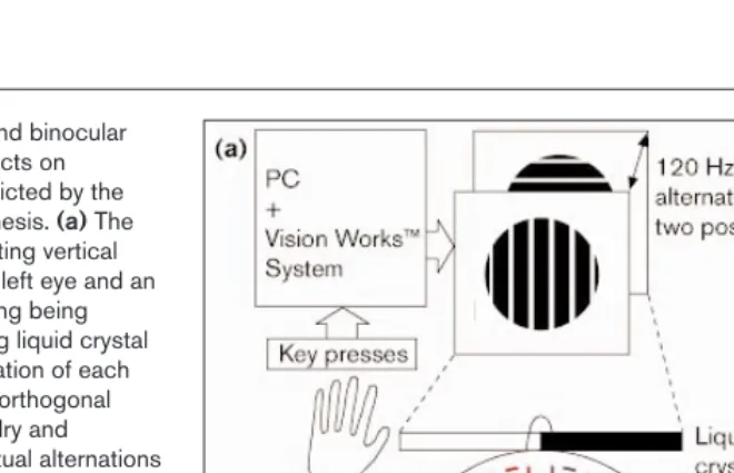
Set-up for caloric stimulation and binocular rivalry experiments and the effects on perceptual predominance predicted by the interhemispheric switch hypothesis. (a)The rivalry set-up shows a right-drifting vertical grating being presented to the left eye and an upward-drifting horizontal grating being presented to the right eye using liquid crystal shutters to restrict the presentation of each image to its intended eye. The orthogonal gratings induced binocular rivalry and subjects reported their perceptual alternations using response keys on a keyboard. The caloric stimulation procedure involved irrigating the external ear canal with iced water until subjects reported vertigo and examiners observed nystagmus. The stimulation acts through the semicircular canals and brainstem, and results in activation of contralateral structures known to be involved in attentional processing and binocular rivalry. (b,c)Expected effects on rivalry alternations from unilateral hemisphere activation (according to the interhemispheric switch hypothesis) depicted by theoretical frequency histograms. These represent the frequency (y axis) of horizontal and vertical perceptual intervals in seconds (x axis) during the rivalry viewing period. In (b), there is no
baseline predominance of either horizontal or vertical percepts so unilateral hemisphere activation might be expected to induce either a horizontal (bottom left) or vertical (bottom right) predominance. In (c), there is a baseline predominance of the horizontal percept that
horizontal drifting gratings. Positron emission tomo-graphy (PET) [26] and fMRI [27] studies have shown that caloric stimulation causes activation in contralateral hemispheric structures that are known to be involved in attentional processing [28] and binocular rivalry [14] (for example, temporo-parietal, insular and anterior cingulate cortex). In a clinical context, this technique can tem-porarily ameliorate left-sided neglect and anosognosia (denial of disease) associated with right hemisphere damage [20,29]. This ability of caloric stimulation to uni-laterally activate the same hemispheric structures impli-cated in attentional processing and binocular rivalry suggests that, if rivalry is mediated by interhemispheric switching, caloric stimulation should alter the baseline perceptual predominance of one image relative to the other (Figure 1). Within-hemisphere competition at any level does not predict an effect from such unilateral hemisphere activation.
We next tested predictions that binocular rivalry occurs at the same level as reversible figures, by assessing the effect of caloric stimulation during viewing of the Necker cube, a line diagram with ambiguous perspectives. Similar effects of caloric stimulation on binocular rivalry and Necker cube alternations would be further support for the notion that these phenomena have a common neural mechanism [6,17,30]. If unilateral hemisphere activation induces a change in the baseline predominance of either perspective of the Necker cube, this would indicate that interhemispheric switching also mediates the alternations of this bistable perceptual phenomenon.
[image:3.858.227.557.98.263.2]Finally, as the longer time course of caloric stimulation in relation to rivalry does not allow a direct assessment of the switching process itself, we used unilateral single-pulse transcranial magnetic stimulation (TMS), with its high tem-poral precision, to assess whether this could perturb the rivalry process. The predictions for this experiment are: first, disruption of a hemisphere’s designated percept (by TMS applied to temporo-parietal cortex) would occur only if the TMS is applied during perceptual dominance of that image; and second, disruption of a hemisphere’s designated image should have little effect on perceptual alternations if the TMS is applied when that image is perceptually suppressed (Figure 2). Thus, a phase-specific pattern of interference effects is expected from unilateral TMS if binocular rivalry is indeed an interhemispheric switching phenomenon.
Results
Binocular rivalry
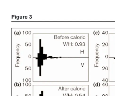
The effect of caloric-induced left hemisphere activation on two subjects’ rivalry alternations with drifting vertical and horizontal gratings is demonstrated in Figure 3 where it can be seen that the stimulation produces a change in image predominance, reflected by the V/H ratio, the ratio of total time spent perceiving the vertical and horizontal gratings, excluding mixed percepts. The experimental design used is shown in Figure 4. In individuals, the effect ranged from strong to absent (Figure 5a,b), perhaps because of variation in the duration and efficacy of the procedure. The group analysis compared the absolute magnitude of change in the log-transformed V/H ratio between two pre-stimulation blocks of rivalry (a measure of the random fluctuation in
Figure 2
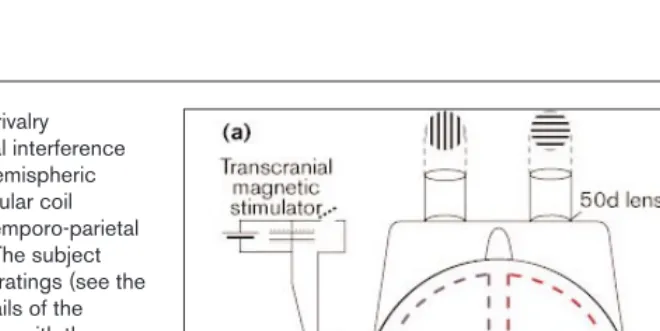
Set-up for TMS and binocular rivalry experiments and the perceptual interference effects predicted by the interhemispheric switch hypothesis. (a)The circular coil delivers a single pulse to the temporo-parietal region of the left hemisphere. The subject viewed orthogonal stationary gratings (see the Materials and methods for details of the display used to avoid interaction with the intense magnetic field) and reported their perceptual alternations using two response keys, one of which triggers the magnetic stimulation. (b)The time course of perceptual alternations shows the predicted disruptive effects of TMS triggered by a switch to the horizontal percept. If the left hemisphere adopts the horizontal percept, TMS applied to this hemisphere when the horizontal image is perceptually dominant will disrupt this percept and allow the vertical percept to assume dominance. The theoretical frequency histogram (right) therefore depicts very short horizontal interval durations. (c)When the stimulation is delivered under identical conditions, but at the opposite phase of the
perceptual switch (that is, triggered when the subject reports a switch to vertical), disruption of the left hemisphere should have little effect as it is the right hemisphere that is
responsible for the vertical percept. Thus, the theoretical frequency histogram (right) for this contingency shows normal interval durations. Although not shown by theoretical frequency
V/H ratio) with the change between the block immediately before, and immediately following, the stimulation (a measure of the experimental effect plus random variation).
The left hemisphere activation group demonstrated a statis-tically significant greater change in the V/H ratio following stimulation than was observed in baseline viewing (Figure 5a,b). This effect had largely diminished by the fifth block of rivalry (that is, 10–20 minutes following stimu-lation). Predominance comparisons were not significant for a control group of twelve subjects who underwent the entire protocol minus the caloric stimulation (Figure 5a). Right-hemisphere activation did not induce a change in image predominance above baseline fluctuations (Figure 5a).
As a control for possible effects on image predominance from ongoing, undetected eye movements induced by the caloric stimulation, the experiments were repeated with rivalling oblique gratings. Any effect from horizontal eye
movements would be spread equally across two orthogonal oblique gratings and could not therefore affect image pre-dominance. The results of these experiments were the same as those for horizontal and vertical gratings. Left-hemisphere activation significantly changed predominance above baseline fluctuations (Figure 5c,d), and the effect had diminished by the fifth block of rivalry. Right-hemi-sphere activation again did not induce a significantly greater change in predominance above baseline fluctuations, and the control condition was also non-significant (Figure 5c).
To assess the direction of change in image predominance following left-hemisphere activation, we looked at the twelve subjects with the largest caloric-induced shifts. For horizontal and vertical rivalry, of the twelve leftmost sub-jects shown in red in Figure 5a (excluding the first subject, who was left-handed), nine showed caloric-induced shifts towards perception of the horizontal grating while three showed shifts favouring the vertical grating. Similarly, in the oblique experiments, of the twelve sub-jects showing the largest predominance shifts (the left-most subjects in red in Figure 5c), nine favoured the rightward tilted (45°) grating, and three favoured the left-ward tilted (–45°) orientation following stimulation.
Figure 4
Design of caloric stimulation experiments. There were six blocks of rivalry, each representing approximately 7 min of viewing. The first block was considered training and discarded before analysis. Blocks 2 and 3 were pre-stimulation blocks, while 4–6 were post-stimulation blocks. The predominance ratio was calculated by dividing the total time spent perceiving the vertical gratings by the total time spent perceiving the horizontal gratings, excluding mixed percepts. Similar ratios were calculated for the oblique rivalry and Necker cube experiments. The ratios were log-transformed before analysis. There was random variation in these predominance ratios between two pre-stimulation blocks. Therefore, to show an effect of caloric pre-stimulation, there must be greater absolute magnitude of change in the
[image:4.858.56.300.92.296.2]predominance ratio between blocks 3 and 4 (random variation plus experimental effect) compared with the random variation seen between blocks 2 and 3. Thus, the graphs to the right in Figure 5 show the ∆log predominancefor blocks 2 and 3 and for blocks 3 and 4. Subtracting the predominance changes seen between blocks 2 and 3 from those between blocks 3 and 4 removes the baseline noise and is labelled ∆(∆log predominance) in the graphs to the left in Figure 5.
Figure 3
Summary statistics for all caloric stimulation experiments are presented in Table 1.
Necker cube
In the Necker cube experiments, the effect of left-hemi-sphere activation was dramatic in two subjects out of the 28. Each of the two subjects had normal baseline perceptual alternations, but demonstrated a virtually complete inability to see one of the two possible perspectives following caloric stimulation. One of these subjects is illustrated in Figure 6c. His post-stimulation perception alternated between one clear perspective and the ‘undecided’/indeterminate option where no depth was perceived in the line diagram.
Other subjects showed predominance shifts following left-hemisphere activation (for example, Figure 6a,b) similar to, and generally more pronounced than, the effect seen with binocular rivalry. The group analysis of these remaining 26 subjects showed that left-hemisphere activation caused a significant change in perspective predominance greater than baseline fluctuations (Figure 5e,f), and that the effect had diminished by the fifth block of data collection. Both control and sham stimulation conditions yielded non-signifi-cant changes, and right-hemisphere activation also did not change Necker cube perspective predominance above base-line fluctuations (Figure 5e). Of the twelve subjects with the largest predominance shifts, seven demonstrated shifts in predominance towards one perspective while the remain-ing five subjects showed shifts in the opposite direction.
Transcranial magnetic stimulation
As left-hemisphere activation showed a clear effect on both rivalry and reversible figure alternations, we concentrated
on this hemisphere for the TMS experiments. Figure 7 shows that application of a TMS pulse to the temporo-pari-etal region of the left hemisphere had a disruptive effect on binocular rivalry which was, as predicted, phase-spe-cific. TMS applied just as the percept was switching from vertical to horizontal caused a reversion to vertical indi-cated by shortened horizontal interval durations, but there was no disruptive effect when the TMS pulse was timed to occur at the opposite perceptual switch. The data for all subjects are shown in Table 2 where it can be seen that this pattern occurred in three subjects. In two other sub-jects, TMS delivered on a switch from horizontal to vertical caused a reversion to horizontal, indicated by shortened vertical interval durations, but there was no similar percep-tual disruption when TMS was delivered on a switch to horizontal in these same subjects. Clear phase-specific dis-ruptive effects of TMS thus occurred in five out of the seven subjects we tested, despite the difficulties associated with simultaneously establishing a threshold stimulation intensity and an optimal location.
Discussion
Interhemispheric switching mediates perceptual rivalry
Our results demonstrate that unilateral (left) hemisphere activation by caloric stimulation influences the alternation patterns of binocular rivalry with drifting vertical and hori-zontal gratings and with stationary oblique gratings. A change in the perceptual predominance of the rivalling images following unilateral hemispheric activation is pre-dicted by the interhemispheric switch hypothesis of binocular rivalry and cannot be explained by models based on within-hemisphere competition. This interhemispheric switch hypothesis is consistent with suggestions that it is the stimulus representations rather than the eyes that rival during binocular rivalry [7–13] and that rivalry is occurring high in the visual pathway [3–17].
In further support of an interhemispheric switch model of binocular rivalry, we have demonstrated a phase-specific disruptive effect of unilateral (left) transcranial magnetic stimulation on perceptual alternations. One stimulation contingency caused perceptual disruption while stimula-tion at the opposite phase had little effect even though delivered under identical conditions. These results cannot be explained by within-hemisphere models but are pre-dicted by the hypothesis proposed here.
[image:5.858.57.298.118.329.2]A similar effect of caloric-induced left-hemisphere activa-tion on the predominance of perceived perspectives of the Necker cube was demonstrated and supports the notion of a common neural mechanism for both binocular rivalry and reversible figures. The data presented here can be explained if both types of perceptual rivalry are mediated by an interhemispheric switch mechanism. Thus, we suggest that, in perceptual rivalry, each hemisphere adopts one image or perspective, and perceptual alternations
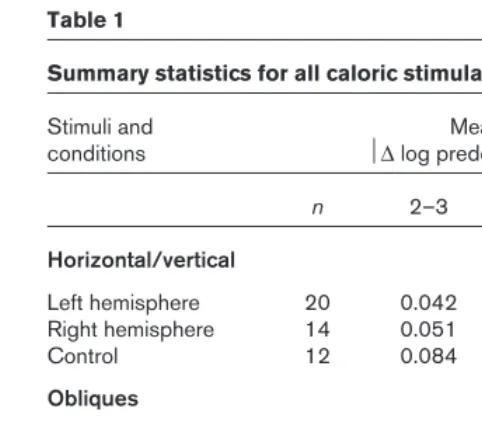
Table 1
Summary statistics for all caloric stimulation experiments.
Stimuli and Mean
conditions ∆log predominance
n 2–3 3–4 p*
Horizontal/vertical
Left hemisphere 20 0.042 0.084 < 0.05 Right hemisphere 14 0.051 0.057 0.72
Control 12 0.084 0.062 0.21
Obliques
Left hemisphere 20 0.059 0.099 < 0.05 Right hemisphere 20 0.057 0.067 0.35
Control 20 0.083 0.095 0.58
Necker cube
Left hemisphere 26 0.089 0.228 < 0.05 Right hemisphere 16 0.124 0.143 0.80 Sham stimulation 10 0.152 0.170 0.59
Control 26 0.130 0.131 0.46
reflect hemispheric alternations and therefore competition between the hemispheres for visual awareness.
The lack of a change in predominance above baseline fluctuations for the right-hemisphere activation group in all three caloric stimulation experiments may be explained in the following way. A recent fMRI study of humans undergoing binocular rivalry found right-sided fronto-pari-etal activation during perceptual transitions [14]. This finding suggests that these regions are involved in gating perceptual alternations or selecting the neuronal represen-tations for access to visual awareness. This idea is sup-ported by reports that right-sided frontal lesions cause the perception of only one of the two possibilities in reversible figures [31]. The finding of right fronto-parietal activation during perceptual rivalry also emphasises that regions
involved in the gating or selection process may be func-tionally quite distinct from the visual regions responsible for the alternative image representations [14,17]. Left-ear cold caloric stimulation might activate both the fronto-parietal gating region and the visual regions in the right hemisphere and this dual activation may be responsible for the lack of predominance change in this group.
[image:6.858.231.558.96.486.2]The directions of shifts in predominance induced by left-hemisphere activation also raise interesting issues. There appears to be a predilection for the horizontal grating to be adopted by the left hemisphere although this was not always the case. The direction of predominance change in the oblique rivalry experiment was also biased, towards the right-tilted (45°) orientation. It is interesting to note that both the horizontal grating and the right-tilted oblique
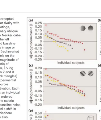
Figure 5
Effect of caloric stimulation on perceptual alternations during (a,b)binocular rivalry with drifting horizontal and vertical gratings,
(c,d)binocular rivalry with stationary oblique gratings, and (e,f)viewing of the Necker cube. In all experiments, activation of the left hemisphere significantly changed baseline perceptual predominance of one image or perspective relative to the other (red inverted triangles in left panels). The panels on the right demonstrate the absolute magnitude of change in the log-transformed ratio of perceptual predominance (that is, ∆log predominance) between blocks 2 and 3 (baseline random fluctuation; pink triangles) and between blocks 3 and 4 (experimental effect plus random fluctuation; purple triangles) for left-hemisphere activation. Each point along the x axis represents an individual subject’s data, and subjects are ordered according to the magnitude of the caloric effect. There was considerable baseline noise but a majority of subjects showed a shift in predominance following left-hemisphere activation in all experiments (see also Table 1). Effects seemed to be stronger with the Necker cube than with binocular rivalry, and three Necker subjects had such strong effects they could not be shown on this graph (one is described in the key below panel e; for a description of the other two, see Figure 6c). The left panels show the data for left- and right-hemisphere activation and the control condition that did not involve stimulation. Each point in these plots was calculated by subtracting the predominance change between blocks 2 and 3 from that between blocks 3 and 4, that is,
∆(∆log predominance). Thus, points above the zero line represent individuals who showed greater predominance change following stimulation than in baseline viewing, while points below the zero line indicate greater random change in predominance than
that seen following stimulation. The subjects were arranged in descending order of magnitude and, therefore, in the oblique rivalry and Necker cube experiments, the data point
grating were presented to the right eye. Thus, eye-of-pre-sentation may influence which hemisphere adopts which image. This is consistent with evidence that there is a higher proportion of binocular neurons with a dominant input from the contralateral eye [32]. Nevertheless, as an individual’s predominance does not completely reverse in preliminary experiments in which the eye-of-presentation has been reversed, we cannot yet rule out some combina-tion of eye-of-origin and higher-order effects. Thus, the horizontal grating may often be adopted by the left hemi-sphere due to a cultural bias for horizontal scripts and the left-lateralisation of sentence reading [33]. The direction of predominance shifts for the Necker cube experiments suggest that, with these stimuli (which do not involve sepa-rate presentation to the eyes), there may be an arbitrary designation of perspective to hemisphere. Future experi-ments might repeat stimulation in the same subject to elu-cidate whether the designation of image, or perspective, to hemisphere, is fixed or varies within an individual.
Hemifields and hemispheres
In thinking about our model of interhemispheric switching, it is important not to be limited by spatially symmetric notions of hemifield representations in V1. It has been sug-gested that the coherence rivalry demonstrated by Diaz-Caneja [8,9] rules out the possibility that rivalry occurs between each cerebral hemisphere [6]. However, the 1.5° stimulus used in the rivalry experiments reported here pro-duces bilateral activation even in V1 (where binocular overlap is around 1° in the foveal region of higher primates) and in the middle temporal visual area (where overlap is around 5°). Moreover, the Diaz-Caneja experiment says nothing about interhemispheric competition at higher
processing levels. The binocular neurons in the inferotem-poral cortex, whose activity correlates with monkeys’ reported percepts [4], can process information presented to either hemifield as indicated by their properties of bilateral receptive fields and ipsilateral field loss following section of the posterior corpus callosum and anterior commissure [34].
Thus, rivalry between the hemispheres at a level beyond V1 is compatible with Diaz-Caneja’s results and may actually help to explain the phenomenon of coherence rivalry. Diaz-Caneja’s experiments [8,9] and the patchwork experiments of Kovacs et al.[7] suggest that the brain is able to group or bind coherent image segments irrespective of their eye-of-origin. How might the brain achieve such reorganisation of presented image components into rivalling coherent images? The interhemispheric switch model suggests that the brain groups or binds the segments of each coherent image in sep-arate hemispheres. Thus, the perceptual resources of each hemisphere may be independently and alternately employed to achieve this kind of synthetic ability.
Eye movements
[image:7.858.57.564.114.240.2]In the horizontal and vertical rivalry experiment, despite the care we took to delay post-stimulation testing until nystag-mus had ceased, it is at least possible that the observed pre-dominance shifts actually result from ongoing, undetected horizontal nystagmus that acts to reduce the spatial fre-quency and contrast of the vertical grating. The fact that three subjects had increased predominance of the vertical grating after caloric stimulation makes this explanation unlikely. Moreover, the results of the oblique rivalry experi-ment strongly argue against this interpretation. Results for the Necker cube experiments are also difficult to explain by
Table 2
Median interval durations and significance levels for individual TMS subjects.
Control TMS on switch to H TMS on switch to V Significance level
Subject V H Vu Hs Vs Hu Hsversus HuVsversus Vu
1 3.48 2.72 6.20 0.80 3.14 4.62 < 0.00001 n.s.
2 5.80 5.74 5.86 0.40 3.84 5.76 < 0.00001 n.s.
3 1.83 1.93 1.33 0.91 1.28 1.39 < 0.01 n.s.
4 4.11 4.82 4.81 4.90 0.65 5.38 n.s. < 0.01
5 3.76 5.06 2.67 8.75 0.81 6.26 n.s. < 0.01
6 1.92 1.22 1.76 0.79 2.15 1.02 n.s. n.s.
7 1.62 4.24 1.13 3.60 2.44 5.25 n.s. n.s.
V, vertical; H, horizontal; s, stimulated; u, unstimulated; n.s., non-significant. Individual subject data demonstrate the phase-specific effect of TMS during binocular rivalry. The bolded median interval durations, all less than 1 sec, show perceptual disruption of the interval immediately following TMS (Hsand Vs; compare these intervals with the same subject’s control intervals). Note that subjects 1–3 had perceptual disruption of the horizontal intervals when TMS was triggered by a switch to horizontal, but no perceptual disruption when TMS was triggered by a switch to vertical (see also Figure 7). Subjects 4 and 5, on the other hand, experienced perceptual disruption when
eye movements. Finally, in the TMS experiments, the stimulation was delivered under exactly the same conditions for both stimulation contingencies, and any effect due to eye movements should therefore be seen in both contingen-cies. This was clearly not the case, as illustrated in Figure 7.
Brainstem oscillator or corpus callosum?
The highly developed corpus callosum connecting the human hemispheres may immediately suggest itself for a
[image:8.858.61.558.98.238.2]key role in the proposed interhemispheric switch. We think that this is unlikely and predict that split-brain sub-jects would still experience perceptual alternations. We suggest that the primary mechanism of interhemispheric switching involves different subcortical bistable oscillator circuits related either to the short-period perceptual alter-nations studied here or to long-period alternating hemi-spheric activity [21,22]. The suggestion that a subcortical bistable oscillator mediates interhemispheric switching is
Figure 6
Effect of caloric vestibular stimulation on perceptual alternations of a reversible figure, the Necker cube. (a,b)Left hemisphere activation (right-ear caloric stimulation) shifts a baseline perspective predominance (A/B) from 1.3 to 0.85 (perspective A, lower square face closer to observer; perspective B, upper square face closer to observer; ratio calculated as for binocular rivalry and excludes indeterminate percepts). This represents a 3 to 4 ∆log predominanceof 0.185. Overall, subjects demonstrated shifts in both directions following stimulation, indicating that, unlike for binocular rivalry, designation of perceptual configuration to hemisphere may be arbitrary. (c)Raw time series data for a single subject demonstrating the normal baseline perceptual alternations, with roughly equal time spent experiencing each perspective, followed by the effect of caloric stimulation, which virtually eliminated the ability to
perceive one of the two perspectives. The subject alternated between perspective A and the ‘undecided’ response option (where no depth was perceived in the line diagram) following left-hemisphere activation. This more dramatic effect may be related to the fact that this subject received prolonged iced-water irrigation compared with other subjects. The same effect was seen in one other subject, so the effects of caloric stimulation on the predominance of Necker cube perspectives varied from infinity (two subjects) to the more graded effects seen in the 26 subjects shown in Figure 5e,f. The effect of unilateral (left) hemisphere activation on Necker cube alternations is further evidence that binocular rivalry and reversible figures have a common neural mechanism and suggests to us that this mechanism is interhemispheric switching.
Figure 7
Effect of left hemisphere TMS on a single subject’s binocular rivalry alternations (subject 1 in Table 2). There was a marked disruption of percepts when left-hemisphere TMS was contingent on one direction of perceptual switch but not when the contingency was at the opposite phase. (a)Control session with no TMS. (b)TMS delivered when the subject signalled a switch from the vertical to the horizontal percept, caused an immediate reversion to the vertical percept,
indicated by a dramatic shortening of the horizontal interval durations.
[image:8.858.58.557.519.653.2]based on both comparative considerations and clinical evi-dence in humans.
Bistable oscillators are well studied in invertebrates [35], and interhemispheric switching has been observed in the brains of birds [23] and fish [24] that lack a corpus callo-sum. Moreover, in human patients with midline cerebellar or brainstem damage, a roughly 90 second oscillator has been described that shows side-to-side alternation of eye movements [25]. This oculomotor alternation, known as periodic alternating nystagmus, is believed to be a brain-stem phenomenon and is accompanied by perceptual alternations during binocular rivalry consistent with our proposals concerning interhemispheric switching (S.M.M. and J.D.P., unpublished observations).
The role of the brainstem in mediating synchronous neural activity [36] will be particularly interesting if tem-poral correlation [37] of neurons with similar preferred stimuli is shown to be important at high levels of the visual pathway during binocular rivalry. A brainstem oscil-lator might increase response synchronisation of neurons with similar preferred stimuli in one hemisphere, before switching its output to the opposite hemisphere to coordi-nate the activity of neurons preferring the other image. Thus, simultaneous bilateral recordings from single neurons and pairs of neurons high in the visual cortex during rivalry in alert monkeys would enable testing of the interhemispheric switch hypothesis through analysis of both the rate and temporal correlation of neural activity. Other means of verifying the hypothesis include looking for the presence of alternating patterns of cerebral activa-tion (and coherence) with electroencephalography, MEG or fMRI. It will be necessary for such studies to analyse signals derived while one percept is dominant separately from those generated during its suppression.
Conclusions
We have presented a readily testable neurophysiological model of binocular rivalry and reversible figure alternations on the basis of the perceptual interference effects that we have observed following unilateral hemisphere activation and disruption. Our results suggest that, during perceptual rivalry, each hemisphere adopts one of the competing images or perspectives, and perceptual alternations corre-spond to hemispheric alternations. The interhemispheric switch hypothesis has clinical relevance because of the find-ings that patients with bipolar disorder (manic depression) have a slow switch rate for both binocular rivalry [21] and reversible figures [38]. Our model may therefore offer a link between such findings and the emerging picture of hemi-spheric asymmetries in the generation and treatment of mood disorders (reviewed in [21]; and see [39–41]). Finally, the hypothesis of interhemispheric switching raises new issues for the scientific study of consciousness. At any one time during perceptual rivalry, the perceived visual scene
may depend on neural activity in only one hemisphere’s higher visual regions.
Materials and methods
Horizontal and vertical binocular rivalryEighteen right-handed and two left-handed, male and female subjects ranging from 18 to 54 years of age underwent cold caloric stimulation of the right ear (left hemisphere). Fourteen right-handed male and female subjects of similar age had left-ear (right-hemisphere) stimulation. Twelve control subjects underwent the full protocol minus the stimulation. Written, informed consent was obtained according to a protocol approved by the University of Queensland’s Medical Research Ethics Committee.
The stimuli were presented in a circular patch and subtended 1.5° of visual angle with a spatial frequency of 8 cycles/degree moving at 4 cycles/sec. Contrast of the gratings was 0.9. Subjects sat 3 m from the monochrome computer monitor (green, P46 phosphor, persis-tence = 500 nsec) and recorded their perceptual alternations by press-ing one of three keyboard response buttons for vertical, horizontal or mixed percepts. The latter were removed before analysis. Baseline per-ceptual alternations were recorded for half an hour. This was followed by the caloric stimulation (or a rest period in the control group) and a further half-hour of rivalry data was then collected. Each half-hour session was divided into three blocks, consisting of four 100 sec trials. Each trial was separated by a 30 sec rest period, and each block by a 2 min rest period.
Oblique binocular rivalry
Twenty right-handed males aged 18–25 were tested on three separate occasions. Each session involved half an hour of baseline rivalry viewing and was then followed by: first, 5 min rest; second, right-ear caloric stimulation; and, third, left-ear caloric stimulation. The two caloric ses-sions were counterbalanced. A further half-hour of rivalry data was then collected. The set-up was the same as for the horizontal and vertical rivalry experiments. A rightward tilted (45°) grating was presented to the right eye and a leftward tilted (–45°) grating to the left eye. The stimulus characteristics were otherwise the same as for the horizontal and verti-cal gratings except that the oblique gratings were stationary.
Necker cube
Twenty-eight right-handed males, aged 18–25, underwent control ses-sions and left-hemisphere activation by right-ear caloric stimulation. Two of the left-hemisphere activation subjects were unable to see one of the two possible perspectives following stimulation. Their extreme results meant that they were not included in the subsequent group analysis even though they offer striking support for the interhemispheric switch hypothesis. Sixteen subjects also underwent right-hemisphere activation by left-ear caloric stimulation while the remaining ten sub-jects underwent sham caloric stimulation with body-temperature water (and, thus, no vestibular stimulation). Following control sessions, the order of subsequent sessions was counterbalanced. The Necker cube was presented on a matt white surface 100 cm from the subject and at eye level. The cube subtended 7.6°×7.4° (height×width) of visual angle and had a central fixation cross (0.5°×0.5°). Subjects were asked to maintain gaze on the fixation point and to record their percep-tual alternations using a keyboard with a response key for each of the percepts and a third option for ‘undecided’ or indeterminate percepts or if their gaze strayed from the fixation point. The latter were removed before analysis. Alternations were recorded for half an hour, divided into three blocks each with three 100 sec trials. Each trial was sepa-rated by a 60 sec break, and each block by a 4.5 min break. Subjects then had (i) 5 min rest (control); (ii) sham stimulation using water at body temperature; or (iii) cold caloric stimulation of the right or left ear. A further half-hour of data was then collected.
Caloric stimulation
canal into the vertical plane; the mid-sagittal plane was vertical. The tubing was inserted into the external auditory canal until it was adjacent to the tympanum. Iced water was then instilled until the subject reported vertigo and the examiner observed nystagmus (usually follow-ing 10–30 ml of iced-water irrigation). Subjects demonstrated nystag-mus with the brisk phase in the direction contralateral to the ear stimulated. We did not have good control over the duration or the intensity of the procedure because of the pain and nausea that the experimenter was reluctant to prolong. All subjects have been included in the results irrespective of the judged efficacy of the procedure. Post-stimulation data collection began when all visible signs of nystagmus and subjective vertigo had ceased. Sham caloric stimulation was administered by irrigation with water at body temperature.
Transcranial magnetic stimulation
Single-pulse TMS was applied to the left temporo-parietal cortex using a 90 mm circular coil (Magstim 200™, The Magstim Company). The centre of the coil was positioned approximately 13 cm from the nasion and 12 cm from the mid-sagittal line and oriented to induce current flow in a posterio-anterior direction in the temporo-parietal cortex. The coil itself was held firmly against the scalp by one of the experimenters, while the feeder cables connecting the main stimulator to the coil were supported by an overhead gantry. Magnetic stimuli were triggered when the subject signalled a perceptual switch either to the vertical percept, in one trial, or to the horizontal percept in the other. The inten-sity of stimulation was varied between 0.66 and 1.1 T according to the subject. The rivalry apparatus used in these experiments consisted of two 1 cm (diameter) by 2 cm translucent plastic tubes, each with a 50 d lens at the proximal end, viewing a 1 mm (diameter) square wave grating (8 cycles) on translucent paper at the distal end (Figure 2). The tubes were positioned by the subject on the face-plate of a safety mask so that the gratings viewed by each eye were orthogonal in orientation and viewed at the same location.
Acknowledgements
This work was supported by the National Health and Medical Research Council of Australia and the Australian Research Council. We thank Andrew Tilley for helpful discussions and especially thank all subjects for their participation.
References
1. Wade NJ: Early studies of eye dominances.Laterality1998, 3:97-108. 2. Blake R: A neural theory of binocular rivalry.Psychol Rev1989,
96:145-167.
3. Leopold DA, Logothetis NK: Activity changes in early visual cortex reflect monkeys’ percepts during binocular rivalry.Nature1996,
379:549-553.
4. Sheinberg DL, Logothetis NK:The role of temporal cortical areas in perceptual organization.Proc Natl Acad Sci USA1997,
94:3408-3413.
5. Sengpiel F: Binocular rivalry: ambiguities resolved.Curr Biol1997,
7:R447-R450.
6. Logothetis NK: Single units and conscious vision.Phil Trans R Soc Lond B 1998, 353:1801-1818.
7. Kovacs I, Papathomas TV, Yang M, Feher A: When the brain changes its mind: interocular grouping during binocular rivalry.
Proc Natl Acad Sci USA1996, 93:15508-15511.
8. Diaz-Caneja E: Sur l’alternance binoculaire.Annales D’Oculistique 1928, 165:721-731.
9. Ngo TT, Miller SM, Liu GB, Pettigrew JD: Binocular rivalry and perceptual coherence.Curr Biol2000, 10:R134-R136.
10. Logothetis NK, Leopold DA, Sheinberg DL:What is rivalling during binocular rivalry?Nature1996, 380:621-624.
11. Andrews TJ, Purves D: Similarities in normal and binocularly rivalrous viewing.Proc Natl Acad Sci USA1997, 94:9905-9908. 12. Campbell FW, Gilinsky AS, Howell ER, Riggs LA, Atkinson J: The
dependence of monocular rivalry on orientation.Perception1973,
2:123-125.
13. Atkinson J, Campbell FW, Fiorentini A, Maffei L: The dependence of monocular rivalry on spatial frequency.Perception1973, 2:127-133. 14. Lumer ED, Friston KJ, Rees G: Neural correlates of perceptual
rivalry in the human brain.Science1998, 280:1930-1934.
15. Tong F, Nakayama K, Vaughan JT, Kanwisher N: Binocular rivalry and visual awareness in human extrastriate cortex.Neuron1998,
21:753-759.
16. Srinivasan R, Russel DP, Edelman GM, Tononi G: Increased synchronization of neuromagnetic responses during conscious perception.J Neurosci1999, 19:5435-5448.
17. Leopold DA, Logothetis NK: Multistable phenomena: changing views in perception.Trends Cog Sci1999, 3:254-264. 18. Zaidel E, Clarke JM, Suyenobu B: Hemispheric independence: a
paradigm case for cognitive neuroscience.In Neurobiology of Higher Cognitive Function.Edited by Scheibel AB, Wechsler A. New York: The Guilford Press; 1990:297-355.
19. Luck SJ, Hillyard SA, Mangun GR, Gazzaniga MS: Independent hemispheric attentional systems mediate visual search in split-brain patients.Nature1989, 342:543-545.
20. Ramachandran VS: Phantom limbs, neglect syndromes, repressed memories, and freudian psychology.Int Rev Neurobiol1994,
37:291-333.
21. Pettigrew JD, Miller SM: A ‘sticky’ interhemispheric switch in bipolar disorder?Proc R Soc Lond B 1998, 265:2141-2148. 22. Shannahoff-Khalsa D: The ultradian rhythm of alternating cerebral
hemispheric activity.Int J Neurosci1993, 70:285-298. 23. Suthers RA: Peripheral control and lateralization of birdsong.
J Neurobiol1997, 33:632-652.
24. Pettigrew JD, Collin SP, Ott M: Convergence of highly-specialized behaviour, eye movements and visual optics in the sandlance (Teleostei) and the chameleon (Reptilia).Curr Biol1999,
9:421-424.
25. Baloh RW, Honrubia V, Konrad HR: Periodic alternating nystagmus.
Brain1976, 99:11-26.
26. Bottini G, Sterzi R, Paulesu E, Vallar G, Cappa SF, Erminio F, et al.:
Identification of the central vestibular projections in man: a positron emission tomography activation study.Exp Brain Res 1994, 99:164-169.
27. Vitte E, Derosier C, Caritu Y, Berthoz A, Hasboun D, Soulie D:
Activation of the hippocampal formation by vestibular stimulation: a functional magnetic resonance imaging study.Exp Brain Res 1996, 112:523-526.
28. Posner MI, Petersen SE: The attention system of the human brain.
Annu Rev Neurosci1990, 13:25-42.
29. Vallar G, Bottini G, Rusconi ML, Sterzi R: Exploring somatosensory hemineglect by vestibular stimulation.Brain1993, 116:71-86. 30. Walker P: Stochastic properties of binocular rivalry alternations.
Percept Psychophys1975, 18:467-473.
31. Meenan JP, Miller LA: Perceptual flexibility after frontal or temporal lobectomy.Neuropsychologia1994, 32:1145-1149.
32. LeVay S, Connolly M, Houde J, Van Essen DC: The complete pattern of ocular dominance stripes in the striate cortex and visual field of the macaque monkey.J Neurosci1985, 5:486-501.
33. Bavelier D, Corina D, Jezzard P, Padmanabhan S, Clark VP, Karni A, et al.: Sentence reading: a functional MRI study at 4 Tesla.J Cogn Neurosci1997, 9:664-686.
34. Gross CG, Rodman HR, Gochin PM, Colombo MW: Inferior temporal cortex as a pattern recognition device. In Computational Learning and Cognition: Proceedings of the Third NEC Research Symposium.Edited by Baum E. 1993:44-73.
35. Marder E, Calabrese RL: Principles of rhythmic motor pattern generation.Physiol Rev1996, 76:687-717.
36. Munk MHJ, Roelfsema PR, Konig P, Engel AK, Singer W: Role of reticular activation in the modulation of intracortical synchronization.Science 1996, 272:271-274.
37. Engel AK, Roelfsema PR, Fries P, Brecht M, Singer W: Role of the temporal domain for response selection and perceptual binding.
Cereb Cortex1997, 7:571-582.
38. Hunt J, Guilford JP: Fluctuation of an ambiguous figure in dementia praecox and in manic-depressive patients.J Abnorm Soc Psychol1933, 27:443-452.
39. Bejjani B-P, Damier P, Arnulf I, Thivard L, Bonnet A-M, Dormont D, et al.: Transient acute depression induced by high-frequency deep-brain stimulation.New Engl J Med1999, 340:1476-1480. 40. Pascaul-Leone A, Rubio B, Pallardo F, Catala MD: Beneficial effect
of rapid-rate transcranial magnetic stimulation of left dorsolateral prefrontal cortex in drug-resistant depression.Lancet1996,
348:233-237.