THE INFLUENCE OF SEX HORMONES
ON CARDIAC AND SKELETAL MUSCLE FUNCTION
IN THE
MDX
MOUSE MODEL OF
DUCHENNE MUSCULAR DYSTROPHY
BY
CONNIE BOOKLESS
BSc (with Distinction) PGDipAppSci PhD
Submitted for the completion of the degree of
DOCTOR OF PHILOSOPHY
At
The Department of Biological and Physical Sciences
Faculty of Sciences
University of Southern Queensland
ABSTRACT
Duchenne Muscular Dystrophy (DMD) is a fatal recessive genetic
human disease affecting one in 3500 live male births. DMD is progressive,
there is no cure, and patients typically die of respiratory or cardiac failure in
their second decade of life. Clinical disease symptoms are exacerbated at
the onset of puberty and the physiological basis of this is unknown. The
mdx
mouse is the preferred experimental animal model of DMD, although aspects
of the model remain poorly understood. This dissertation characterises
physiological and histological features of the dystrophic
mdx
mouse in
response to manipulations of hormonal status including testosterone
treatment, surgical castration, and oestrogen treatment. Sex-specific
differences in the
mdx
were also examined. Furthermore, physiological and
histological features of the dystrophic
mdx
mouse model throughout the
mdx
lifespan were evaluated. Cardiac muscle contractility, left atrial response to
exogenous calcium, and the contractile properties of both fast-twitch (EDL)
and slow-twitch (SOL) skeletal muscles were examined in male
mdx
mice
ranging from 14 to 330 days of age. Testosterone treatment produced a
non-significant trend towards a dose-dependent decrease in both basal and
maximal left atrial contractility in the
mdx
. Surgical castration produced no
significant cardiac effects within mouse strains. The
mdx
castrates had a
45% lower maximum atrial force of contraction than control castrates
(p<0.05). Conversely, oestrogen treatment significantly improved cardiac
contractility in the
mdx
. An increase in basal left atrial contractility was
evident at doses of 0.08 mg/kg/day (p<0.05) and 0.16 mg/kg/day (p<0.01)
and in maximum left atrial contractility at a dose of 0.16 mg/kg/day (p<0.01).
Gender studies showed cardiac forces in
mdx
were not different between
males and females at any age tested and that both sexes in
mdx
had a
dampened cardiac responsiveness to exogenous calcium. Skeletal muscle
function studies showed that castration produced a 25% increase in
mdx
EDL specific force generation (p<0.01) and no increase in SOL forces.
Oestrogen treatment produced a non-significant trend towards increased
EDL forces and a 29% increase in SOL specific force at a dose of 0.16
mg/kg/day (p<0.05). Gender studies revealed no differences between male
and female
mdx
in terms of skeletal muscle force production. Further to the
hormonal investigations, lifespan characterisation studies revealed that the
mdx
mouse showed reduced basal and maximal left atrial contractility
specifically at ages 14 and 90 days (p<0.05). Skeletal muscle studies
showed that specific tetanic force production was significantly lower than
controls at 19 (p<0.05), 21, 23, 27, and 330 days of age (p<0.01) for EDL
muscles and at 19, 21, and 23 days of age in SOL muscles (p<0.01). These
studies further improve our understanding of the
mdx
mouse as an
experimental model of DMD and emphasises that the model is most
appropriate a specific ages for specific muscles. These studies further
illustrate that testosterone does not improve cardiac contractility in the
mdx
CERTIFICATION OF DISSERTATION
This thesis is my own work, except where otherwise acknowledged, and the
work is original and has not been previously submitted for any other award at
any other University or Institution of Tertiary Education.
______________________________ _________________
Signature of Candidate
Date
ENDORSEMENT
_____________________________ _____________
ACKNOWLEDGEMENTS
This was a very personal and challenging journey and I wish to
acknowledge and thank wholeheartedly the following people for their
generosity in time and spirit that made the completion of this important work
and subsequent dissertation possible. Firstly, to my supervisor Professor
Andrew Hoey who has continued to inspire me from my earliest
undergraduate studies with his passion for learning, his inspirational lecturing
style, and ethical approach to research. To my husband Dr Gregory Bookless
and our children, I love you and thank you. My friend and colleague Dr
Christel van Erp, never change. To Dr Renee Cornford-Nairn and the
members of the Muscle Research Laboratory USQ who always encouraged
perserverance even in the toughest times. Special thanks go to Christine
Bartlett (USQ Office of Research and High Degrees) who was just wonderful
in her tireless encouragement, to Julie Murphy and Sandra Sharp whose
professionalism and dedication in the USQ Animal House breeding facility
ensured that my experimental animals were always cared for with the utmost
concern for their welfare. In addition, the technical expertise and knowledge
imparted by the following people greatly enhanced my skills and facilitated
the completion of this project; Ilona Iberle (USQ Librarian), Paul Addison (UQ
Histologist), Dr Peter Dunn (USQ Statistician), Oliver Kinder (USQ
Technician), Maggie Fryer, Anne Woodroffe and Julie Pember (USQ IT
Services), Debbie and Steven White (USQ Administrators), Chris de Byl, Phil
Hallas, and Sammy Quatromani (USQ IT Services). I wish to make special
mention of Mrs Debbie Robbins and Mrs Pat Furlong whose tireless work for
the Duchenne Muscular Dystrophy Parent Project is an inspiration to us all. It
is an honour to have known you and your courage and dedication moves
mountains. Finally, and most importantly my love goes to every boy with
DMD. You are the reason I strove for excellence and I hope that my work, in
some small way, might just contribute to a better understanding of this
devastating disease and may help to make your lives a little better.
“Motion is life…..”
GLOSSARY OF ABBREVIATIONS
AAV
adeno-associated virus
ACE
angiotensin converting enzyme
AO
antisense
oligonucleotide
AR
androgen
receptor
BW
body
weight
C57
C57BL/10ScSn mouse
Ca
2+calcium
[Ca
2+]
i
intracellular calcium concentration
CHF
congestive
heart
failure
CK
creatine
kinase
CNF
centronucleated
fibre
CO
2carbon dioxide
CRC
concentration response curve
CSA
cross-sectional
area
CV
cardiovascular
cxmd
canine X-linked muscular dystrophy
DFZ
deflazacort
DGC
dystrophin-associated glycoprotein complex
DHT
dihydrotestosterone
DMD
Duchenne muscular dystrophy
DNA
deoxyribonucleic acid
ECG
electrocardiogram
EDL
extensor digitorum longus muscle
eNOS
endothelial nitric oxide synthase
EOM
extraocular muscles
ER
oestrogen
receptor
EtOH
ethanol
FOC
force
of
contraction
FSH
follicle
stimulating
hormone
FXMD
feline X-linked muscular dystrophy
g
gram
GI
gastrointestinal
GnRH
gonadotrophin
releasing
hormone
GRMD
Golden Retriever muscular dystrophy
H&E
haematoxylin
and
eosin
HFMD
hypertrophic feline muscular dystrophy
HR
heart rate
HW
heart
weight
HW%
heart weight as a percentage of body weight
Hz
Hertz
IGF-1
insulin-like growth factor 1
iNOS
inducible nitric oxide synthase
IP
intraperitoneal
IQ
intelligence
quota
K
+potassium
ion
kb
kilobase(s)
kDa
kilo Dalton(s)
KPSS
Krebs physiological salt solution
LA
left atria/left atrium
Lf
absolute
muscle
length
LH
leutinising
hormone
LIF
leukaemia inhibitory factor
Lo
muscle
length
LV
left
ventricle/ventricular
LV+S
left ventricle and septum
M
molar
MDSC
muscle-derived stem cell
mdx
muscular
dystrophy
X-linked
mg
milligram
min
minute(s)
mL
millilitre
mM
millimolar
mN
millinewton
msec
millisecond(s)
µm
micrometre
n
number of animals in experimental group
Na
+sodium
ion
NOS
nitric oxide synthase
nNOS
neuronal nitric oxide synthase
NMJ
neuromuscular junction
O
2oxygen
PCR
polymerase
chain
reaction
%
percent
PK
pyruvate
kinase
Po
tetanic force
Pt
twitch force
Po:Pt
tetanus to twitch ratio
RA
right atria/right atrium
REM
rapid eye movement
rpm
revolutions
per
minute
RV
right
ventricular
free
wall
RyR
ryanodine
receptor
SC
subcutaneous
SEM
standard error of the mean
SERCA
sarcoplasmic reticulum calcium-ATPase
SHR
spontaneously hypertensive rat
SOL
soleus muscle
sPo
tetanic force normalised for cross-sectional area
sPt
twitch force normalised for cross-sectional area
SR
sarcoplasmic
reticulum
TGF
transforming growth factor
TPSS
Tyrode physiological salt solution
U/L
units of active enzyme per litre of serum
USQ
University of Southern Queensland
VC
vital
capacity
CONTENTS
ABSTRACT ...
ICERTIFICATION OF DISSERTATION ...
IIACKNOWLEDGEMENTS ...
IIIGLOSSARY OF ABBREVIATIONS ...
IVCONTENTS ...
IXLIST OF TABLES ...
XVLIST OF FIGURES ...
XVII1
LITERATURE REVIEW ... 1
1.1 History of Duchenne Muscular Dystrophy (DMD) ... 1
1.2 Genetics and inheritance ... 3
1.3 Detection, diagnosis and clinical disease features ... 5
1.3.1 Genetic testing ... 5
1.3.2 Serum creatine kinase (CK) and pyruvate kinase (PK) levels ... 6
1.3.3 Muscle biopsy ... 7
1.3.4 Disease onset ... 7
1.3.5 Initial clinical signs ... 8
1.3.6 Skeletal muscle involvement ... 8
1.3.7 Biomechanics of movement in DMD ... 9
1.3.8 Gowers’ manoeuvre ...10
1.3.9 Cardiac muscle involvement ...11
1.3.10 Skeletal system involvement ...13
1.3.11 Respiratory muscle involvement ...14
1.3.12 Smooth muscle involvement ...16
1.3.13 Intellectual and behavioural impairment ...16
1.4 Muscle and system sparing ...17
1.5 Female DMD carriers ...19
1.5.2 Female DMD carrier cardiac and skeletal muscle involvement ...20
1.6 Life expectancy and mortality ...22
1.7 Physical therapies ...23
1.7.1 Prolongation of ambulation ...23
1.7.2 Orthopaedic management of scoliosis ...24
1.7.3 Prevention of pulmonary mortality ...25
1.7.4 Prevention of cardiac mortality...26
1.8 Dystrophin and the dystrophin-associated glycoprotein complex (DGC) ...27
1.8.1 Neuronal nitric oxide synthase and nitric oxide synthase ...29
1.9 Animal models of DMD ...30
1.9.1 The X-linked muscular dystrophy (mdx) mouse ...31
1.9.2 Alternative mouse models ...34
1.9.3 Feline disease models ...35
1.9.4 Canine disease models ...36
1.9.5 Zebrafish model ...38
1.9.6 The Caenhorhabiditis elegans worm ...39
1.10 Pathology and histology of DMD ...39
1.10.1 Calcium homeostasis...41
1.10.2 Centronucleated fibres...42
1.11 Therapeutic agents and strategies in DMD treatment and research ...43
1.11.1 Dampening the inflammatory response ...43
1.11.2 Corticosteroids ...44
1.11.3 Natural remedies ...47
1.11.4 Tumour necrosis factor-α ...47
1.12 Nitric oxide based therapeutics ...47
1.12.1 L-arginine ...48
1.12.2 Utrophin upregulation ...48
1.13 Improving protein synthesis ...49
1.13.1 Anabolic steroids ...49
1.14 Decreasing proteolysis through reducing calpain activity ...49
1.15 β2-adrenergic agonists ...50
1.16 Increasing proliferation and regeneration of muscle cells ...50
1.16.1 Myostatin inhibition ...50
1.16.2 Leukaemia inhibitory factor ...51
1.17 Gene based therapies ...57
1.17.1 Aminoglycoside antibiotic therapy ...58
1.17.2 Antisense oligonucleotides ...59
1.18 Sex hormones and DMD ...59
1.18.1 Testosterone ...60
1.18.2 Oestrogens ...64
1.19 Aims of study ...67
2
METHODOLOGY ... 68
2.1 Ethical considerations ...68
2.2 Experimental animals ...68
2.3 Treatment of animals - allometric scaling ...68
2.4 Individual trial methodologies ...69
2.4.1 Testosterone trial ...69
2.4.2 Sex (gender) trial ...69
2.4.3 Castration trial ...69
2.4.4 Oestrogen trial ...70
2.4.5 Influence of age trial ...71
2.5 Euthanasia ...71
2.6 Dissection of tissues ...71
2.7 Organ bath (functional) experiments ...72
2.8 Skeletal muscle experiments ...72
2.9 Cardiac muscle experiments ...74
2.10 Histology protocol ...75
2.11 Histological analysis ...76
3
TESTOSTERONE TRIAL ... 78
3.1 Influence of testosterone on body weight ...78
3.2 Influence of testosterone on testes weight ...78
3.3 Influence of testosterone on cardiac characteristics ...79
3.3.1 Cardiac morphometry ...79
3.3.2 Cardiac function ...80
3.4 Influence of testosterone on skeletal muscle morphometry ...83
3.5 Discussion ...84
4
SEX (GENDER) TRIAL ... 89
4.1 Influence of sex on body weight and heart weight ...89
4.2 Influence of sex on cardiac characteristics ...90
4.2.1 Cardiac morphometry ...90
4.2.2 Cardiac function ...92
4.3 Influence of sex on EDL characteristics ...96
4.3.1 EDL morphometry ...96
4.3.2 EDL function ...97
4.3.3 EDL histology ...99
4.4 Influence of sex on SOL characteristics ... 101
4.4.1 SOL morphometry ... 101
4.4.2 SOL function ... 102
4.4.3 SOL histology ... 104
4.5 Discussion ... 106
5
CASTRATION TRIAL ... 114
5.1 Influence of castration on body weight ... 114
5.2 Influence of castration on cardiac characteristics ... 114
5.2.1 Cardiac morphometry ... 114
5.2.2 Cardiac function ... 115
5.3 Influence of castration on skeletal muscle morphometry ... 117
5.4.1 EDL function ... 118
5.4.2 EDL histology ... 119
5.5 Influence of castration on SOL characteristics ... 120
5.5.1 SOL function ... 120
5.5.2 SOL histology ... 121
5.6 Discussion ... 122
6
OESTROGEN TRIAL ... 127
6.1 Influence of oestrogen on body weight ... 127
6.2 Influence on oestrogen on testes weight ... 127
6.3 Influence of oestrogen on cardiac characteristics ... 128
6.3.1 Cardiac morphometry ... 128
6.3.2 Cardiac function ... 129
6.4 Influence of oestrogen on skeletal muscle morphometry ... 130
6.5 Influence of oestrogen on EDL characteristics ... 131
6.5.1 EDL function ... 131
6.5.2 EDL histology ... 132
6.6 Influence of oestrogen on SOL characteristics ... 132
6.6.1 SOL function ... 132
6.6.2 SOL histology ... 133
6.7 Discussion ... 135
7
INFLUENCE OF AGE TRIAL ... 152
7.1 Influence of age on body weight ... 152
7.2 Influence of age on testes weights ... 152
7.3 Influence of age on cardiac characteristics ... 153
7.3.1 Cardiac morphometry ... 153
7.3.2 Cardiac function ... 157
7.4 Influence of age on skeletal muscle morphometry ... 161
7.5 Influence of age on EDL characteristics ... 169
7.5.2 EDL histology ... 170
7.6 Influence of age on SOL characteristics ... 172
7.6.1 SOL function ... 172
7.6.2 SOL histology ... 173
7.7 Discussion ... 175
8
CONCLUSIONS AND FUTURE DIRECTIONS ... 182
9
REFERENCES ... 187
10
APPENDIX I ... 243
10.1 Krebs physiological salt solution (KPSS) recipe ... 243
10.2 Tyrode’s physiological salt solution (TPSS) recipe ... 243
10.3 Haematoxylin stain (Mayer’s Haematoxylin) recipe ... 244
10.4 Eosin stain recipe ... 244
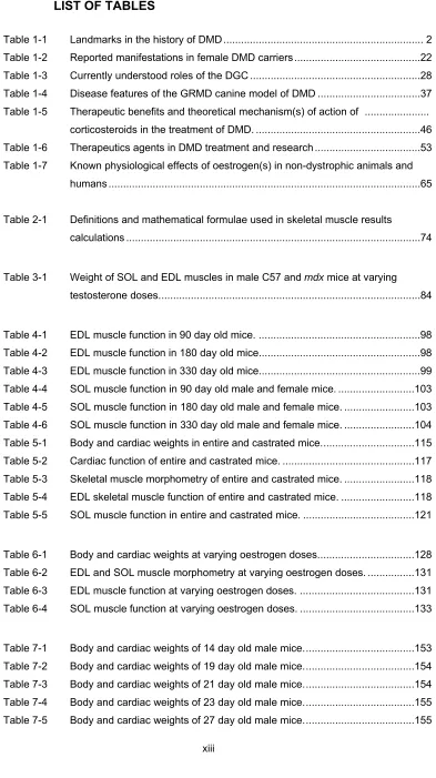
LIST OF TABLES
Table 1-1 Landmarks in the history of DMD ... 2
Table 1-2 Reported manifestations in female DMD carriers ...22
Table 1-3 Currently understood roles of the DGC ...28
Table 1-4 Disease features of the GRMD canine model of DMD ...37
Table 1-5 Therapeutic benefits and theoretical mechanism(s) of action of ... corticosteroids in the treatment of DMD. ...46
Table 1-6 Therapeutics agents in DMD treatment and research ...53
Table 1-7 Known physiological effects of oestrogen(s) in non-dystrophic animals and humans ...65
Table 2-1 Definitions and mathematical formulae used in skeletal muscle results calculations ...74
Table 3-1 Weight of SOL and EDL muscles in male C57 and mdx mice at varying testosterone doses. ...84
Table 4-1 EDL muscle function in 90 day old mice. ...98
Table 4-2 EDL muscle function in 180 day old mice...98
Table 4-3 EDL muscle function in 330 day old mice...99
Table 4-4 SOL muscle function in 90 day old male and female mice. ... 103
Table 4-5 SOL muscle function in 180 day old male and female mice. ... 103
Table 4-6 SOL muscle function in 330 day old male and female mice. ... 104
Table 5-1 Body and cardiac weights in entire and castrated mice. ... 115
Table 5-2 Cardiac function of entire and castrated mice. ... 117
Table 5-3 Skeletal muscle morphometry of entire and castrated mice. ... 118
Table 5-4 EDL skeletal muscle function of entire and castrated mice. ... 118
Table 5-5 SOL muscle function in entire and castrated mice. ... 121
Table 6-1 Body and cardiac weights at varying oestrogen doses. ... 128
Table 6-2 EDL and SOL muscle morphometry at varying oestrogen doses. ... 131
Table 6-3 EDL muscle function at varying oestrogen doses. ... 131
Table 6-4 SOL muscle function at varying oestrogen doses. ... 133
Table 7-1 Body and cardiac weights of 14 day old male mice. ... 153
Table 7-2 Body and cardiac weights of 19 day old male mice. ... 154
Table 7-3 Body and cardiac weights of 21 day old male mice. ... 154
Table 7-4 Body and cardiac weights of 23 day old male mice. ... 155
Table 7-6 Body and cardiac weights of 34 day old male mice. ... 155
Table 7-7 Body and cardiac weights of 40 day old male mice. ... 156
Table 7-8 Body and cardiac weights of 90 day old male mice. ... 156
Table 7-9 Body and cardiac weights of 180 day old male mice. ... 156
Table 7-10 Body and cardiac weights of 330 day old male mice. ... 157
Table 7-11 Skeletal muscle morphometry of 14 day old male mice. ... 161
Table 7-12 Skeletal muscle morphometry of 19 day old male mice. ... 162
Table 7-13 Skeletal muscle morphometry of 21 day old male mice. ... 162
Table 7-14 Skeletal muscle morphometry of 23 day old male mice. ... 163
Table 7-15 Skeletal muscle morphometry of 27 day old male mice. ... 163
Table 7-16 Skeletal muscle morphometry of 34 day old male mice. ... 164
Table 7-17 Skeletal muscle morphometry of 40 day old male mice. ... 164
Table 7-18 Skeletal muscle morphometry of 90 day old male mice. ... 165
Table 7-19 Skeletal muscle morphometry of 180 day old male mice. ... 165
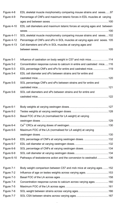
LIST OF FIGURES
Figure 1-1 Guillaume Benjamin Amand Duchenne de Boulogne and the first observed
case of DMD ... 2
Figure 1-2 Pathways of X-linked recessive inheritance in DMD ... 4
Figure 1-3 Schematic diagram of the dystrophin gene. ... 5
Figure 1-4 Pseudohypertrophy of the calf muscles in DMD ... 9
Figure 1-5 Classical Gowers’ manoeuvre (sign). ...11
Figure 1-6 ECG pattern in a nine year old boy with DMD ...12
Figure 1-7 Progressive body posture changes in DMD ...13
Figure 1-8 Severe scoliosis of the spine in a boy with DMD ...14
Figure 1-9 Dystrophin and the dystrophin-associated glycoprotein complex (DGC) ...28
Figure 1-10 Vicious cycle of muscle degeneration in DMD ...30
Figure 1-11 C57BL/10ScSn (C57) and X-linked muscular dystrophy (mdx) mice...32
Figure 1-12 Human skeletal muscle biopsies of unaffected and DMD affected individuals. ...40
Figure 1-13 H&E stained sections of DMD affectedhuman skeletal muscles. ...41
Figure 1-14 H&E stained SOL muscles in a C57 and an mdx mouse. ...43
Figure 1-15 Vector based therapeutic strategies in DMD ...58
Figure 1-16 Oestrogen activation of eNOS. ...66
Figure 3-1 Body weights of C57 and mdx mice at varying testosterone doses. ...78
Figure 3-2 Testes weights of C57 and mdx mice at varying testosterone doses. ...79
Figure 3-3 Left atrial weights of C57 and mdx mice at varying testosterone doses. ...80
Figure 3-4 Basal force of contraction of left atria at varying testosterone doses. ...81
Figure 3-5 Concentration response curves to calcium at varying testosterone doses. ...82
Figure 3-6 Maximum force of contraction of the LA at varying testosterone doses. ...83
Figure 4-1 Influence of sex on body weight and heart weight between strains and sexes. ...90
Figure 4-2 Effects of age on cardiac tissue weights between strains and sexes. ...92
Figure 4-3 Basal FOC of LA (normalised for LA weight) in mdx and C57 males and females across ages. ...93
Figure 4-4 Concentration response curves to calcium comparing male and female 90 day old mice. ...94
Figure 4-5 Concentration response curves to calcium comparing male and female 180 day old mice. ...94
Figure 4-6 Concentration response curves to calcium comparing male and female mice 330 day old. ...95
Figure 4-8 EDL skeletal muscle morphometry comparing mouse strains and sexes. ...97
Figure 4-9 Percentage of CNFs and maximum tetanic forces in EDL muscles at varying ages and between sexes. ... 100
Figure 4-10 EDL cell diameters and maximum tetanic forces at varying ages and between sexes. ... 100
Figure 4-11 SOL skeletal muscle morphometry comparing mouse strains and sexes. ... 102
Figure 4-12 Percentage of CNFs and sPo in SOL muscles at varying ages and sexes. 105 Figure 4-13 Cell diameters and sPo in SOL muscles at varying ages and between sexes. ... 105
Figure 5-1 Influence of castration on body weight in C57 and mdx mice. ... 114
Figure 5-2 Concentration response curves to calcium in entire and castrated mice. .... 116
Figure 5-3 EDL percentage CNFs and sPo for entire and castrated mice. ... 119
Figure 5-4 EDL cell diameter and sPo between strains and for entire and castrated mice... 120
Figure 5-5 SOL percentage CNFs and sPo between strains and for entire and castrated mice... 121
Figure 5-6 SOL cell diameters and sPo between strains and for entire and castrated mice... 122
Figure 6-1 Body weights at varying oestrogen doses. ... 127
Figure 6-2 Testes weights at varying oestrogen doses. ... 128
Figure 6-3 Basal FOC of the LA (normalised for LA weight) at varying oestrogen doses. ... 129
Figure 6-4 Ca2+CRCs at varying doses of oestrogen. ... 130
Figure 6-5 Maximum FOC of the LA (normalised for LA weight) at varying oestrogen doses. ... 130
Figure 6-6 EDL percentage of CNFs at varying oestrogen doses. ... 132
Figure 6-7 EDL cell diameter at varying oestrogen doses. ... 132
Figure 6-8 SOL percentage of CNFs at varying oestrogen doses. ... 134
Figure 6-9 SOL cell diameter at varying oestrogen doses. ... 134
Figure 6-10 Pathways of testosterone action and the conversion to oestradiol ... 136
Figure 7-1 Body weight comparison between C57 and mdx mice at varying ages. ... 152
Figure 7-2 Influence of age on testes weights across varying ages. ... 153
Figure 7-3 Basal FOC of the LA across ages. ... 158
Figure 7-4 Concentration response curves to calcium across varying ages. ... 160
Figure 7-5 Maximum FOC of the LA across ages. ... 161
Figure 7-6 SOL weight between strains across varying ages. ... 166
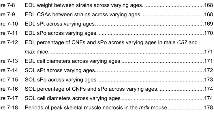
Figure 7-8 EDL weight between strains across varying ages. ... 168
Figure 7-9 EDL CSAs between strains across varying ages. ... 168
Figure 7-10 EDL sPt across varying ages. ... 169
Figure 7-11 EDL sPo across varying ages. ... 170
Figure 7-12 EDL percentage of CNFs and sPo across varying ages in male C57 and mdx mice. ... 171
Figure 7-13 EDL cell diameters across varying ages ... 171
Figure 7-14 SOL sPt across varying ages. ... 172
Figure 7-15 SOL sPo across varying ages. ... 173
Figure 7-16 SOL percentage of CNFs and sPo across varying ages. ... 174
Figure 7-17 SOL cell diameters across varying ages ... 174