EFFECT OF TWO DIFFERENT CONCENTRATIONS OF
PAPAIN SOLUTION ON ORTHODONTIC BRACKET
BONDING WITH TWO DIFFERENT ADHESIVES –
AN IN VITRO STUDY
DISSERTATION
Submitted to The Tamil Nadu Dr. M.G.R Medical University
in Partial Fulfillment of the Requirement for the Degree of
Master of Dental Surgery
Branch V
ORTHODONTICS AND DENTOFACIAL ORTHOPEDICS
Certified that the dissertation entitled: “Effect of two different concentrations of
papain solution on orthodontic bracket bonding with two different adhesives –
An in vitro study ” is a bonafide record of the work done by Dr.Rajkumar.R. under our guidance during his post graduate study during the period of 2012-2015 under THE TAMIL NADU DR. M.G.R MEDICAL UNIVERSITY, CHENNAI, in partial fulfilment for the degree of MASTER OF DENTAL SURGERY IN ORTHODONTICS AND DENTOFACIAL ORTHOPEDICS, BRANCH V. It has not been submitted (partial or full) for the award of any other degree or diploma.
.
.
Dr.Anilkumar.P (Guide) Professor& HeadDepartment of Orthodontics and
Dentofacial Orthopedics
Sree Mookambika Institute of
Dental Sciences,
Kulasekharam,KanyaKumari
District-629161
Dr. Shino.P.Mathew(Co-guide)
Professor
Department of Orthodontics
and Dentofacial Orthopedics
Sree Mookambika Institute of
Dental Sciences,
Kulasekharam,KanyaKumari
District-629161
SREE MOOKAMBIKA INSTITUTE OF DENTAL
SCIENCES, KULASEKHARAM
ENDORSEMENT BY THE PRINCIPAL / HEAD OF THE INSTITUTION
This is to certify that this dissertation titled
“
Effect of two different
concentrations of papain solution on orthodontic bracket bonding
with two different adhesives”
– An in vitro study
is a bonafide research
work done by
Dr. Rajkumar.R
under the guidance of Dr.Anilkumar.P.
MDS, Professor and Head, Department of Orthodontics and Dentofacial
Orthopedics, Sree Mookambika Institute
of Dental Sciences,
Kulasekharam.
Dr. Elizabeth Koshi MDS
PRINCIPAL
Sree Mookambika Institute of Dental Sciences. V.P.M Hospital Complex,
Padanilam, Kulasekharam,
I
take
this
opportunity
to
thank
our
Chairman
Dr .C. K Velayudhan Nair. MS, Chairman & Dr. Rema.V.Nair MD.,
Director,
Sree
Mookambika
Institute
of
Dental
Sciences,
ACKNOWLEDGEMENT
First and foremost, I thank God almighty for his abundant blessings, wisdom, health and strength to complete my research work.
I extend my profound sense of gratitude to Dr.Anilkumar.P. MDS, Professor and head of our department for his support and help in all the academics. He has always made himself available to clarify my doubts. inputs and consistent encouragement I received throughout the dissertation work. This feat was possible only because of the unconditional support provided by him. I consider it as a great opportunity to do my postgraduate programme under his guidance. Thank you Sir, for all your help and support.
I am extremely grateful to Dr.Shino.P.Mathew MDS, Professor, Department of Orthodontics and Dentofacial Orthopedics, Sree Mookambika Institute of Dental Sciences for his valuable guidance and scholarly inputs. I have benefited greatly from his attention to the finer details and insistence on quality during the three years of my postgraduate study as well as during the thesis work.
I consider it a great privilege and an honour to express my profound gratitude to Dr.Amal.S.Nair, Reader, Department of Orthodontics, SMIDS, Kulashekaram for his inspiring guidance, sound advice, help and kind support through out the course of this study as well as during my thesis work.
SMIDS, Kulashekaram for his valuable help and contribution in my thesis work
Our Principal Dr. Elizabeth Koshi MDS has been very encouraging and supportive, and I express my gratitude to her.
I duly acknowledge and extend my sincere thanks to Dr.Roy Joseph, Polymer Processing Laboratory ,Sree Chitra Tirunal Institute Of Medical
Sciences and Technology, for allowing me to do the shear test in the lab under his guidance.
I also thank my senior Dr.Anjana , my fellow postgraduate, Dr.Rahul.M. and juniors ,Dr.Smitha, Dr.Anitha, Dr.Harsha,Dr.Thasneem for their co- operation and support during my study, for the stimulating discussions and suggestions which helped me a lot in the completion of the work.
CONTENTS
Sl. No:
Index
Page No:
1
List of Abbreviations
i
2
List of Tables
ii
3
List of Graphs
iii
4
List of Figures
iv
5
List of Annexures
v
6
Abstract
vi-vii
7
Introduction
1-7
8
Aim and Objectives
8
9
Review of Literature
9-27
10
Materials and Methods
28-35
11
Results
36-43
12
Discussion
44-51
13
Summary and Conclusion
52-54
14
Bibliography
viii-xviii
i
RMGIC- Resin modified glass ioinomer cement.
SBS - Shear Bond Strength
MPa-Megapascal
SEM- Scanning Electron Microscope
NaOHCL- sodium hypochlorite
ii
LIST OF TABLES
Table No: Title
Table 1 Mean Max.SBS values of different groups
Table 2 Comparison of mean Max.SBS values of group-I with other groups
Table 3 Comparison of mean Max.SBS values of group-II with other groups
Table 4 Comparison of mean Max.SBS values of group-III with other groups
Table 5 Comparison of mean Max.SBS values of group-IV with other groups
Table 6 Comparison of mean Max.SBS values of group-V with other groups
Table 7 Comparison of mean Max.SBS values of group-VI with other groups
iii
LIST OF GRAPHS
Graph No: Title
Graph 1 Mean Max.SBS values of different groups
Graph2 Comparison of mean Max.SBS values of group-I with other
groups
Graph3 Comparison of mean Max.SBS values of group-II with other
groups
Graph 4 Comparison of mean Max.SBS values of group-III with
other groups
Graph 5 Comparison of mean Max.SBS values of group-IV with
other groups
Graph 6 Comparison of mean Max.SBS values of group-V with other
groups
Graph 7 Comparison of mean Max.SBS values of group-VI with
other groups
Graph 8 Multiple comparison of mean Max.SBS values between the
iv
LIST OF FIGURES
Figure No: Name of figure
Figure 1 Group I Samples (15) for Transbond XT
Figure 2 Group II Samples (15) for 8% Papain and Transbond XT
Figure 3 Group III Samples (15) for 10% Papain and Transbond XT
Figure 4 Group IV Samples (15) for Fuji Ortho LC
Figure 5 Group V Samples (15) for 8% Papain and Fuji Ortho LC
Figure 6 Group VI Samples (15) for 10% Papain and Fuji Ortho LC
Figure 7 Papain powder
Figure 8 Papain powder
Figure 9 8% and 10% Papain solution
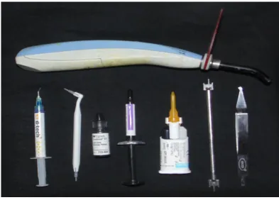
Figure 10 Armamentarium
Figure 11 Instron Machine
Figure 12 Shear testing on Instron
Figure 13 Ion sputter unit
Figure14 Scanning Electron Microscope( SEM)
Figure 15 SEM of normal enamel(1500X)
Figure 16 SEM of normal enamel(3000X)
Figure 17 SEM of 37% phosphoric acid etched enamel(1500X)
Figure 18 SEM of 37% phosphoric acid etched enamel(3000X)
Figure 19 SEM of 8% Papain and 37% phosphoric acid etched(1500X)
Figure 20 SEM of 8% Papain and 37% phosphoric acid etched(3000X)
Figure 21 SEM of 10% Papain and 37% phosphoric acid etched(1500X)
[image:11.595.102.509.123.771.2]v
LIST OF ANNEXURES
Annexure No: Contents
Annexure 1 Declaration from Senthil Papain and food Products
Annexure 2 Institutional Research Committee
Annexure 3 Institutional Human Ethics Committee Certificate
Annexure 4 Test Report from Sree Chitra Tirunal Institute For Medical Sciences And Technology
Abstract
vi Introduction: The purpose of this study is to verify de-proteinization of the enamel surface with two different concentrations of papain solution 8% and 10% before acid etching with 37% phosphoric acid which increases the shear bond strength of brackets using two adhesives- Transbond XT and Fuji Ortho LC, a Resin Modified Glass Ionomer Cement.
vii Results: Statistical analysis of the data indicates that, there are significant differences between shear bond strength obtained with deproteinisation with 10% papain and without deproteinisation with both Transbond XT and Fuji Ortho LC. That is with Group I and Group III, with Group IV and Group VI. There is increase in the shear bond strength with deproteinisation with 8% papain with both Transbond XT and Fuji Ortho LC, but the values are not statistically significant.
Conclusion: From this study the following conclusions were made; 10% deproteinisation with papain before acid etching shows statistically significant increase in the shear bond strength values with both Transbond XT and Fuji Ortho LC when compared with the groups without deproteinisation . Therefore deproteinisation with 10% papain and bonding with Fuji Ortho LC can be routinely used as an alternative to Transbond XT inorder to prevent White spot lesions.
1 Contemporary orthodontic treatment requires a successful clinical bond between the orthodontic bracket and the tooth surface to withstand the mechanical and thermal effects of the oral environment. Bonding can be accomplished with various enamel conditioning procedures which can either be chemical or mechanical. The direct bonding of orthodontic brackets has revolutionized and improved the clinical practice of orthodontics. However, there is a need to further improve the bonding procedure to save time and to minimize enamel loss without jeopardizing the ability to maintain clinically useful bond strength.
Buonocore(1) introduced the use of micromechanical bonding between a dental material and the enamel surface in 1955 by treating enamel surface with 85% phosphoric acid. Phosphoric acid etching has been used for tooth enamel preparation and for resin bonding and also for orthodontic attachment. An irregular surface of enamel is created mostly by dissolving hydroxyapatite which allows the flow of fluid adhesive components which locks the adhesive, leading to micromechanical retention. Achieving a low bond failure rate should be a high priority objective. Replacing loose brackets is inefficient, time-consuming, and costly. Consequently, a continuous search is on for higher bond strengths, better adhesives, simpler procedures, and materials. However, most bond failures result from inconsistencies in the bonding technique and not because of the bonding resins, inadequate bond strengths, or quality of the brackets being used.
Introduction
2 bonding over the indirect procedure were improved bond strength, and the bonding adhesive constantly filled out the entire contact surface of the brackets.(3)
Enamel consists of 96 % inorganic content by weight. The remaining 4 % consists of 3% water and organic content 1% (proteins and lipids).(4) Protein content of normal enamel is 0.04-0.7%.(5) These organic elements make it difficult for adhesive components to adhere to the tooth enamel surface, diminishing its shear bond strength.
Enamel lacks collagen, as found in dentin and bone, but it contains two unique
classes of proteins - amelogenins and enamelins, and other proteins like ameloblastin
and sheathlin.(6) These proteins help in the development of enamel by serving as a
framework for minerals to form on.(6)
3 permeability membrane, preventing direct contact between acids and the tooth surface, thus reducing the dissolution rate of dental hard tissue.
Therefore conditioning of the enamel surface should be done in order to eliminate the organic components that hinder effective enamel etching. It is highly likely that, despite our best efforts, the organic layer cannot be entirely removed without considering the proteins embedded in the crystals forming the enamel. Studies have shown that it is this layer of external organic matter that prevents the acid to effectively etch the surface, resulting in inconsistent patters of etching and a non-reliable area for orthodontic bonding.(8)
Etching of enamel with 37% phosphoric acid after eliminating the organic elements from the enamel surface probably produces longer adhesive tags that penetrate the enamel. It is important to realize that the action of phosphoric acid on the enamel surface occurs mostly on mineralized tissues (inorganic matter). Unfortunately, this acid does not eliminate the organic matter. Proof of this is the “collagen network” resulting from demineralization of dentin by phosphoric acid where the collagen fibers are left intact.(8) The longer tags greatly increase the mechanical retention of adhesives to the enamel. Therefore elimination of organic substances from the enamel surface before acid etching increases the resistance to debonding of orthodontic bracket by providing a better acid etching pattern on enamel.
Different studies have been conducted in an effort to improve the retention
between enamel surface and the restorative materials. Bonding to enamel depends
Introduction
4
and composition of the enamel surface. (9,10,11) Studies done by Espinosa et al,(12) and
Justus et al.(13) used sodium hypochlorite for deproteinizing the enamel surface with the intention to remove the organic components that interfered with effective enamel
etching and it shows that there is an increase in the etched surface area to double
when compared with conventional phosphoric acid etching alone. Deproteinization of
enamel involves the removal of organic content i.e. proteins from the enamel. It
showed that prior deproteinization by sodium hypochlorite doubled significantly
enamel’s retentive surface to 94.47%. This technique seemed effective in removing
organic elements from the enamel structure as well as the acquired pellicle thereby
conserving the tooth structure and improving its adhesive properties. According to this study, the sodium hypochlorite (NaOCl) eliminates the organic matter present on the enamel surface by dissolving it. Among substances with similar properties, papain is outstanding.
5 Papain is an enzyme that is similar to human pepsin, and is used in food technology, pharmaceutical and cosmetic industries.(16) Guzman and Guzman(17) performed clinical studies on patients with skin lesions caused by burns, observing that the enzymatic action of papain was considered excellent in areas with necrotic and purulent processes. Udok and Storojuk(18) also verified that papain aided cleansing necrotic tissue and secretions, shortening the period of tissue repair. Dawkins et al.(19) showed that Papain has bactericidal and bacteriostatic properties which inhibit the growth of gram positive and gram negative organism.
Introduction
6 In 2003, a Brazilian formulation was introduced and commercially denominated as "Papacárie" (Fórmula e Ação, São Paulo, SP, Brazil) and thus papain was introduced into dentistry. The product, Papacarie, is used in the chemical removal of caries. It is used with the aim of removing infected tissue without causing any damage to any healthy structure in the mouth, and requires neither cutting instruments.(23,24)
Papain-gel has been utilized as a chemo-mechanical material for caries removal due to its ability to preserve underlying sound dentin. However, little is known about the effect of the papain enzyme on intact type I collagen fibrils that compose the dentin matrix. Intact nonmineralized type I collagen fibrils are partially degraded by a papain-gel. (25)Papain when applied to the contaminated dentine has proteolytic, chlorinating and oxidating properties on the affected collagen, without acting on the sound dentine. It is able to remove the smear layer, which facilitates the penetration of adhesives, thereby enhancing the adhesional properties of restorative materials, without compromising on the shear bond strength.(26,27)
Recently, Pithon et al. (28) in 2012 suggested the use of 10% papain as a deproteinizing agent before acid etching and verified that this removal of organic elements intensified the bond strength.
The most common and popular adhesives used in orthodontics for bonding
procedures are bis GMA and Resin Modified Glass Ionomer Cements. Bisphenol A glycidyl dimethacrylate, more commonly known as Bowen’s resin or bis
7 dimensional stability of epoxy. The eventual addition of filler particles to these resins to form composites greatly enhanced the strength of this material.
Wilson and Kent introduced glass polyalkenoate, or Glass Ionomer Cement (GIC), to dentistry in 1972. Glass Ionomer Cement contains a powder similar to that of silicate cement and a polyacrylic liquid similar to that of polycarboxylate cement. It bonds chemically to enamel, cementum, dentin, nonprecious metals, and plastics.(30) The dry field necessary for composite resin bonding is not necessary for this type of cement. Antonucci et al(31) introduced resin modified glass ionomer cements (RMGICs) in 1988.
8
The purpose of this study is to verify de-proteinization of the enamel surface with two different concentrations of papain solution 8% and 10% before acid etching with 37% phosphoric acid which increases the shear bond strength of brackets using two adhesives- Transbond XT and Fuji Ortho LC, a Resin Modified Glass Ionomer Cement.
1. Comparision of shear bond strength of metal brackets with deproteinization by two different concentrations of papain solution and with that of control group using a Universal testing Machine.
9 Buonocore (1955) outlined a simple method of increasing the adhesion of filling materials to enamel surfaces in which he employed 85% phosphoric acid for 30 seconds and phosphomolybdate oxalic acid treatment to alter the enamel surface chemically and concluded that phosphoric acid gave better results and was simpler to use.(3)
Review of Literature
10
Burrow etal (1990) concluded in this study that the etching does not remove much of the organic debris, and there was no difference between a liquid or gel etchant. Agitation of the etchant did not aid pellicle removal.(32)
Legler etal (1990) in his study of effects of phosphoric acid concentration and etch duration on enamel depth of etch reported that no significant differences among the shear bond strengths resulting from the application of an orthodontic bonding resin to enamel surfaces etched with three phosphoric acid (H3PO4) concentrations, each for three etch durations in the the facial surfaces of 45 extracted human maxillary permanent central incisors.(9)
Bhad etal (1995)their study of Scanning electron microscopic study and shear bond strength measurement with 5% and 37% phosphoric acid. Detected the etch pattern with scanning electron microscopy and the shear bond strength with a Hounsefield tensometer (Nene Instruments Ltd., Northampton, England) by using 37% and 5% phosphoric acid (H3PO4) shows that with 5% H3PO4 there was minimal
enamel loss compared with 37% H3PO4 and there was no significant difference in
shear bond strength, when enamel surface was etched with 5% H3PO4 and 37%
H3PO4. (10)
11
utilized. They concluded that the light-cured composites had a higher shear bond strength than the RMGIC.(67)
Rixetal (2001) in this study compared 3 orthodontic adhesives in the areas of shear-peel bond strength, location of adhesive failure, and extent of enamel cracking before bonding and after debonding of orthodontic brackets. The adhesives included a composite resin control (Transbond XT; 3M/Unitek, St Paul, Minn), a resinmodified glass ionomer cement (Fuji Ortho LC; GC America Corp, Alsip, Ill), and a polyacid-modified composite resin under dry and saliva-contaminated conditions (Assure; Reliance Orthodontic Products Inc, Itasca, Ill), from that Transbond XT displayed significantly greater shear-peel bond strength than Fuji Ortho LC and Assure, although the bond strengths for all 3 adhesives were clinically acceptable. There was no significant difference in mean shear peel bond strengths between Assure-wet (salivacontaminated) and Assure-dry (non-contaminated) protocols.Fuji Ortho LC and Assure-wet tended to display adhesive failure at the enamel/adhesive interface while Assure-dry and Transbond XT tended to display cohesive failure within the adhesive. The greatest frequencies for enamel fracture upon debonding occurred in the groups showing the highest bond strengths (Transbond XT and Fuji Ortho LC).(33)
Review of Literature
12
resin glass ionomer adhesive system can be formulated and employed to release fluoride and possess a high enough bond strength to minimize untimely bracket debonding.(75)
Newman etal (2001) in his study of Comparative assessment of light-cured
resin-modified glass ionomer and composite resin adhesives states that the ideal adhesive system is one that prevents decalcifications and has sufficient bond strength to withstand untimely impact forces on bonded brackets, a new light-cured resin-modified glass ionomer adhesive was compared with the conventional adhesive systems. The effects of the new adhesive, with a system of etching and using adhesive promoters on the tooth enamel, as well as microetching the brackets, were analyzed. The new adhesive system is indicated where prevention of decalcification and increased bond strength in noncompliant patients are indicated. (34)
13
greatest area of etched enamel surface was occupied by type C (etched, but enamel prisms not evident). It was concluded that there is a significant difference in the acid-etch patterns achieved on different tooth types, which suggests that bond-strength studies should be performed with a single tooth type or that an equal number of different tooth types be included.(11)
Smith etal (2003) in the study to assess the shear bond strengths of resinreinforced glass ionomer Fuji Ortho LC and GC Fuji Ortho cements under differing conditions and compare their bonding performance with that of conventional resin composite bonding systems has shown that glass ionomer cements provide sufficiently high shear bond strengths to retain orthodontic brackets under clinical conditions and, at the same time, could provide a reservoir of fluoride, thereby reducing the cariogenic potential of plaque-retaining orthodontic attachments Both glass ionomer cements were thus acceptable for orthodontic bonding. Transbond XT had the highest mean shear bond strength irrespective of the incubation period.(68)
Dawkins etal (2003) A study on antibacterial effects of carica papaya fruit on common wound organisms found that no significant difference was found in bacterial sensitivity between immature, mature and ripe fruits. Also found that carica papaya seeds contain anti-bacterial activity that inhibits growth of positive and gram-negative organisms which was independent of stage of fruit maturity which could be useful in treating chronic skin ulcers to promote healing.(19)
Review of Literature
14
brackets, after orthophosphoric acid–etching of enamel, is strongly encouraged because obtained bond strengths are within the range of clinical use and are not different from those attained by light cured composite resins. Also stated that RMGICs are fluoride-releasing materials that are able to stand wet conditions, and enamel is less damaged after debracketing.(69)
Pascotto etal (2004) In vivo effect of a resin-modified glass ionomer cement on enamel demineralization around orthodontic brackets in vivo study showed that a resin-modified glass ionomer cement could reduce enamel demineralization around a bonded bracket mainly in a tooth area at high caries risk. It is suggested that its use as a bonding material should be encouraged during orthodontic treatment. (76)
Summers etal (2004) Comparison of bond strength between a conventional resin adhesive and a resinmodified glass ionomer adhesive: An in vitro and in vivo study In vitro results showed significantly greater shear bond strengths when brackets were bonded with 37% phosphoric acid and composite resin (Light Bond) compared with RMGI (Fuji Ortho LC) bonded with 10% polyacrylic acid. Significantly greater shear bond strengths can be obtained 24 hours after bonding brackets for both materials.The in vivo results showed no significant difference in bracket failure rates between Fuji Ortho LC and Light Bond after 1.3 years. Clinically, Fuji Ortho LC adhesive has adequate bond strength to withstand the occlusal forces of chewing and biting.(35)
15 Campoy etal (2005) his study evaluates the effect of saliva contamination at different stages of the bonding brackets procedure using the self-etching primer Adper Prompt L-Pop (3M ESPE, Minneapolis, Minn) and the resin orthodontic adhesive system Transbond XT (3M). The greatest bond strength values were obtained when contamination did not occur. Significant differences were observed between the bond strengths of the control group and the group inwhich contamination occurred before the application of Adper PLP. Significant differences were also observed between the control group and the group in which saliva contamination occurred before and after the application of the SEP.(36)
Matheus Melo Pithon etal in (2006).A study on 'Metallic Brackets Bonded with Resin-reinforced Glass Ionomer Cements under Different Enamel Conditions' to assess the shear bond strength of metallic orthodontic brackets bonded with either Fuji Ortho or Ortho Glass LC resin-reinforced glass ionomer cements to enamel surfaces under different conditions, namely, enamel without etching, enamel conditioned with 37% phosphoric acid and enamel conditioned with Transbond Plus Self Etching Primer (TPSEP) concluded that regardless of the enamel treatment, Fuji Ortho LC yielded shear strength values superior to those from OrthoGlassLC.(37)
Review of Literature
16
be useful when the enamel surface is contaminated with water before the application of bonding materials.(78)
Godoy-Bezera etal (2006) A study done to evaluate the shear bond strength of resin-modified glass ionomer cement in a saliva-contaminated environment, using different enamel pretreatments like 10% polyacrylic acid , 37% phosphoric acid ,no enamel etching,rinsing and drying with oil-free compressed air concluded that enamel pretreatment with 37% phosphoric acid increased RMGIC bond strength values, with no statistically significant differences from those obtained with the resin composite used as a control and that etching the enamel surface with 10% polyacrylic acid, or not etching it, yielded the lowest shear bond strength values, with no difference between them.(70)
17
primer was also negatively affected by blood contamination, although it was suitable for bonding with saliva contamination.(38)
Lopes etal (2007) did a study to assess the shear bond strength of an adhesive restorative system on sound and demineralized dentin after the use of a papain-based agentand he concluded that the use of a papain-based gel to remove dental caries did not interfere in the bond strength of restorative materials to dentin.(27)
Cacciafesta etal (2007) on their study compared the in-vitro fluoride release rates from 9 orthodontic adhesives in distilled water, of that adhesives tested, 4 were bracket bonding agents Fuji Ortho LC ,Enlight LV, ConTec LC and Transbond XT , among the bracket bonding adhesives, statistically significant differences were found in fluoride release rates with Fuji Ortho LC releasing the most fluoride to reduce white spot formation.(77)
Bishara etal (2007) To compare the effects of a standardized thermocycling protocol on the shear bond strength (SBS) of two adhesive systems: a resin-modified glass ionomer and a composite resin used with a new self-etching primer. Following thermocycling, the SBS of a new self-etching primer adhesive system and a resin-modified glass ionomer adhesive are not significantly different from each other and are at clinically acceptable levels.(39)
Review of Literature
18
composite resin. • The new self-etch conditioner has the added benefit of not needing to be rinsed off and may reduce technique sensitively in the bonding process.(40)
Evandro Piva etal in (2008) A study on' Papain-based gel for biochemical caries removal: influence on microtensile bond strength to dentin' investigated the influence of a papain-based gel (Papacárie) for chemo-mechanical caries removal on bond strength to dentin which showed that for the self-etching adhesive system tested, the papain-based gel reduced the microtensile bond strength to carious dentin.(41)
Soumya etal (2008) A study on assessment of the shear bond strength of an adhesive restorative system on sound and demineralized dentin after the use of a papain-based agent concluded that a papain-based chemomechanical agent does not interfere in the shear bond strength of restorative materials when a total etch adhesive system is required so that this agent can safely be used as a method for caries removal when employing conventional adhesive systems.(42)
19 Budassori etal 2008 A case report on chemo-mechanical removal of caries in an adolescent patient using a papain gel stated that use of papacarie is recommended as a solution for the treatment of patients seeking alternative to conventional methods. They also stated that removal of carious tissue with papacarie proved to be efficient, easy to perform and comfortable for the patient.(23)
Romano etal 2009 The aim of this study was to assess the shear bond strength of orthodontic brackets in different enamel surfaces using the Transbond Plus Color Change composite (TPCC-3M Unitek).IBracket bonding using TPCC showed adequate adhesion for clinical use, and the type of enamel preparation had no influence.(43)
Bertassoni LE etal in 2009 A study on' Papain-gel degrades intact nonmineralized type I collagen fibrils' to define structural changes that occur in intact type I collagen fibrils after an enzymatic treatment with a papain-gel gave a novel evidence that intact nonmineralized type I collagen fibrils are partially degraded by a papain-gel.(25)
Review of Literature
20
XT is used for resin bracket bonding, it shows a certain tolerance to wet conditions only when used in combination with TSEP.(44)
Retamoso etal2009 in their study to evaluate the influence of saliva contamination on shear bond strength and the bond failure pattern of 3 adhesive systems (Transbond XT, AdheSE and Xeno III) on orthodontic metallic brackets bonded to human enamel. Each system was tested under 2 different enamel conditions: no contamination and contaminated with saliva.. The control and contaminated groups showed no significant difference in shear bond strength for the same adhesive system. Saliva contamination showed little influence on the 24-h shear bond strength of orthodontic brackets.(45)
Espinosa etal 2010 studied about Resin Replica in Enamel Deproteinization and its Effect on Acid Etching and the goal of this in vitro study was to identify the topographical features of deproteinized (NaOCl) and etched with phosphoric acid (H3PO4) enamel surface, compared to phosphoric acid surface alone with a Resin Replica model shows that conventional H3PO4 enamel etching has significant limitations, etching less than 46% of the total enamel’s surface.• Enamel deproteinization prior to phosphoric acid etching almost doubles enamel’s retentive surface to 73%.• The topographical features of the replica resin penetration surface increases significantly with type I-II etching pattern, when deproteinization is done with 5.25%NaOCl for 1 minute prior phosphoric acid etching.(46)
21
composite resin and a RMGI in 76 extracted human premolars concluded that applying 5.25% NaOCL to the enamel surface eliminates the organic elements allowing the acid etchant to penetrate more effectively into the enamel, creating type 1 and 2 etching patterns. They also showed that significantly greater bracket SBS can be obtained with RMGI if the enamel surface is wetted for 1 minute with 5.25% NaOCL, before etching.and when 5.25% NaOCL is used to deproteinize the enamel surface, brackets bonded with RMGI have comparable SBS to brackets bonded with composite resin.(13)
Brown etal 2011 A study which intended to develop novel BAG-containing composite resins which would release ions into simulated body fluid (SBF) at pH 4 and pH 7 that would result in a change in the pH of the surrounding environment to be used for bonding of orthodontic brackets concluded that combining BAG into resin adhesives may result in a smart material that provides a reservoir of crucial ions for remineralization or for the protection of enamel from demineralization by raising the pH of cariogenic environments and may also decrease critical pH, resulting in decreased mineral loss from teeth.(47)
Review of Literature
22
treatments resulted in greater bond strength in BisCem specimens while acid etching alone did not improve the performance of the material.(48)
Flavio etal 2011 Another study was done to evaluate the microtensile bond strength of adhesive systems to caries-affected dentin formed in situ after the use of a papain gel and their results showed that the application of the chemomechanical and mechanical methods to demineralized dentin did not affect the bond strength values and SEM analysis showed no interference of papain-based gel in the formation of hybrid layer.(27)
Harleen etal 2011 in an invitro study about enamel deproteinization with 5.25% sodium hypochlorite before acid etching and its effect on the shear bond strength with two adhesives -AdperTM Single Bond 2 adhesive and Filtek Z-350 XT composite resin, the mean shear bond strength value for AdperTM was 13.51 +/- 5.726 MPa and for Filter Z was 15.06 +/- 6.220 MPa. No significant effect of sodium hypochlorite enamel deproteinization on the shear bond strength of Adper Single Bond 2 adhesive and Filtek Z-350 XT composite resin before acid etching was observed in this study.(49)
23
efficient alternative for deproteinization of the tooth enamel surface before bonding orthodontic brackets with RMGIC.(28)
Ljima etal 2012 A study to determine if a new experimental resin-based material containing Portland cement (PC) can help prevent enamel caries while providing adequate SBS and a caries-preventive effect equivalent to that of the RMGIC adhesive system.ing adequate shear bond strength (SBS) concluded that the new system provides adequate SBS and a caries-preventive effect equivalent to that of the RMGIC adhesive system.(50)
Kohda etal 2012 A study done to determine if the enamel around orthodontic brackets is significantly altered after demineralization followed by application of adhesives with and without fluoride-releasing ability in 108 non-carious human premolars concluded that fluoride-releasing adhesives may prevent demineralization of enamel around brackets during orthodontic treatment. They also suggested that nanoindentation is a useful method for investigating mechanical properties in small regions of enamel, and these properties should be relevant to demineralization.(71)
Amri etal 2012 concluded that Papain has revealed to be an enzymatic protein of significant biological and economic importance. It is through the unique structure of papain that provides functionality and helps explain how this proteolytic enzyme works and also makes it valuable for a variety of purposes.(14)
Review of Literature
24
antiviral,antifungal and antibacterial properties. And also used for digestive problems, toothpaste and meat tenderizers.(16)
Gabriel etal 2013 A study was done to evaluate the capacity of 2% chlorhexidine gel associated with 8% papain gel in comparison with 5.25% sodium hypochlorite in bovine pulp tissue dissolution and they concluded that 8% papain in gel either alone or in association with chlorhexidine, was able to dissolve bovine pulp tissue.(51)
Martins etal 2013 A study was performed histological evaluation of the
antiinflammatory and healing properties of Papacarie applied to oral ulcers against
non treated control group .They showed that oral ulcers treated with a papain-based
gel exhibited the same inflammatory reaction and healing aspects as those of the non-
treated control group.(52)
Dayanand etal 2013 A study on evaluation of in vitro total proteolytic activity from various plant lattices belongs to families such as apocynaeceae, asclepiadaceae, caricaceae, euphorbiaceae, moraceae at different pH conditions and compared with latex of Carica papaya as standard protease activity stated that lattices of these selected plant families contain protease activity as common biological activity.(20)
25
demineralised enamel concluded that in demineralised enamel, microleakage occurred mainly at the enamel-adhesive than the adhesive bracket interface, proposing a greater risk of enamel demineralisation upon occurrence of microleakage, enamel deproteinisation with a 5% sodium hypochlorite solution failed to reduce microleakage under brackets bonded to demineralised enamel, use of Transbond Plus self-etching primer for preparation of demineralised enamel significantly increased microleakage at the adhesive-bracket interface on both sides of the brackets and use of 2% NaF on hypomineralised enamel before the bracket bonding procedure is an effective way to decrease microleakage.(53)
Pithon etal 2013 A study done to verify the hypothesis that enamel deproteinization with papain gel at concentrations of 2%, 4%, 6%, 8%, and 10% increases shear bond strength as concentration increases concluded that concluded that enamel deproteinization with 8% and 10% papain gel increases shear bond strength of orthodontic brackets bonded with RMGIC. They also stated that papain gel at concentrations of 2%, 4%, 6%, 8%, and 10% increased the RMGIC bond to the enamel surface, which is clinically relevant, as they minimize the occurrence of cracks and fractures on the tooth surface when orthodontic accessories are removed.(73)
Review of Literature
26
treated or non-treated with NaOCl.Also showed that when enamel was deproteinized, a larger amount of cement remained on the enamel surface, showing a behaviour similar to that of the resin composite.(72)
Sharma etal (2013) in the study of comparative evaluation of the retention of metallic brackets bonded with resin modified glass ionomer cements under different enamel conditions- sand blasting and 5.25% sodium hypochlorite concludes that the bond failure rate of RMGIC on unprepared and deproteinised enamel surface was significantly higher than that of Transbond XT which was used as the control group.(54)
J Kumar etal 2014 A study done to compare the clinical efficiency of chemomechanical caries removal using carisolv and papacarie - a papain gel in 40 patients concluded that carisol and papacarie were both clinically efficient for carious dentin removal and that papacarie was marginally better in the tested clinical parameters, i.e., time taken and volume of carious tissue excavated.(55)
27 Gurel etal 2014 A study was done to compare the shear bond strength (SBS) of the adhesive pre-coated II (APC II) adhesive coated appliance system with that of Transbond XT composite resin and they concluded that although SBS for APC II was found to be higher than that of Transbond XT, the APC II system has been proven to be efficient for clinical use.(57)
Caglalogru 2014 A study was done to evaluate the force required to cause debanding when polyacid-modified composite resin, resin-modified glass ionomer, glass ionomer of various commercial products are used as the luting agents concluded that all evaluated cements yielded comparable shear bond strengths and no significant difference in mean shear debonding forces of bands bonded with conventional GIC, RMGIC, or modified composite resin.But their mode of failure differed significantly in that bond failure for bands cemented with modified composite resin occurred predominantly at the cement/band interface, whereas failure for GIC and RMGIC specimens occurred mostly at the enamel/cement interface.(58)
Pradeep kumar. Etal 2014 Papacarie is an emerging chemo-mechanical caries removal agent which on interaction with the exposed collagen causes dissolution of dentin minerals and makes the dentin softer and hence facilitates removal of caries. Papain can safely be used as a method for caries removal when employed along with conventional adhesive systems.(59)
28
This experimental invitro study has been approved by Institutional Human Ethics Committee in Sree Mookambika Institute Of Medical Sciences Kulasekaram,
Ref. No. SMIMS/IHEC/2014/A/20.
And the study has been conducted here at Sree Mookambika Institute Of Dental Sciences. Shear Bond Testing has been done at Sree Chithra Institute For Medical Sciences And Technology, BIO MEDICAL TECHNOLOGY WING, Poojappura, Thiruvananthapuram.
Materials
1. Freshly extracted 90 premolars 2. Isotonic saline
3. Cold cure acrylic-powder and liquid 4. Etchant- 37% Phosphoric acid (D-Tech) 5. Transbond XT Primer (3M Unitek)
6. Transbond XT composite, (3M Unitek Monrovia Calif) 7. Resin Modified Light Cure GIC- GC FUJI ORTHO LC.
8. MBT -3M Gemini SERIES premolar brackets 0.022 “ bracket base surface area of 9.806 mm2.
9. Papain powder obtained from Senthil Papain And Food Products (P) Limited, Senguptha Street Ramnagar Coimbatore, Tamilnadu and here by enclosed authentication letter from the company with composition of the powder. 10.Papain solution – 8% and 10%.
Materials and Methods
29
10% concentration obtained by 10mg in 100ml of sterile distilled water. Dilution of papain powder to 8% and 10% was done at the lab of Senthil Papain And Food Products using a standardised procedure.
Instruments
1. MBT gauge 2. Bracket Holder
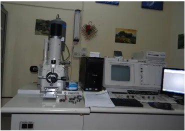
3. Sputter coater( Hitachi E 1010 ion sputter) 4. Scanning Electron Microscope ( Hitachi S. 2400)
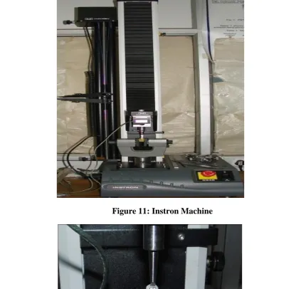
5. Visible light curing unit( Woodpecker LED Light, DC-5.0V) 6. Universal testing machine, (INSTRON model-3345).
Inclusion criteria: Premolar teeth extracted for orthodontic purpose. Exclusion criteria: Teeth with enamel defects
Teeth with morphological defects Teeth with decalcification and caries Teeth that were previously bonded. Cracks caused by extraction forceps.
Methodology:
30
this, the teeth were randomly divided into six groups (n = 15) that are denominated as follows:
Scientific basis of sample size used in the study: Calculated by
2 P2) -(P1 7.84 x 2Pq ; 2 P2 P1 is
P ; P= Prevalence, P1 and P2 are the
mean or proportions or percentage of any one groups. q is 1-p
Detailed description of the groups:
Groups Colour code Sample code Deproteinization Adhesive
Group I Orange XT Nil Bonding with Transbond XT after 37% phosphoric acid etching
Group II Blue XTPAP8 8% Papain Bonding with Transbond XT after 37% phosphoric acid etching
Group III Yellow XTPAP10 10% Papain Bonding with Transbond XT after 37% phosphoric acid etching
Group IV Red RM Nil Bonding with Fuji Ortho LC after 37% phosphoric acid etching
Group V Green RMPAP8 8% Papain Bonding with Fuji Ortho LC after 37% phosphoric acid etching
Materials and Methods
31
All the samples from the six group are bonded with metal brackets. Group I
serves as the control group to which Group II and Group III are compared whether they are statistically significant or not. While Group IV is the control groups for
Group V and Group VI to which they are compared.
Deproteinization Procedure :
Samples from Group II and Group V were deproteinized with 8% papain solution for 60 seconds separately and then rinsed with running water for a period of 60 seconds and dried.
Samples from Group III and Group VI were deproteinized with 10% papain solution for 60 seconds separately and then rinsed with running water for a period of 60 seconds and dried.
Bonding Procedure:
All the Samples from Group I to Group VI are acid etched using 37% phosphoric acid by applying it to the enamel surface for a time period of 30 seconds then rinsed with running water for 60 seconds and gently dried with oil free air spray.
32
uniform thickness of adhesive, adjusted to the final position and pressed firmly. The excessive adhesive was removed from the periphery of the tooth surfaces. Each side of the tooth was light cured for a period on 10 seconds (total of 40 seconds).
For the samples from the Groups IV ,V and VI, Fuji Ortho LC was placed on the bracket mesh covering the entire base of the bracket without bubbles or voids, and the bracket was then applied to the tooth using sufficient force to produce a “flash” of excess adhesive around the bracket to ensure a uniform thickness of adhesive, adjusted to the final position and pressed firmly. The excessive adhesive was removed from the periphery of the tooth surfaces. Each side of the tooth was light cured for a period on 10 seconds (total of 40 seconds).
Shear bond testing:
Materials and Methods
33
The shear force at a crosshead speed of 1mm/minute was transmitted to the bracket by a shear blade. The force required to shear the bracket causing bonding failure was recorded in Newtons and the bond strengths were calculated in megapascals (MPa). Samples collected were tested and stored in PC- software origin 6.1- Origin Lab, California,USA. the readings of shear bond strengths recorded in Newtons was converted into Megapascals by following equation:
Debonding force in newtons Shear bond strength in megapascals =
Bracket base area in mm2
Method of statistical analysis
34 SEM (SCANNING ELECTRON MICROSCOPY):
A qualitative study was carried out to observe with Scanning Electron Microscope (SEM), the type of etch pattern with and without the use of papain before etching. Four extracted, intact, human premolars were selected for Scanning Electron Microscopic study of normal enamel, acid etched enamel and papain treated enamel with 8% and 10%. Samples are selected to study the topographic feature of normal enamel, 37 % phosphoric acid etched enamel and papain treated enamel with 8% and 10% before acid etching.
Sample No.1 Normal Enamel
Sample No.2 Acid etched with 37% phosphoric acid
Sample No.3 8% papain solution plus 37% phosphoric acid
Sample No.4 10% papain solution plus 37% phosphoric acid
Sample No.1- The buccal surface of premolar is viewed under scanning electron microscope for the normal enamel morphology.
Materials and Methods
35 Sample No. 3- The buccal surfaces of the premolars were deproteinized with 8%. papain for 60 seconds followed by rinsing, drying, and acid etching with 37% phosphoric acid for 30 seconds. Subsequently, the acid was rinsed off, the enamel was dried, and observed with scanning electron microscope to determine the etch pattern.
Sample No.4- The buccal surfaces of the premolars were deproteinized with 10% papain for 60 seconds followed by rinsing, drying, and acid etching with 37% phosphoric acid for 30 seconds. Subsequently, the acid was rinsed off, the enamel was dried, and observed with scanning electron microscope to determine the etch pattern.
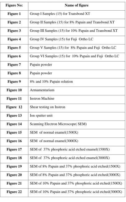
Figure 1: Samples (15) of Group I
Figures
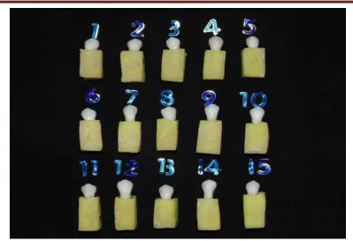
Figure 3: Samples (15) of Group III
Figure 5: Samples (15) of Group V
[image:58.595.138.500.439.727.2]
Figures
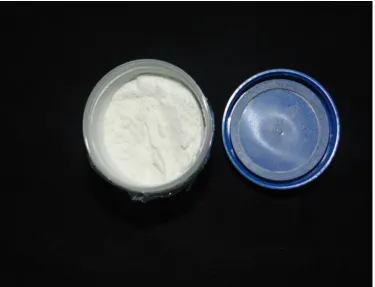
[image:59.595.130.506.439.726.2]Figure 7: Papain powder
Figure 9: 8% and 10% Papain Solution
Figures
Figure 11: Instron Machine
[image:61.595.105.522.96.502.2]Figure 13:Ion Sputter Unit
36 Statistical analysis: The data expressed in MEAN±SD. Statistical Package for Social Sciences (SPSS 20.0) version was used for statistical analysis. One way ANOVA applied for analysis. Post Hoc followed Dunnet t test used to find statistical significant between and within the groups. P value less than 0.05 (P<0.05) considered statistically significant at 95% confidence interval.
Table-1: Mean Max.SBS values of different groups
Groups Sample Code Max.SBS (MPa) (MEAN±SD)
Group-I XT 16.04±1.34
Group-II XTPAP8 17.59±1.13
Group-III XTPAP10 18.35±2.35
Group-IV RM 13.82±1.51
Group-V RMPAP8 15.69±1.40
Group-VI RMPAP10 16.05±1.57
Table-2: Comparison of mean Max.SBS values of group-I with other groups
Groups Max.SBS (MPa) (MEAN±SD) P value
Group-I 16.04±1.34
Group-II 17.59±1.13 0.80
Group-III 18.35±2.35* 0.04
Group-IV 13.82±1.51* 0.05
Group-V 15.69±1.40 0.78
Group-VI 16.05±1.57 0.97
[image:64.595.116.521.506.752.2]Results
[image:65.595.121.511.116.359.2]37 Table-3: Comparison of mean Max.SBS values of group-II with other groups
Groups Max.SBS (MPa) (MEAN±SD) P value
Group-II 17.59±1.13
Group-I 16.04±1.34 0.80
Group-III 18.35±2.35 0.73
Group-IV 13.82±1.51* 0.05
Group-V 15.69±1.40* 0.05
Group-VI 16.05±1.57 0.95
(*P<0.05 significant compared group-II with other groups)
Table-4: Comparison of mean Max.SBS values of group-III with other groups
Groups Max.SBS (MPa) (MEAN±SD) P value
Group-III 18.35±2.35
Group-I 16.04±1.34* 0.04
Group-II 17.59±1.13 0.73
Group-IV 13.82±1.51* 0.02
Group-V 15.69±1.40* 0.04
Group-VI 16.05±1.57* 0.05
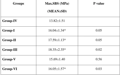
[image:65.595.112.520.460.696.2]38 Table-5: Comparison of mean Max.SBS values of group-IV with other groups
Groups Max.SBS (MPa)
(MEAN±SD)
P value
Group-IV 13.82±1.51
Group-I 16.04±1.34* 0.05
Group-II 17.59±1.13* 0.05
Group-III 18.35±2.35* 0.02
Group-V 15.69±1.40 0.56
Group-VI 16.05±1.57* 0.03
(*P<0.05 significant compared group-IV with other groups)
Table-6: Comparison of mean Max.SBS values of group-V with other groups
Groups Max.SBS (MPa) (MEAN±SD) P value
Group-V 15.69±1.40
Group-I 16.04±1.34 0.78
Group-II 17.59±1.13* 0.05
Group-III 18.35±2.35* 0.04
Group-IV 13.82±1.51 0.56
Group-VI 16.05±1.57 0.69
[image:66.595.106.527.486.720.2]Results
[image:67.595.110.522.121.358.2]39 Table-7: Comparison of mean Max.SBS values of group-VI with other groups
Groups Max.SBS (MPa) (MEAN±SD) P value
Group-VI 16.05±1.57
Group-I 16.04±1.34 0.97
Group-II 17.59±1.13 0.95
Group-III 18.35±2.35* 0.05
Group-IV 13.82±1.51* 0.03
Group-V 15.69±1.40 0.69
(*P<0.05 significant compared group-VI with other groups)
Table-8: Multiple comparison of mean Max.SBS values between the groups
Groups Max.SBS (MPa) (MEAN±SD)
Group-I 16.04±1.34
Group-II 17.59±1.13
Group-III 18.35±2.35*
Group-IV 13.82±1.51*,#,$
Group-V 15.69±1.40#,$
Group-VI 16.05±1.57$,ǁ
[image:67.595.169.462.458.687.2]40 Shear Bond Strength (SBS)
The descriptive statistics, including mean, standard deviation, and maximum values for the Transbond XT without papain,with 8%, 10% papain and Fuji Ortho LC without papain ,with 8% and 10% papain, are presented in Table 1.
The mean SBS for the brackets bonded using Transbond XT, with enamel deproteinization by 8% papain was 17.59±1.13 MPa and by 10% papain was 18.35±2.35 MPa, and the mean SBS for the brackets bonded using Transbond XT in the control group (without enamel deproteinization) was 16.04±1.34 MPa.
The mean SBS for the brackets bonded using Fuji Ortho LC, with enamel deproteinization by 8% papain was 15.69±1.40 MPa, and with 10% papain was 16.05±1.57 MPa and the mean SBS for the brackets bonded using Fuji Ortho LC in the control group (without enamel deproteinization) was 13.82±1.51 MPa.
Table 2 shows that Group I is statistically significant (P<0.05 ) when compared with Group III and Group IV.
Table 3 shows that Group II is statistically significant (P<0.05 ) when compared with Group IV and Group V.
Table 4 shows that Group III is statistically significant (P<0.05 ) when compared with Group I, Group IV,Group V and Group VI.
Results
41
Table 6 shows that Group V is statistically significant (P<0.05 ) when compared with Group II and Group III.
Table 7 shows that Group VI is statistically significant (P<0.05 ) when compared with Group III and Group IV.
Scanning Electron Microscope Study of Enamel Morphology
A qualitative study was carried out to observe with Scanning Electron Microscope (SEM), the type of etch pattern with and without the use of papain before etching. Four extracted, intact, human premolars were selected for Scanning Electron Microscopic study of normal enamel, 37 % phosphoric acid etched enamel for 30 seconds and enamel deproteinized with 8% papain solution followed by 37% phosphoric acid etched for 30 seconds and enamel deproteinized with 10% papain solution followed by 37% phosphoric acid etched for 30 seconds .
Sample No.1 Normal Enamel
Sample No.2 Acid etched with 37% phosphoric acid Sample No.3 8% papain solution plus 37%
phosphoric acid
Sample No.4 10% papain solution plus 37% phosphoric acid
[image:69.595.155.466.471.651.2]42 Sample No.1- The buccal surface of premolar is viewed under scanning electron microscope for the normal enamel morphology shows no surface indendations.(Figure No.15,16)
Sample No.2-The buccal surface of premolar when viewed under scanning electron microscope shows more of Silverstone’s Type I etching pattern ( honeycomb pattern) and less of Type II etch pattern.(Figure No. 17,18)
Sample No. 3- The buccal surfaces of the premolars when observed under scanning electron microscope shows more than 70% Silverstone’s Type I etching pattern and remaining of Type II etch pattern. Both types of etching pattern shows a greater increase in the surface area and also shows a qualitatively rougher enamel surface when compared with 37% phosphoric acid etching alone.(Figure No.19,20)
Sample No.4- The buccal surfaces of the premolars were observed under scanning electron microscope shows more than 70% Silverstone’s Type I etching pattern and remaining of Type II etching pattern. Both types of etching pattern shows a greater increase in the surface area and also shows a qualitatively rougher enamel surface when compared with 37% phosphoric acid etching alone. Deproteinization with 10% papain probably produces a slightly more rougher surface compared with deproteinization by 8% papain. (Figure No.21,22)
All the samples prepared for observation were viewed at 1500x and 3000x magnification with Scanning Electron Microscope.
Results
43
with enamel deproteinised with 8% and 10% papain produced a qualitatively greater enamel surface area than the enamel in which papain was not used .
Method of statistical analysis
The purpose of the present study was to check the effect of two different concentrations of papain solution before etching and followed by bonding with two different adhesive. All the measurements were tabulated and subjected to statistical analysis. Descriptive statistics, including the mean, standard deviation, and minimum and maximum values, were calculated for each group. Multiple comparisons of the shear bond strengths for the different etching types were performed with the One Way ANOVA test. The mean shear bond strength values of different groups was compared by using SPSS (20.0) version software. Data was analysed by ANOVA Post hoc test followed by Duccan’s t test. P value less than 0.05 considered statistically significant at 95% confidence interval. Statistical Package for Social Sciences (SPSS version 20.0) used for statistical analysis.
The shear bond strength was recorded in Newtons and was converted into Megapascals by the following equation
Bond strength in Newtons Bond strength in Megapascals =
Bracket base area in mm2
Graph-1: Mean Max.SBS values of different groups
Graphs
Graph-3: Comparison of mean Max.SBS values of group-II with other groups
Graph-5: Comparison of mean Max.SBS values of group-IV with other groups
Graphs
Graph-7: Comparison of mean Max.SBS values of group-VI with other groups
Scanning Electron Microscopic Images
[image:76.595.132.497.107.382.2]Figure 15: Normal Enamel (1500X)
[image:76.595.144.491.441.711.2]Figures
[image:77.595.141.493.430.715.2]Figure 17: After 37% Phosphoric acid etched enamel surface (1500X)
Figure 19: After 8% Papain and 37% phosphoric acid etched enamel surface (1500X)
Figures
Figure 21: After 10% Papain and 37% phosphoric acid etched enamel surface (1500X)
Discussion
44
Enamel consists of 96 % inorganic content by weight. The remaining 4 % consists of 3% water and organic content 1% (proteins and lipids).(4) Protein content of normal enamel is 0.04-0.7%.(5) These organic elements make it difficult for adhesive components to adhere to the tooth enamel surface, diminishing its shear bond strength. Enamel contains proteins such as amelogenins, enamelins, and also other proteins like ameloblastin and sheathlin.
All solid surfaces exposed to the oral cavity are covered by a proteinaceous layer referred to as the acquired salivary pellicle.(7) The bond strength between orthodontic adhesives and enamel may also be compromised by the presence of the pellicle at the time when they are being bonded. Functionally, it plays a role in mineral homeostasis of tooth enamel. This structure is formed by selective adsorption of proteins, peptides, and other molecules present in oral fluid . It is an organic film free of bacterial colonization that covers oral hard and soft tissues, composed of proteins, glycoproteins, enzymes, and mucins or their derivatives. Many of these proteins contain high levels (35-40%) of proline, and are therefore, designated as proline-rich proteins (PRPs), which comprise almost 70% of the total protein content of human parotid saliva. The presence of proteins covering enamel serves as a diffusion barrier or a selective permeability membrane, preventing direct contact between acids and the tooth surface, thus reducing the dissolution rate of dental hard tissue.
45
in the crystals forming the enamel. Studies have shown that it is this layer of external organic matter that prevents the acid to effectively etch the surface, resulting in inconsistent patters of etching and a non-reliable area for orthodontic bonding.(8)
Etching of enamel with 37% phosphoric acid after eliminating the organic elements from the enamel surface produces longer adhesive tags that penetrate the enamel. It is important to realize that the action of phosphoric acid on the enamel surface occurs mostly on mineralized tissues (inorganic matter). Unfortunately, this acid does not eliminate the organic matter. Proof of this is the “collagen network” resulting from demineralization of dentin by phosphoric acid where the collagen fibres are left intact.(8) The longer tags greatly increase the mechanical retention of adhesives to the enamel. Therefore elimination of organic substances from the enamel surface before acid etching increases the resistance to debonding of orthodontic bracket by providing a better acid etching pattern on enamel.
Discussion
46
Transbond XT, a light cure composite was specifically developed for bonding orthodontic attachments to the enamel. Transbond XT bonding system (3M Unitek) contains a liquid sealant and an adhesive paste. The latter is a composite that contains Bis GMA, Bis EMA, and quartz/silica fillers. The main advantages offered by this material are: reduced working time, no need of mixing, and good adhesion to enamel, thus being largely used in clinical orthodontics and experimental studies as controls.(60)Because of these advantages Transbond XT composite was used in the present study.
47
conditions.(68,69,70,71)Studies done by Newmann, Pascotto, shamsi, Reichneder showed that Fuji Ortho LC has adequate bond strength.(72,73,74,75) Hence Fuji Ortho LC was used in this study.
The present study evaluates two contemporary adhesive systems marketed for use to bond 3M 0.022” premolar orthodontic brackets—a RMGIC, Fuji Ortho LC( GC INTERNATIONAL), and a primer composite resin system, Transbond XT( 3M
UNITEK).This study was aimed to verify whether the hypothesis that deproteinization of the enamel surface with 8%, and 10% papain solution for 60 seconds increases the shear bond strength of brackets bonded with Transbond XT and Fuji Ortho LC .
In this study the highest shear bond strength was found in Group III where deproteinization was done with 10% Papain solution before etching with 37% phosphoric acid and bonding with Transbond XT which was then followed by the
Group II where deproteinization was done with 8% Papain solution, 37% phosphoric acid etching and bonded with Transbond XT( Group II). This agrees with the previous study by Pithon etal where deproteinization with 10% papain increases the shear bond strength.(28)
Discussion
48
In the present study, brackets bonded using Fuji Ortho LC after deproteinization with 10% papain solution followed by 37% phosphoric acid etching (Group VI) shows a better mean shear bond strength when compared with deproteinization with 8% papain followed by 37% phosphoric acid etching, bonded with Fuji Ortho LC(Group V). This agrees with the findings by Pithon etal, where 10% papain with Fuji Ortho LC increases the shear bond strength.(77) and also with Periere etal where NaOHCL was used as deproteinising agent. (78)
The scanning el