A CLINICAL STUDY OF FETO
OUTCOME IN PREGNANCIES WITH
ABNORMAL LIQUOR VOLUME
Dissertation submitted
THE TAMILNADU
DR. M.G.R MEDICAL UNIVERSITY, CHENNAI
With partial fulfillment of the regulations
For the award of the degree of
M.S (OBSTETRICS AND GYNAECOLOGY)
INSTITUTE OF OBSTETRICS AND GYNAECOLOGY
MADRAS MEDICAL COLLEGE
A CLINICAL STUDY OF FETO-MATERNAL
OUTCOME IN PREGNANCIES WITH
ABNORMAL LIQUOR VOLUME
Dissertation submitted
To
THE TAMILNADU
DR. M.G.R MEDICAL UNIVERSITY, CHENNAI
With partial fulfillment of the regulations
For the award of the degree of
(OBSTETRICS AND GYNAECOLOGY)
Branch - II
INSTITUTE OF OBSTETRICS AND GYNAECOLOGY
MADRAS MEDICAL COLLEGE
CHENNAI.
APRIL 2014
MATERNAL
OUTCOME IN PREGNANCIES WITH
DR. M.G.R MEDICAL UNIVERSITY, CHENNAI
(OBSTETRICS AND GYNAECOLOGY)
CERTIFICATE
This is to certify that the dissertation titled
“A CLINICAL
STUDY
OF
FETO-MATERNAL
OUTCOME
IN
PREGNANCIES WITH ABNORMAL LIQUOR VOLUME”
is a bonafide work done
by Dr.PUNITHAVATHI .J.in the
Institute of Obstetrics and Gynaecology (Madras Medical
College) Egmore, Chennai in partial fulfillment of the
university rules and regulations for the award of MS degree in
Obstetrics and Gynaecology under my guidance and
supervision during the academic year 2011-2014.
DIRECTOR AND PROFESSOR DEAN
Institute of Obstetrics & Gynaecology Madras Medical College & Madras Medical College, Rajiv Gandhi
Egmore, Chennai – 8. Govt.General Hospital Chennai – 3
GUIDE
PROF.DR.UMASHANTHI M.D., DGO
Institute of Obstetrics and Gynaecology Madras Medical College,
DECLARATION
I solemnly declare that this dissertation titled
"
“A
CLINICAL
STUDY
OF
FETO-MATERNAL
OUTCOME IN PREGNANCIES WITH ABNORMAL
LIQUOR VOLUME”
was done by me at Institute of
Obstetrics and Gynaecology , Madras Medical College during
the year 2011 - 2014 under the guidance and supervision of
Prof.DR.UMASHANTHI M.D.,DGO. This dissertation is submitted
to The Tamil Nadu Dr.M.G.R. Medical University towards the
partial fulfillment of requirements for the award of M.S. Degree in
Obstetrics and Gynaecology (Branch -II)
Place : Signature of the candidate
Date :
Dr.PUNITHAVATHI.J. M.B.B.S., MS Post Graduate Student
Institute of Obstetrics and Gynaecology Madras Medical College, Chennai -3
Prof.DR.UMASHANTHI M.D.,DGO.
Guide
Institute of Obstetrics and Gynaecology
ACKNOWLEDGEMENT
I gratefully acknowledge and sincerely thank the
Prof.Dr.V.KANAGASABAI, MD, DEAN
Madras Medical
College
and
Rajiv
Gandhi
Govt.
General
Hospital,
Chennai-600003 for permitting me to conduct the study and use
the facilities of the institution for my study.
I am grateful to the Director and Superintendent,
Prof.Dr.MEENAUMACHANDER MD., DGO,
Institute of
Obstetrics and Gynaecology, Egmore, Chennai for helping me
all through the study.
I sincerely thank
Prof.Dr.UMASHANTHI
MD., DGO.,for
being my guide and helping me all through the study.
I
express
my
sincere
thanks
to
Prof. Dr.GEETHAPRASAD M.D., D.G.O.,
for all the
guidance throughout the work.
I am greatly thankful to
Prof.Dr.D.TAMILSELVI, M.D.
D.G.O.,
for helping me in this study.
I am bound by ties of gratitude to my respected teacher
I express my sincere thanks to
Prof.Dr.KRISHNAVENI
M.D., D.G.O.,
for all the guidance throughout the work.
I express my sincere thanks to
Prof.Dr.USHARANI
M.D.,
D.G.O.,
for all the guidance throughout the work.
I wish to express my sincere thanks to Assistant Professors
of our department for their support during the study.
I thank the secretary and chairman of Institution Ethics
Committee, Rajiv Gandhi Government General Hospital and
Madras Medical College, Chennai.
I would be failing in my duty if I don’t place my sincere
thanks to those patients who were the subject of my study.
1
ABSTRACT
A CLINICAL STUDY OF FETO-MATERNAL OUTCOME IN
PREGNANCIES WITH ABNORMAL LIQUOR VOLUME
AIMS AND OBJECTIVES:
To study the obstetric and perinatal outcome in pregnancies complicated with abnormal liquor volume and to detect the etiological factors responsible for causing abnormal liquor volume.
MATERIALS AND METHODS:
This descriptive study was conducted at Institute Of Obstetrics and Gynaecology, egmore, Chennai from October 2012 to September 2013. In this study, pregnant women with singleton, gestational age between 28- 42 weeks with oligohydramnios (AFI ≤ 5) and polyhydramnios (≥ 25) were taken as a study population. They were subjected to detailed history, clinical examination and ultrasound examination with Doppler and they were followed up through out the pregnancy and their fetal and maternal outcome were studied.
RESULTS:
2
Idiopathic polyhydramnios (58%) were the first common cause of polyhydramnios, the second were congenital anomalies( 22%). Incidence of PROM(44.5%), preterm labour(14%), cord prolapse(6%),atonic PPH (4%), retained placenta(2%) were common in polyhydramnios group. Perinatal mortality were high in polyhydramnios than in oligohydramnios group. CONCLUSION:
Isolated oligo and polyhydramnios in term gestation has better perinatal outcome compared to early onset and with associated conditions like hypertensive diseases of pregnancy, GDM, IUGR. A detailed history, clinical examination and relevant investigations should be done to identify the various etiological factors in all cases of abnormal liquor volume, to get better foetal outcome as well as to avoid the maternal complications.
KEYWORDS:
TABLE OF CONTENTS
SI.NO
TITLES
PAGE NO
1.
Introduction
1
2.
Aims and Objectives
4
3.
Review of literature.
5
4.
Materials and Methods
31
5.
Observation and Results
35
6.
Discussion
64
7.
Summary
72
8.
Conclusion
75
9.
Bibliography
10.
Annexures
1
INTRODUCTION
As our ancestors crawled out of the ocean to life on land, We too,
float in the amniotic fluid until birth. The Amniotic fluid starts its origin
from the maternal plasma by transudation as early as from the seventh
week of gestation. Its amount varies throughout the pregnancy. The
Amniotic fluid performs several functions during the intrauterine life. It
helps to shape the fetal skeleton normally by creating the physical space,
promotes fetal lung maturation and protects the umbilical cord from the
compression during labour. Too much or too little amount of amniotic
fluid is the most common clinically detectable intrinsic abnormality1
which was the of basis of our study.
Before the era of the invent of ultrasound use in obstetrics, the
amniotic fluid volume was assessed clinically by the bimanual palpation
and symphysio-fundal height which was found to be unreliable
subsequently. In 1950, Prof.sir. Ian donald2 was the first to demonstrate
and document the application of ultrasound to medical diagnosis. In
modern obstetrics, ultrasound is an integral part of the obstetrician’s
armamentarium- almost an extension of the examining finger, because of
2
The Amniotic fluid volume assessment is an integral part of the
antepartum fetal surveillance because of its abnormality is an indicator of
poor perinatal outcome. Various ultrasound methos has been proposed for
the detection of amniotic fluid, among which the amniotic fluid
index(AFI) is the most widely used method. J.P. phelan3 and colleagues
in 1987 proposed this method. According to him, the amniotic fluid
volume was categorized as follows,
Normal 8-24 cm
Borderline 5-8 cm
Oligohydramnios ≤ 5
Polyhydramnios ≥ 25
Oligohydramnios is recently defined as AFI below 5th percentile
for the gestational age. Post dated pregnancy, uteroplacental insufficiency,
congenital anomalies especially renal abnormalities, meconium passage,
fetal heart rate abnormalities, low 5 minute APGAR and increased
NICU admission are associated with Oligohydramnios4. Other studies
are also shown that it is associated with increased perinatal morbidity
and mortality. Hence antepartum fetal surveillance is mandatory in
pregnant women with Oligohydramnios. Hence Oligohydramnios in term
3
Polyhydramnios is defined as AFI > 95th percentile for gestational
age. More than fifty percent of women with polyhydramnios, the etiology
was unknown. Congenital fetal anomalies accouts for 20%, among which
anencephaly occurs in 50% of the cases. Gestational diabetes, congenital
infections also leads to the development of polyhydramnios. An increased
risk of congenital abnormalities and perinatal mortality are associated
with increasing severity of polyhydramnios5. Severe polyhydramnios
(AFI ≥35 cm) is commonly associated with major congenital anomaly in
31% of cases.
So amniotic fluid volume assessment is an useful method to
identify the fetus at risk for adverse obstetric and perinatal outcome.
Therefore the present study was conducted to find out the perinatal and
maternal outcome and to identify the possible causes of abnormal liquor
4
AIMS AND OBJECTIVES
1. To study the obstetric outcome in pregnancies with
oligohydramnios and polyhydramnios.
2. To determine the perinatal outcome in pregnancies complicated
with oligohydramnios and polyhydramnios.
5
REVIEW OF LITERATURE
AMNIOTIC FLUID
Sources and Circulation:
During the intrauterine development, the foetus is surrounded by
the amniotic fluid . The precise site of origin of amniotic fluid is not well
understood till now. Both maternal and foetal factors contributes to the
development of liquor amnii6,7. It is produced from the sources listed
below6,
1. Transudation of maternal plasma across the amnion and chorion
2. Transudation from foetal circulation through umbilical cord and
placental membranes
3. Transudation of foetal serum through the permeable foetal skin
before keratinisation
4. Secretion from the amniotic epithelium
5. Foetal urine is the major source after 20 weeks of pregnancy
6. Foetal lung fluid that enters amniotic cavity
6
In first trimester inward transfer of solutes along with passive
diffusion of water from extracellular fluid through the amnion and the
permeable skin of the foetus is the likely source of amniotic fluid. After
20th week, increasing stratification and cornification of the skin prevents
diffusion, the foetal urine becomes the main source of amniotic fluid
thereafter. During 4th- 5th weeks of gestation, foetal kidneys start to
develop, by 8th to 11th weeks it begin to excrete urine and by 20thweek
produces most of the amniotic fluid. Daily urine production depends upon
the weight of the foetus, approximately 30% of foetal weight. The
excreted urine via the amniotic fluid is recycled back to the foetus by
swallowing, it is approximately 25% of foetal weight, hence it will not
serve real excretory or homeostatic function. Therefore foetal urine
output should be adequate to maintain amniotic fluid volume. An another
important contributor of AFV is foetal lung fluid8.
Brace9 et al 1997 described the factors involved in regulation of
amniotic fluid volume,
Flow out of the amniotic sac,
1. Foetal swallowing ( 500-1000ml/ day)
2. Intramembranous flow across the placenta and umbilical cord
7
3. Transmembranous flow from amniotic cavity into the uterine
circulation (10ml/day)
Flow into the amniotic sac,
1. Foetal urination (800-1200ml/day)
2. Foetal lung liquid secretion (170ml/day)
3. Oral-nasal secretions (25ml/day)
Various conditions which affects these factors results in abnormal
liquor volume during pregnancy.
Volume of Amniotic Fluid
8:
Amount of AFV varies throughout the pregnancy. It increases from
1ml at seven weeks to 25ml at ten weeks, 400ml at 20 weeks reaches
about 1 litre at 36 weeks . Thereafter it decreases progressively to about
800ml at term, as the pregnancy continues post term, further reduction
occurs to the extent of 200ml at 42 weeks.
Abhilash sandhyala and Radswiki et al studied the rate of change
of amniotic fluid during each gestation. It raises from 10ml/ week at 8
weeks to 25 ml/week at 13 weeks reaches a maximum at 21 weeks of
about 60 mls/week and then decreases and reached 0 at 33 weeks. AFV
8
AFV during the first half of pregnancy. Upto 30 weeks of gestation, ratio
of amniotic fluid to fetal volume increase and then declines
Queenan10 et al 1991 also described the correlation of AFV with
fetal and placental weight in grams.
Gestation age in weeks
16 28 36 40
Amniotic fluid in ml 200 1000 900 800
Fetas weight in grams
100 1000 2500 3300
Placenta in grams 100 200 400 500
Physical Features of Amniotic Fluid:
Amniotic fluid is slightly alkaline in nature with pH of 7-7.5.
Lower electrolyte concentration of fetal urine makes it hypotonic and it
contains more urea, creatinine and uric acid compared to maternal serum.
With increasing gestational age, fetal urine osmolality decreases. Specific
gravity of liquor amnii is low6,7
The colour of the amniotic fluid changes during the normal course
of pregnancy. Before 20 weeks it ranges from a pale straw colour to deep
yellow depending upon the amount of bilirubin. Before 20 weeks
9
the rhesus hemolytic disease in the fetus. After that the bilirubin
concentration decreases. Normal amniotic fluid is colourless by 36 weeks
of gestation. White floccules sometimes appear in the fluid during the last
4-5 weeks due to the presence of desquamated fetal cells and free lipid
material( vernix caseosa)6.
Abnormal colouring usually results from contamination with
meconium or blood, but it may also be due to bilirubin. High bilirubin
levels after 30 weeks is considered as abnormal6.
Chemical Composition of Amniotic Fluid
6,7:
The chemical composition of amniotic fluid is identical to maternal
plasma in first half of pregnancy, as pregnancy advances it is changed
markedly due to the addition of fetal urinary metabolites.
The main content of amniotic fluid is water constitute 98.1-99%,
the solid part forms the minor component of about 1-2 %. Solid
component includes organic, inorganic and other suspended particles
Organic Components:
Protein -0.5mg,
Non protein nitrogen-24mg
Uric acid-4-5 mg,
10
Creatinne 2.2mg/ 100ml of amniotic fluid,
Urea-30 mg,
Total lipids- 50 mg,
Bilirubin,
Enzymes
Hormones-Cortisone, human chorionic gonodotrophin, human
placental lactogen, pregnanediol, 17-OH corticosteroids, estriol.
Inorganic Components:
Sodium, potassium, chloride and calcium. Sodium and chloride
concentration decreases as pregnancy advances but potassium remains
unchanged.
Others:
Cells from bladder, vagina and respiratory tract
Vernix caseosa
Exfoliated squamous epithelial cell from fetal skin and lanugo hair
11
Evaluation of Amniotic Fluid:
A diagnosis of an amniotic fluid abnormality may be suspected by
physical examination like uterine fundal height & dates variation, but the
diagnosis is generally made by the examination of the fluid compartments.
Ultrasound evaluation is widely used technique among the various tests
available to detect AFV. Being a non invasive method, makes it ideal for
large scale use and repeat AFV determination in suspected amniotic fluid
abnormalities. AFV by USG is done either by simple visual estimation or
by biometric assessment. It is a semiquantitative method, never represent
a true quantitative method11,12.
Various methods used are,
1. Dye dilution test13: It is considered as gold standard for assessment
of amniotic fluid volume. However this is an invasive technique
requiring amniocentesis and therefore not suitable for clinical
practice which often needs repeated evaluation. In this technique a
known volume of dye like aminohippurate sodium is injected into
the amniotic cavity through amniocentesis. A sample of dye is
taken after 20 mints which is analysed with spectrometry for
degree of dilution. It reflects the actual AFV but invivo dye
12
2. Ultrasound evaluation of the amniotic fluid
Subjective method: It is based on the visualisation of AF
pockets without measurements. The results are reported as either
normal, low or high14. Examination by an experienced
sonographer is necessary to reduce the intraobserver variation
which is common in this method15.The results of this method is
comparable with objective methods like AFI, SDVP, 2DP and
dye dilution method.
Single deepest vertical pocket(SDVP): Manning et al 16in 1981
described the concept of measuring the depth of maximum
vertical pocket(MVP).They defined severe oligohydramnios as
MVP <1cm, reduced liquor as MVP 1-2 cm.
In 1984 chamberline et al16 defined the normal amount of
amniotic fluid as the largest vertical pocket measuring 2-8cm,
oligohydramnios as SDVP <2cm and polyhydramnios as SDVP
>8 cm. While measuring SDVP ultrasound transducer probe
should be right angle to the uterine contour without loops of cord
13
Amniotic fluid index (AFI): This method was proposed by
phelan et al3 in 1987. It is a more objective and reproducible
method as it estimates the amniotic fluid in four quadrants. The
uterus is arbitrarily divided into four quadrants by the umbilicus
transversely and linea nigra vertically. The deepest vertical
pocket with no loops of cord and free of foetal parts in each
quadrant is measured and it is summed up to give the AFI.
Pockets are measured perpendicular to the floor with the patient
in supine position. An AFI of 5-18 cm is considered normal, AFI
of 18cm or greater is polyhydramnios or less than 5cm is
oligohydramnios. Recently oligohydramnios has been defined as
less than 3rd and 5th percentile and hydramnios more than 95th
and 97th percentile for gestational age17. The reliability of
correctly identifying oligo- or polyhydramnios using the
percentiles is similar to SDVP(2-8) and AFI (5-18).
Two diameter pocket method (2-DP): It is an another semi
quantitative method to assess the AFV which was described by
magnan et al18 in 1992. He multiplied the depth of largest
vertical pocket to its transverse diameter. According to this
method normal AFV is 2-DP 15.1-50cm2, 2-DP <15cm2 defined
14
Though the accuracy of ultrasound indices is good to diagnose
normal amount liquor amnii, the sensitivity for both oligohydramnios and
polyhydramnios remains poor19. All these measurements suffer from
methodological limitations of two dimensional ultrasound and
interference from foetal movements and loops of cord.
Functions of Amniotic Fluid
6,7:
Amniotic fluid acts as a shock absorber to protect the growing
foetus from any external injury
Prevents adhesion formation between fetal parts and amniotic
sac
Supplying nutrients
Facilitating growth and development of musculoskeletal system,
lungs and gastrointestinal tracts
Promotes surfactant synthesis
Provides thermally stable environment
During labour it helps in dilatation of cervix by forming a wedge
the bag of membranes
The amniotic fluid in the intact membranes prevents interference
with placental circulation by preventing the umbilical cord
15
Antiseptic and bactericidal action of AF prevents ascending
infection into the uterus
Clinical Importance of Amniotic Fluid
6,7:
Amniocentesis has to be done to collect amniotic fluid for the
following clinical purposes,
For the detection of developmental abnormalities and genetic
diseases in the fetus
To assess the fetal lung maturity
To check fetal renal maturity
Hyaluronic acid which is rich in AF promotes bone healing
Prostaglandins and hypertonic saline are injected in the amniotic
cavity for the induction of abortion
Artificial rupture of membranes is a one of the method for the
induction and augmentation of labour
Detection of abnormal liquor volume either excess or low by
16
POLYHYDRAMNIOS:
Excessive amniotic fluid of more than 2000-2200ml is defined as
polyhydramnios1,6,7. The incidence of polyhydramnios is 1%-2%,
independant of race and ethinicity20. Multiparous women has increased
risk to develop polyhydramnios than primi.
Definitions of hydramnios according to various study:
Chamberlin et al21,22 SDVP > 8 cm
Phelan et al3 AFI > 25 cm
Carlson et al23 AFI > 2SD of the mean for late 2nd
and 3rd trimester (24cm)
Moore et al17 > 95th to 97
th
percentile for gestational age
Classification:
Based on the severity, Hill24, Biggio25 and Golan26 classified the
polyhydramnios as mild, moderate and severe. Harman CR27 et al studied
the perinatal mortality and anomalies associated with different types of
17
Types SDVP in cm AFI in cm % Perinatal Mortality
in 1000 Anomalies(%)
Mild 8-11 25-30 80 50 ≤ 6
Moderate 12-15 30-35 15 190 ≤ 45
Severe >16 >35 5 540 ≤ 65
Based on the onset, it is further classified as acute and
chronic
1,6.
Acute polyhydramnios: it is a rare condition with acute onset and
the accumulation of fluid within a few days. It often manifests before 20
weeks, associated with monozygotic twins and chorioangioma of the
placenta. Usually spontaneous abortion occurs, slow amnioreduction can
be done for maternal distress. It often needs repeated amniocentesis.
Chronic polyhydramnios: It is the most common type with gradual
increase in fluid over few weeks. It usually occurs after 32 weeks.
Causes of hydramnios:
Polyhydramnios can be due to excessive production of liquor amnii
or due to defective absorption. The degree of hydramnios as well as its
prognosis is often related the cause. Both maternal and fetal causes leads
18
Its various causes are as follows:
1. Idiopathic: In 66% of cases, cause is unknown
2. Fetal causes:
Congenital anomalies28 -
Anencephaly (50%) –It is a most common fetal congenital anomaly causing polyhydramnios. Increased urination caused by impaired ADH secretion, decreased swallowing reflex and increased transudation from the exposed meninges are the possible causes of hydramnios.
Open spina bifida- Increased transudation from the exposed meninges
Esophageal and duodenal atresia (15%) - Decreased swallowing of the liquor
Facial clefts and neck masses- by interfering with normal swallowing
Congenital diaphragmatic hernia
Fetal bartter syndrome
Fetal muscular dystrophy
Fetal sacrococcygeal teratoma
Fetal vein of galen aneurysm
19
Hydrops fetalis due to Rh isoimmunisation, cardiothoracic anomalies and fetal cirrhosis
Multiple pregnancy due to large placenta- 10 times the incidence, It is more commmon in monoamniotic twins affecting the second sac
3. Placental causes:
Placental chorioangioma due to increased transudation 4. Maternal causes:
Diabetes (30%)- Due to fetal hyperglycemia causing fetal diuresis and hydramnios
Cardiac or renal diseases due to increased transudation from edematous placenta
Clinical Presentation:
Symptoms1,6,7:
Depending upon the rapidity of its onset and degree of hydramnios,
the clinical presentations will vary. Acute polyhydramnios will manifest
like acute abdominal catastrophe like pain abdomen, nausea, vomiting. In
gradual onset, the patient may present with increased abdominal girth,
breathlessness on supine posture, digestive discomfort, swelling of the
20
Mirror syndrome or ballantyne syndrome occurs in hydrops foetalis with
hydramnios.
Signs1,6,7:
Dyspnoea on supine position
Signs of preeclampsia –hypertension, albuminuria, edema.
The foetus is freely ballottable
Fluid thrill is present
Foetal parts are difficult to palpate, foetal heart sounds are not
easily audible
Malpresentations are common
Evaluation
29:
Ultrasonography :
It is helpful in the diagnosis of hydramnios
To exclude the other causes of hydramnios.
To detect associated congenital anomalies
21
Blood Investigations:
Glucose tolerance test should be done to all women to exclude
gestational diabetes.
Blood grouping and typing. If USG shows foetal hydrops,
maternal antibody screen for D, C, Kell and Duffy antigen
should be done to exclude alloimmunisation. Further
evaluation for non immune hydrops can be done if antibody
testing is negative. These include serology testing for syphilis,
IgG and IgM for rubella, toxoplasma, parvovirus and
cytomegalovirus.
Invasive testing like amniocentesis can be performed for foetal
karyotyping
Differential Diagnosis
6,7:
1. Multiple pregnancy – it can be excluded from polyhydramnios
by 1. Fundal height is more than the period of gestation 2. Too
many foetal parts 3. Fluid thrill absent 4. USG will confirm
the diagnosis
2. Large ovarian cyst complicating pregnancy – 1. The gravid
uterus is felt separately from the cyst 2. The cervix is pushed
22
3. Maternal ascites- 1. Presence of shifting dullness 2.
Resonance in the midline due to floating gut whereas in
hydramnios it is dull 3.Size of the uterus will be normal
4. Retoverted gravid uterus with full bladder
5. Hydatiform mole
6. Concealed abruption
Complications
1,6,7:
Fetal Complications:
Perinatal morbidity and mortality is increased in polyhydramnios.
Most cases of mild hydramnios are idiopathic and carry a low risk for
undiagnosed anomalies compared to severe hydramnios. Premature
delivery and congenital anomalies are the main foctors responsible for
morbidity and mortality. Other factors are cord prolapse, hydrops foetalis,
operative delivery and abruption
Maternal Complications:
During Pregnancy:
1. Abruptio placentae is most dreadly complication of
hydramnios
2. Gestatioal hypertension
23
4. PROM
5. Premature delivery either spontaneous or induced
6. Cardio respiratory embarrassment
During Labour:
1. Increased incidence of cord prolapse
2. Dysfunctional labour
3. Uterine inertia
4. Increased operative delivery
5. Increased cesarean delivery
6. Postpartum hemorrhage
7. Retained placenta
Postpartum Period:
1. Subinvolution is common
2. Puerperal sepsis due to increased operative interference and
24
Management :
Conservative Management with close observation
will suffice in most of the cases of minor degree of
Polyhydramnios
Moderate type of Polyhydramnios can be managed
until labour starts.
Severe type often requires hospitalization, due to
maternal respiratory distress, significant abdominal
pain or premature uterine contractions. In this
condition therapeutic amniocentesis is required.
Serial amnioreduction is required in conditions with
fetal abnormality or twin-twin transfusion syndrome
with severe polyhydramnios.
25
Risks of Amniocentesis are fetal loss (1.2%),
preterm labour, premature rupture of membranes,
placental
abruption,
chorioamnionitis,
Rh
isoimmunisation and fetal pneumothorax.
Prostaglandin synthetase inhibitors: Among the PG
synthetase inhibitors, indomethacin is the most
commonly used drug. It reduces the amniotic fluid
volume by decreasing the urine production from the
fetal kidneys, decreasing the production of lung fluid
and increased removal of fluid from the lungs as
well as increased movement across fetal membranes.
Dose is 1.4-3 mg/kg daily. (25mg 4-6 hourly to 75
mg 12 hourly). Maternal side effects are GIT
disturbances, rectal irritation, transient prenal
insufficiency and cholestatic jaundice.
26
27
OLIGOHYDRAMNIOS:
Oligohydramnios is the condition in which the amount amniotic
fluid is reduced to <200 ml at term. Incidence vary between 0.5 - 5%.
Definitions based on USG measurements are,
Manning16 et al MVP < 1cm
Chamberline 21,22et al SDVP <2cm
Phelan3 et al AFI <5cm
Jeng30 et al AFI <8cm
Conditions Associated with Oligohyadramnios
1,6,29:
Maternal Causes:
1. Preterm premature rupture of membranes- 3-17 %
2. Uteroplacental insufficiency
3. Preeclampsia
4. Postdated pregnancy
5. Autoimmune disorders
28
Fetal Causes:
1. Chromosomal abnormalities- triploidy, turner syndrome,
trisomy 18 - 4.4-30.7%
2. Intrauterine growth restriction
3. Intra uterine fetal demise
4. Fetal infections
5. Congenital anomalies- 7-37%
Bilateral renal agenesis
Multicystic dysplastic kidneys Bladder outflow tract obstruction Infantile polycystic kidney disease Musculoskeletal
Cardiac
Digestive tract anomalies. Placental Causes:
1. Abruptio placentae
2. TTTS
Idiopathic: Failure of secretion from amnion cells
Clinical Implications
1,26:
Maternal outcome is not affected adversely due to oligohydramnios
per se. However maternal morbidity is indirectly increased by higher rate
29
Perinatal mortality and morbidity is significantly increased in
oligohydramnios particularly if it occurs remote from term. Congenital
anomalies, pulmonary hypoplasia, concurrent early onset of preeclampsia
and IUGR are the main contributors to this adverse perinatal outcome.
Recent studies and reviews suggests that isolated oligohydramnios in
pregnancies with appropriately grown fetuses, reassuring fetal heart
pattern and no maternal disease, does not increase the incidence of
adverse perinatal outcome and therefore does not justify induction of
labour.
Diagnosis
1,6,26:
1. Uterine size is smaller than expected
2. Decreased fetal movements
3. Fetal parts are easily palpable
4. USG is confirmatory
Management
It depends on
1. Gestational Age
2. Severity of oligohydramnios
3. Fetal status & well being
30
Treatment includes adequate rest
Hydration therapy – either oral or intravenous fluids (2 litres per days) to
improve amniotic fluid volume.
Amnioinfusion is done to prevent cord compression during labour & in
case of meconium stained liquor to avoid meconium aspiration syndrome
in the fetus.
Induction of labour / LSCS is based on fetal lung maturity, presence of
31
MATERIALS AND METHODS:
This descriptive study was carried out in the Department of
Obstetrics and Gynaecology at Institute of Obstetrics and Gynaecology,
Egmore, Chennai from October 2012 to September 2013, to evaluate the
feto-maternal outcome in pregnancies complicated with abnormal liquor
volume. Pregnant women with abnormal liquor volume who attended the
antenatal clinic regularly and those who fulfilled the following criteria
were included in this study as a study population.
Cases were selected according to the following inclusion and
exclusion criteria.
Inclusion Criteria for Oligohydramnios:
1. Singleton pregnancy
2. Ultrasound finding of AFI ≤ 5cm
3. Gestational age of 28-42 weeks
4. With intact membranes
Exclusion Criteria for Oligohydramnios
1. Intrauterine Fetal demise
2. Prelabour rupture of membranes
32
Inclusion Criteria for Polyhydramnios:
1. Singleton pregnancy
2. Ultrasound finding of AFI ≥ 25cm
3. Gestational age of 28 - 42weeks
Exclusion Criteria for polyhydramnios
1. Multifetal gestation
2. Pregnancies before 28 weeks
Approval from the Institutional Ethical Committee was sort. After
getting the informed consent from the pregnant women who fulfilling the
above criteria were taken up for the study.150 cases of Oligohydramnios
and 50 cases of Polyhydramnios were selected as a study population. The
study population were subjected to a detailed history taking including age
of the patient, parity, last menstrual period, previous menstrual history,
obstetric history, past medical and surgical history, family history,
personal history were taken. Followed by complete physical examination
including general examination (including Ht, Wt, BMI, BP recorded at
each visit, presence of anemia and pedal edema), cardiovascular and
33
line investigations like urine albumin, sugar& deposits, Hb%, OGCT,
blood grouping and typing, HIV,VDRL, and HbsAg test were done.
Obstetrical Ultrasound with Doppler was done for the study
population using a real time scanner with 3.5-5MHz transducer by the
same person to avoid the inter observer variation. Fetal presentation,
gestational age, liquor status, placental localization, and anomalies if
present were noted. Amniotic fluid index was measured by the Phelan’s
technique. In this technique uterus was divided into four quadrants by an
imaginary lines drawn through the linea nigra and the umbilicus.
Maximum deepest vertical pocket from the each quadrant was measured.
Sum total of the four measurement gives the value of AFI. Women with
AFI of ≥ 25 were taken as Polyhydramnios group and AFI of ≤ 5 were
taken as Oligohydramnios group. These women were closely monitored
throughout their antenatal, intrapartum and postpartum periods.
Age of the patient, distribution of parity, Gestational age at
delivery, possible etiological factors causing abnormal liquor volume,
rate of induction, liquor colour, No of vaginal, cesarean and instrumental
deliveries, indications for the cesarean section, intrapartum complications
like cord prolapse, preterm labour, PROM, malpresentation, abruptio
placentae, immediate postpartum complications like atonic PPH, retained
34
Neonatal Intensive Care Unit (NICU) admission, perinatal morbidity and
mortality were also noted. All these datas were entered in the preformed
proforma and these different variables were tabulated and analyzed by
SPSS 11.5.
STATISTICAL METHODS:
T test, chi square and fisher test were used for the statistical
0 10 20 30 40 50 60 ≤19 20 8 6 P e rc e n ta g e
Age Distribution in Abnormal Liquor Volume OBSERVATION AND RESULTS
AGE DISTRIBUTION IN ABNORMAL LIQUOR VOLUME
This table shows, the age distribution in the study population. Among the
150 oligohydramnios group, 57.3% were in the age of 20
the 50 polyhydramnios group, 42% were in the age of 26 AGE IN YEARS AFI NO OF CASES N-150
≤19 12
20 -25 86
26 – 30 42
31 -35 8
above 35 2
35
20 -25 26 – 30 31 -35 above 35 57.3 28 5.3 1.3 34 42 8 10
Age in years
Age Distribution in Abnormal Liquor Volume
AFI ≤5
AFI ≥25
OBSERVATION AND RESULTS
TABLE-1
AGE DISTRIBUTION IN ABNORMAL LIQUOR VOLUME
age distribution in the study population. Among the
150 oligohydramnios group, 57.3% were in the age of 20-25 yrs. Among
the 50 polyhydramnios group, 42% were in the age of 26-30 yrs.
CHART 1 GROUP
AFI ≤5 AFI ≥25
%
NO OF CASES N-50
%
8.0 3
57.3 17 34
28.0 21 42
5.3 4
1.3 5 10.0
AFI ≤5
AFI ≥25
AGE DISTRIBUTION IN ABNORMAL LIQUOR VOLUME
age distribution in the study population. Among the
0 10 20 30 40 50 60 70 Primi 66 32 P er ce n ta g e
Parity Distribution in Abnormal Liquor
PARITY DISTRIBUTION IN ABNORMAL LIQUOR VOLUME
OBSTETRIC
SCORE NO OF
CASES N-150
Primi 99
G2 – 3 51
G4 -5 0
This table shows the parity distribution in abnormal liquor volume. 66%
of the cases in oligohydramnios group were
56% were multigravida in polyhydramnios group.
36
G2 – 3 G4 -5
34 0 32 56 12 Obstetric Score
Parity Distribution in Abnormal Liquor Volume
AFI ≤ 5
[image:44.612.109.507.165.316.2]AFI ≥ 25
TABLE -2
PARITY DISTRIBUTION IN ABNORMAL LIQUOR VOLUME
GROUP
AFI ≤ 5 AFI ≥ 25
NO OF CASES 150 % NO OF CASES N-50
66 16
34 28
0 6
This table shows the parity distribution in abnormal liquor volume. 66%
of the cases in oligohydramnios group were primigravida, compared to
56% were multigravida in polyhydramnios group.
CHART 2
≤ 5
AFI ≥ 25
PARITY DISTRIBUTION IN ABNORMAL LIQUOR VOLUME
%
32 56 12
This table shows the parity distribution in abnormal liquor volume. 66%
37
TABLE-3
GESTATIONAL AGE AT DELIVERY IN ABNORMAL LIQUOR
VOLUME
GROUP
AFI ≤ 5 AFI ≥ 25
GESTATIONAL AGE IN WEEKS
NO OF
CASES %
NO OF
CASES %
28 -32 weeks 12 8 7 14
33 -37 weeks 44 29.33 11 22
>37 weeks 94 62.66 32 64
This table shows, the gestation age at delivery in the study population. In
oligohydramnios group, 62.66% were in the gestation age of >37 weeks,
29.33% between 33-37 weeks and 8% between 28-32 weeks. In the
polyhydramnios group also, 64% were in >37 weeks, 22% between 33-37
weeks, 14% between 28-32 weeks. According to this table, incidence of
term and preterm delivery in abnormal liquor volume is not statistically
0 10 20 30 40 50 60 70
28 -32 8
14
P
e
rc
e
n
ta
g
e
Gestational Age in Weeks
Gestational Age at Delivery in Abnormal Liquor
38
CHART 3
33 -37 >37
29.33
62.66
22
64
Gestational Age in Weeks
Gestational Age at Delivery in Abnormal Liquor Volume
AFI ≤ 5
AFI ≥ 25
AFI ≤ 5
Maternal Conditions Associated with
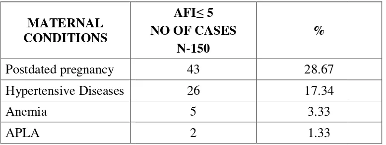
Postdated pregnancy
MATERNAL CONDITIONS ASSOCIATED WITH
OLIGOHYDRAMNIOS MATERNAL CONDITIONS Postdated pregnancy Hypertensive Diseases Anemia APLA
This table shows maternal conditions associated with oligohydramnios in
which 28.67% were postdated pregnancy, 17.34% hypertensive disease
(among which 15.38% were gestational hypertension, 84.61% were
preeclampsia), 3.33% of anemia
present. 39 28.67% 17.34% 3.33% 1.33%
Maternal Conditions Associated with Oligohydramnios
[image:47.612.105.494.193.339.2]Postdated pregnancy Hypertensive Diseases Anemia APLA
TABLE-4
MATERNAL CONDITIONS ASSOCIATED WITH
OLIGOHYDRAMNIOS
AFI≤ 5 NO OF CASES
N-150
%
43 28.67
26 17.34
5 3.33
2 1.33
table shows maternal conditions associated with oligohydramnios in
which 28.67% were postdated pregnancy, 17.34% hypertensive disease
(among which 15.38% were gestational hypertension, 84.61% were
, 3.33% of anemia and 1.33% of APLA syndrome were
CHART 4
MATERNAL CONDITIONS ASSOCIATED WITH
table shows maternal conditions associated with oligohydramnios in
which 28.67% were postdated pregnancy, 17.34% hypertensive disease
(among which 15.38% were gestational hypertension, 84.61% were
40
TABLE-5
MATERNAL CONDITIONS ASSOCIATED WITH
POLYHYDRAMNIOS
MATERNAL CONDITIONS
AFI ≥ 25
NO OF CASES N- 50
%
GDM 8 16
Gestational Hypertension 2 4
Preeclampsia 3 6
Rh Negative 1 2
Chorioangioma of Placenta 1 2
GDM NO. OF CASES
N-60
%
Controlled 5 62.5
Uncontrolled GDM 3 37.5
This table shows the polyhydramnios associated maternal
conditions.Among the 50 cases, GDM were present in 16% (out of which
62.5% were controlled GDM, 37.5% were uncontrolled), Preeclampsia in
6%, gestational hypertension in 4%, Rh negative pregnancy in 2% and
0 GDM Gestational Hypertension Preeclampsia Rh Negative Chorioangioma of Placenta
Maternal Conditions Associated
41
2 4 6 8 10 12 14
4 6 2
2
Percentage Maternal Conditions Associated
with Polyhydramnios CHART 5
0 0.5 1 1.5 2 Multicystic dysplastic kidney 0.67 P er ce n ta g e
Congenital Anomalies Associated with
CONGENITAL ANOMALIES ASSOCIATED WITH
OLIGOHYDRAMNIOS
CONGENITAL ANOMALIES
Multicystic dysplastic kidney Infantile PCKD
Single umblical artery Micro cephaly
In 50 oligohydramnios patients, only 4% had congenital anomalies.
Among which infantile
Single umbilical artery 0.67%, microcephaly 0.67%.
42 Multicystic dysplastic kidney Infantile PCKD Single umblical artery Micro cephaly 0.67 2 0.67 0.67
[image:50.612.137.480.503.706.2]Congenital Anomalies Associated with Oligohydramnious
TABLE -6
CONGENITAL ANOMALIES ASSOCIATED WITH
OLIGOHYDRAMNIOS
CONGENITAL AFI ≤ 5
NO OF CASES N-6
%
Multicystic dysplastic kidney 1 0.67
3 2.0
1 0.67
1 0.67
In 50 oligohydramnios patients, only 4% had congenital anomalies.
Among which infantile polycystic kidney disease 2%, MCKD 0.67%,
Single umbilical artery 0.67%, microcephaly 0.67%.
CHART 6
CONGENITAL ANOMALIES ASSOCIATED WITH
In 50 oligohydramnios patients, only 4% had congenital anomalies.
43
TABLE-7
CONGENITAL ANOMALIES ASSOCIATED WITH
POLYHYDRAMNIOS
CONGENITAL ANOMALIES
AFI ≥ 25
NO OF CASES N-11
%
Anencaphaly 4 8
Diaphragmatic Hernia 1 2
Duodenal atresia 1 2
Non Immnue Hydrops 1 2
Spinabifida 2 4
Hydrocephalus with
meningomyelocele 2 4
In 50 polyhydramnios cases, Total of 22% had congenital anomalies.
Among which anencephaly was the common anomaly accounts 8% ,
spina bifida 4%, hydrocephalus with meningomyelocele 4%,
Diaphragmatic hernia 2%, Duodenal atresia 2% and Non immune
0 2 4 6 8
8
P
e
rc
e
n
ta
g
e
Congenital Anomalies Associated with
44
2 2 2
4 4
Congenital Anomalies Associated with Polyhydramnios
45
TABLE -8
ETIOLOGICAL FACTORS IN OLIGOHYDRAMNIOS
ETIOLOGY
AFI ≤ 5 NO OF CASES
N-150
%
Postdated Pregnancy 43 28.67
IUGR 19 12.66
Hypertensive Diseases 26 17.34
Congenital Anomalies 6 4.00
Idiopathic 56 37.33
This table shows the etiological factors in oligohydramnios group.
According to this table 37.33% were isolated oligohydramnios with no
identifiable cause, 28.67% were post dated pregnancy, 12.66% were
IUGR, 17.34% were hypertensive diseases, 4% were congenital
0 10 20 30 40
Postdated Pregnancy
28.67
P
e
rc
e
n
ta
g
e
Etiological Factors in Oligohydramnios
46
CHART 8
IUGR Hypertensive Diseases
Congenital Anomalies
Idiopathic
12.66
17.34
4
47
TABLE -9
ETIOLOGICAL FACTORS IN POLYHYDRAMNIOS
ETIOLOGY
AFI ≥ 25 NO OF CASES
N-50
%
Idiopathic 29 58
Congenital Anomalies 11 22
GDM 8 16
Rh- Isoimmunisation 1 2
Chorioangioma of the placenta 1 2
This table shows the various etiological factors causing polyhydramnios
in the study population. Exact cause of polyhydramnios were not
detected in 58% of the cases. 22% had congenital anomalies, 16% had
GDM, 2% had Rh isoimmunisation and 2% chorioangioma of the
0 10 20 30 40 50 60
58
P
e
rc
e
n
ta
g
e
Etiological Factors in Polyhydramnios
48
CHART 9
22
16
2 2
18% 6%
Severity of Polyhydramnios SEVERITY OF POLYHYDRAMNIOS
TYPES (AFI)
Mild (25-30) Moderate (31-35) Severe (>35)
This table shows the severity of polyhydramnios.
polyhydramnios, 18% were moderate and
polyhydramnios group.
49
76% 6%
Severity of Polyhydramnios
Mild (AFI 25-30)
Moderate (AFI 31-35)
[image:57.612.103.511.167.257.2]Severe (AFI >35) TABLE -10
SEVERITY OF POLYHYDRAMNIOS
NO OF CASES N-50
%
38 76
9 18
3 6
This table shows the severity of polyhydramnios. 76% were in mild
polyhydramnios, 18% were moderate and 6% were in severe
CHART 10
35)
76% were in mild
0 10 20 30 40 50 60 70 80
AFI ≤ 5
34.67 P e rc e n ta g e
Onset of Labour in Abnormal Liquor Volume
ONSET OF LABOUR IN ABNORMAL LIQUOR VOLUME
ONSET OF LABOUR
N- 150
Spontaneous 52
Induced 98
This table shows the onset of labour in abnormal liquor volume. Among
the oligohydramnios group, 65.33% were induced compared to 20% in
polyhydramnios group, which was highly significant with p value of
0.0001.
50
AFI ≤ 5 AF I≥ 25
80
65.33
20
Onset of Labour in Abnormal Liquor Volume
Spontaneous
Induced TABLE -11
ONSET OF LABOUR IN ABNORMAL LIQUOR VOLUME
GROUP
AFI ≤ 5 AFI ≥ 25
150 % N-50 %
52 34.67 40 80
98 65.33 10 20
table shows the onset of labour in abnormal liquor volume. Among
oligohydramnios group, 65.33% were induced compared to 20% in
polyhydramnios group, which was highly significant with p value of
CHART 11
Spontaneous ONSET OF LABOUR IN ABNORMAL LIQUOR VOLUME
% 80 20
table shows the onset of labour in abnormal liquor volume. Among
oligohydramnios group, 65.33% were induced compared to 20% in
[image:58.612.100.493.165.262.2]51
TABLE-12
MODE OF DELIVERY
MODE OF DELIVERY
GROUP
AFI AFI ≥ 25
N- 150 % N-50 %
SPVD 45 34 36 72
Instrumental delivery 10 6.66 5 10
cesarean 89 59.33 9 18
This table shows mode of delivery in abnormal liquor volume. In
oligohydramnios group 59.33% were underwent cesarean section
compared 18% in polyhydramnios group which is statistically significant
with p value of 0.000. In polyhydramnios group 82% delivered vaginally
compared to 40.66% in oligohydramnios group which is also statistically
0 10 20 30 40 50 60 70 80
SPVD 34
72
P
e
rc
e
n
ta
g
e
52
CHART 12
Instrumental delivery
cesarean 6.66
59.33 72
10
18 Mode of Delivery
AFI ≤ 5
AFI ≥ 25
AFI ≤ 5
53
TABLE-13
INDICATIONS OF CESAREAN SECTION
Among the indications for cesarean section in abnormal liquor volume,
76.40% were due to fetal distress, 11.23% due to CPD, 8.98% due to
failed induction, 3.37% due to malpresentation in oligohydramnios group.
In polyhydramnios group, 33.33% were due to CPD, 22.22% were due to
malpresentation and fetal distress, 11.11% were due to cord prolapsed
and failed induction. INDICATIONS
GROUP
AFI ≤ 5 AFI ≥ 25
N-89 % N-9 %
Fetal Distress 68 76.40 2 22.22
CPD 10 11.23 3 33.33
Failed Induction 8 8.98 1 11.11
Mal presentation 3 3.37 2 22.22
0
10 20 30 40 50 60 70
80 76.4
11.23 22.22
33.33
P
e
rc
e
n
ta
g
e
Indications of Cesarean Section
54
CHART 13
11.23 8.98
3.37 0
33.33
11.11 22.22
11.11 Indications of Cesarean Section
AFI ≤ 5
0 20 40 60 80 100
AFI ≤
42.66 57.33 P e rc e n ta g e
Colour of Liquor in Abnormal Liquor Volume
COLOUR OF LIQUOR IN ABNORMAL LIQUOR VOLUME
COLOUR OF LIQUOR
Clear Meconium
This table shows the colour of
had meconium stained liquor
in polyhydramnios group, which is statist
0.000.
55
AFI ≤ 5 AFI ≥ 25
42.66
88 57.33
12
Colour of Liquor in Abnormal Liquor Volume
Meconium
[image:63.612.104.494.188.318.2]Clear TABLE -14
COLOUR OF LIQUOR IN ABNORMAL LIQUOR VOLUME
GROUP
AFI ≤ 5 AFI ≥ 25
N- 150 % N-50 %
64 42.66 44 88
86 57.33 6 12
This table shows the colour of liquor in Abnormal liquor volume
had meconium stained liquor in oligohydramnios group compared to 12%
in polyhydramnios group, which is statistically significant with p value
CHART 14
Meconium COLOUR OF LIQUOR IN ABNORMAL LIQUOR VOLUME
%
88 12
in Abnormal liquor volume. 57.33%
compared to 12%
4% 2%
14%
Labour Complications in Abnormal Liquor
LABOUR COMPLICATIONS IN POLYHYDRAMNIOS
LABOUR COMPLICATIONS PROM Cord Prolapse Atonic PPH Retained Placenta Preterm Labour
This table shows intrapartum and postpartum
Polyhydramnios cases. A
labour, 6% Cord Prolapse, 4% Atonic PPH, 2% Retained Placenta.
56
20%
6%
[image:64.612.139.478.497.701.2]Labour Complications in Abnormal Liquor Volume PROM Cord Prolapse Atonic PPH Retained Placenta Preterm Labour
TABLE - 15
COMPLICATIONS IN POLYHYDRAMNIOS
AFI ≥ 25
NO OF CASES N-50
%
10 20
3 6
2 4
1 2
7 14
s table shows intrapartum and postpartum complications in
cases. Among which 20% were PROM , 14% Preterm
abour, 6% Cord Prolapse, 4% Atonic PPH, 2% Retained Placenta.
CHART - 15
Retained Placenta
Preterm Labour
COMPLICATIONS IN POLYHYDRAMNIOS
complications in
ch 20% were PROM , 14% Preterm
57
TABLE -16
FETAL OUTCOME IN ABNORMAL LIQUOR VOLUME
FETAL OUTCOME
GROUP
AFI AFI ≥ 25
N- 150 % N-50 %
Alive 139 92.7 36 72
Perinatal Death 11 7.3 14 28
This table shows Fetal Outcome in pregnancies with Abnormal Liquor
Volume. In Oligohydramnios group 92.70% were alive compared to 72%
in polyhydramnios group. Perinatal death was 28% in polyhydramnios
group compared to 7.3% in oligohydramnios group which is statistically
92.7
0 10 20 30 40 50 60 70 80 90 100
AFI ≤ 5
P
e
rc
e
n
ta
g
e
Perinatal outcome in abnormal liquor volume
58
72
7.3 28
AFI ≤ 5 AFI ≥25
Perinatal outcome in abnormal liquor volume
Alive
59
TABLE -17
5 MINUTES APGAR IN ABNORMAL LIQUOR VOLUME
5 MINUTES APGAR
GROUP
AFI AFI ≥ 25
N- 150 % N-39 %
<7 58 38.66 11 28.20
≥7 92 61.33 28 71.79
This table shows the 5 minutes apgar status in abnormal liquor volume. 5
minutes apgar score in oligohydramnios group was <7 in 38.66%
compared to 28.20% in polyhydramnios group which is not statistically
60
TABLE -18
BIRTH WEIGHT IN ABNORMAL LIQUOR VOLUME
BIRTH WEIGHT IN kg
GROUP
AFI AFI ≥ 25
N- 150 % N-39 %
≤ 2.5 81 54 6 15.38
2.6-3 45 30 10 25.64
3.1-3.5 19 12.66 15 38.46
3.6-4 5 3.33 5 12.82
> 4 - - 3 7.69
This table shows the birth weight distribution in abnormal liquor
volume.In oligohydramnios group, 54% were ≤ 2.5 kg, 45.99% were
between 2.6-4 kg, no babies were born above 4 kg. In contrast
polyhydramnios group delivered 76.92% babies with birth weight
0 10 20 30 40 50 60
≤ 2.5 2.6 54
30
15.38
P
e
rc
e
n
ta
g
e
Birth weight in abnormal liquor volume
61
2.6-3 3.1-3.5 3.6-4 > 4 30
12.66
3.33
0 25.64
38.46
12.82
7.69
Birth weight in kg
Birth weight in abnormal liquor volume
AFI ≥ 5
62
TABLE -19
IUGR IN ABNORMAL LIQUOR VOLUME
GROUP
AFI AFI ≥ 25
N- 150 % N-50 %
IUGR 28 18.66 - -
According to this table, 18.66% of babies were IUGR in oligohydramnios
NICU ADMISSION IN ABNORMAL VOLUME
NICU Admission
This table shows the N
volume. Compared to polyhydramnios group
admitted in NICU in oligohydramnios
significant with p value of 0.0001.
28 %
[image:71.612.128.469.456.662.2]63
TABLE -20
NICU ADMISSION IN ABNORMAL VOLUME
GROUP
AFI ≤ 5 AFI ≥ 25
N- 150 % N-39 %
76 50.66 14 28
Number of NICU admission in abnormal
Compared to polyhydramnios group , 50.66% of babies were
NICU in oligohydramnios group which is statistically
significant with p value of 0.0001.
50.66 %
NICU Admission
AFI ≥ 5
AFI ≥ 25
%
28
of NICU admission in abnormal liquor
babies were
group which is statistically
64
DISCUSSION
Various studies have been presented to know the perinatal
morbidity and mortality in pregnancy with abnormal liquor volume. In
the same way our study was tried to reveal the fetomaternal outcome in
abnormal liquor volume in our Institute of Obstetrics and Gynaecology,
Egmore, Chennai.
In our study, 57.3% were in the age of 20-25 yrs in
oligohydramnios group, 42% were in the age of 26-30 yrs in
polyhydramnios group. This is comparable to Guin et al4 study in 2011
In our study, among the parity distribution, 66% of the cases in
oligohydramnios group were primigravida, but there was no significant
relation of age and parity with oligohydramnios according to the study
done by Casey et a31, Chauhan et al32,33, Magann et al15. In
polyhydramnios group majority of the women were multigravida which is
comparable to study by Guin et al4 .
In our study, majority of the women in the gestation age of >37
65
Maternal conditions associated with abnormal liquor volume:
Oligohydramnios:
In our study, 28.67% were postdated pregnancy as compared to
10.7% in Guin et al4 study.
In our study, 17.34% were hypertensive disease of pregnancy as
compared to 38.46% in Chandra et al34 study, 3.5% in Guin et al4 study
and 8% in Preshit et al35 study.
In our study, anemia were present in 3.33% of cases. APLA were
present in 1.33%.
Polyhydramnios:
In our study, GDM were present in 16% as compared to Guin et
al4 study where 20% cases were GDM and 5% cases were GDM in
Vaid et al36 study.
In our study, 10% cases were hypertensive diseases as compared
to 17.7% in Guin et al4 study and 13% in Vaid et al36 study.
In our study, 2% cases were Rh negative pregnancy as compared
to Guin et al4 where Rh -ve pregnancy were 4.4% , 1% in Lyndon M Hill
et al37 study.
66
Congenital anomalies in abnormal liquor volume:
Oligohydramnios
In our study, total of 4% cases had congenital anomalies as
compared to 12.9% in Guin et al study, 5.8% in Anil Shetty et al38.
In our study, infantile PCKD were 2% as compared to Guin et al4
study where 7.5% were PCKD. In our study 0.67% were MCKD, single
umbilical artery and microcephaly.
Polyhydramnios.
In our study, total of 22% had congenital anomalies which was
comparable to Guin et al4 study where 31.1% were associated with
congenital anomalies.
In our study, anencephaly was the common anomaly that account
for 8% as compared to 6% in Guin et al4 study and 65.96% in Vaid et
al36 study.
Spina bifida were present in 4% cases in our study as compared to
67
Hydrocephalus with meningomyelocele were in 4% cases in our
study as compared to Guin et al4 study where 10% cases were
hydrocephalus, 10.63% in Vaid36 et al study.
Diaphragmatic hernia were present in 2% in our study, Duodenal
atresia were 2% in our study as compared 4% in Guin et al4 study.
In our study, Non immune hydrops were present in 2% cases as
compared to 7% in Nicole Damato et al39 study.
Etiological factors in abnormal liquor volume:
Oligohydramnios
In our study, 37.33% were isolated oligohydramnios with no
identifiable cause as compared to 52% in Krishna jagatia et al.
In our study, 28.67% were postdated pregnancy as compared to
10.7% in Guin et al4 study.
In our study, 17.34% were hypertensive disease of pregnancy as
compared to 38.46% in Chandra et al34 study, 3.5% in Guin et al4 study
and 8% in Preshit chate et al35 study.
In our study, 12.66% cases were IUGR which was comparable
68
In our study, 4% cases were congenital anomalies as compared to
12.9% in Guin et al4 study, 5.8% in Anil Shetty et al38 study.
Polyhydramnios:
In our study, exact cause of polyhydramnios were not detected in
58% which is comparable with Brady et al40 study.
In our study, total of 22% had congenital anomalies which was
comparable to Guin et al4 study where 31.1% were associated with
congenital anomalies.
16% had GDM in our study which was comparable to 20% in
Guin et al4 study.
In our study, 2% had Rh isoimmunisation as compared to 4.4% in
Guin et al4 study, 1% in Lyndom M Hill et al27.
In our study, chorioangioma of the placenta were present in 2% of
cases.
Severity of polyhydramnios:
In our study , 76%, 18%, 6% patients were mild, moderate and
severe polyhydramnios respectively, which