1
Dissertation submitted to
The Tamil Nadu Dr. M.G.R.Medical University
In partial fulfilment of the requirements for the degree of
MS BRANCH – II
OBSTETRICS AND GYNAECOLOGY
KILPAUK MEDICAL COLLEGE
THE TAMILNADU DR. M.G.R. MEDICAL UNIVERSITY CHENNAI,
2
BONAFIDE CERTIFICATE
This is to certify that the Dissertation entitled “PREVALENCE OF METABOLIC SYNDROME AMONG PATIENTS WITH POLYCYSTIC OVARIAN SYNDROME” is the bonafide original work of Dr. S.S. Meera, postgraduate, Department of Obstetrics and Gynaecology KMCH, Chennai 10 in partial fulfilment of the requirements of the Tamil Nadu Dr. M.G.R. Medical University for award of MS degree Branch II (Obstetrics & Gynaecology) to be held in April 2014 . The period of postgraduate study and training was from May 2011 to April 2014.
Prof Dr.T.K. SHAANTHY GUNASINGH Professor
Department of Obstetrics and Gynaecology
Kilpauk Medical College and Hospital, Chennai – 10.
Prof Dr.A.KALA
Professor & HOD
Department of Obstetrics and Gynaecology
Kilpauk Medical College and Hospital, Chennai – 10.
Dr. P. RAMAKRISHNAN MD,DLO The Dean
3
ACKNOWLEDGEMENT
I gratefully acknowledge and sincerely thank Prof. Dr. P. Ramakrishnan MD.DLO., Dean. Kilpauk Medical College, Chennai, for granting me permission to utilize the facilities of the Institution for my study.
I take this opportunity to express my deepest sense of gratitude to Prof. Dr. A. Kala MD.D.G.O Head of the Department of obstetrics and Gynaecology Kilpauk Medical College, Chennai for encouraging me and rendering timely suggestions and guiding me through out the course of this work.
I am extremely thankful to Prof. Dr. T.K.Shaanthy Gunasingh MD.D.G.O for her extensive support, advice and guidance in the analysis and successful completion of this study.
I sincerely thank Dr.Geetha MD.D.G.O Dr.Jikki
4 I wish to thank Mr. S. Padmanaban, statistician for his useful inputs.
I wish to express my gratitude to my parents, friends and colleagues who have always been a source of love, support and encouragement.
7 TABLE OF CONTENTS
S.NO. TITLE PAGE NO.
1 Introduction 1
2 Aim of the study 3
3 Review of literature 4
4 Materials and methods 48
5 Observation, discussion and Analysis
55
6 Summary and conclusion 77
7 Bibliography
8
Annexures
Proforma
Master chart
8
LIST OF ABBREVIATIONS
1. PCOS - Polycystic Ovarian Syndrome 2. USG - Ultra Sonogram
3. LH - Leutenizing Hormone
4. FSH - Follicle Stimulating Hormone 5. GnRH - Gonadotropin Releasing Hormone 6. SHBG - Serum Hormone Binding Globulin
7. ESHRE - European Society for Human Reproduction and Embryology
8. AEPCOS - Androgen Excess Polycystic Ovarian
Syndrome
9. NICHD - National Institute Of Child Health and Human Development
9
INTRODUCTION
In recent times, there has been a tremendous change in the lifestyle of people. These changes do have an impact on the health of the people and make them prone to certain diseases and its complications. Polycystic ovarian syndrome (PCOS) is one such disease. It is a multisystem endocrine syndrome . This is due to the ovarian effect resulting in menstrual disturbances and various metabolic disturbances like hyperandrogenism and obesity.
PCOS is more common between 15-25yrs1 of age. It has genetic and familial tendency. PCOS includes chronic anovulation, polycystic ovaries on ultrasound (USG) , hyperandrogenism, raised LH levels , low FSH levels, elevated fasting insulin, low serum binding hormone globulin. PCOS may be autosomal dominant inherited. Prevalence of PCOS in reproductive age group is 5-10%2.
11
AIM OF THE STUDY
12
REVIEW OF LITERATURE
Polycystic ovaries were recognized from mid 18th century. They were known as multicystic ovaries or sclerotic ovaries. In the early 20th century, polycystic ovaries were considered as a result of inflammation due to infection and congestion.
Michael L. Leventhal and Irving F. Stein together described a syndrome associated with anovulation28 in 1935. They described that patients with PCOS had hirsuitism and amenorrhea. They proposed the theory that the thickened ovarian capsule prevented the follicles from reaching and escaping the surface of the ovary .
The characteristic polycystic ovaries results when chronic anovulation is present for a long period of time. About 75% of anovulatory women will have polycystic ovaries.The histologic picture of Stein- Leventhal ovary 30 is as follows
Cross sectional area is twice as normal ovaries. Sub -cortical stroma is 5% thicker.
Developing and atretic follicles are double in number. Tunica is 50% thicker and has more collagen.
13 PATHOPHYSIOLOGY
PCOS is a complex multisystem disorder, where numerous genetic and environmental factors contribute to its pathophysiology. Polycystic ovaries and clinical features of PCOS are due to disturbances in the development of follicle causing anovulation.
Gonadotropin Secretion And Action:
PCOS women have increased serum Leutenising Hormone(LH), low Follicle Stimulating Hormone (FSH) levels, and raised LH: FSH ratios. Both increased pulse frequency and amplitude of LH contributes for raised LH levels. The pulse frequency of LH is not as constant as seen in normal ovulatory women , which is one pulse per hour. The bioactivity25 of LH in PCOS women is also more. The response of LH to exogeneously given GnRH is also raised in women with PCOS.
14 Thus excessive LH32 secretion is an important cause of disrupted development of follicle resulting in anovulatory cycles.
INSULIN RESISTANCE
The association between hyperandrogenism and glucose intolerance was first described in 1921 by Theirs and Archard8 . They reported a diabetic women with beard . Insulin resistance is a feature of variety of conditions like type 2 diabetes, obesity, pregnancy, stress, and PCOS. The role of insulin in the pathogenesis of polycystic ovarian syndrome was first suggested in 1980 . In that study significant correlation was demonstrated between testosterone and basal levels of insulin. They also demonstrated correlation after an oral glucose load between testosterone and insulin levels
15 utilization and increased hepatic gluconeogenesis causing elevated blood glucose and compensatory hyperinsulinemia.
High circulating insulin levels cause hyperandrogenism in PCOS women by two mechanisms . Insulin acts on theca cells of ovarian stroma through insulin receptors and stimulates the production of androgens from ovaries. It inhibits hepatic Serum Hormone Binding Globulin (SHBG) production.
Actions of insulin are mediated through its receptors 26 by two intracellular pathways. Metabolic effects are mediated through phosphatidyl inositol 3-kinase pathway. Proliferative action is mediated through mitogen – activated protein kinase pathway. In women with PCOS activation of mitogen pathway by insulin is increased . there is also resistance in the metabolic pathway of insulin mediated by phosphatidyl-inositol 3-kinase.
“ Thus insulin actions can be selectively inhibited and enhanced at the same time via different signaling pathways” . This
16 Insulin and androgens together lower SHBG causing free androgen levels which in turn aggravates insulin resistance. Thus insulin resistance and hyperinsulinemia are the cause of hyperandrogenism.
Insulin and hyperinsulinemia are important factors in the pathophysiology of PCOS. 20-50%8 of PCOS women have no insulin resistance and prevalence of PCOS is low among women who have insulin resistance. After ovarian wedge resection sensitivity of insulin is not changed. Though insulin resistance and hyperinsulinemia contribute major portion in the pathophysiology of PCOS they are not the primary cause in all PCOS women.
HYPERANDROGENISM
17 ovaries and the rest of the testosterone are produced from peripheral conversion of androstenedione.21
Important reason for excess production of androgen from ovaries in PCOS women is due to increased LH secretion , increased bioactivity of LH, hyperinsulinemia and obesity. Other factors include increased volume of theca cells due to increased ovarian stroma, increased sensitivity to LH and overexpression of LH receptors.
18 New follicles continue to grow and get arrested before full maturation which results in multiple small follicular cysts. These follicular cysts are surrounded by hyperplastic theca cells. Increased ovarian stroma is contributed by the atretic follicles.
Adrenal androgens like androstenedione , DHEA , DHEA-S are also increased in PCOS patients . Even when the synthesis of androgens by ovaries were inhibited by GnRH agonists, adrenal androgens remained higher in PCOS women when compared with normal women. Adrenal androgens donot have any intrinsic activity. There is peripheral conversion of adrenal androgens to testosterone. This peripheral conversion contributes to the pathophysiology of polycystic ovarian syndrome.
19 DIAGNOSIS OF PCOS
Polycystic ovarian syndrome is not a specific disease. It is a syndrome characterized by group of signs and symptoms. Clear cut definition of PCOS is important because of the complications associated with PCOS. Women with PCOS are at increased risk of getting , cardiovascular disease obesity , endometrial cancer, dyslipidemia, diabetes mellitus, hypertension. It is also important to diagnose PCOS because of its health implications in other family members and also due to the need of life long treatment
In earlier times the disease was described based on hirsuitism , menstrual disturbances and enlarged ovaries. Only in recent times the role of insulin resistance in the pathopysiology of PCOS have been recognized. The attention is now being focused on the metabolic consequences of the disease.15
CRITERIA FOR PCOS
20 Institute of Child Health and Human Development , Androgen excess and PCOS society criteria and American Society for Human Reproduction (ASRM) held in Rotterdam, the Netherlands.
The first to define PCOS was National Institue Of Child Health and Human Development(NICHD) in 1990.
1) Hyperandrogenism 2) Menstrual dysfunction
3) Exclusion of other disorders with same clinical symptoms
The second was defined in a conference in 2003 conducted by the European Society for Human Reproduction and Embryology (ESHRE) and American Society for Reproductive Medicine (ASRM) in Rotterdam28, The Netherlands . It concluded that atleast two of the three major criteria should be present.
1) Oligomenorrhoea
2) Clinical or biochemical hyperandrogenism 3) Polycystic ovaries (USG) , excluding other
21 The third was defined by Androgen Excess and Polycystic ovarian syndrome Society (AE-PCOS) in 2006.
1) Hyperandrogenism
2) Oligomenorrhoea or anovulation and or polycystic ovaries
3) Exclusion of other androgen excess disorders.
All these criterias were designed only to refine the diagnosis of PCOS,and for purpose of research. NICHD criteria says that both hyperandrogenism and menstrual dysfunction are necessary for diagnosis of PCOS.
The Rotterdam criteria 2003 says that polycystic ovaries, are evidence of ovulatory dysfunction and should be included in the criteria. It also highlights that hirsuitism and menstrual abnormalities can be either present or absent. “AE-PCOS 2006”
criteria says that polycystic ovaries can be included but still hyperandrogenism must be present for any patient to be categorized as PCOS.
22 Evidence-based Methodology Workshop on PCOS in December 2012 concluded that the Rotterdam criteria should be adopted for now because it is the most inclusive”27
FEATURES OF PCOS Hyperandrogenism
Hyperandrogenism can be either clinical or biochemical. Hirsuitism , acne and androgenic alopecia contribute to clinical hyperandrogenism . All these features are due to the effect of androgens on the pilosebaceous unit. Clinical and biochemical hyperandrogenism are poorly related as the pilosebaceous unit’s
sensitivity is not same among all individuals.
Clinical Hyperandrogenism :
Clinical hyperandrogenism is characterized by hirsuitism, acne , virilization , temporal balding and decreased muscle mass.
HIRSUITISM
23 hair grows. Hair follicle, sebaceous gland, and arrector pili muscle together constitute the pilosebaceous unit. This pilosebaceous unit is sensitive to hormones , especially the more potent androgen , Dihydrotestosterone(DHT). 5 alpha - reductase converts testosterone to dihydrotestosterone . It is DHT which is responsible for stimulation of hair follicles, so local levels of 5 alpha – reductase also influence the stimulation of hair follicle.
Human hair is of two types. Vellus and terminal hair. Vellus is fine, soft, and short . It is not pigmented . It is usually present over face, back and chest. Terminal hair is dark, coarse and long. It is pigmented and is present over the scalp, pubis and axilla. Excess growth of the terminal hair on face, and body in women is called hirsuitism. It is due to the excess amount of androgen.
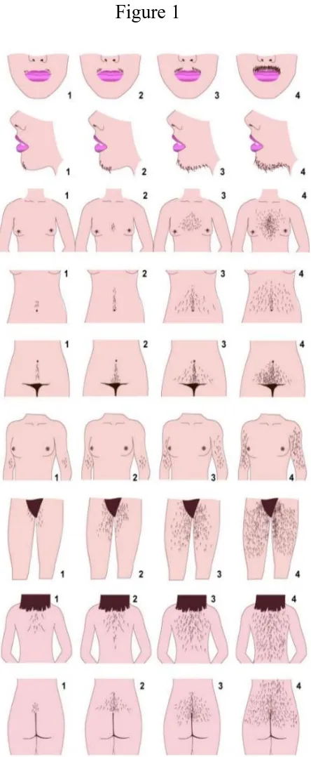
24 The most common cause of excessive androgens in women is PCOS. Hirsuitism is graded by modified Ferriman –Gallwey score . 9 androgen sensitive areas are selected and a score of 1-4 is given according to the extent of hirsuitism. A score of 6-831 defines hirsuitism.
The growth of hair is not continuous. It is cyclic. It undergoes three phases. The resting phase is called quiescent phase, growth phase is called as anagen and the phase of involution is called as catagen. In the anagen phase hair is short and loose. Only in the anagen phase hair grows to its maximum. Androgens decreases the time spent by hair in anagen phase in scalp whereas it increases the amount of time spent by hair in anagen phase in all other places like forearm and face.
Nine androgen sensitive areas are upper lip, chin, inter mammary area, lower abdomen, midline from umbilicus to pubic symphysis, forearm, back of thigh, interscapular region and gluteal region.
25 contributed by the testosterone which is produced from peripheral conversion. The marker for peripheral metaboliasm of androgen is 3 alpha androstanediol glucuronide. This 3 alpha androstanediol correlates wellwith 5 alpha reductase activity. In women with hirsuitsm with normal androgen levels, 5alpha reductase is increased.
26 MODIFIED FERRIMAN –GALLWEY SCORE
[image:26.595.199.421.188.727.2]A score of 1-4 is given. Scores between 6-8 is considered hirsuitism.
27 Other causes of hirsuitism include
Classical congenital adrenal hyperplasia
Non classical congenital adrenal hyperplasia
Cushing syndrome
HAIR –AN syndrome 12 (Hyperandrogenism, insulin resistance, and Acanthosis nigricans)
Androgen secreting neoplasm
Idiopathic hirsuitism
Hyperprolactinemia
Pregnancy luteoma
Drugs like Phenytoin, Minoxidil and Diazoxide.
Acne , alopecia, temporal balding, and seborrhea are other clinical signs of androgen excess. Virilization is present if there is severe amount of excess androgen. Virilization signs include deepening of voice, breast atrophy, increased muscle mass, and clitoromegaly.
28 Figure 2
Hirsutism
Excessive overgrowth of terminal hair is called hirsuitism.
29 The physical signs of hyperandrogenism reflect the severity of androgen excess. All women with hirsuitism do have an increased production of androgens, both testosterone and androstendione
BIOCHEMICAL HYPERANDROGENISM
Dehydroepiandrosterone sulfate (DHEA-S), Testosterone, Dehydroepiandrosterone sulfate (DHEA), Dihydrotestosterone(DHT) and Androstenedione are the major circulating androgens in women. Circulating androgen levels and the sensitivity of hair follicles to the circulating androgens cause hirsuitism.
DHEA-S is produced exclusively by adrenal glands . DHEA is produced by both ovaries and adrenals . DHEA is also produced from the peripheral conversion of DHEA-S.
Androstenedione is produced equally by ovaries and adrenals. DHEA-S, DHEA and Androstenedione have to be converted to testosterone for them to exert their androgenic actions.
30 production of testosterone. It is the free testosterone which is responsible for the androgenic actions. 80% of testosterone is bound to SHBG , 19% to albumin and only 1% is free. Any condition altering the amount of SHBG will affect the amount of free testosterone. Most of the serum immunoassays measure both the free and bound testosterone. Serum total concentration of testosterone doesnot reflect the amount of androgen excess as testosterone is converted to DHT which is more potent androgen and has a longer of duration than testosterone .
DHEA-S is the marker for adrenal androgens. It remains stable through out day and night. Though DHEA-S can be taken as marker of hyperandrogenism in patients with PCOS the sensitivity and specificity is very less for it to be considered as the diagnostic marker of hyperandrogenism.
31 OVULATORY DYSFUNCTION
Most of the women with PCOS have menstrual disturbances. About 80%10 of them have ovulatory dysfunction. The most common abnormalities are oligoamenorrhoea and amenorrhoea. The normal inter -menstrual period is 21- 35 days.
Oligomenorrhoea is defined as absence of menses for a period of 35-182 days. Amenorrhoea is defined as absence of menses for a period of more than 182 days.
Very few women have polymenorrhoea. Polymenorrhoea is regular cycles occurring less than 25 days. Only 2%8 of PCOS women have polymenorrhoea. About 15%8 of women with polycystic ovarian syndrome have normal menstrual cycles. Other common conditions with anovulatory state are hyperthyroidism, hypothyroidism, hyperprolactinemia.
32 POLYCYSTIC OVARIES :
Polycystic ovaries appear macroscopically as enlarged and
lobulated and have a very thick capsule with no adhesions.
On USG they appear as multiple follicles ,usually more
than or equal to 12 measuring about 2-9mm in diameter.
They are located along the border giving them a necklace
pattern
ovarian volume is increased > 7 ml.
Atretic follicles are also visible .
Stromal hyperplasia , thecal hyperplasia and atretic follicles
contribute to the increased ovarian volume5.
Rotterdam criteria takes into consideration only the number of follicles whereas the others define PCOS by considering both number of follicles and also the ovarian volume .
33 Although polycystic ovaries are present in most women with PCOS, the important point to be noted is, polycystic ovaries are not necessary for the diagnosis of PCOS.
POLYCYSTIC OVARIES ON USG
Twelve or more follicles .
Each follicle 2-9mm in diameter.
Ovaraian volume >10cm3
[image:33.595.159.453.428.698.2]Arranged peripherally
Figure 3
34 OTHER FEATURES OF PCOS
Insulin Resistance:
Most of women with PCOS have insulin resistance. When compared with lean women insulin resistance is more in obese women . Insulin resistance can be demonstrated by the serum fasting insulin levels. Levels more than 30microunits/ml are suggestive of insulin resistance. Insulin sensitivity can be identified by fasting glucose/insulin ratio. Sensitivity is reasonable if the ratio is less than 4.5 .12
Another method to assess the insulin sensitivity is by Homeostatic model assessment of insulin resistance (HOMA-IR) .
Product of fasting insulin and fasting glucose divided by constant 22.5 gives HOMA-IR. Normal values are 3-3.9. Values greater than this indicate insulin resistance.
Insulin resistance can also be assessed by QUICKI20 , quantitative insulin sensitivity check index.
35 tolerance test is used to diagnose glucose tolerance and diabetes mellitus.
OBESITY
Obesity is an important feature of PCOS. 70%8 of PCOSwomen are obese and the remaining are lean. Menstrual dysfunction, infertility and hirsuitism are higher in obese PCOS women when compared with lean PCOS women.
Obesity itself is related to insulin resistance. Intraabdominal obesity is highly correlated with insulin resistance . Visceral fat is metabolically more active than subcutaneous fat .Visceral fat is sensitive to lipolysis and releases large amount of fatty acids. These fatty acids produce large amount of cytokines like leptin, interleukin -6, and tumor necrosis factor alpha. All these cytokines are involved in insulin resistance.
36 OVARIAN EFFECTS OF OBESITY
Obesity is related to insulin resistance . Insulin stimulates production of androgens from ovaries . Multiple growth factors like insulin growth factor are more in obese women. These growth factors stimulate the production of androgens. They also inhibit aromatization of androgens to estrogens
Hypothalamic pituitary Action
Insulin resistance is associated with the effects of hypothalamus. Adipokines like leptin play very important role in regulating the function of ovary. This can be demonstrated in patients with anorexia nervosa . secretion of gonadotropin is suppressed in them . Gonadotropin suppression causes loss of ovulatory function.
Metabolic effects:
37 well as have additive effects. Dyslipidemia is common in obese women who have PCOS than lean PCOS women.
Reproductive effects:
Obesity as such doesnot have any effect on the reproductive functions, except for causing anovulation and hirsuitism. Obesity and PCOS are associated with endometrial cancer. Obesity is associated with breast cancer.
INFERTILITY
Due to chronic anovulation , women with PCOS donot conceive . Infertility is common in women with PCOS. Endometrial abnormalities and quality of oocyte also contribute to the causes of infertility in addition to anovulation
POLYCYSTIC OVARIAN SYNDROME AND ENDOMETRIAL CANCER
38 Polycystic Ovarian Syndrome and
Endometrial Cancer
Ovary adrenal
Estradiol androgens
estrones
Endometrium
Atypical hyperplasia
carcinoma
39 OTHER ANDROGEN EXCESS DISORDERS
PCOS is the diagnosis of exclusion, so all other conditions presenting with chronic anovulation and hyperandrogenism should be ruled out.
Thyroid disorders
Women with thyroid disorders have menstrual dysfunction , so serum thyroid stimulating hormone should be done to rule out thyroid disorders in all anovulatory women. Both hypothyroidism and hyperthyroidism patients have anovulatory cycles.
Hyperprolacinemia
Women with hyperprolactinemia have menstrual dysfunction. It is the most common cause of secondary amenorrhoea , hence women with menstrual dysfunction should be tested for prolactin levels.
Androgen secreting tumors
40 150ng/dl is suggestive of malignancy. Rapidly progressing virilization and hirsuitism also suggests androgen producing tumor.
Cushing syndrome
Patients with cushing syndrome have all clinical features seen in women with PCOS. Along with this , they have signs and symptoms suggestive of hypercortisolism like hypertension, striae, atrophy of muscle weakness. The best screening test is the Overnight dexamethasone suppression test .
Idiopathic Hirsuitism
41 Cushing Syndrome
42 METABOLIC DISTURBANCES :
Most of the women with PCOS have metabolic derangements including dyslipidemia, insulin resistance and Obesity. They are, at increased for developing cardiovascular disease and type 2 diabetes mellitus. Insulin resistance along with obesity increase the risk of developing cardiovascular disease.
Many women with PCOS have metabolic syndrome. Metabolic syndrome is also called as metabolic syndrome X. Insulin resistance syndrome , syndrome X are the names given to it . It is also called as Revans syndrome ,named after Gerald Revan.
It is due to disruption in the normal process of energy utilization and storage resulting in metabolic abnormalities which include dyslipidemia, hypertension, insulin resistance, high fasting glucose, obesity. Atleast three of the five criteria should be present to diagnose metabolic syndrome.
43 syndrome are genetics19, physical inactivity, aging and proinflammatory factors.
There are different classifications to define metabolic Syndrome. The objective of all the classification is to provide a simple diagnostic and clinical tool to identify people who have increased of developing cardiovascular disease and type 2 diabetes mellitus.
National cholesterol Education Program Adult Treatment Panel(III)17 defines metabolic syndrome, if there are three out of the following five features
1) Waist circumference more than 88cm in females
2) Hypertension systolic BP more than 130 mmHg and diastolic BP more than 85 mmHg.
3) Triglycerides more than 150mg/dl
4) High Density Lipoprotein (HDL) less than 50mg/dl
44 WHO World Health Organisation defines metabolic Syndrome as that , where along with insulin resistance any of the two following criteria be present.
1) Hypertension systolic BP more than 140 mmHg and Diastolic BP more than 90 mmHg.
2) Triglycerides more than 150mg/dl
3) High Density Lipoprotein (HDL) less than 50mg/dl
4) Obesity BMI > 30kg/m2. or waist to hip ratio > 0.85 in females
5) Microalbuminuria
International Diabetes Federation defines metabolic syndrome as central obesity , waist circumference more than 88cm in females and more than 104cm in males plus any of the two following criteria
1) Hypertension systolic BP more than 130 mmHg and
Diastolic BP more than 85 mmHg
2) Triglycerides more than 150mg/dl
3) High Density Lipoprotein (HDL) less than 50mg/dl
45 NCEP ATP III guidelines differ from that of WHO criteria .In WHO criteria insulin resistance is calculated , which is a cumbersome process if the study is applied to large population. In NCEP ATP III instead of insulin resistance fastin blood glucose is calculated which will give a better idea of glucose metabolism . It is of more help in predicting the progress of glucose regulation abnormalitieslike impaired glucose tolerance and diabetes mellitus.
All the factors have their own individual risk of developing cardiovascular disease. The combination of these factors further augment the chance of developing coronary heart disease and diabetes mellitus.
PCOS women have insulin resistance . Obesity further accelerates insulin resistance . In women with PCOS there is increased amount of triglycerides, decreased amount of HDL. The amount of LDL doesnot vary significantly among PCOS patients.
46 Obesity, by itself is an individual risk factor for developing heart disease.
The final point in metabolic syndrome is hypertension . It is not found in all patients and requires some duration of time to develop. Women with Polycystic ovarian syndrome presenting with insulin resistance develop impaired glucose tolerance and also type 2 diabetes mellitus in due course of time.
TREATMENT OF PCOS
Treatment of PCOS includes life style modification, which includes diet and exercise, like moderate exercises for 30 minutes .losing weight upto 5% provides good results.
Drugs includes estrogen progesterone contraceptives, metformin and anti androgens .
47 Metformin regularizes menstrual cycle and is the most important treatment in young women presenting with anovulatory cycles . Treatment with Metformin decreases the risk of developing diabetes and heart diseases.
The most appropriate candidates for treatment with Metformin are PCOS women with impaired glucose tolerance and patients with evidence of insulin resistance. Clomiphen citrate , Metformin, and GnRH agonists form the main stay of treatment in PCOS patients with infertility.
TREATMENT OF HIRSUITISM
For women with PCOS presenting with acne, hirsutuism, management options include cosmetic measures, medical treatment and surgical treatment.
Cosmetic measures
48 Medical management:
Combined oral contraceptive pills and GnRH agonists suppresses pituitary LH . COC pills also suppress the production of ovarian testosterone. Medroxy progesterone acetate suppresses pituitary LH, ovarian testosterone and also inhibits 5 alpha reductase . ketoconazole blocks adrenal and ovarian androgens . anti androgens like spironlactone, cyproterone acetate , flutamide inhibits androgen binding to receptors. Finasteride inhibits 5alpha reductase
Surgical management :
Clitoral reduction is done in women with clitoromegaly. Androgen secreting tumours are managed by hysterectomy and oophorectomy. Adrenal adenomas are treated by adrenalectomy.
INFERTILITY :
49 Metformin alone or in combination with other insulin senitizing agents improve the menstrual function in most women with PCOS.
Clopmiphen citrate, selective estrogen receptor modulator has both estrogen agonist and antagonist activity. Due to negative feedback of estrogen there is rise in the levels of GnRH , which in turn causes follicular development.
50 TREATMENT OF OBESITY
Treatment of obesity includes lifestyle therapy, pharmaceuticals like metformin, anti obesity drugs and bariatric surgery. Lifestyle28 therapy remains the main mode of treatment of obesity. Lifestyle therapy includes both dietary component exercise and increased physical inactivity.
Aerobic exercises for a period of 150 minutes in a week is recommended. It is done in divided sessions.
Diet therapy includes restriction of calories to about 500kcal. This restriction of 500 kcal/day reduces the weight by 1pound per week29. Diet low in carbohydrate is also associated with profound weight loss.
51 surgery is done for morbidly obese patients with PCOS. Studies have proved that long term survival is superior with bariatric surgery when compared without surgery31.
TREATMENT OF METABOLIC SYNDROME
The main objective to treat metabolic syndrome is to prevent the development of coronary heart disease, and type 2 diabetes mellitus. Other risk factors contributing to the development of cardiovascular disease like smoking should be stopped. Long term and short term complications can be prevented by appropriate life style modifications. These include
Weight loss upto 10% . BMI of less than 25kg/m2 should be
maintained.
Moderate physical exercise of about 30 minutes
Diet should include reduced amount of salt, saturated fatty
acids and more amount of polyunsaturated fatty acids.
Each individual has to be treated according to the features
52 Treatment of insulin resistance
Diet and exercise forms the main stay of treatment. In patients having impaired glucose tolerance the use of insulin sensitizing agents like metformin are used. Metformin and other insulin sensitizing agents slow down the progress of both insulin resistance and impaired glucose tolerance to diabetes mellitus. Acarbose is also used. Patients with type 2 diabetes mellitus are treated with insulin.
Treatment of Lipid Abnormalities
Lipid disturbances respond well to exercise and weight loss along with drug therapy. The main aim is to treat increased LDL levels. Later increased triglycerides and reduced HDL levels are treated. Statin group of drugs along with niacin , and fibrate are used to treat lipid abnormalities.Fibrates decrease the LDL levels , increase HDL levels and also decrease TGL levels
Prevention of thromboembolic disorders
53 Treatment of hypertension
Hypertension forms an important risk factor for developing metabolic syndrome. Maintaining blood pressure at adequate levels improves the general outcome.
Angiotensin converting enzyme inhibitors and Angiotensin receptor blockers are used to treat hypertension.
Prevention
54 ACANTHOSIS NIGRICANS
[image:54.595.120.493.229.520.2]Linear velvety pigmented lesions due to insulin resistance. It appears on nape of neck and axilla.
55 POLYCYSTIC OVARIES
Laparascopic finding of polycystic ovaries
[image:55.595.102.480.286.554.2]They appear as enlarged and pearly white with multiple follicles
56
METHODOLOGY
STUDY DESIGN
Prospective , descriptive, cross sectional study to determine the prevalence of metabolic syndrome in patients with PCOS.
STUDY PLACE
Gynaecology Outpatient deparment, Department of Obstetrics and Gynaecology Government kilpauk Medical college Hospital, Chennai -10
STUDY POPULATION
All patients who satisfied the inclusion and exclusion criteria below, after obtaining consent were included in the study and those in exclusion criteria were deleted from the study.
INCLUSION CRITERIA
1. Women in the age group 18-45 years
2. Patients presenting with menstrual abnormalities
57 3. Women presenting with clinical evidence of
hyperandrogenism like hirsuitism, acne, temporal balding]
4. Women with ( acanthosis nigricans )
5. USG evidence of polycystic ovaries
EXCLUSION CRITERIA
1. Pregnancy
2. Hyperprolactinemia
SAMPLE SIZE
Sample size was calculated from the formula
N = z2p (1-q)/ E2
N is sample size
Z is constant , point corresponding to significant level of 5% z = 1.96
P is prevalence of metabolic syndrome (assumed) is 14% p= .14
Q = 1-P = ( 1-.14)= .86
E is maximum likely error 5%
N = 1.96*1.96*.14*.86/.05*.05 = 185
58 ETHICAL CLEARANCE
Ethical committee clearance was obtained from the Ethical committee , Kilpauk medical college. All the patients included in the study were informed about the study and consent was obtained
before eliciting history and collecting sample for lab tests.
STUDY PROCEDURES
Patients attending gynaecology department in the age group 18-45 were screened for menstrual complaints. Patients with complaints of oligomenorhoea and amenorrhoea were enrolled in the study. Detailed history was taken and clinical examination done . During examination, clinical evidence of hyperandrogenism if present was noted. Hirsutism was
established by using the modified Ferriman–Gallwey score.
Transvaginal ultrasonography was systematically performed
in logic mindray, using the 7.5 MH z transvaginal probe. Ovarian
volume measurements were carried out by using three
perpendicular dimensions and applying the equation for the
59
three dimensions and those with a mean diameter of 2–9 mm
counted.
Other disorders like hypothyroid, and hyperprolactinemia causing menstrual abnormalities were ruled out by measuring serum TSH levels and prolactin levels. Patients who satisfied Rotterdam criteria of PCOS were taken as study participants.
Rotterdam criteria for PCOS is as follows , atleast two of the three major criteria should be present.
1) Oligomenorrhoea
2) Clinical or biochemical hyperandrogenism
3) Polycystic ovaries (USG) , excluding other androgen excess
Clinical Measurements
60 Height assessment was done using a height measuring rod without shoes and recorded to the nearest 0.5 centimeters.
BMI for each person was calculated by dividing weight (kilograms) with height squared(meter).
BMI of 30kg/m2 and over, was taken as obesity.
Waist circumference was also taken using a non-stretchable tape measure at level of the uppermost edge of the hip bone on a light clothed abdomen with the tape parallel to the ground and recorded to the nearest 0.5 centimeters. The measurement above 88cm was considered as central obesity. This was according to the IDF & NCEP ATP III criteria
Blood Pressure Measurements:
Blood pressure was taken from the arm (brachial artery) from all patients in the first encounter by using digital sphygmomanometer.
61 The systolic pressure of above or equal to140mmHg and diastolic pressure above or equal to 90mmHg was regarded as a high blood pressure.
Those who found to have high BP were referred to a physician for further evaluation and possible treatment
After an overnight fasting, blood samples for high density lipoproteins (good cholesterol), serum triglycerides and blood glucose was collected. Five millilitres of venous blood was taken from the antecubital fossa and placed in empty sterile tubes. The samples were transported to the laboratory.
DIAGNOSTIC CRITERIA
In this study National Cholesterol Education Programme ATP III criteria of metabolic syndrome was used. According to this criteria atleast three of the following five criteria should be present
1) Waist circumference more than 88cm in females and more than 104cm in males.
2) Hypertension systolic BP more than 130 mmHg and
62 3) Triglycerides more than 150mg/dl
4) High Density Lipoprotein (HDL) less than 50mg/dl
5) Fasting blood glucose more than 110mg/dl
Patients with high blood pressure were referred to physician. Patients with increased fasting blood glucose, high TGL, low HDL levels were advised to repeat the blood investigations and patients with persistence high values were referred to physician.
DATA ANALYSIS
63 AGE GROUP AND METABOLIC SYNDROME (MS)
[image:63.595.96.516.293.660.2]The association between age and prevalence of metabolic syndrome was evaluated. Patients were divided into four age groups.
Table 1 AGE GROUP AND METABOLIC SYNDROME (MS)
Age group
Metabolic syndrome Present Absent Total Count
1 %within MS Total %
0 0 0 11 6.7% 5.7% 11 6.7% 5.7% Count
2 %within MS Total %
6 20.7% 3.1% 57 34.5% 29.4% 63 32.5% 32.5% Count
3 %within MS Total %
19 65.5% 9.8 % 87 52.7% 44.8% 106 54.6% 54.6% Count
4 %within MS Total %
4 13.8% 2.1% 10 6.1% 5.2 % 14 7.2 % 7.3% TOTAL COUNT
% within MS
64 Age of 20 -25 were in the first group, in second group patients were in the age group 26-30,third group had patients between age 31-35 , fourth group had patients in the age group 36-40years.
Majority of patients , about 107 of them were in the age group of 31-35 years. About 63 patients fell in the age group of 26-30years. 14 were in the age group 36-40 years and 11 were in the age group 21-25.
Among 107 patients, 19 had metabolic syndrome. Out of 63 patients, 6 had metabolic syndrome. Out of 14 patients 11 had metabolic syndrome. The age group analysis was made using chi -square test.
P value was found to be 0.103
65 Figure - 7
0 10 20 30 40 50 60 70 80 90
20-25 26-30 31-35 36-40
N U M BE R O F P A T IE N T S AGE (YEARS)
AGE AND METABOLIC SYNDROME
66 Polycystic ovaries and Metabolic Syndrome
[image:66.595.114.515.354.634.2]Among 194 patients 185 patients had USG evidence of polycystic ovaries. Out of this 20 patients had metabolic syndrome. Polycystic ovaries were absent in 9 patients , but all the 9 patients had metabolic syndrome.
TABLE 2
Polycystic ovaries and Metabolic Syndrome
Polycystic ovaries(USG) Metabolic Syndrome Absent Present Total Count
Absent %within MS Total %
0 0 0 9 31% 4.6% 9 4.6% 4.6% Count
Present %within MS Total %
165 100% 85.1% 20 69% 10.3% 185 95.4% 95.4% TOTAL COUNT
% within MS Total %
67 Relationship between the usg evidence of polycystic ovaries and metabolic syndrome was evaluated using chisquare test and Fisher’s exact test . The
p value was found to be <.05 .
[image:67.595.98.523.231.585.2]The relationship was found to be statistically significant
TABLE 3
Chi-Square Tests
Value Df Asymp. Sig.
(2-sided)
Exact Sig.
(2-sided)
Exact Sig. (1-sided)
Pearson Chi-Square 53.698a 1 .000
Continuity Correctionb 46.912 1 .000
Likelihood Ratio 36.923 1 .000
Fisher's Exact Test .000 .000
Linear-by-Linear Association
53.421 1 .000
68 Figure – 8
[image:68.595.101.501.122.732.2]0 20 40 60 80 100 120 140 160 180 ABSENT PRESENT N u mb e r o f p a ti e n ts
Polycystic ovaries( USG)
Polycystic ovaries and Metabolic
Syndrome
69 HIRSUITISM AND METABOLIC SYNDROME.
[image:69.595.113.516.305.611.2]Hirsuitism was present in 101 patients and absent in 93 patients. Among 101 patients 28 patients had metabolic syndrome. In 93 patients only 1 patient had metabolic syndrome.
TABLE 4
Hirsuitism and Metabolic Syndrome
Hirsuitism Metabolic Syndrome
Absent Present Total Count
Absent %within MS Total %
92 55.8% 47.4% 1 3.4% .5% 93 47.9% 47.9% Count
Present %within MS Total %
73 44.2% 37.6% 28 96.6% 14.4% 101 52.1% 52.1% TOTAL COUNT
% within MS % of Total
70 Relationship between hyperandrogenism and metabolic syndrome using hirsuitism was evaluated by chi-square test.
P-value was found to be less than .05.
[image:70.595.103.507.276.560.2]The association was found to be statistically significant .
TABLE 5 Chi-Square Tests
Value df
Asymp. Sig. (2-sided)
Exact Sig.
(2-sided)
Exact
Sig. (1-sided)
Pearson Chi-Square 27.042a 1 .000 Continuity Correctionb 24.987 1 .000 Likelihood Ratio 33.366 1 .000
Fisher's Exact Test .000 .000
Linear-by-Linear Association
26.903 1 .000
71 Figure - 9
Hirsuitism and Metabolic Syndrome
72 ACANTHOSIS NIGRICANS AND METABOLIC SYNDROME
[image:72.595.113.517.352.675.2]Among 194 patients , acanthosis nigricans which is an evidence of insulin resistance was present in 14 patients. Out of 14, ten patients had metabolic syndrome.
TABLE 6
Acanthosis Nigricans and Metabolic Syndrome
Acanthosis Nigricans Metabolic Syndrome Absent Present Total Count
Absent %within MS Total %
161 97.6% 83% 19 65.5% 9.8% 180 92.8% 92.8% Count
Present %within MS Total %
4 2.4% 2.1% 10 34.5% 5.2% 14 7.2% 7.2% TOTAL COUNT
% within MS % of Total
73 The association between acanthosis nigricans and metabolicsyndrome was evaluated using chisquare test.
[image:73.595.95.510.281.649.2]p- value was found to be < .05. The relationship was to be statistically significant.
TABLE 7 Chi-Square Tests
Value Df
Asymp.
Sig.
(2-sided)
Exact Sig.
(2-sided)
Exact Sig.
(1-sided)
Pearson Chi-Square 37.859a 1 .000
Continuity Correctionb 33.223 1 .000
Likelihood Ratio 25.549 1 .000
Fisher's Exact Test .000 .000
Linear-by-Linear Association
37.664 1 .000
74 Figure 10
Acanthosis Nigricans and Metabolic Syndrome 0 20 40 60 80 100 120 140 160 180 ABSENT PRESENT N u mb e r o f p a ti e n ts
Acanthosis NigricansT
75 PCOS AND METABOLIC SYNDROME
[image:75.595.96.515.355.706.2]Patients with polycystic ovarian syndrome were divided into three groups . The first group had patients who satisfied all three criteria of Rotterdam (ie) 1.hyperandrogenism, 2.anovulation 3. usg evidence of polycystic ovaries .
TABLE 8
PCOS and Metabolic Syndrome
Age group
Metabolic syndrome Present Absent Total Count
5 %within MS Total %
0 0 0 11 6.7% 5.7% 11 6.7% 5.7% Count
6 %within MS Total %
6 20.7% 3.1% 57 34.5% 29.4% 63 32.5% 32.5% Count
7 %within MS Total %
19 65.5% 9.8 % 87 52.7% 44.8% 106 54.6% 54.6% TOTAL COUNT
% within MS
76 Second group had patients with anovulation and usg evidence of polycystic ovaries.Third group constituted patients with anovulation and hyperandrogenism. Number of patients with metabolic syndrome in first , second and third group were 20, 1, and 8 respectively.
[image:76.595.99.513.523.709.2]The risk of developing metabolic syndrome between the three groups were assesed using chisquare test . There was difference in risk between the three groups . The difference was found to be stastistically significant. From the above analysis it can be concluded that clinical hyperandrogenism is important risk factor in developing metabolic syndrome.It can also be concluded that all patients with usg evidence of polycystic ovaries donot necessarily develop, metabolic syndrome.
TABLE 9 Chi-Square Tests
Value df
Asymp. Sig. (2-sided) Pearson Chi-Square 62.741a 2 .000
Likelihood Ratio 55.782 2 .000 Linear-by-Linear Association .061 1 .805
77 Figure 11
PCOS and Metabolic Syndrome
0 10 20 30 40 50 60 70 80 90 100
GROUP 1 GROUP 2 GROUP 3
78 Relationship between patients with and without metabolic syndrome was analysed. It was analysed using independent t test.
Table 10 Group Statistics
MS N Mean Std.
Deviation
Std. Error Mean
Age 1 29 32.41 2.872 .533
0 165 30.66 3.652 .284
BMI 1 29 24.28 1.099 .204
0 164 22.40 1.849 .144 Waist
circumference
1 29 93.03 3.311 .615
0 165 87.02 5.534 .431 Sys BP 1 29 130.52 15.256 2.833
0 165 109.76 7.647 .595 Dias BP 1 29 83.10 10.037 1.864
0 165 70.85 5.988 .466
FBS 1 29 92.69 18.517 3.438
0 165 83.32 7.414 .577 TGL 1 28 147.11 10.874 2.055
0 165 138.75 7.371 .574
HDL 1 29 47.07 3.741 .695
79 The variables taken for analysis were age group, systolic BP, diastolic BP, HDL, TGL, waist circumference, BMI and fasting blood glucose level.
Except age group, all other variables contributed to the difference between patients with and without metabolic syndrome and they were statistically significant .
Of all these variables, waist circumference had a strong correlation towards metabolic syndrome. All patients with metabolic syndrome had increased waist circumference.
80 Table 11
Independent Samples Test
t-test for Equality of Means
df Sig. (2-tailed)
Mean Difference
AGE Equal variances assumed 192 .015 1.753
Equal variances not assumed 45.539 .006 1.753
BMI Equal variances assumed 191 .000 1.873
Equal variances not assumed 60.481 .000 1.873
WAIST
CIRCUMFERENCE
Equal variances assumed 192 .000 6.016
Equal variances not assumed 59.787 .000 6.016
SYS BP Equal variances assumed 192 .000 20.760
Equal variances not assumed 30.517 .000 20.760
DIAS BP Equal variances assumed 192 .000 12.255
Equal variances not assumed 31.592 .000 12.255
FBS Equal variances assumed 192 .000 9.375
Equal variances not assumed 29.596 .012 9.375
TGL Equal variances assumed 191 .000 8.362
Equal variances not assumed 31.343 .000 8.362
HDL Equal variances assumed 192 .000 -7.719
81 Figure 12
0 5 10 15 20 25 30 35 40 N o o f p a ti e n ts in %
Individual Risk Factors of Metabolic
Syndrome
82 Individual risk factors of each component for metabolic syndrome is as follows
Hypertension 11.7%
High FBS 12%
Increased waist circumference 36%
Low HDL 17.6%
83
DISCUSSION
The main objective of this study is to determine the
prevalence of metabolic syndrome in women diagnosed as PCOS
according to the Rotterdam criteria. The main findings are that the
overall prevalence of syndrome is 16.1%. Moreover, within this
anovulatory cohort, the hyperandrogenic PCOS phenotype is associated
with the highest risk of metabolic abnormalities.
In comparison to other studies with similarly diagnosed PCOS
women, the prevalence of metabolic abnormalities in our study group is
lower.prevalence is 35–44% in American (Shroff et al., 2007) and
Australian anovulatory PCOS women (Cussons et al., 2008),
comparable to the 16% reported in
Taiwanese anovulatory PCOS women (Chen et al., 2006), but
higher than the 8.2% reported in Southern Italian women with
anovulatory PCOS (Carmina et al., 2006).
84 Low level of HDL-C and impaired fasting glucose were the most frequent (17.6% and 11.4% respectively) components of metabolic syndrome. This could make the population to have greater odds of having cardiovascular disease and type 2 diabetes .
Systemic hypertension was also found to be one of the most components of MS by 11.7%. The findings were similar in studies done in Nigeria and Cameroon.
85
SUMMARY AND CONCLUSION
This prospective cross sectional descriptive study evaluated 194 patients with PCOS by both clinical and labarotory tests, following the inclusion and exclusion criteria at Government kilpauk Medical college
In this prospective study of 194 patients with PCOS, according to Rotterdam criteria the prevalence of metabolic syndrome was found to be 16.1%.
In this study patients with PCOS were classified into three groups.
First group had patients who satisfied all three criteria like menstrual abnormalities, hyperandrogenism, and USG evidence of Polycystic ovaries .
Second group were patients with menstrual irregularities and usg evidence of polycystic ovaries.
86 Prevalence of metabolic syndrome in each group are as follows.
Prevalence in first group 21% Prevalence in second group 1%
Prevalence in third group is 100%
Out of 194 patients 29 patients had metabolic syndrome. Out of 29 patients, I patient met all five criterias. 10 out of 29 patients met 4 criterias.
18 patients met 3 criterias.
Odds ratio for acanthosi nigricans with reference to metabolic syndrome was found to be 21.
Odds ratio for hirsuitism with reference to metabolic syndrome was found to be 35
Hyperandrogenism is important risk factor in developing metabolic syndrome.
87 Hypertension 11.7%
FBS 12%
Increased waist circumference 36% HDL 17.6%
TGL 8.8%
All patients with metabolic syndrome had increased waist circumference.
Hence all PCOS women with clinical hyperandrogenism and increased waist circumference should be screened for metabolic syndrome.
Women at risk of developing metabolic syndrome should be adviced about lifestyle modifications .
88
BIBLIOGRAPHY
1. Azziz R, Woods KS, Reyna R, Key TJ, Knochenhauer ES, Yildiz BO (June 2004). "The Prevalence and Features of the Polycystic Ovary Syndrome in an Unselected Population". Journal of Clinical Endocrinology &Metabolism 89 (6): 2745–9.doi: 10.1210/jc.2003-032046.
2. Asuncion M, Calvo RM, San Millan JL, Sancho J, Avila S, Escobar-Morreale HF: A prospective study of the prevalence of the polycystic ovary syndrome in unselected Caucasian women from Spain.J Clin Endocrinol Metab 2000, 85:2434-2438.
3. Alberti KG, Zimmet P, Shaw J. The metabolic syndrome—a new worldwide definition. Lancet 2005;366:1059–1062.
89 5. Balen AH, Laven JS, Tan SL, Dewailly D. Ultrasound assessment of the polycystic ovary: international consensus definitions. Hum Reprod Update 2003;9:505–5143.Barber TM, McCarthy MI,Wass JA, Franks S.
6. Broekmans FJ, Knauff EA, Valkenburg O, Laven JS, Eijkemans MJ, Fauser BC. PCOS according to the Rotterdam consensus criteria: change in prevalence among WHO-II anovulation and association with metabolic factors. BJOG 2006;113:1210–1217
7. Chen MJ, Yang WS, Yang JH, Hsiao CK, Yang YS, Ho HN. Low sex Hormone-binding globulin is associated with low high-density Lipoprotein cholesterol and metabolic syndrome in women with PCOS. Reprod 2006;21:2266–2271.
8. Clinical Gynecologic Endocrinology and Infertility. Self. Assessment and study guide. David B. Seifer and Leon Speroff Linkinghub.elsevier.com/retrieve/pii/0301211595900209
90 10. Dewailly D, Catteau-Jonard S, Reyss AC, Leroy M, Pigny P. Oligoanovulation with polycystic ovaries but not overt hyperandrogenism. J Clin Endocrinol Metab 2006;91:3922–3927.
11. Dokras A, Bochner M, Hollinrake E, Markham S, Van Voorhis B, Jagasia DH. Screening women with polycystic ovary syndrome for metabolic syndrome. Obstet Gynecol 2005;106:131–137.
12. Dumesic DA, Abbott DH, Padmanabhan V. Polycystic ovary syndrome and its developmental origins. Rev Endocr Metab Disord 2007;8:127–141.
13. Dunaif A. Insulin resistance and the polycystic ovary syndrome: Mechanism and implications for pathogenesis. Endocr Rev. 1997;18:774–800.
14. Eckel RH, Grundy SM, Zimmet PZ. The metabolic syndrome. Lancet 2005; 365:1415–1428.
91 16. Ehrmann DA, Liljenquist DR, Kasza K, Azziz R, Legro RS, Ghazzi MN. Prevalence and predictors of the metabolic syndrome in women with polycystic ovary syndrome. J Clin Endocrinol Metab 2006;91:48–53.
17. Elting MW, Korsen TJ, Bezemer PD, Schoemaker J. Prevalence of diabetes mellitus, hypertension and cardiac complaints in a follow-up study of a Dutch PCOS population. Hum Reprod 2001;16:556–560. Expert Panel on the Diagnosis, Evaluation, and Treatment of High Blood Cholesterol in Adults. Executive Summary of The Third Report of The National Cholesterol Education Program (NCEP) Expert Panel on Detection, Evaluation, And Treatment of High Blood Cholesterol in Adults (Adult Treatment Panel III). JAMA 2001;285:2486–2497.
18. Escobar-Morreale HF, Luque-Ramirez M, San Millan JL. The molecular-genetic basis of functional hyperandrogenism and the polycystic ovary syndrome. Endocr Rev. 2005;26:251–82
92 20. Hahn S, Tan S, Elsenbruch S, Quadbeck B, Herrmann BL, Mann K, Janssen OE. Clinical and biochemical characterization of women with polycystic ovary syndrome in North Rhine-Westphalia. Horm Metab Res 2005;37:438–444.
21. Hatch R, Rosenfield RL, Kim MH, Tredway D. Hirsutism: Implications, etiology, and management. Am J Obstet Gynecol. 1981;140:815–30. [PubMed
22. Indian J Endocrinol metab.2013 Jan-feb;17(1) :134-145.20i10.4103/2230-8210.107858
23. Knowler WC, Barrett-Connor E, Fowler SE, et al. Diabetes Prevention Program Research Group. Reduction in the incidence of type 2-diabetes with lifestyle intervention or metformin. N Engl J Med. 2002;346(6):393–403. [PMC free article][PubMed]