recombinant baculoviruses. LEF-1 was purified to near homogeneity and found to have primase activity in an
indirect assay employingEscherichia coliDNA polymerase I (Klenow enzyme) and poly(dT) template. The
LEF-1 primase products were also directly characterized by electrophoresis in 20% polyacrylamide–8 M urea gels and agarose gels. Primer synthesis was time dependent, and products of several hundred nucleotides or more were observed from the M13 single-stranded DNA (ssDNA) template. The LEF-1 primase was absolutely
dependent on divalent cations (Mg2ⴙ), and optimal activity was supported by 10 mM MgCl
2. An alkaline pH
(8.8 to 9.4) was optimal, whereas monovalent salt (KCl) was inhibitory. Mutation of an invariant aspartic acid in a putative primase domain caused LEF-1 activity to be abolished. Upon ultracentrifugation in glycerol gradients, LEF-1 was found to have a sedimentation coefficient of 3S that is consistent with its being present as a monomer. Elution profiles of LEF-1 and LEF-2 from ssDNA-cellulose and DEAE resin suggested that LEF-2 may bind to both DNA and LEF-1.
The Baculoviridae are a large and diverse family of rod-shaped, enveloped, occluded viruses that are pathogenic for invertebrates, particularly members of the Insecta. They have been reported from over 600 species, most of which are mem-bers of the Lepidoptera, Diptera, and Hymenoptera (34). Two genera of baculoviruses have been characterized, and they include the nucleopolyhedroviruses (NPVs) (47), which have numerous virions within large polyhedron-shaped occlusion bodies, and the granuloviruses (52), which commonly have a single virion within small granular occlusion bodies. Baculovi-rus genomes consist of double-stranded, circular, supercoiled DNA of 100 to 180 kb, depending on the strain of virus (20). Although evidence suggests that baculovirus genomes may rep-licate via a rolling-circle intermediate (31, 42), the mechanisms of initiation, elongation, processing, and maturation have not been determined.
Baculovirus DNA replication has been shown to be associ-ated with discrete replication factories in the nuclei of infected cells (41). In addition, a conserved set of genes that are essen-tial or highly stimulatory for transient DNA replication have been identified for Autographa californica multinucleocapsid NPV (AcMNPV) (26, 33), Orgyia pseudotsugata MNPV (OpMNPV) (reviewed in reference 1), and Lymantria dispar MNPV (44). These include genes encoding a DNA polymerase homolog, a DNA helicase homolog,ie-1, a transactivator of early gene transcription, and late expression factors (LEFs) encoded bylef-1, -2, and-3.The DNA polymerase and DNA helicase homologs were subsequently shown to have activities associated with these enzymes (35, 36). In addition,lef-3
en-codes a product with the properties of a single-stranded DNA (SSB) binding protein (16, 37), interacts with helicase (14), and is required for transfer of helicase to the nucleus (53).
In addition to its ability to transactivate early gene transcrip-tion, the product of ie-1 has been demonstrated to bind to homologous regions which act as replication origins in tran-sient assays (9, 46) and colocalizes with LEF-3 in nuclear replication factories (41). AcMNPV geneslef-1 and lef-2 po-tentially encode basic proteins with isoelectric points (pI) of 8.84 and 9.38, respectively, and calculated molecular masses of 30,780 and 23,926 Da, respectively (4). Possible roles for LEF-1 and LEF-2 have not been elucidated, although LEF-1 contains several motifs related to eukaryotic primases. When the putative primase domain in LEF-1, WVVDAD, was al-tered to WVVQAD, transient DNA replication was abolished (13). In addition, it was found in yeast two-hydrid analyses that LEF-1 and LEF-2 interact (13). These data suggested that LEF-1 may function as a primase and that LEF-2 may be associated with this activity.
In this report we describe the overexpression and purifi-cation of LEF-1 and demonstrate that it has DNA primase activity. The parameters of the LEF-1 primase activity were optimized, and the primase products were characterized by electrophoresis in polyacrylamide and agarose gels. We also discuss a possible role of LEF-2 in the baculovirus replication complex.
MATERIALS AND METHODS
Chemicals and enzymes.Nucleoside triphosphates (NTPs) and deoxy-NTPs
(dNTPs) were purchased from Roche Molecular Biochemicals. 7-Deaza-2⬘
-de-oxyadenosine 5⬘-triphosphate and poly(dT) were from Amersham Pharmacia
Biotech. Radiolabeled nucleotides [␣-32P]ATP, [␥-32P]ATP, and [␣-32P]dATP
were from Perkin-Elmer.Escherichia coliDNA polymerase I (Klenow Exo⫺)
and other modifying enzymes were from New England Biolabs.
Cells.Spodoptera frugiperda9 (Sf9) cells were cultured in Sf900II serum-free medium (Gibco-BRL) supplemented with 10% fetal bovine serum, penicillin G
* Corresponding author. Mailing address: N. K. Koltzov Institute of Developmental Biology, Russian Academy of Sciences, Vavilov St. 26, Moscow 117808, Russia. Phone: (7-095)1358847. Fax: (7-095)1358012. E-mail: [email protected].
† Technical report no. 11820 from the Oregon State University Ag-ricultural Experiment Station.
2287
on November 8, 2019 by guest
http://jvi.asm.org/
(50 U/ml), streptomycin (50g/ml; Whittaker Bioproducts), and Fungizone (amphotericin B; 375 ng/ml; Flow Laboratories) as previously described (17).
Transfer plasmids and virus construction.The transfer plasmids were con-structed as follows. Plasmids pHSEpiHisLef-1 and pHSEpiHisLef-2 (45) were
digested withBglII, treated with calf intestinal phosphatase, and modified using
two oligonucleotides that were phosphorylated by treatment with polynucleotide
kinase and ATP, gel purified, annealed (48), and inserted into theBglII site. The
oligonucleotide names and sequences are 5bamhahisc (GATCGGGATC CG ACCATGAG CTCCCGATAC CCATACGACG TCCCAGATTA CGCCCG GCAT CATCATCATC ATCATCACC) and 3bamhahisc (GATCGGTGAT G ATGATGATG ATGCCGGGCG TAATCTGGGA CGTCGTATGG GTATC GGGAG CTCATGGTCG GATCCC). This resulted in the creation of an ATG translation start site followed by the HA epitope and HIS tag upstream of each
open reading frame (ORF) but downstream of a newBamHI site (Fig. 1).
Clones were screened usingBamHI digestion (the correct orientation is about
70 nucleotides [nt] smaller than the reverse orientation), and presumptive cor-rect plasmids were sequenced to confirm the corcor-rect orientation of the insert.
The primers used for sequencing were AcMNPV nt 10971 to 10990 (lef1-330;
CGGTATACATGACTCTTGAC) and nt 3480 to 3462 (lef2-373, AACCTCT TCCTGTACATAC) (4). The ORFs were then removed by digestion with
BamHI andNotI, gel purified, and inserted into a pFastbac1 vector (Gibco-BRL)
digested with the same enzymes and gel purified. Recombinant baculoviruses were produced using the Bac-to-Bac baculovirus expression system (Gibco-BRL) following the manufacturer’s instructions.
A virus, vfbHAHISLef-1D76Q, in which the conserved primase motif WVV
DAD was altered to WVVQAD was constructed by removing anMluI-NdeI
fragment from a mutant plasmid described previously (13) and inserting it into pfbHAHISLef-1 digested with the same enzymes. The insertion of the mutant sequence was confirmed by DNA sequence analysis. The recombinant virus
expressing the mutantlef-1gene was then generated as described above.
Purification of *LEF-1.Sf9 cells at a density of 1⫻106to 1.5⫻106/ml in
shaker flasks were coinfected with the recombinant baculoviruses vfbHAHISLef-1 and vfbHAHISLef-2 at a multiplicity of infection (MOI) of about 5 and incu-bated with shaking for 46 h at 28°C. LEF-1 was purified routinely from a 100-ml or 50-ml culture of infected cells by liquid chromatography sequentially on Ni-nitrilotriacetic acid (NTA)-agarose (Qiagen), DEAE-Toyopearl 650 (Toso-Haas), and heparin-Sepharose CL-6B (Amersham Pharmacia Biotech) columns.
The infected cells (100 ml) were pelleted by centrifugation for 5 min at 500⫻g
and resuspended in 8 ml of lysis buffer containing 50 mM Tris-HCl (pH 8.5), 200 mM KCl, 1% Nonidet P-40, 5 mM 2-mercaptoethanol, and a set of protease
inhibitors (1 mM phenylmethylsulfonyl fluoride, 1M pepstatin, 5g of
leu-peptin per ml, 5g of aprotinin per ml, and 2g of E64 per ml). After extraction
for 15 min at 4°C on a rotating shaker, the preparation was clarified by
centrif-ugation at 150,000⫻gfor 30 min. The supernatant was loaded onto an
Ni-NTA-agarose column (0.8 ml) equilibrated with buffer A (20 mM Tris-HCl [pH 8.5], 0.5 M KCl, 10% [vol/vol] glycerol, 5 mM 2-mercaptoethanol, 20 mM imidazole). The column was washed successively with 10 ml of buffer A, 4 ml of the same buffer containing 1 M KCl, 3 ml of buffer A, and finally with 3 ml of buffer B (20 mM Tris-HCl [pH 8.5], 100 mM KCl, 10% [vol/vol] glycerol, 5 mM 2-mer-captoethanol, 50 mM imidazole).
Proteins were eluted from the column with 4 ml of buffer B containing 150 mM imidazole. The sample was immediately mixed with an equal volume of dilution buffer containing 20 mM Tris-HCl (pH 7.5), 30% (vol/vol) glycerol, 2 mM dithiothreitol (DTT), 2 mM EDTA, and the set of protease inhibitors to the final concentrations listed above, and then passed at a rate of 4 ml per h through a
DEAE-Toyopearl column (0.5 by 2.5 cm) equilibrated with a 1:1 mixture of buffer B and dilution buffer. The flowthrough was loaded on a 0.5-ml column of heparin-Sepharose. The column was washed with several volumes of buffer C (0.1 M KCl, 10 mM Tris-HCl [pH 7.5], 20% [vol/vol] glycerol, 1 mM DTT, 1 mM EDTA, and the set of protease inhibitors), and HAHISLEF-1 (*LEF-1) was then eluted with several volumes of the same buffer containing 0.2 M KCl. In some experiments, the heparin-Sepharose column was successively processed with 1-ml portions of buffer C containing KCl in final concentrations of 0.1, 0.15, 0.20, 0.25, 0.30, 0.40, and 0.5 M. Proteins from each fraction were analyzed by sodium dodecyl sulfate–12% polyacrylamide gel electrophoresis (SDS–12% PAGE), fol-lowed by staining with silver or Coomassie brilliant blue. The presence of *LEF-1 was monitored by Western blotting with monoclonal antibody HA.11 (see be-low). Gelatin (0.5 mg per ml) was added to the fractions containing *LEF-1. The fractions were combined or dialyzed separately against buffer D (50 mM KCl, 10 mM Tris-HCl [pH 7.5], 50% [vol/vol] glycerol, 1 mM DTT, 0.1 mM EDTA), and
stored at⫺20°C for periods of 2 to 3 weeks or at⫺80°C for longer terms.
PAGE and Western blotting.SDS–12% PAGE was performed as described by Laemmli (29). Gels were either fixed and stained or electrophoretically trans-ferred to PVDF-Plus transfer membrane (Micron Separations Inc) using a Trans-blot SD semidry transfer cell (Bio-Rad) according to the manufacturer’s guidelines. Western blots were probed with a 1:1,000 dilution of monoclonal antibody HA.11 (BAbCO), washed, incubated with a 1:3,000 dilution of goat anti-mouse immunoglobulin G (IgG) conjugated to horseradish peroxidase (Bio-Rad Laboratories), and developed by using BM chemiluminescence blotting substrate (POD) (Roche Molecular Biochemicals) according to the manufactur-er’s instructions.
Assays for endonuclease and topoisomerase activity.Reactions were carried out in three mixtures of different composition. Reaction mixture 1 contained 25 mM Tris-HCl (pH 7.5), mixture 2 contained 25 mM Tris-HCl (pH 7.5) and 50 mM KCl, and mixture 3 contained 25 mM Tris-HCl (pH 8.8). All reactions
contained 0.3g of DNA (replicative form I [RFI]) of plasmid pHSEHLEF-1
(45), 10 mM MgCl2, 50g of bovine serum albumin (BSA) per ml, 1 mM DTT,
and 1 mM ATP. *LEF-1 (0.1g in 3l of buffer D) was added to a final volume
of 15l. After incubation for 1 h at 37°C, reactions were terminated by the
addition of EDTA to 15 mM and SDS to 0.5%, and the samples were treated
with proteinase K (100g/ml) for 30 min at 37°C. The samples were then
analyzed by electrophoresis in a 0.7% agarose gel, followed by staining with ethidium bromide, and examined for the conversion of RFI into RFII and RFIII or topoisomers.
Primase assays. (i) Indirect assay. Reaction mixtures (25 l) contained
25 mM Tris-HCl (pH 8.0), 5 mM MgCl2, 20g of poly(dT) per ml, 100g of
BSA per ml, 1 mM DTT, 1 mM ATP, 2.5M 7-deaza-dATP, 2.5M [␣-32P]
dATP (10Ci per ml), 5 U ofE. coliDNA polymerase I (Klenow Exo⫺) per ml,
and various amounts of *LEF-1 added with 5l of buffer D. Reactions were
carried out at 30°C for 60 min and were terminated by chilling on ice and adding
5l of 0.5 M EDTA saturated with sodium pyrophosphate. The samples were
transferred onto pieces of Whatman 3MM paper, which were washed in four changes of cold 5% trichloroacetic acid–1% sodium pyrophosphate. The acid-insoluble radioactivity was measured by Cerenkov counting.
(ii) Direct assays.Reaction mixtures (10l) contained 25 mM Tris-HCl (pH
9.1), 10 mM MgCl2, 25g of poly(dT) per ml, 100g of BSA per ml, 1 mM DTT,
5M [␣-32P]ATP (100Ci per ml), and various amounts of *LEF-1 added with
3l of buffer D. In experiments with M13mp9 DNA (25g per ml) instead of
poly(dT), the reaction mixtures were supplemented with 50M each GTP, CTP,
and UTP. Reactions were carried out at 30°C for 2 h and were terminated by
FIG. 1. Schematic diagram of plasmids used to construct recombinant baculoviruses expressing LEF-1 and LEF-2. The two oligomers used to engineer the plasmids pHSEpiHisLef-1 and pHSEpiHisLef-2 (45) are shown. They were inserted into aBglII site and resulted in the elimination of this site and the creation of an upstreamBamHI site. The inserts were then removed withBamHI andNotI and inserted into pFastBac1 as shown. The orientation of a genericlefgene is indicated by the open arrow.
on November 8, 2019 by guest
http://jvi.asm.org/
chilling on ice and adding 7l of stop solution (95% formamide, 20 mM EDTA, 0.05% each bromophenol blue and xylene cyanol). In initial experiments, the reactions were inactivated by heating at 75°C for 5 min and treated with
pro-teinase K (100g/ml) at 37°C for 30 min prior to the addition of the stop
solution. We routinely omitted the proteinase K treatment because it did not affect electrophoretic pattern under subsequent analysis of the reaction products in a polyacrylamide gel.
After heat denaturation for 5 min at 75°C, a portion of each reaction was loaded onto a 20% polyacrylamide–8 M urea slab gel (17 by 14.7 by 0.08 cm). Electrophoresis was performed in TBE (Tris-borate-EDTA) buffer (48) at 600 V for 2 to 2.5 h until the bromophenol blue had migrated 3 to 4 cm above the bottom of the gel. Size standards, generated by partial alkaline hydrolysis of
5⬘-end32P-labeled A
10(12), were electrophoresed in a parallel lane on the same
gel. The gel was transferred onto a polymer support and exposed to X-ray film
at⫺80°C with an intensifying screen.
For analysis of RNA synthesized by *LEF-1 on single-stranded M13 DNA,
the mixture containing 25g of M13mp9 ssDNA per ml, 25 mM Tris-HCl
(pH 9.1), 10 mM MgCl2, 5M (100Ci/ml) [␣-32P]ATP, 50M each CTP,
GTP, and UTP, 100g of BSA per ml, and 1 mM DTT was assembled on ice.
*LEF-1 (1.3g in 39l of buffer D) was added to a final volume of 130l,
and 30-l portions were taken from the mixture immediately (time zero) or
after incubation for 30, 60, and 120 min at 30°C. Reactions were terminated by the addition of EDTA to 15 mM and SDS to 0.5%, and the samples were
treated with proteinase K (100g/ml) for 30 min at 37°C. An excess of
unincorporated label was removed by spinning the samples in multispin tubes (AxyGen, Inc.) equipped with Bio-Gel P-4 medium (Bio-Rad Laboratories) according to the manufacturer’s instructions. Polynucleotides were precipi-tated in cold 75% ethanol, then dissolved in TE (Tris-EDTA) buffer, and analyzed by electrophoresis in a 1.2% neutral agarose gel in Tris-acetate buffer. The gel was transferred onto Whatman 3MM paper, dried under
vacuum, and exposed to X-ray film as described above. Size standards (0.3g
of M13mp9 DNA and 1.5g of a 0.24- to 9.5-kb RNA ladder [Gibco-BRL])
were electrophoresed in parallel lanes on the same gel and visualized by ethidium bromide staining.
Sedimentation in glycerol gradients.Linear 15 to 30% glycerol gradients were prepared in buffer E (0.4 M KCl, 10 mM Tris-HCl [pH 7.5], 1 mM DTT, 1 mM EDTA). Protein sample (0.1 ml), after dialysis against buffer E containing 5% glycerol, was layered over 4.9 ml of a glycerol gradient prepared in nitrocellulose tubes for an SW 50.1 rotor (Beckman). BSA (66 kDa, 4.3S), aldolase (158 kDa, 8.3S), and catalase (220 kDa, 11.3S) (0.2 mg of each) were centrifuged in individual tubes as sedimentation standards. After centrifugation in the SW 50.1 rotor at 48,000 rpm and 4°C for 22 h, the gradients were fractionated from the bottom with a peristaltic pump. The presence of *LEF-1 was monitored by SDS–12% PAGE followed by Western blotting with monoclonal antibody HA.11 (see above).
RESULTS
Purification of *LEF-1.In order to facilitate the
character-ization of LEF-1, the LEF-1 ORF was fused at its N terminus with the HA epitope and a HIS6 tag and overexpressed in a
recombinant baculovirus under the control of the polyhedrin promoter. The recombinant virus is called vfbHAHISLef-1 (Fig. 1), and the recombinant protein HAHISLEF-1 is desig-nated *LEF-1 in this report. The HA epitope allows monitor-ing of LEF-1 by Western blot analysis, whereas the HIS6tag
allows purification of LEF-1 with Ni-NTA resin. Evidence sug-gests that LEF-1 may form a stable complex with another essential replication protein (LEF-2) in infected cells (13). Association with LEF-2 may be required for LEF-1 function as well as the proper processing of LEF-1 during the infec-tion cycle. Therefore, we also overexpressed LEF-2 with a recombinant virus, vfbHAHISLef-2, that was constructed in the same way as vfbHAHISLef-1 (Fig. 1). For our studies, Sf9 cells were coinfected with both baculoviruses (vfbHAHISLef-1 and vfbHAHISLef-2), thereby ensuring expression of both *LEF-1 and *LEF-2.
Although infection with the recombinant viruses vfbHA HISLef-1 and vfbHAHISLef-2 induced accumulation of both proteins in infected cells, only a minor portion of the cellular pool of *LEF-1 and *LEF-2 appeared soluble upon extraction under nondenaturing conditions. An increase in KCl concen-tration in the lysis buffer up to 1.0 M elevated the yield of both proteins in the clarified extract (stage 1) but caused solubili-zation of chromatin, which markedly hampered further purifi-cation. We found that lysis buffer containing 0.2 M KCl was optimal for purification of *LEF-1.
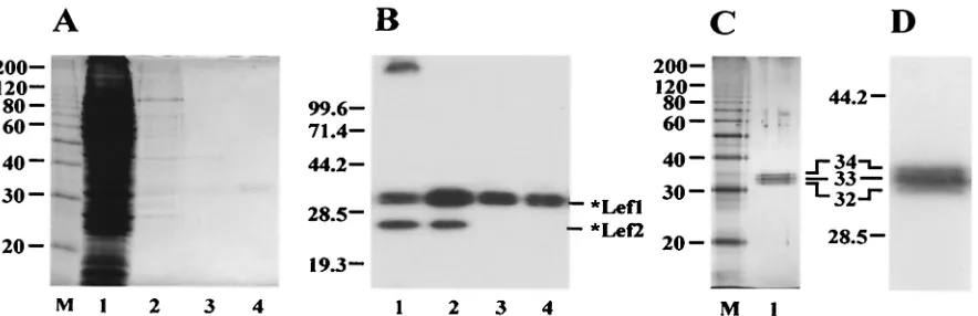
The purification procedure was monitored by SDS-PAGE followed by Coomassie staining (Fig. 2A) and Western blotting with antibody HA.11 (Fig. 2B). Most contaminating proteins were removed from the extract by chromatography on an Ni-NTA column (stage 2) (Fig. 2A, lane 2). However, this fraction contained contaminating proteins and a prominent soluble *LEF-2 component. Further chromatography on a DEAE
col-FIG. 2. Purification of *LEF-1 from infected Sf9 cells. The samples obtained at different stages of the purification procedure were analyzed by SDS–12% PAGE, followed by staining with Coomassie brilliant blue (A) or by Western blotting with monoclonal antibody HA.11 (B). Lane 1, the high-speed supernatant (stage 1), 5l (A) and 2.5l (B); lane 2, the sample collected from Ni-NTA (stage 2), 10l (A) and 5l (B); lane 3, the flowthrough from DEAE-Toyopearl (stage 3), 10l (A) and 5l (B); lane 4, the final preparation from heparin-Sepharose (stage 4), 10l (A) and 5l (B); lane M, 10-kDa protein ladder. (C and D) SDS–12% PAGE of purified *LEF-1 followed by silver staining (C) or by Western blotting with monoclonal antibody HA.11 (D). (C) Lane 1, *LEF-1, 60 ng; lane M, 10-kDa protein ladder. (D) *LEF-1, 15 ng. A magnified section of the blot with *LEF-1 is shown. The molecular masses of protein markers (in kilodaltons) are shown on the left sides of panels A to D.
on November 8, 2019 by guest
http://jvi.asm.org/
[image:3.587.74.516.72.215.2]umn caused the separation of the *LEF-1 sample into two fractions: *LEF-1 associated with *LEF-2 was retained on the column, whereas the *LEF-1 fraction free of *LEF-2 appeared in the flowthrough (stage 3) (Fig. 2B, lane 3).
Besides contaminating proteins, the DEAE column re-moved nucleic acids from the sample. After final chromatog-raphy on a heparin-Sepharose column (stage 4), *LEF-1 was essentially free of contamination (Fig. 2A, lane 4). SDS-PAGE followed by silver staining (Fig. 2C) confirmed that *LEF-1 was purified to near homogeneity. Western blot analysis did not reveal contamination of the *LEF-1 samples with traces of *LEF-2 (Fig. 2B, lanes 3 and 4). The *LEF-1 purification protocol requires 1 day and yields about 30g of pure protein from a 100-ml culture of infected cells.
At all stages of purification, *LEF-1 was present as a set of three polypeptides with apparent molecular masses of 32, 33, and 34 kDa (Fig. 2B, C, and D). These values are close to the calculated molecular mass of *LEF-1 (33.4 kDa). The reason for the observed microheterogeneity of *LEF-1 remains un-clear. All three polypeptides were immunoreactive with anti-body HA.11 (Fig. 2D). Therefore, they retained intact N ter-mini with the HA epitope. Two smaller polypeptides were unlikely to have been produced by limited proteolysis of the largest one during purification because the extracts analyzed immediately after extraction of infected cells showed the same tripartite pattern of *LEF-1 as the purified samples. Treatment of samples with calf intestinal phosphatase had no effect on the electrophoretic pattern of *LEF-1 when it was analyzed by SDS-PAGE, suggesting that the heterogeneity was not due to phosphorylation (data not shown). However, this result does not completely exclude modification by phosphorylation, e.g., if the phosphates are inaccessible to the enzyme.
Primase activity of *LEF-1. Before purified *LEF-1 was
assayed for primase activity, the *LEF-1 samples were tested for the presence of endonucleases and topoisomerases (see Materials and Methods). No conversion of RFI DNA into RFII and RFIII and no appearance of DNA topoisomers was observed after incubation of plasmid DNA with purified *LEF-1 (data not shown). This indicates that the *LEF-1 sam-ples are essentially free of endonuclease and topoisomerase activity.
Purified *LEF-1 showed a primase activity in both the indi-rect and diindi-rect assays (Fig. 3). The indiindi-rect assay is based on the ability of primase to initiate DNA synthesis on an ssDNA template in the absence of exogenous primers. A typical reac-tion mixture contains Klenow enzyme, [␣-32P]dATP, poly(dT)
template, and ATP. There is no DNA synthesis and label incorporation in this system in the absence of primase. If RNA transcripts are synthesized by primase from ATP, they serve as primers for DNA polymerization by Klenow enzyme, thus re-sulting in the incorporation of [32P]dAMP into nascent DNA,
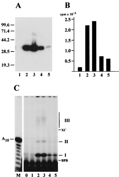
which is monitored by trichloroacetic acid (TCA) precipitation and counting in a scintillation counter. The *LEF-1 fractions collected from a heparin-Sepharose column at the final stage in purification (Fig. 3A) were able to support DNA synthesis by Klenow enzyme on poly(dT) (Fig. 3B). The primase activity correlated well with the amount of *LEF-1 protein in the column fractions.
In the direct primase assay, the radioactive products synthe-sized by *LEF-1 in the presence of [␣-32P]ATP and poly(dT)
were analyzed by electrophoresis in a 20% polyacrylamide–8 M urea gel (Fig. 3C). *LEF-1 produced three radioactive prod-ucts of different electrophoretic mobilities, called prodprod-ucts I, II, and III (Fig. 3C). The amount of all three products corre-lated with the amount of *LEF-1 (Fig. 3A) and the primase activity (Fig. 3B) in the column fractions. Treatment with 0.1 N NaOH caused degradation of product III, confirming that it is RNA (data not shown). In contrast, products I and II were insensitive to alkali, and their structure is currently under in-vestigation.
To find an optimum for the primase activity of LEF-1, we varied the pH and concentration of KCl and MgCl2 in the
[image:4.587.305.533.71.410.2]reaction mixture (Fig. 4A). *LEF-1 primase activity was highly
FIG. 3. Correlation of primase activity with *LEF-1. The fractions eluted from heparin-Sepharose at KCl concentrations of 100 mM (1), 150 mM (2), 200 mM (3), 250 mM (4), and 300 mM (5) were analyzed by the following methods: (A) SDS–12% PAGE, followed by Western blotting with monoclonal antibody HA.11; (B) indirect primase assay with Klenow enzyme, followed by determination of the acid-insoluble radioactivity; (C) direct primase assay on the poly(dT) template, fol-lowed by electrophoresis in a 20% polyacrylamide–8 M urea gel. The assays were performed as described in Materials and Methods. Por-tions of 2.5l (A), 5l (B), and 3l (C) were taken from the fractions for analysis. Lane 0 in panel C represents a control reaction lacking a column fraction. The molecular masses of protein markers (in kilodal-tons) are shown on the left side of panel A. The migration of xylene cyanol (XC), bromophenol blue (BPB), and the primase products (I, II, and III) is indicated on the right side of panel C. Lane M in panel C represents oligonucleotides generated by partial alkaline hydrolysis of 5⬘-end32P-labeled A
10.
on November 8, 2019 by guest
http://jvi.asm.org/
dependent on pH and reached a maximum at alkaline condi-tions (pH 8.8 to 9.4) (Fig. 4A, lanes 2 to 8). The effect of KCl and MgCl2 was assayed at pH 7.5, 8.3, and 9.1. The results
obtained at pH 9.1 are shown in Fig. 4A (lanes 9 to 11 and 12 to 16). At all three pHs, KCl was inhibitory. The primase activity of *LEF-1 was absolutely dependent on the presence of divalent cations in the reaction mixture and reached a max-imum at 10 mM MgCl2. The magnesium dependence shown in
Fig. 4A (lanes 12 to 16) clearly indicated that the optima for synthesis of products I and III are different. The amount of high-molecular-weight product III reached a maximum at 5 to 10 mM MgCl2, whereas the amount of product I was maximal
at 10 to 20 mM. At the optimum conditions, *LEF-1 remained enzymatically active during a 2-h incubation at 30°C (Fig. 4B). The template dependence of *LEF-1 activity was analyzed in experiments with poly(dT) and ssDNA of phage M13mp9 (Fig. 5). The synthesis of products II and III by *LEF-1 was absolutely dependent on the presence of DNA template in the reaction mixture. In contrast, the synthesis of product I was highly stimulated in the presence of poly(dT), but was not absolutely dependent on the presence of DNA template (Fig. 5A and B, lane 1). The lack of strict template dependence for
product I suggests that it might be synthesized by a mecha-nism other than the regular template-dependent polymeriza-tion. The synthesis of long RNA transcripts (product III) on poly(dT) and M13 DNA templates indicated that LEF-1 is capable of extensive nucleotide polymerization (Fig. 5A and B, lanes 3 and 4). However, the synthesis of nascent poly(A) chains on the poly(dT) template appeared to be less efficient than synthesis from M13 DNA templates. This may be because elongation of the poly(A) chain on the poly(dT) template can be blocked by the poly(dT) template’s folding back to form Hoogsteen base pairs with the nascent poly(A) chain in the transient triplexes (38). The trapping of nascent poly(A) in such triplex structures prevents efficient elongation of poly(A) chains by the enzyme. Natural single-stranded DNAs do not form regular triplexes with nascent RNA, although stable sec-ondary structures in DNA may serve as physical obstacles for polymerization. In the presence of M13 DNA and only one precursor, [␣-32P]ATP, *LEF-1 synthesized the radioactive
products I and II, but not product III (Fig. 5, lane 5). Addition of three other precursors, CTP, GTP, and UTP, allowed syn-thesis of long RNA polymers, some of which did not enter the 20% polyacrylamide gels that were employed in this assay (Fig.
FIG. 4. Optimization of primase activity of *LEF-1 on poly(dT). (A) Three parameters of the reaction mixture used in the direct primase assay were varied: the pH of Tris-HCl buffer (lanes 2 to 8), the KCl concentration (lanes 9 to 11), and the MgCl2concentration (lanes 12 to 16). The
reaction shown in lane 1 contained 50 mM Tris-HCl (pH 9.1), 15 mM KCl, and 10 mM MgCl2, but did not contain *LEF-1. Other reactions
contained 200 ng of *LEF-1. The reactions contained the following: lanes 2 to 8, 15 mM KCl, 10 mM MgCl2, and 50 mM Tris-HCl buffer at the
indicated pH; lanes 9 to 11, 30 mM Tris-HCl (pH 9.1), 10 mM MgCl2, and KCl at the indicated concentration; lanes 12 to 16, 30 mM Tris-HCl
(pH 9.1), 15 mM KCl, and MgCl2at the indicated concentration. Other ingredients in the reaction mixture [25g/ml poly(dT), 5M (100Ci/ml)
[␣-32P]ATP, 100g of BSA per ml, and 1 mM DTT] were not varied. After incubation for 2 h at 30°C, the reactions were terminated and the
samples were processed for electrophoresis in a 20% polyacrylimide–8 M urea gel as described in Materials and Methods. (B) Time course of reaction catalyzed by *LEF-1 on poly(dT) at the optimal conditions. The reaction mixture containing 25 mM Tris-HCl (pH 9.1), 10 mM MgCl2,
25g of poly(dT) per ml, 5M (100Ci/ml) [␣-32P]ATP, 100g of BSA per ml, and 1 mM DTT was assembled on ice. *LEF-1 (600 ng in 18
l of buffer D) was added to a final volume of 60l, and 10-l portions were taken from the mixture immediately (time zero) and after incubation for 30, 60, 90, and 120 min at 30°C. The samples were processed for further analysis in a 20% polyacrylamide–8 M urea gel as described in Materials and Methods. The migration of xylene cyanol (XC), bromophenol blue (BPB), and the primase products (I, II, and III) is indicated on the sides of panels A and B. Lanes M in panel A represent oligonucleotides generated by partial alkaline hydrolysis of 5⬘-end32P-labeled A
10.
on November 8, 2019 by guest
http://jvi.asm.org/
5, lane 4). The long transcripts appeared to be a major product of the enzymatic activity of *LEF-1 in the complete system containing M13 DNA and four NTPs.
To estimate the size of the RNA transcripts made on M13 DNA, the reaction products were subjected to electrophoresis in a 1.2% agarose gel (Fig. 6). Prior to thermal denaturation, the radioactive products synthesized by *LEF-1 were associ-ated with M13 DNA (lanes 2 to 4). Heat treatment caused liberation of heterogeneous radioactive polymers mostly in a range of several hundred bases in size (lanes 6 to 8). Thus, *LEF-1 is capable of synthesis in vitro of long RNA transcripts that greatly exceed the size of primers synthesized by DNA primases inside the cell.
Primase domain mutation in *LEF-1.The ability to
synthe-size RNA transcripts on poly(dT) and natural ssDNA is in agreement with the primase function of LEF-1 predicted ear-lier (13) that was based on the presence of conserved primase
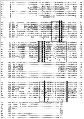
motifs in baculovirus LEF-1 sequences. The conserved motif of Pri-type primases comprising two aspartate residues separated by a single hydrophobic amino acid and preceded by three hydrophobic amino acids (W[I/V][I/L/V]D[A/I/V]D) is found in the LEF-1 sequence of AcMNPV and several other baculo-viruses (Fig. 7).
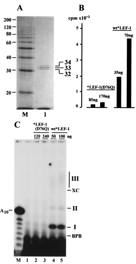
To confirm that the enzymatic activity observed in *LEF-1 samples was actually dependent on the predicted primase do-main, we made a conserved change from aspartate to glu-tamine at amino acid 76 (REWVV DAD to REWVV QAD) in LEF-1 and expressed this construct in a recombinant baculo-virus. In the previous study, we found that this mutation did not prevent interaction with LEF-2, but it eliminated the abil-ity of LEF-1 to function in the transient-replication assay (13). The mutant *LEF-1(D76Q), fused at the N terminus with the HA epitope and HIS6tag, was overexpressed in Sf9 cells and
purified by the same method as the wild-type (wt) *LEF-1. When purified to near homogeneity, *LEF-1(D76Q) demon-strated three polypeptides with the same mobility as those from our wt *LEF-1 construct (Fig. 8A). However, the activity of the mutant *LEF-1(D76Q) in the indirect assay with Kle-now enzyme was very low (Fig. 8B). In the direct assay, the mutant *LEF-1(D76Q) did not synthesize any of the three products, I, II, or III, typical of wt *LEF-1 (Fig. 8C). Thus, the enzymatic activity of LEF-1 requires the intact primase domain in the protein. These data confirm the dependence of DNA synthesis by Klenow enzyme in the indirect assay on RNA primers provided by *LEF-1. The minor stimulation of DNA synthesis caused by *LEF-1(D76Q) in the indirect assay may
FIG. 5. Template dependence of synthesis catalyzed by *LEF-1. The direct primase assay was carried out in 10-l reaction mixtures containing 25g of poly(dT) (lanes 2 and 3) or 25g of ssDNA of phage M13mp9 (lanes 4 and 5) per ml. Lanes 1 and 2 represent control reactions lacking DNA template and *LEF-1, respectively. Other re-actions contained 200 ng of *LEF-1. CTP, GTP, and UTP, each at a concentration of 50M, were added to the reaction shown in lane 4. Other ingredients in the reaction mixtures were 25 mM Tris-HCl (pH 9.1), 10 mM MgCl2, 5M (100Ci/ml) [␣-32P]ATP, 100g of BSA
per ml, and 1 mM DTT. After incubation for 2 h at 30°C, the reactions were terminated and the samples were processed for electrophoresis in a 20% polyacrylamide–8 M urea gel as described in Materials and Methods. Panel B represents lanes 1 to 5 of panel A after a longer exposure to X-ray film. The migration of xylene cyanol (XC), bromo-phenol blue (BPB), and the primase products (I, II, and III) is indi-cated on the sides of panels A and B. Lane M in panel A represents oligonucleotides generated by partial alkaline hydrolysis of 5⬘-end32
[image:6.587.49.278.72.353.2]P-labeled A10.
FIG. 6. RNA synthesis catalyzed by *LEF-1 on single-stranded DNA of phage M13mp9. Reactions were conducted as described in Materials and Methods. Aliquots were removed after 30, 60, and 120 min. Prior to electrophoresis in a 1.2% agarose gel, each sample was divided in two portions. The first portion was loaded directly onto the gel (lanes 1 to 4), while the second portion was loaded after heating for 3 min at 100°C (lanes 5 to 8). After electrophoresis, the gel was transferred onto Whatman 3MM paper, dried under vacuum, and exposed to X-ray film. M13 DNA and an RNA ladder were loaded onto separate lanes and visualized by staining with ethidium bromide. The migration of M13 DNA and RNA markers is indicated on the left and right sides of the gel, respectively.
on November 8, 2019 by guest
http://jvi.asm.org/
be due to a nonspecific stabilization effect of the added protein on Klenow enzyme.
Sedimentation analysis of *LEF-1 in glycerol gradients.To
elucidate the molecular structure of LEF-1, we analyzed the
[image:7.587.116.471.73.578.2]sedimentation of the purified protein in a 15 to 30% glycerol gradient under conditions that prevent nonspecific aggregation (Fig. 9). *LEF-1 sedimented in the gradient slower than BSA (4.3S, 66 kDa) and had a sedimentation coefficient of about
FIG. 7. Sequence alignment of LEF-1 proteins of baculoviruses AcMNPV (ac) (4), OpMNPV (op) (2),Choristoneura fumiferana multinucleo-capsid NPV (cf) (5),Lymantria disparmultinucleocapsid NPV (ld) (28),Spodoptera exignamultinucleocapsid NPV (se) (21),Heliothus armigera single-nucleocapsid NPV (8),Plutella xylostellagranulovirus (px) (18),Xestia c-nigrumgranulovirus (xc) (19), andCulex nigripalpusgranulovirus (cuni) (40). Putative primase domains are boxed. Invariant amino acids in the putative primase domains are in white letters against black, and invariant aspartates in these domains are indicated by asterisks. Identical amino acids are shown at the bottom of each alignment, and dots indicate conservative changes. The last line within the boxed domains shows invariant amino acids from selected archaeal and eukaryotic primases (3). Dashes indicate gaps in the alignment. The numbers on the left and right indicate the amino acid sequence coordinates. The alignment was produced using MacVector DNA analysis software.
on November 8, 2019 by guest
http://jvi.asm.org/
3S. This value suggests that the *LEF-1 samples consist of a mixture of protein monomers. The sedimentation rate of *LEF-1 in the glycerol gradients was much lower than that of AcMNPV RNA polymerase, another viral enzyme capable of synthesizing RNA products. In our experiments, the sedimen-tation pattern of purified AcMNPV RNA polymerase was con-sistent with a molecular mass of 300 to 400 kDa (data no shown), which is slightly less than has been reported previously (15).
Chromatographic behavior of LEF-2. LEF-2 presumably
forms a functional complex with LEF-1 in infected cells (13), and it has been shown to be essential for plasmid replication in the transient-replication assay (26, 33). However, the function of LEF-2 in baculovirus replication is not known. The primase activity found in the purified *LEF-1 samples which were es-sentially free of contaminating *LEF-2 clearly indicated that LEF-2 is not required for the primase activity of LEF-1 in vitro. Some of our indirect data suggest that LEF-2 may serve as an accessory factor needed for incorporation of LEF-1 into replication complexes assembled on viral DNA.
We routinely overexpressed HIS-tagged proteins LEF-1 and LEF-2 together, and both proteins were expressed in Sf9 cells at almost equivalent levels. However, it was more difficult to solubilize *LEF-2 than *LEF-1, and thus the samples after passage through an Ni-NTA column appeared to enriched for *LEF-1 (Fig. 2B, lane 2). At the next chromatographic step, *LEF-2 bound tightly to a DEAE resin, whereas *LEF-1 appeared mostly in the flowthrough (Fig. 2B, lane 3). It was possible to elute *LEF-2 and a portion of *LEF-1 from DEAE-Toyopearl only by using buffers with relatively high concentrations of monovalent salt (0.3 to 0.5 M KCl) (data not shown). Optical densitometry revealed the presence of nucleic acids in this high-salt fraction. Because all free proteins were removed from the DEAE resin at lower salt concentration, this result suggested that *LEF-2 and *LEF-1 might be associated with traces of viral DNA.
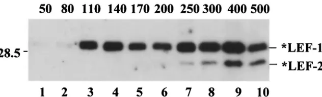
In another experiment, the sample of *LEF-1 and *LEF-2 collected from the Ni-NTA column was subjected to chroma-tography on an ssDNA-cellulose column (Fig. 10). Stepwise elution resulted in the *LEF-1 pool’s being divided in two fractions. One fraction of *LEF-1 was free of *LEF-2 and eluted at a relatively low salt concentration (110 to 170 mM KCl), whereas the other fraction contained both proteins and eluted at much higher salt concentrations (300 to 500 mM KCl). This result suggests that LEF-1 might associate with LEF-2 to form a stable complex with single-stranded DNA and probably with other replicative proteins associated with DNA.
DISCUSSION
[image:8.587.45.278.77.529.2]The essential baculovirus replication protein LEF-1 was originally predicted to serve as a DNA primase, because a mutation in a conserved primase-like domain failed to support transient DNA replication (13). Therefore, we initiated a bio-chemical study of this protein to determine if it possesses primase activity. Upon testing a number of fractions obtained from AcMNPV-infected Sf9 cells in an indirect assay employ-ing Klenow enzyme and poly(dT) template (see Materials and Methods), a strong primase activity was found associated with host cell DNA polymerase␣(data not shown). However, we
FIG. 8. Elimination of primase activity by a mutation in a putative primase domain of LEF-1. The mutated *LEF-1(D76Q) with the as-partate-to-glutamine change at amino acid 76 was purified from Sf9 cells as described in Materials and Methods. (A) SDS–12% PAGE of the purified *LEF-1(D76Q) followed by staining with Coomassie bril-liant blue. Lane 1, *LEF-1(D76Q), 400 ng; lane M, 10-kDa protein ladder. (B) The indirect primase assay of *LEF-1(D76Q) and wild-type (wt) *LEF-1. The amount of proteins in each reaction is shown above the bars. After reaction for 1 h at 30°C, the samples were processed for determination of the acid-insoluble radioactivity as de-scribed in Materials and Methods. (C) The direct primase assay on the poly(dT) template of *LEF-1(D76Q) (lanes 2 and 3) and wt *LEF-1 (lanes 4 and 5). The amount of protein in each reaction is shown above the lanes. Lane 1 represents a control reaction lacking any protein. Reactions were carried out for 2 h at 30°C, and the samples were processed for electrophoresis in a 20% polyacrylamide–8 M urea gel as described in Materials and Methods. The migration of xylene cyanol (XC), bromophenol blue (BPB), and the primase products (I, II, and III) is indicated on the right side of panel C. Lane M in panel C represents oligonucleotides generated by partial alkaline hydrolysis of 5⬘-end32P-labeled A
10.
on November 8, 2019 by guest
http://jvi.asm.org/
were unable to detect a virus-induced DNA primase in extracts from infected cells.
To increase the LEF-1 level in infected cells, we overex-pressed it in recombinant baculovirus under the control of the strong polyhedrin promoter. In this construct, the HA epitope and a HIS6 tag were fused to LEF-1 at the N terminus. A
similar construct was found to retain its activity in transient replication and transcription assays (45). This construct al-lowed the rapid and efficient purification of the recombinant protein, *LEF-1, to homogeneity, as judged by SDS-PAGE (Fig. 2). Biochemical analyses revealed the presence of pri-mase activity by *LEF-1, providing evidence for its role as a primase in baculovirus replication that had been predicted earlier. The list of virally encoded enzymes involved in bacu-lovirus DNA replication now includes a DNA primase in ad-dition to the DNA polymerase (35, 39, 50) and DNA helicase (32, 36) characterized previously. A set of similar viral enzymes are required for genome replication by theHerpesviridae, an-other family of large DNA viruses (7).
The identification of LEF-1 as a DNA primase is based on its ability to synthesize primers from a poly(dT) template, which then allows initiation of DNA synthesis by exogenous DNA polymerase (Klenow enzyme) (Fig. 3B). The synthesis of
primers was confirmed in a direct assay involving examination of the products by gel electrophoresis (Fig. 3C). Furthermore, analysis of a mutant LEF-1 confirmed the dependence of its activity on an intact domain conserved in archaeal and eukary-otic primases (Fig. 7 and 8).
The *LEF-1 primase activity is absolutely dependent on divalent cations (Fig. 4A), which may be directly involved in catalysis (see below). The most striking property of *LEF-1 is its ability to synthesize in vitro polynucleotides of a thousand nucleotides or more on ssDNA of phage M13 (Fig. 6). In this respect *LEF-1 resembles DNA primase from the archaeon Pyrococcus furiosus(6). The RNA transcripts synthesized by *LEF-1 greatly exceed the size of primers synthesized by known DNA primases during DNA replication inside eukary-otic cells. If baculovirus replication is similar to that of other systems, some other viral or host cell factors may limit the size of RNA transcripts synthesized by LEF-1 to produce physio-logically relevant RNA primers.
[image:9.587.138.446.72.201.2]Two distinct types of primases that lack sequence or struc-tural relatedness have been characterized. The Pri-type pri-mases are found in eukaryotes and archaea, whereas the DnaG type are found in eubacteria (23). Baculovirus LEF-1 contains three motifs (for AcMNPV, residues 69 to 78, 114 to 139, and
FIG. 9. Sedimentation analysis of *LEF-1. *LEF-1 (7g in 100l) was layered over 4.9 ml of a 15 to 30% glycerol gradient in buffer containing 0.4 M KCl, 10 mM Tris-HCl (pH 7.5), 1 mM DTT, and 1 mM EDTA and centrifuged in an SW 50.1 rotor at 48,000 rpm and 4°C for 22 h. The gradient was fractionated from the bottom into 16 fractions, and 5-l portions from each fraction were analyzed by SDS–12% PAGE, followed by Western blotting with monoclonal antibody HA.11. The positions of the standards centrifuged in separate tubes are shown by arrows: catalase (220 kDa, 11.3S), aldolase (158 kDa, 8.3S), and BSA (66 kDa, 4.3S).
FIG. 10. Chromatography of *LEF-1 and *LEF-2 on ssDNA-cellulose. A sample of *LEF-1 and *LEF-2 was obtained from a 50-ml culture of Sf9 cells infected with the recombinant viruses vfbHAHISLef-1 and vfbHAHISLef-2 by purification on an Ni-NTA column. After dialysis against buffer containing 25 mM KCl, 10 mM Tris-HCl (pH 7.5), 20% (vol/vol) glycerol, 1 mM DTT, and 1 mM EDTA, the sample was loaded onto an 0.5-ml column of ssDNA-cellulose (Sigma), which was processed with 1-ml portions of the same buffer containing KCl in the concentrations indicated above the lanes. Aliquots (5l) from each fraction were subjected to SDS–12% PAGE, followed by Western blotting with antibody HA.11. A portion of the blot with *LEF-1 and *LEF-2 is shown. The position of the 28.5-kDa protein marker is indicated on the left side.
on November 8, 2019 by guest
http://jvi.asm.org/
[image:9.587.134.450.576.671.2]189 to 214) that are conserved in eukaryotic and archaeal primases (boxed in Fig. 7), suggesting that it is a Pri-type primase. These motifs presumably contribute to the structure of the major domain, which contains the primase active site (3). The lef-1 gene of AcMNPV encodes a protein of 266 amino acids with a calculated molecular mass of 30,780 Da. Other knownlef-1genes of baculoviruses encode proteins as small as 216 amino acids (Fig. 7). These represent the smallest RNA-synthesizing enzymes characterized so far. Due to its small size, baculovirus LEF-1 may be a valuable tool for struc-tural research of Pri-type primases as well as for biochemical and mechanistic studies. The first conserved LEF-1 motif (REW[I/V][I/L/V]D[A/I/V]D) contains two aspartate residues separated by a single hydrophobic amino acid (DXD) and preceded by three hydrophobic amino acids. This motif is also found in primases from a number of herpesviruses, phage T7, Saccharomyces cerevisiae, archaea, and mammals (3, 24). Ex-perimental data suggest that the two proximal aspartates in this motif are important for enzymatic activity. In herpes simplex virus type 1 primase (UL52), a conserved aspartate-to-glu-tamine (IILDLD to IILQLD) change at amino acid 628 com-pletely eliminated the primase activity associated with the UL5-UL8-UL52 complex (25). In the catalytic subunit p49 of mouse primase, two aspartates (D109 and D111) of the respec-tive domain and one distal aspartate (D306) are essential for primase activity and are thought to form the metal-binding core of the active site (10).
Aspartates D76 and D78 in the first conserved motif of AcMNPV LEF-1 and the distal invariant aspartate D193 in the third motif presumably play the same role. In agreement with this prediction, a conserved change from aspartate to glu-tamine at amino acid 76 (REWVVDAD to REWVVQAD) in AcMNPV LEF-1 completely abolished the LEF-1 primase ac-tivity (Fig. 8). Because this mutation eliminated the ability of LEF-1 to function in transient-replication assays (13), the es-sential function of LEF-1 in baculovirus replication is directly connected with the primase activity. Interestingly, the three aspartates in mouse primase (D109, D111, and D306) align precisely with similar residues in the 31-kDa domain of DNA polymerase, suggesting that Pri-type primases are probably members of the Pol X polymerase family (24). The three as-partates (D95, D97, and D280) in the archaeon Pyrococcus furiosus(Pfu) primase could be superimposed with the active-site residues of four different DNA polymerases, including human DNA polymerase , indicating similar three-dimen-sional arrangements in all five structures (3). These data sug-gest that Pri-type primases including baculovirus LEF-1, use the common two-metal ion mechanism of DNA polymerases (49).
Another conserved motif in DNA primases is the Zn2⫹ binding domain located near the active site. In bacterial DnaG-type primases, this domain is thought to play a role in template sequence recognition (43). ThePfuprimase structure confirms the presence of a Zn2⫹ ion at a location quite close to the putative active site, suggesting that this ion may also play a role in the activity of Pri-type primases (3). However, the zinc binding site (C/HX2–5C/HX11–13C/HX2–5C/H) and its deriv-atives are absent in some archaeal primases (3) and in bacu-lovirus LEF-1. This raises doubts about whether the zinc ion is essential for the function of Pri-type primases.
Although *LEF-1 itself has relatively low affinity for ssDNA and was eluted from ssDNA-cellulose at 110 to 170 mM KCl, a portion of the cellular pool of *LEF-1 copurified with *LEF-2 as a fraction possessing a high affinity for ssDNA (Fig. 10). Since it has been demonstrated that LEF-1 interacts with LEF-2 (13), this observation suggests that LEF-2 may stabilize binding of LEF-1 to DNA template and probably to other baculovirus replication proteins. In this respect, LEF-2 resem-bles subunit p58 of eukaryotic DNA primase. Although p58 is not required for enzymatic activity of the catalytic subunit p49, it binds to ssDNA, tethers p49 to the 180-kDa subunit of DNA polymerase␣, and stabilizes p49 (11).
All known DNA primases act in close association with either replicative DNA polymerases or DNA helicases. Both subunits of simian primase interact with host cell replication protein A and simian virus 40 large T antigen, which serves as the heli-case in virus replication (51).E. coliprimase (DnaG) interacts with the replicative DnaB helicase, SSB protein, and DNA polymerase III holoenzyme (27). In T7 phage, whose helicase and primase are homologous to the equivalent eubacterial pro-teins (22), the COOH-terminal region of the primase is directly linked to the NH2-terminal region of the helicase, forming a
single polypeptide. Replication of baculoviruses may proceed by a mechanism similar to that of herpesviruses. In herpesvi-ruses, the 114-kDa primase subunit UL52 is tightly associated with UL5 and UL8, forming a heterotrimeric primosome pos-sessing DNA-dependent ATPase, DNA helicase, and DNA primase activities (for a review, see reference 30). The func-tional partner of the LEF-1 and LEF-2 complex in the repli-some of baculoviruses is not known. Further analysis of the complexes of LEF-1 and LEF-2 with ssDNA observed in this study may provide a more detailed picture of the structure and organization of the replication machinery of baculoviruses.
ACKNOWLEDGMENTS
The suggestions of Sam Bennett, Rebecca Russell, and Margot Pearson related to this project and the gift of plasmids from Lois Miller, Joyce Wilson, and Lorena Passarelli are gratefully acknowl-edged.
This research was supported by grants from the NSF (9982536) and NIH (GM60404) to G.F.R., a supplement from the NSF Central and Eastern Europe and Program, and a grant from the Russian Founda-tion for Basic Research (00-04-49237) to V.S.M.
ADDENDUM IN PROOF
After this paper was submitted, Eugene Koonin informed us that his group had detected a low level of homology between baculovirus LEF-2 and the large subunit (p58) of eukaryotic DNA primases (E. V. Koonin, Y. I. Wolf, A. S. Kondrashov, and L. Aravind, J. Mol. Microbiol. Biotechnol. 2:509–512, 2000). We appreciate this information, which is in agreement with our suggestion that LEF-2 plays an accessory role in baculovirus DNA primase activity, analogous to that of p58 in eukaryotic primases.
REFERENCES
1.Ahrens, C. A., D. J. Leisy, and G. F. Rohrmann.1996. Baculovirus DNA
replication, p. 855–872.InM. De Pamphilis (ed.), DNA replication in
eu-karyotic cells. Cold Spring Harbor Laboratory Press, Cold Spring Harbor, N.Y.
2.Ahrens, C. H., R. Russell, C. J. Funk, J. T. Evans, S. H. Harwood, and G. F. Rohrmann.1997. The sequence of theOrgyia pseudotsugata
multinucleocap-sid nuclear polyhedrosis virus genome. Virology229:381–399.
on November 8, 2019 by guest
http://jvi.asm.org/
J. Gen Virol.82:241–257.
9.Choi, J., and L. A. Guarino.1995. The baculovirus transactivator IE1 binds to viral enhancer elements in the absence of insect cell factors. J. Virol.
69:4548–4551.
10.Copeland, W., and X. Tan.1995. Active site mapping of the catalytic mouse
primase subunit by alanine scanning mutagenesis. J. Biol. Chem.270:3905–
3913.
11.Copeland, W., and T. Wang.1993. Enzymatic characterization of the indi-vidual mammalian primase subunits reveals a biphasic mechanism for
initi-ation of DNA repliciniti-ation. J. Biol. Chem.268:26179–26189.
12.Donis-Keller, H., A. Maxam, and W. Gilbert.1977. Mapping adenines,
gua-nines, and pyrimidines in RNA. Nucleic Acids Res.4:2527–2538.
13.Evans, J. T., D. J. Leisy, and G. F. Rohrmann.1997. Characterization of the interaction between the baculovirus replication factors, LEF-1 and LEF-2.
J. Virol.71:3114–3119.
14.Evans, J. T., G. S. Rosenblatt, D. J. Leisy, and G. F. Rohrmann.1999. Characterization of the interaction between the baculovirus ssDNA-binding
protein (LEF-3) and putative helicase (P143). J. Gen. Virol.80:493–500.
15.Guarino, L. A., B. Xu, J. Jin, and W. Dong.1998. A virus-encoded RNA
polymerase purified from baculovirus-infected cells. J. Virol.72:7985–7991.
16.Hang, X., W. Dong, and L. A. Guarino.1995. Thelef-3gene ofAutographa californicanuclear polyhedrosis virus encodes a single-stranded
DNA-bind-ing protein. J. Virol.69:3924–3928.
17.Harwood, S. H., L. Li, P. S. Ho, A. K. Preston, and G. F. Rohrmann.1998.
AcMNPV late expression factor-5 interacts with itself and contains a zinc
ribbon domain that is required for maximal late transcription activity and is
homologous to elongation factor TFIIS. Virology250:118–134.
18.Hashimoto, Y., T. Hayakawa, Y. Ueno, T. Fugita, Y. Sano, and T. Matsu-moto.2000. Sequence analysis of thePlutella xylostellagranulovirus genome.
Virology275:358–372.
19.Hayakawa, T., R. Ko, K. Okano, S. Seong, C. Goto, and S. Maeda.1999.
Sequence analysis of theXestia c-nigrumgranulovirus genome. Virology
262:277–297.
20.Hayakawa, T., G. F. Rohrmann, and Y. Hashimoto.2000. Patterns of
ge-nome organization and content in lepidopteran baculoviruses. Virology278:
1–12.
21.Ijkel, W. F. J., E. A. van Strien, J. G. M. Jeldens, R. Broer, D. Zuidema, R. W. Goldbach, and J. M. Vlak.1999. Sequence and organization of the Spodopt-era exiguamulticapsid nucleopolyhedrovirus genome. J. Gen. Virol.80:3289– 3304.
22.Ilyina, T., A. Gorbalenya, and E. Koonin.1992. Organization and evolution
of bacterial and bacteriophage primase-helicase systems. J. Mol. E vol.34:
351–357.
23.Keck, J., and J. Berger.2001. Primus inter pares (first among equals). Nat.
Struct. Biol.8:2–4.
24.Kirk, B., and R. Kuchta.1999. Arg304 of human DNA primase is a key contributor to catalysis and NTP binding: primase and the family X
poly-merases share significant sequence homology. Biochemistry38:7727–7736.
25.Klinedinst, D. K., and M. D. Challberg.1994. Helicase-primase complex of herpes simplex virus type 1: a mutation in the UL52 subunit abolishes
primase activity. J. Virol.68:3693–3701.
26.Kool, M., C. Ahrens, R. W. Goldbach, G. F. Rohrmann, and J. M. Vlak.1994.
Identification of genes involved in DNA replication of theAutographa
cali-fornicabaculovirus. Proc. Natl. Acad. Sci. USA91:11212–11216. 26a.Koonin, E. V., Y. I. Wolf, A. S. Kondrashov, and L. Aravind.2000. Bacterial
homologs of the small subunit of eukaryotic DNA primase. J. Mol.
Micro-biol. Biotechnol.2:509–512.
27.Kornberg, A., and T. A. Baker.1992. DNA replication, 2nd ed. W. H. Freeman and Company, New York, N.Y.
34.Martignoni, M. E., and P. J. Iwai.1986. A catalog of viral diseases of insects, mites, and ticks, 4th ed. USDA Forest Service publication PNW-195. U.S. Department of Agriculture, Portland, Ore.
35.McDougal, V. V., and L. A. Guarino.1999. Autographa californica nuclear polyhedrosis virus DNA polymerase: measurements of processivity and
strand displacement. J. Virol.73:4908–4918.
36.McDougal, V. V., and L. A. Guarino.2000. TheAutographa californica
nu-clear polyhedrosis virus p143 gene encodes a DNA helicase. J. Virol.74:
5273–5279.
37.Mikhailov, V.2000. Helix-destabilizing properties of the baculovirus
single-stranded DNA-binding protein (LEF-3). Virology270:180–189.
38.Mikhailov, V., and D. Bogenhagen.1996. Termination within oligonucleotide (dT) tracts in template DNA by DNA polymerase gamma occurs with for-mation of a DNA triplex structure and is relieved by mitochondrial
single-stranded DNA-binding protein. J. Biol. Chem.271:30774–30780.
39.Mikhailov, V., K. Marlyev, J. Ataeva, P. Kullyev, and A. Atrzhev.1986.
Characterization of 3⬘35⬘exonuclease associated with DNA polymerase of
silkworm nuclear polyhedrosis virus. Nucleic Acids Res.14:3841–3857.
40.Moser, B., J. Becnel, S. White, C. Afonso, G. Kutish, S. Shanker, and E. Almira.2001. Morphological and molecular evidence thatCulex nigripalpus
baculovirus is an unusual member of the family Baculoviridae. J. Gen Virol.
82:283–297.
41.Okano, K., V. S. Mikhailov, and S. Maeda.1999. Colocalization of baculo-virus IE-1 and two DNA-binding proteins, DBP and LEF-3, to viral
repli-cation factories. J. Virol.73:110–119.
42.Oppenheimer, D. I., and L. E. Volkman.1997. Evidence for rolling circle
replication of Autographa californica M nucleopolyhedrovirus genomic
DNA. Arch. Virol.142:2107–2113.
43.Pan, H., and D. Wigley. 2000. Structure of the zinc-binding domain of
Bacillus stearothermophilusDNA primase. Struct. Fold. Des.8:231–239. 44.Pearson, M. N., and G. F. Rohrmann.1998. Characterization of a
baculo-virus-encoded ATP-dependent DNA ligase. J. Virol.72:9142–9149.
45.Rapp, J. C., J. A. Wilson, and L. K. Miller.1998. Nineteen baculovirus open reading frames, including LEF-12, support late gene expression. J. Virol.
72:10197–10206.
46.Rodems, S. M., and P. D. Friesen.1995. Transcriptional enhancer activity of
hr5 requires dual-palindrome half sites that mediate binding of a dimeric
form of the baculovirus transregulator IE1. J. Virol.69:5368–5375.
47.Rohrmann, G. F.1999. Nuclear polyhedrosis viruses, p. 146–152.InR. G. Webster and A. Granoff (ed.), Encyclopedia of virology, 2nd ed. Academic Press, London, England.
48.Sambrook, J., E. F. Fritsch, and T. Maniatis.1989. Molecular cloning: a laboratory manual, 2nd ed. Cold Spring Harbor Laboratory, Cold Spring Harbor, N.Y.
49.Steitz, T., S. Smerdon, J. Jager, and C. Joyce.1994. A unified polymerase
mechanism for nonhomologous DNA and RNA polymerases. Science266:
2022–2025.
50.Tomalski, M. D., J. Wu, and L. K. Miller.1988. The location, sequence, transcription, and regulation of a baculovirus DNA polymerase gene.
Virol-ogy167:591–600.
51.Weisshart, K., H. Forster, E. Kremmer, B. Schlott, F. Grosse, and H. Nasheuer.2000. Protein-protein interactions of the primase subunits p58 and p48 with simian virus 40 T antigen are required for efficient primer
synthesis in a cell-free system. J. Biol. Chem.275:17328–17337.
52.Winstanley, D., and D. O’Reilly.1999. Granuloviruses, 2nd ed. Academic Press, London, England.
53.Wu, Y., and E. B. Carstens.1998. A baculovirus single-stranded DNA bind-ing protein, LEF-3, mediates the nuclear localization of the putative helicase
P143. Virology247:32–40.