A Dissertation on
Assessment of Sentinel Lymph Node Using Methylene Blue Dye in Carcinoma Breast
In Government Royapettah Hospital
Submitted to
The Tamilnadu Dr. MGR Medical University
In partial fulfilment of the requirement
For the award of degree of
M.Ch. (SURGICAL ONCOLOGY) BRANCH VII
KILPAUK MEDICAL COLLEGE
THE TAMILNADU Dr. MGR MEDICAL UNIVERSITY CHENNAI, TAMILNADU
BONAFIDE CERTIFICATE
This is to certify that Dr. J. SAKTHIUSHADEVI,bonafide student of M.Ch. Surgical Oncology (August 2012 to July 2015) in the Department of Surgical
Oncology, Government Royapettah Hospital, Chennai – 600 014 has done this
dissertation on “Assessment of Sentinel Lymph Node Using Methylene Blue Dye in Carcinoma breast” under my guidance and supervision in partial fulfilment of the regulations laid down by The Tamilnadu Dr. MGR. Medical University,
Chennai for M.Ch. Surgical Oncology Examination to be held in August 2015.
Dean
Kilpauk Medical College, Chennai
Prof. R .Rajaraman MS, MCh Head, Centre for Oncology
Prof&Head , Department of Surgical Oncology Govt. Royapettah Hospital &
DECLARATION
I solemnly declare that the dissertation titled “Assessment of Sentinel Lymph Node Using Methylene Blue Dye in Carcinoma breast” was done by me at Department of Surgical Oncology, center for Oncology, Kilpauk medical college
and Govt. Royapettah Hospital, Chennai between August 2012 to January 2015
under the guidance and supervision of Prof.R.Rajaraman. The Dissertation is
submitted to The Tamil Nadu Dr.MGR Medical University towards the partial
fulfillment for the award of M.Ch degree
(Branch VII) in Surgical Oncology.
Dr.SakthiUshaDevi. J
M Ch Post graduate student
Dept. of Surgical Oncology
Kilpauk Medical college
ACKNOWLEDGEMENTS
It is my pleasure and privilege to record my deep sense of gratitude to
Prof. Dr. R. Rajaraman M.S., M.Ch, Professor& Head of the Department, Centre for oncology, Government Royapettah Hospital, Kilpauk Medical College,
Chennai, for his constant encouragement, motivation and guidance given to me in
bringing forth this piece of work.
Special gratitude is due to the Associate Professor of our department,
Dr. S. Subbiah M.S., M.Ch, for the kindness and help rendered.
I am extremely grateful to Assistant professors Department of Surgical
Oncology, Dr.Gopu M.SMCh, Dr.Sujay Susikar M.S,MCh, Dr.Syed Afroze Hussain M.S,MCh, Government Royapettah Hospital, and Chennai for their constant support, valuable comments, and suggestions.
I am also thankful to Professors of Pathology at Government Royapettah
Hospital, present and past for their generous help and co-operation rendered
towards the study.
I thank my fellow post graduates, technical staff, and paramedical staff of
our department for their generous assistance throughout this study. I owe my
CONTENTS Page No
1 INTRODUCTION 1
2 REVIEW OF LITERATURE 2
3 AIM OF THE STUDY 59
4 MATERIALS AND METHODS 60
5 STATISTICAL ANALYSIS& RESULTS 64
6 DISCUSSION 77
7 CONCLUSION 85
8 ANNEXURES
I Originality Report
II Ethical Justification and Ethics Committee Approval
ABSTRACT
Title : Assessment of Sentinel Lymph Node Using Methylene Blue Dye in Carcinoma Breast Key words :Breast cancer, sentinel node biopsy, methylene blue dye, axillary dissection
1 Introduction:
Management of axilla is an integral part of treatment of carcinoma breast.
Axillary lymph node dissection has a well-established role in regional disease
control and it provides information about the histopathological status which has
significant prognostic and therapeutic implications(1).However only around 30
percent of the of clinically node negative patients prove to be histopathologically
node positive which means that 70 percent of clinically node negative patients
undergo axillary dissection and are exposed to its morbidities like neuropathies,
seromas and upper extremity lymphedema. This can be avoided with a sentinel
lymph node (SLN) biopsy. Published data till date use vital blue dye and /or
99m
Technetium labelled colloid with gamma probe for the identification of sentinel
lymph nodes. A combination of the two techniques has been found to be the best
and is recommended for optimal outcome. Blue dye guided SLN identification
may be the only available option in countries with low resources due to prohibitive
price of gamma probes. This study was done to analyse methylene blue dye uptake
after peri-tumoural injection and compare tumour positivity in nodes stained and
2 REVIEW OF LITERATURE
Addressing the axillary nodes is an integral component in the loco regional
management of early breast cancer. A formal axillary lymph node dissection was
until recently the standard procedure of choice for the management in the majority
of the patients irrespective of primary tumour characteristics. Around 25-30% of
patients have nodal disease at the time of diagnosis. For these patients removal of
lymph nodes minimises the chance of loco regional relapse and can provide crucial
information for guiding systemic adjuvant treatments. Moreover, axillary lymph
node status remains the single most important prognostic factor in breast cancer (2).
Nonetheless, for node negative patients axillary lymph node dissection represents
over treatment and can be associated with significant morbidity. Sentinel node
biopsy (SNB) permits accurate axillary staging of patients with early breast cancer
with clinically node negative axilla. Sentinel node biopsy has less morbidity and
fewer complications than axillary lymph node dissection (3).
Anatomy of the breast(4)
The adult female breast is located within the superficial fascia of the anterior
chest wall. The base of the breast extends from the second rib above to the fifth or
sixth rib below, and from the sternal border medially to the mid-axillary line
laterally. Two-thirds of the base of the breast lies anterior to the pectoralis major
muscle, the remainder lies over the serratus anterior muscle. A small part may lie
3
In about 95 percent of women there is a prolongation of the upper lateral
quadrant toward the axilla. This tail (of Spence) of breast tissue enters a hiatus (of
Langer) in the deep fascia of the medial axillary wall. This is the only breast tissue
found normally beneath the deep fascia.
Skin
The epidermis of the areola and the nipple is distinguished from that of the
surrounding skin by the colour imparted by blood vessels carried close to the
surface in long dermal papillae. In females at puberty, and with each pregnancy,
there is an increase in the melanin content of the basal cells, further darkening the
area. The dermis of the skin merges with the superficial fascia, which envelops the
parenchyma of the breast.
Superficial Fascia
The superficial fascia enveloping the breast is continuous with the
superficial abdominal fascia (of Camper) below, and the superficial cervical fascia
above. Anteriorly, it merges with the dermis of the skin.
Deep Fascia
The deep pectoral fascia envelops the pectoralis major muscle and is
continuous with the deep abdominal fascia below. It attaches to the sternum
medially and to the clavicle and axillary fascia above and laterally. Along the
lateral border of the pectoralis major muscle, the anterior lamina of the deep
4
inferiorly, with the fascia of the serratus anterior. A posterior extension of this
fascia is continuous with the fascia of the latissimus dorsi and forms the so-called
suspensory ligament of the axilla.
Anatomy of the axilla
The axilla is defined as a pyramidal space having an apex, a base, and four
walls. The apex is a triangular space bordered by the clavicle, the upper border of
the scapula, and the first rib, which is sometimes called the cervico-axillary canal.
The base consists of the axillary fascia beneath the skin of the axillary fossa. The
anterior wall is composed of three muscles (the pectoralis major, the pectoralis
minor, and the subclavius) and the clavipectoral fascia, which envelops the muscles
and fills the spaces between them. The posterior wall is formed by the scapula and
three muscles: the Subscapularis, the Latissimus dorsi, and the Teres major. The
medial wall consists of the lateral chest wall, with the second to sixth ribs, and the
serratus anterior muscle. The lateral wall is the narrowest of the walls, being
formed by the bicipital groove of the humerus.The axilla contains lymph nodes, the
axillary sheath, which covers blood vessels and nerves, and the tendons of the long
5 FIGURE-1 Lateral wall of the axilla
Morphology of breast
Each breast is composed of between 15 and 20 lobes, some larger than
others, within the superficial fascia, which is loosely connected with the deep
fascia. Between the superficial and deep fascia is the retromammary
(submammary) space, which is rich in lymphatics.Each lobe has a duct terminating
at the nipple. These lobes, together with their ducts, are anatomical but not surgical
units. In the fat-free area under the areola, the dilated portions of the lactiferous
ducts (the lactiferous sinuses) are the only sites of actual milk storage. Intraductal
papillomas may develop here. The suspensory ligaments of Cooper form a network
of strong connective tissue fibres passing between the lobes of parenchyma and
6
Occasionally the superficial fascia is fixed to the skin in such a way that ideal
subcutaneous total mastectomy is impossible. With malignant invasion, portions of
the ligaments of Cooper may contract, producing a characteristic fixation and
retraction or dimpling of the skin. This must not be confused with the retraction of
‘peau d’orange’ secondary to lymphatic obstruction.
Vascular system of the breast
1. Thoracic Artery
The internal thoracic (or internal mammary) artery is a branch of the
subclavian artery that parallels the lateral border of the sternum behind the internal
intercostal muscles.
2. Branches of the Axillary Artery
Four branches of the axillary artery may supply the breast. They are, in order
of appearance: (1) the supreme thoracic branch, (2) the pectoral branches of the
thoracoacromial artery, (3) the lateral thoracic arteries, and (4) unnamed mammary
branches.
3. Intercostal Arteries
The lateral half of the breast may also receive branches of the 3rd, 4th, and
7 Anatomy of the lymphatics(5)
The lymphatic system consists of a fluid (lymph), vessels that transport the
lymph and organs that contain lymphoid tissue
Lymph is a fluid similar in composition to blood plasma. It is derived from
blood plasma as fluids pass through capillary walls at the arterial end. As the
interstitial fluid begins to accumulate, it is picked up and removed by tiny
lymphatic vessels and returned to the blood. As soon as the interstitial fluid enters
the lymph capillaries, it is called lymph. Returning the fluid to the blood prevents
edema and helps to maintain normal blood volume and pressure.
Lymphatic vessels carry fluid away from the tissues. The smallest lymphatic
vessels are the lymph capillaries, which begin in the tissue spaces as blind-ended
sacs. Lymph capillaries are found in all regions of the body except the bone
marrow, central nervous system, and tissues that lack blood vessels (epidermis).
The wall of the lymph capillary is composed of endothelium in which the simple
squamous cells overlap to form a simple one-way valve. This arrangement permits
fluid to enter the capillary but prevents lymph from leaving the vessel. The
microscopic lymph capillaries merge to form lymphatic vessels. Small lymphatic
vessels join to form larger tributaries, called lymphatic trunks, which drain large
regions. Lymphatic trunks merge until the lymph enters the two lymphatic ducts
namely the right lymphatic duct and the thoracic duct. The right lymphatic duct
8
the rest. Like veins, the lymphatic tributaries have thin walls and have valves to
prevent backflow of blood. There is no pump in the lymphatic system like the heart
in the cardiovascular system. The pressure gradients to move lymph through the
vessels come from the skeletal muscle action, respiratory movement, and
contraction of smooth muscle in vessel walls.
The lymphatic organs include lymph nodes, tonsil, spleen and thymus.
Lymphatic organs are characterized by clusters of lymphocytes and other cells,
such as macrophages, enmeshed in a framework of short, branching connective
tissue fibres. Lymph nodes are small bean-shaped structures that are usually less
than 2.5 cm in length. They are widely distributed throughout the body along the
lymphatic pathways where they filter the lymph before it is returned to the blood.
Lymph nodes are not present in the central nervous system. There are three
superficial regions on each side of the body where lymph nodes tend to cluster.
These areas are the inguinal nodes in the groin, the axillary nodes in the armpit, and
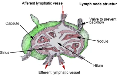
9 FIGURE-2 Lymph node structure
A typical lymph node is surrounded by a connective tissue capsule and is
divided into compartments called lymph nodules. The lymph nodules are dense
masses of lymphocytes and macrophages, separated by spaces called lymph
sinuses. The afferent lymphatics enter the node at different parts of its periphery,
which carry lymph into the node; entering the node on the convex side. The lymph
moves through the lymph sinuses and enters an efferent lymphatic vessel, located at
10 Anatomy of the axillary lymph nodes
The lymphatic vessels of the upper limb, most of those from the breast, and
cutaneous lymph vessels of the trunk above the level of umbilicus drain to the
axillary lymph nodes. The primary route of lymphatic drainage of the breast is
through the axillary lymph nodes. Unfortunately the boundaries for this group of
lymph nodes found in the axilla are not well demarcated. Thus there has been
considerable variation in the names given to lymph node groups.
Anatomical classification of axillary lymph node groups
1. LATERAL - Above the axillary vein, drain the upper limb
2. ANTERIOR - Along the inferior border of the pectoralis minor drain most
of the breast
3. POSTERIOR - Subscapular nodes, drain the shoulder
4. CENTRAL - Near the base of the axilla receive lymph from the preceding
three groups and form the most common palpable group.
5. APICAL – Lies medial to the axillary vein and superior to the pectoralis
minor. Receive lymph from all the other groups sometimes directly from the breast.
They drain into two or three subclavian trunks which enter the jugular-subclavian
venous confluence or join a common lymphatic duct or empty into lower deep
11
Surgical classification of axillary lymph node groups
1. Axillary vein group
2. External mammary group,
3. Scapular group,
4. Central group,
5. Subclavicular group,
6. Inter pectoral or Rotter s group
Surgeons also define the axillary lymph nodes with respect to their
relationship with the pectoralis minor muscle.
Level I - Lymph nodes located lateral or below the lower border of the
pectoralis minor muscle and include external mammary, scapular and axillary vein
nodal groups
Level II- Lymph nodes deep or posterior to the pectoralis minor and include
central and some of the subclavicular lymph node group
Level III - Lymph nodes medial or superior to the upper border of the
12 Lymphatic system of the breast
The lymphatics of the breast form an extensive and complex network of
periductal and perilobular vessels which drain principally to the axillary nodes. The
lymphatic vessels occur in three interconnecting groups (6)
1. Primary set of channels within the gland
2. Sub areolar plexus
3. A plexus on the deep surface of the breast
Although the internal mammary nodes were recognized by Handley as a
primary route for lymphatic drainage from medial and central zones of the
breast(7), the majority of the breast cancer metastasise to the axillary nodes
irrespective of the index quadrant(8). Fewer than 10% of the node positive tumours
exclusively affect the internal mammary nodes (9). Accessory pathways of
lymphatic drainage routes assume importance in more advanced states of disease
when the axillary drainage routes become obstructed (10) and include the following
routes,
1. Sub sternal crossover (contralateral internal mammary chain)(11)
2. Presternal cross over (contralateral breast)
3. Mediastinal (12)
13 Axillary lymph node dissection (ALND)
ALND refers to removal of lymph nodes in the axilla. A complete ALND
refers to extirpation of the lymph nodes from all the three levels. A partial ALND
refers to removal of lymph nodes from level I and level II, and Axillary sampling
indicates only resection of level I nodes.
The axilla is a pyramidal space with an apex directed into the root of the
neck; base bounded in front by the anterior axillary fold, which consists of the two
pectoral muscles, the subclavius muscle and behind by the posterior axillary fold,
formed by Teres major and Latissmus dorsi muscle and medially by the chest wall.
The axillary tissue is composed of adipose tissue and lymph nodes.
Steps of axillary dissection
1. Definition of pectoralis muscles
The lateral edge of the pectoralis major muscle is retracted medially
to expose the pectoralis minor muscle and allow interpectoral dissection of Rotter’s
lymph nodes.
2. Identification of Latissmus dorsi
The anterior surface of the Latissmus dorsi muscle is the lateral
border of axillary dissection.
14
The clavipectoral fascia is divided at the level of inferior axillary
sheath to expose the underlying fat pad and axillary lymph nodes within the fat.
4. Axillary vein
Dissection is carried posteriorly until the axillary vein is exposed.( 13)
5. Mobilization of axillary fat pad
The fatty tissue and lymph nodes cleared off the chest wall and the
inferior surface the axillary vein.
6. Identification of nerves
The position of thoraco dorsal nerve and long thoracic nerve can vary
from patient to patient. The best approach is to preserve all neuro vascular
structures until full axillary anatomy can be determined. The thoraco dorsal neuro
vascular pedicle can be identified by dissecting in the mid axilla. The long thoracic
nerve can be identified by blunt dissection just below the medial aspect of the
axillary vein and lateral to the chest wall.
Extent of dissection
In general level I and level II anatomic ALND is the preferred procedure for
axillary assessment (14). Typical level I/II dissection should yield more than 10
axillary nodes.(15-16)
15
maximize local control only if grossly positive lymph nodes are identified intra
operatively at level I and level II. Level III lymph node dissection increases the
morbidity of ALND (17).
Completion of ALND -Once the thoraco dorsal and long thoracic nerves are
identified, contents of the axilla can be removed from the defined boundaries.
Methods of axillary node sampling
The recognition that axillary dissection was principally a staging procedure
with concomitant morbidity led to investigation of alternative procedures for
staging the axilla these included 1. Four node axillary sampling, 2.sentinel node
biopsy
Four node axillary sampling
Axillary sampling was introduced more than two decades ago by Sir
Patrick Forrest (18) the original technique of blind four node sampling of axilla
could stage with estimated accuracy of 97%(19). This blind four node sampling is
not associated with impaired loco regional control or any detriment in overall
survival. (20) For patients found to be node positive on sampling, the axilla can be
irradiated or surgically cleared (21)
16
The sentinel node hypothesis presupposes an orderly spread of cancer cell
to the sentinel node then to the higher echelon nodes. If the sentinel node is
free from metastases, the non-sentinel nodes are presumed to be tumour free.
History of sentinel node concept
The concept of sentinel node biopsy is based on two basic principles: the
existence of an orderly and predictable pattern of lymphatic drainage to a regional
lymph node basin, and the functioning of a first echelon lymph node as an effective
filter for tumour cells. With the wide spread use of sentinel node biopsy, sufficient
data was needed to prove that sequential lymphatic dissemination and entrapment
of tumour cells in first draining lymph nodes does occur.
The concept of selecting a single lymph node to represent a nodal basin is
very old. In 1951 Gould sent a normal appearing node at the junction of anterior
and posterior facial vein for frozen. Intraoperative examination of this lymph node
in its typical anatomical location guided the decision to perform a radical neck
dissection during the following parotidectomies (22).
Two decades later, Cabañas observed the existence of sentinel lymph node
in carcinoma penis around the superficial epigastric vein in 1977 (23). These nodes
were identified with lymphangiography. Kett et al administered contrast medium in
breast lymphatics that were visualised with the aid of areolar blue dye injection
(24). They observed flow to an isolated lymph node, called the ‘Sorgius’ node, and
17
collecting system around the axillary vein. Using breast lymphoscintigraphy in
1980, Christensen et al observed primary draining nodes [25]. Haagensen studied
the route of metastases through the axillary lymph node and stated that the nodes of
the central group are most often exclusively involved [26]. He used the term
sentinel node for specific lymph nodes of the inferior deep cervical group.
Morton et al have used cutaneous lymphoscintigraphy with colloidal gold
since 1977 to identify the lymphatic drainage pattern of melanomas located at
ambiguous sites [27]. In addition to this preoperative procedure, they also
developed a technique for intraoperative mapping to selectively remove lymph
nodes on the direct drainage pathway from the primary melanoma. This sentinel
node was considered to be the first site of metastatic disease. The work of the group
at the John Wayne Cancer Institute initiated one of the most interesting recent
developments in surgical oncology (28). Giuliano applied the concept of a negative
lymph node to eliminate the need for radical dissection (29).
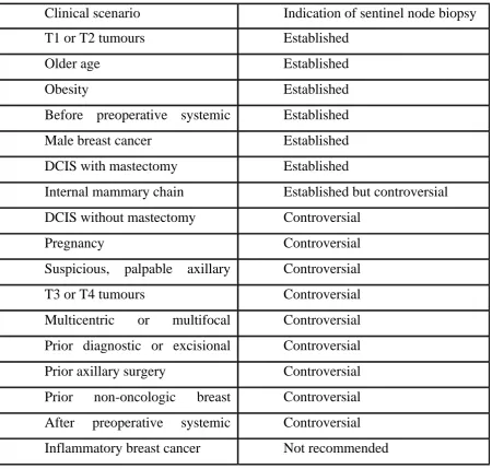
Indications and contraindications of axillary sentinel node biopsy in breast cancer
Clinical Indications of sentinel node biopsy have been changing and still
there is debate on some of them. Sentinel node biopsy is indicated in most of the
women with clinically node negative axilla with invasive or micro invasive breast
cancer (30).Some of the current indications and recommendations are given below
18 TABLE-1
{DCIS - ductal carcinoma in situ; Controversial - indications suggest that the
it is not universally accepted or the evidence behind the practice is limited.}
Contraindications
1.Prior axillary surgery
2.DCIS when lumpectomy is planned
Clinical scenario Indication of sentinel node biopsy
T1 or T2 tumours Established
Older age Established
Obesity Established
Before preoperative systemic Established
Male breast cancer Established
DCIS with mastectomy Established
Internal mammary chain Established but controversial
DCIS without mastectomy Controversial
Pregnancy Controversial
Suspicious, palpable axillary Controversial
T3 or T4 tumours Controversial
Multicentric or multifocal Controversial
Prior diagnostic or excisional Controversial
Prior axillary surgery Controversial
Prior non-oncologic breast Controversial
After preoperative systemic Controversial
19 3.Prior breast surgery
4. Pregnancy
5. Inflammatory breast cancer.
Role of ultrasound in sentinel node evaluation
For women with clinically suspicious axillary nodes, axillary ultrasound
with fine needle aspiration or core needle biopsy provides a mean to identify the
patients who have positive nodes and thus need ALND rather than the SLND.The
efficacy of this approach is variable between centres because the accuracy of
ultrasound examination is operator dependent (32)
Lymphatic mapping techniques for sentinel node biopsy
Given the rapid growth of lymphatic mapping and sentinel node biopsy
surgical groups have developed several variations and many technical aspects are
evolving which include
Choice of mapping label
Radio isotope quantity and processing
Injection site
Timing of injection
The use of preoperative lymphoscintigraphy.
20 Choice of mapping label
Radio isotope
Blue dye
Both radio isotope and blue dye
Radio isotope
Krag and colleagues first described the use of radioisotope alone for breast
cancer in 1993, using technetium-99m sulphur colloid and a hand held gamma
probe(33). The sentinel node identification rate was 98% with a false negative rate
of 11%. Technetium-99 m sulphur colloid is the most commonly used isotope for
lymphatic mapping in United States. In Europe technetium-99 m colloidal albumin
is used. The dose range from -0.1 to 4 mCi
Blue dye
Isosulfan blue dye (Lymphazurin 1%) was studied extensively in
melanoma. The use of Isosulfan blue dye as a single agent in sentinel node biopsy
for carcinoma breast was reported by Giuliano and colleagues(34). The
disadvantage is the risk of life threatening allergic and anaphylactic reactions. The
reported allergic reactions rate ranges from 1% to 3%. They include utricaria,rash,
pruritus, hypotension and anaphylaxis. Overall isosulfan blue dye has excellent
21
Methylene blue dye has also been successful in lymphatic mapping for
breast cancer. Simmons et al used methylene blue in the identification of lymphatic
mapping in breast cancer (35). Methylene blue was compared with Isosulfan blue
by Blessing et al in 2002 and found similar identification rates (36).Methylene blue
is preferred by some authors because of its lower cost and less complications.
Methylene blue must be injected subcutaneously. Inadvertent injection into the
dermis has resulted in severe skin reactions including necrosis and dermolysis.
Combination of blue dye and radioisotope
The combination of blue dye and radioisotope improves the sentinel node
identification rate. Albertini et al first reported the successful use of lymphatic
mapping with blue dye and radioisotopes prospectively (37).The results confirmed
that the combination improves the sentinel node identification rate and dual agent
lymphatic mapping has been accepted universally (38). Some centres have selected
to rely on radioisotope mapping alone, given the potentially life-threatening allergic
reactions of isosulfan blue dye.
Injection site for mapping agents
1. Peritumoural injection.
22 Peritumoural injection
Peritumoural injection replicates the intramammary lymphatic pathway that
has been traversed by metastases. The initial data regarding sentinel node biopsy
used peritumoural injection. For patients who have nonpalpable tumours this
method has proven to be difficult and time consuming as it requires the use of
additional imaging modalities to guide the Peri-tumoural injection of radioisotopes.
Peri-tumoural injections also have a higher potential for shine through where
residual radioactivity from the peri-tumoural injection site creates misleading
background activity detected by the gamma probe in the axilla. It is for these
reasons that alternate injection sites have been tried.
Subareolar and dermal injection
Nearly all breast tissue lymphatics pass through the sub areolar plexus of
Sappey and then to the axillary basin. Non palpable and multicentric tumours can
be studied with sub areolar and dermal injection and they do not have the shine
through effect. Sub areolar and dermal injection of blue dye may cause
considerable skin discoloration which may last for months. In addition up to 10%
of breast cancers may demonstrate non axillary lymphatic drainage with sentinel
nodes found in the internal mammary or supraclavicular nodal basins; so, not all
breast tumours will have the same drainage patterns as the overlying skin and
23 Preoperative lymphoscintigraphy
Patients undergoing lymphatic mapping with radioisotopes most often
receive a
pre-operative lymphoscintigram,to aid in sentinel lymph node (SLN)
identification.
Pre-operative lymphoscintigram, consists of anterior and lateral view and
specific patient positioning to optimize transit time and drainage (40). Scanning is
done after 20 minutes and images are repeated till identification of SLN basin then
the patient is taken to the operating room for SLNB..McMasters and colleagues
(41) evaluated the role of pre-operative lymphoscintigram, in breast cancer. In the
study, a pre-operative lymphoscintigram, was performed in 348 of 588 patients, and
240 patients did not receive scans. The SLN was identified in 221 of the 240
patients who did not undergo preoperative scanning. In these patients, the
false-negative rate was 1.6%. The authors found no significant difference in the SLN
identification rate, false-negative rate, or number of SLNs removed between
patients receiving PL and those proceeding to operation without scanning.
Borgstein and colleagues(42) also studied the role of PL in breast
cancer patients. The authors found that the intraoperative gamma probe was more
sensitive in detecting radioactive nodes in the axilla than the PL, even when
delayed images were obtained. Data have continued to emerge questioning the
24
some centres have abandoned the technique, relying on the intra operative gamma
probe to detect radioactive SLNs.
Timing of radio isotope injection
Lymphatic mapping with radio isotopes is performed either as a 1 or 2 day
procedure.(43) The single day procedure requires isotope injection on the morning
of surgery followed by serial imaging 1 to several hours after injection until the
SLN is identified. The effect of delay on patients and on operating room has led to
use a 2 day mapping procedure, with injection of radio isotopes 1 day before
operation. Based on the current literature, 2 day lymphatic mapping procedure is
safe and reliable method of SLN identification.(44)
Technique
Blue dye
The patient is prepped and draped in the operating room. The surgeon injects
3-5ml of blue dye peritumourally or sub dermally
The axillary fascia is entered through an inferior axillary incision. Careful
search is made for lymphatic channels leading to blue stained node. All blue nodes
and nodes at the end of the blue lymphatic channel are removed .Care must be
taken to identify the most blue node and the blue node most proximal to the tumour
in the axilla , because the dye transit time is rapid and blue staining of distal,
25
blue lymphatic channel as a sentinel node whether or not the node itself appears
blue is the most common technical error (45).
Radioactive colloid (46)
Technetium-99m-labeled colloids with a particle size of 20 –100 nm and an
injection volume of 0.2–1.0 mL generally are used. The amount of radioactivity
has to be determined according to the time between radio-colloid injection and the
surgical procedure to obtain a sufficient radio-colloid uptake in the lymph node and
a high target signal intensity.
If surgery is planned 24 hours after the injection and a probe with a
sensitivity of 10 cps/kBq is used, then activity of 150 –250 megabecquerel (MBq)
will be needed. With a nuclide half-life of 6 hours (Technetium-99) and an
injection time 6 hours prior to SLNB, the amount of radioactivity required is
reduced by 50%. In 1-day protocols, 10 –50 MBq are sufficient. Tracers may be
injected into the peritumoural tissue, intratumourally, intradermally or in the
subareolar region. The patient lies supine for imaging on the gamma-camera bed
3hours after the injection of tracer .Anterior and 45° anterior oblique static
scintigraphic images are obtained by using a dual head gamma camera with low
26
localised on overlying skin on the 45° anterior oblique image and the skin of the
patient on the identical site marked with a small spot of indelible ink.
The first “hot spot” detected on images has to be considered as the sentinel
lymph node. During the operation, the surgeon guided by the skin mark will locate
the lymph node with the highest radioactivity, with the hand held gamma probe. All
nodes with radioactive counts more than 10 times the back ground count,as
measured in the ante cubital fossa using hand held gamma probe are identified as
sentinel nodes.(47). If there are two or more such lymph nodes, all should be
removed. Before sending for histological examination, any lymph node removed
should be re-checked by the probe to demonstrate that they are radioactive.(48)
The disadvantages of SLNB
1. Skip metastasis alter management predictions. 2. Learning curve
3. Radionuclide retention in tissues. 4. Failure to identify sentinel nodes
Pathological evaluation of SLNs
Pathological assessment of the SLN is done both intra operatively and post
operatively. Intraoperative assessment permits axillary surgery in a single session if
27
evaluation. The postoperative examination provides the final histopathologic status
of the SLN for the definitive decision to be taken for further loco regional and
systemic treatment.
Intra operative evaluation
Intra operative examination, at the beginning of SLN era was done only to
evaluate the nodal tissue, because the tissue removed may not represent true lymph
node .Intra operative evaluation is used only if it influences immediate
management. Both frozen sections and imprint have been very efficient in detecting
metastases intra operatively (50)
Frozen section
Each SLN is grossly sectioned at 0.2 cm interval portions. The most
superficial 25% of the thickness of each resulting 0.2 cm SLN tissue section is
processed for frozen section analysis, providing at least three separate levels of
tissue for frozen section analysis. These frozen sections are then hand-stained by
routine Hematoxylin and Eosin (H&E) staining. The remaining tissue of each
resulting 0.2 cm SLN tissue section, encompassing 75% of the thickness of that
tissue, is then sent for routine processing. (51)
Imprint cytology
Excised lymph nodes submitted for intraoperative evaluation are examined
28
evaluation. In the first method, the SLN is bisected along the long axis. Care is
taken to obtain complete cross sections of the maximum diameter, preferably
including the hilum and the marginal sinus. For each lymph node half, a pair of
imprints is made by gently touching the cut surface of the SLN to a glass slide. One
imprint from each pair is air dried and stained with the Diff-Quik stain. The second
imprint from each surface is immediately fixed in 95% ethanol for 3 minutes and
then stained with hematoxylin and eosin (H&E). In the second method, the SLN is
sliced into 4-mm slices and imprints are made of each cut surface, air-dried, and
stained with Diff-Quik(52)
Post-operative examination
The extent and meticulousness of the histopathologic work-up of SLNs
should depend on the relevance of the results for further treatment decisions. The
prognostic significance of micrometastasis, their predictive role for the involvement
of non-SLNs, and the clinical consequences of a false-negative staging due to
undetected micrometastasis still are being debated. However, the clinical
implications of micrometastatic involvement of SLNs do not justify a systematic
search for all micrometastasis. There is no doubt that all macrometastasis should be
detected during the definitive pathologic examination.
1embedding of the lymph node and a homogeneous distribution of the levels
histologically examined. This can be achieved by producing SLN slices of 3 mm
29
negative macroscopic findings, the slices should be examined at regular, 500um
intervals (maximum, 6 steps). This approach provides a theoretical detection rate
for macrometastasis of 100% (53).
Immune histochemistry (IHC)
The use of cytokeratin may facilitate pathological finding. The use of
immunohistochemistry in the evaluation of SLNs is controversial. If the aim is to
correctly stage the disease with the exclusion of micrometastasis then the IHC
should be used.
Molecular pathology
Molecular methods using reverse transcriptase polymerase chain reaction
have been used to detect the tumour cell. Reverse transcriptase-polymerase chain
reaction analysis is a highly sensitive method. However, it is difficult to find a
RNA sequence that is specific for single tumours. Hence, it may be necessary to
use a panel of markers. High sensitivity, however, is associated with the risk of
false-positive result
Results from the American College Of Surgeons Oncology
30
overall survival.(54) Thus routine IHC or PCR is not recommended for the
valuation of SLNs in guidelines published by ASCO 2014,NCCN 3.2014 and
others.(55)
Documentation of histopathological findings
SLNB requires standardized documentation of the histologic findings. Every
SLN must be sent separately for histopathologic examination with a specific
identification number. The location and sequence of removal and the method of
detection (including target count)should be documented by the surgeon. The
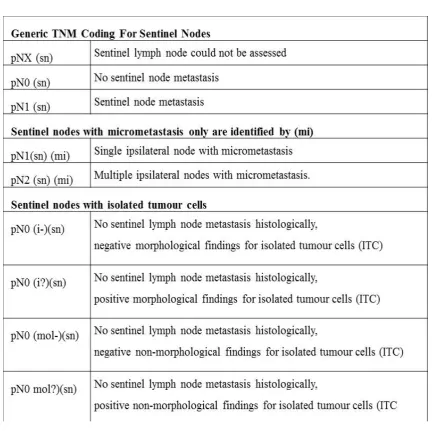
histopathologic results are reported as the pathologic lymph node (pN) category
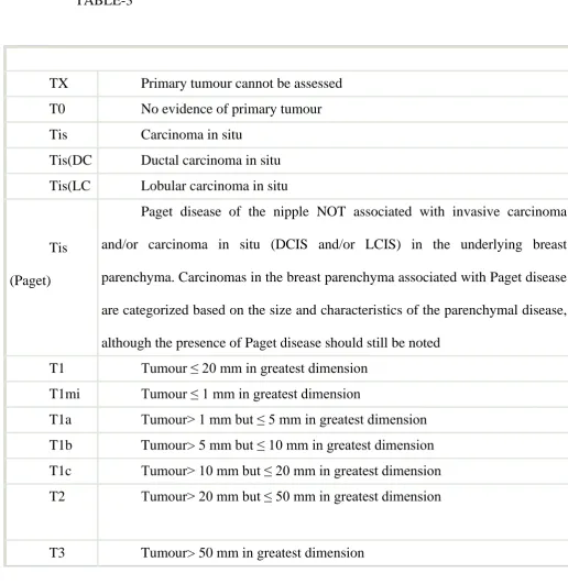
31 STAGING OF CARCINOMA BREAST
International Union Against Cancer (UICC) TNM classification system and
[image:37.595.54.571.216.748.2]by the American Joint Committee on Cancer(AJCC) 7th edition
TABLE-3
TX Primary tumour cannot be assessed
T0 No evidence of primary tumour
Tis Carcinoma in situ
Tis(DC Ductal carcinoma in situ
Tis(LC Lobular carcinoma in situ
Tis
(Paget)
Paget disease of the nipple NOT associated with invasive carcinoma
and/or carcinoma in situ (DCIS and/or LCIS) in the underlying breast
parenchyma. Carcinomas in the breast parenchyma associated with Paget disease
are categorized based on the size and characteristics of the parenchymal disease,
although the presence of Paget disease should still be noted
T1 Tumour ≤ 20 mm in greatest dimension
T1mi Tumour ≤ 1 mm in greatest dimension
T1a Tumour> 1 mm but ≤ 5 mm in greatest dimension
T1b Tumour> 5 mm but ≤ 10 mm in greatest dimension
T1c Tumour> 10 mm but ≤ 20 mm in greatest dimension
T2 Tumour> 20 mm but ≤ 50 mm in greatest dimension
32
T4 Tumour of any size with direct extension to the chest wall and/or to the
skin (ulceration or skin nodules)
T4a Extension to chest wall, not including only pectoralis muscle
adherence/invasion
T4b Ulceration and/or ipsilateral satellite nodules and/or edema (including
peaud’orange) of the skin, which do not meet the criteria for inflammatory
carcinoma
T4c Both T4a and T4b
T4d Inflammatory carcinoma
Regional lymph nodes (N)
Clinical
NX Regional lymph nodes cannot be assessed (eg, previously removed)
N0 No regional lymph node metastasis
N1 Metastasis to movable ipsilateral level I, II axillary lymph node(s)
N2
Metastases in ipsilateral level I, II axillary lymph nodes that are clinically
fixed or matted or in clinically detected* ipsilateral internal mammary nodes in
the absence of clinically evident axillary lymph node metastasis
N2a Metastases in ipsilateral level I, II axillary lymph nodes fixed to one
33
N2b Metastases only in clinically detected* ipsilateral internal mammary
nodes and in the absence of clinically evident level I, II axillary lymph node N3 Metastases in ipsilateral infraclavicular (level III axillary) lymph node(s),
N3a Metastasis in ipsilateral infraclavicular lymph node(s)
N3b Metastasis in ipsilateral internal mammary lymph node(s) and axillary
N3c Metastasis in ipsilateral supraclavicular lymph node(s)
*"Clinically detected" is defined as detected by imaging studies (excluding
lymphoscintigraphy) or by clinical examination and having characteristics highly suspicious for
malignancy or a presumed pathologic macrometastasis on the basis of fine-needle aspiration
(FNA) biopsy with cytologic examination.
Pathologic (pN)*
Pnx
Regional lymph nodes cannot be assessed (for example, previously
34 pN0
No regional lymph node metastasis identified
histologically. Note: Isolated tumour cell clusters (ITCs) are defined as small
clusters of cells ≤ 0.2 mm, or single tumour cells, or a cluster of < 200 cells in a
single histologic cross-section; ITCs may be detected by routine histology or by
immunohistochemical (IHC) methods; nodes containing only ITCs are excluded
from the total positive node count for purposes of N classification but should be
included in the total number of nodes evaluated
pN0(i-) No regional lymph node metastases histologically, negative IHC
pN0(i+
)
Malignant cells in regional lymph node(s) ≤ 0.2 mm (detected by
haematoxylin-eosin [H&E] stain or IHC, including ITC)
pN0(m
ol-)
No regional lymph node metastases histologically, negative molecular
findings (reverse transcriptase polymerase chain reaction [RT-PCR])
pN0(m
ol+)
Positive molecular findings (RT-PCR) but no regional lymph node
metastases detected by histology or IHC
pN1
Micro metastases; or metastases in 1-3 axillary lymph nodes and/or in
internal mammary nodes, with metastases detected by sentinel lymph node
35
pN1mi Micro metastases (> 0.2 mm and/or > 200 cells, but none > 2.0 mm)
pN1a Metastases in 1-3 axillary lymph nodes (at least 1 metastasis > 2.0 mm)
pN1b
Metastases in internal mammary nodes, with micro metastases or macro
metastases detected by sentinel lymph node biopsy but not clinically detected†
pN1c
Metastases in 1-3 axillary lymph nodes and in internal mammary lymph
nodes, with micrometastases or macrometastases detected by sentinel lymph
node biopsy but not clinically detected†
pN2
Metastases in 4-9 axillary lymph nodes or in clinically detected‡ internal
mammary lymph nodes in the absence of axillary lymph node metastases
pN2a
Metastases in 4-9 axillary lymph nodes (at least 1 tumour deposit > 2.0
mm)
pN2b
Metastases in clinically detected‡ internal mammary lymph nodes in the
absence of axillary lymph node metastases
pN3
Metastases in ≥ 10 axillary lymph nodes; or in infraclavicular (level III
axillary) lymph nodes; or in clinically detected‡ ipsilateral internal mammary
lymph nodes in the presence of ≥ 1 positive level I, II axillary lymph nodes; or in
> 3 axillary lymph nodes and in internal mammary lymph nodes, with
36
not clinically detected†; or in ipsilateral supraclavicular lymph nodes
pN3a
Metastases in ≥ 10 axillary lymph nodes (at least 1 tumour deposit > 2.0
mm); or metastases to the infraclavicular (level III axillary lymph) nodes
pN3b
Metastases in clinically detected‡ ipsilateral internal mammary lymph
nodes in the presence of ≥ 1 positive axillary lymph nodes; or in > 3 axillary
lymph nodes and in internal mammary lymph nodes, with micrometastases or
macrometastases detected by sentinel lymph node biopsy but not clinically
detected†
pN3c Metastases in ipsilateral supraclavicular lymph nodes
*Classification is based on axillary lymph node dissection, with or without sentinel lymph
Distant metastasis (M)
M0 No clinical or radiographic evidence of distant metastasis
cM0(i+
)
No clinical or radiographic evidence of distant metastases, but deposits of
molecularly or microscopically detected tumour cells in circulating blood, bone
marrow, or other non regional nodal tissue that are no larger than 0.2 mm in a
patient without symptoms or signs of metastases
M1 Distant detectable metastases as determined by classic clinical and
37 TABLE-4
Management of positive sentinel node.
Isolated tumour cells
The seventh edition of the American Joint Committee on Cancer (AJCC)
tumour node metastasis (TNM) staging system for breast cancer includes a
38
single cells. Small clusters of cells not greater than 0.2 mm, or non-confluent or
nearly confluent clusters of cells not exceeding 200 cells in a single histologic
lymph node cross section, are classified as isolated tumour cells and are considered
prognostically similar to node negative patients.
Malignant cells in regional lymph node(s) no greater than 0.2 mm are
designated as pN0(i+). Isolated tumour cells are not considered an indication for
further axillary surgery, radiation treatment, or adjuvant systemic therapy. The
finding of isolated tumour cells in lymphatics as a result of iatrogenic displacement
from core biopsy procedures has been observed and is not considered to be
clinically significant.(56-57)
Micrometastases — SLND allows the pathologist to perform a more detailed study of few lymph nodes that most likely contain metastases this has the
potential to improve the staging accuracy, but with increase in the rate of
identification of micro metastatic nodal involvement(58). For this reason, there is a
separate designation of pN1mi (>0.2 mm and no greater than 2.0 mm) to indicate
the micrometastasis alone.
Although patients with micrometastasis are considered node positive, this
finding does not change the survival compared with those without
micrometastasis.(59).However some analyses suggest a negative impact of micro
metastases on outcomes including regional recurrence rate. Pathological evaluation
39
or SLND plus ALND for invasive breast cancer detected occult metastases in 16%
of patients (60).
In the management of positive SLN nodes, there is general acceptance of
the following approaches.
Patients with SLN showing isolated tumour cells are considered node
negative and completion ALND is not indicated (61)
In patients with a positive SLN showing micrometastasis or
macrometastasis in three or more nodes detected with standard hematoxylin
& eosin examination , completion ALND is indicated(62). The timing of
the procedure immediate versus delayed does not seem to affect the total
lymph node yield or the rate of long term complications(63)
Completion ALND in patients showing micrometastasis or macrometastasis in less
than three nodes may not be necessary because the SLN is the sole tumour-bearing
node in up to 60 percent of cases overall, and in almost 90 percent of patients who
harbour only micrometastatic disease. These observations have led to speculation
that completion ALND may not be necessary in selected patients with a positive
SLND in less than three nodes because the need for systemic therapy is established
(64) and the risk of an axillary recurrence appears to be low(65-66).
A retrospective review of studies including randomized and prospective
trials with at least two years follow up supports a completion ALND in select
40
1. Palpable or needle proven axillary metastases
2. SLN positive undergoing mastectomy without radiation therapy
3. Three or more SLN positive
Important trials and current guidelines in the management of SLN positive patients.
1. ACOSOG Z –0010
A multicenter prognostic study of sentinel node (SN) and bone marrow
(BM) micrometastases in women with clinical T1/T2 N0 M0 breast cancer
Patients underwent breast conservation surgery and SNB with bilateral iliac
crest BM aspiration. BM and histologically negative SN were evaluated with IHC.
This study evaluated prognostic significance of metastases detected in bone
marrow by IHC and metastases detected in SLN by routine histopathology and
IHC
In multivariate analysis there was no difference in Overall survival (OS),
disease-free survival, and loco regional recurrence in patients tested negative for
metastases in SLN by both IHC& histopathology and positive for metastases
41
In a subset analysis it was seen that there was no significant difference in the
rate of sentinel node identification with the use of blue dye alone or radio colloid
alone or the combination of the two
This study, questions the routine examination of SN by IHC.(68)
2.ACOSOG Z –0011
This study was designed to address the need for completion ALND for
patient with less than three SLN positive. All were treated with breast conservation
surgery and whole breast radiation therapy (69).Women with clinically T1, T2, N0
and a positive sentinel node were randomly assigned into two arms, ALND versus
No ALND. Primary end point was Overall Survival (OS) and secondary end
points were axillary recurrence and surgical morbidities
The results of the trial included, at a median follow-up of 6.3 years, no
significant differences in survival or loco regional recurrence between the SLND
with ALND group versus the SLND alone group. The five-year overall survival
was similar whether women were treated with SLND plus ALND or with SLND
alone (91.9 versus 92.5 percent, respectively) (HR 0.79, 90% CI 0.56-1.10).The
five-year disease-free survival was also similar between the two arms (82.2 versus
83.9 percent, respectively).Recurrence rates in the ipsilateral axilla were similar
between the two arms with four recurrences (0.9 percent) in the SLND alone arm
42
Women in the ALND group had a significantly greater incidence of postoperative
complications than did the SLN-only group (70% vs25%).
The ACOSOG Z-0011 trial was criticized for a number of performance
deficiencies; including target accrual for ACOSOG Z-0011 study was 1900
patients. The study closed prematurely because of low accrual and low event rate
after enrolling 436 patients in the SLND alone arm and 420 in the SLND plus
ALND arm. The trial was not adequately powered to meet the predetermined
statistical survival primary endpoint due to low accrual. Eleven patients assigned to
the SLND only arm did have an ALND and 32 patients assigned to the SLND plus
ALND arm did not have an ALND. Almost 20 percent of patients were lost to
follow-up and 7 percent of patients in the SLND arm were found to be node
negative compared with 1 percent of the ALND arm. There was no breakdown of
the numbers of patients with isolated tumour cell clusters, micrometastasis or
macrometastasis in the two arms. The majority of patients had T1 (almost 70
percent), hormone receptor positive tumours (85 percent). Estrogen receptor status
and adjuvant systemic therapy were independent predictors of survival.
Based upon the apparent lack of regional benefit and low risk of events in
this trial, completion ALND may not be necessary for all women with T1 tumours
that are clinically node negative, treated with breast conservation surgery ,with less
than three positive SLNs, who will be treated with whole breast radiation,
43
This was also shown in a retrospective review of 242 consecutive patients
who met the ACOSOG Z0011 criteria (70).
When completion ALND is omitted in patients with a positive SLND,
whole breast radiotherapy is indicated. If partial breast radiation is planned,
completion ALND should be performed.
3. IBCSG 2301 trial- the international Breast Cancer Study group trial 2301
This trial analysed in patients with micro metastases in SLNs -ALND vs.
No ALND
Patients with SLN micrometastasis (<2 mm) and primary tumours <5 cm in
size were randomized to completion for ALND Vs. no additional axillary surgery
(71). The primary end-points were five-year disease-free survival (DFS) and
overall survival (OS) rates.
The results of the trial showed that with a median follow-up of 49 months,
there was no significant difference in DFS rate for patients treated with an ALND
compared with those treated with a SLND (87 versus 92 percent).There was no
significant difference in OS rate for patients treated with an ALND compared with
those treated with a SLND (97.6 versus 98.0 percent).The results of this trial, when
considered in the context of the ACOSOG Z0011 trial, offer additional support to
44
do well with SLND alone as compared with a completion ALND. However, the data are derived from studies that are underpowered and have relatively short follow-up.
3. AMAROS trial(After Mapping of the Axilla: Radiotherapy Or Surgery)
Radiotherapy or surgery of the axilla after a positive sentinel node in breast
cancer (EORTC 10981-22023AMAROS) (72) was studied
Patients were randomly assigned (1:1) by a computer-generated allocation
schedule to receive either axillary lymph node dissection or axillary radiotherapy in
case of a positive sentinel node, stratified by institution. The primary end point was
non-inferiority of 5-year axillary recurrence, considered to be not more than 4% for
the axillary radiotherapy group compared with an expected 2% in the axillary
lymph node dissection group.
5-year axillary recurrence was 0·43% (95% CI 0·00-0·92) after axillary
lymph node dissection versus 1·19% (0·31-2·08) after axillary radiotherapy. The
planned non-inferiority test was underpowered because of the low number of
events. Lymphoedema in the ipsilateral arm was noted significantly more often
after axillary lymph node dissection than after axillary radiotherapy at 1 year, 3
years, and 5 years.
Results prove Axillary lymph node dissection and axillary radiotherapy after
a positive sentinel node provide excellent and comparable axillary control for
patients with T1-2 primary breast cancer and no palpable lymphadenopathy.
45
4. NCCN guidelines version 3.2014– recommendation
ALND may be avoided in patients who have positive SLN provided all the following criteria are met
1. T1 or T2 tumours
2. 1 or 2 positive sentinel nodes
3. No neo adjuvant chemo therapy
4. Breast conservation therapy
5. Whole breast Radiation Therapy is planned
ALND should be performed even if one of the above criteria is not met
ASCO guidelines 2014(73)
In patient with less than three SLN positive node, completion ALND need
not be performed, provided there is no evidence of bulky metastatic disease, gross
extra capsular extension and the patient is treated with whole breast irradiation.
Complications of axillary lymph node dissection
1.Seroma
2.Arm morbidity
3. Infection
4. Hematoma 5. Nerve injury
1. Seroma – The normal lymphatic drainage of the breast to the axilla can lead to seroma formation after axillary dissection (78, 79). Seromas can be
46
2. Arm morbidity – Lymphedema of the arm is a potential and serious complication of ALND. The rate varies substantially with the level of
dissection and whether or not postoperative radiation therapy is
used. Shoulder stiffness, and numbness and paresthesias in the upper arm are
common complaints following an ALND. Although these symptoms do not
usually interfere with daily living, they may reduce the quality of life (80,
81).
3. Infection– The incidence of postoperative wound infection following axillary dissection varies from 3 to 15 percent in the literature (77).The most
common organisms are usually gram positive (streptococcal or staphylococcal
species) and will respond to treatment with appropriate oral antibiotics. If there is
an underlying seroma, it should be aspirated and cultured to direct antibiotic
treatment.
4.Hematoma – The reported incidence of postoperative hematoma has varied from 2 to 10 percent
5. Nerve injury- The risk of major motor nerve injury following an ALND is <1 percent. Injury to the long thoracic nerve results in a winged scapula.
Injury to the thoracodorsal nerve weakens shoulder abduction and internal rotation.
Injury to the medial pectoral nerve may lead to atrophy of the lateral aspect of the
47
of the intercostobrachial nerve results in numbness and paresthesias on the inner
upper arm.
Complications of sentinel node biopsy
1. Axillary paresthesia- 8.6% 2. Seroma-7.1%
3. Lymphedema-6.9%
4. Decreased range of motion-3.8% 5. Hematoma-1.4%
6.Axillary wound infection-1%
7.Axillary web syndrome.
Axillary paraesthesia was found in 8.6% presumably due to damage to the
inter-costobrachial nerve. Decreased range of motion was reported in 3.8% and
lymphedema in 6.9 %.( 74) Axillary web syndrome is the development of palpable
tender cords in the axilla antecubital fossa or upper arm It occurs after SNB but less
commonly than ALND. The pathogenesis proposed by research groups is
lymphovenous damage, hyper coagulation, superficial venous and lymphatic stasis
as well as disorders and injuries of tissues as a result of the disruption of superficial
lymphatics and vessels during axillary surgery. The condition can be treated by
physiotherapy and massage (75).
SLNB is associated with significantly less subjective and objective
48
biopsy is 2% to7% compared with a lifetime risk of lymphedema with ALND of
25%.Lucci et al12 compared the postoperative complication rate of women
undergoing SLN vs. SLN and ALND in the ACOSOG Z0011study. They found
that women in the ALND group had a significantly greater incidence of
postoperative complications than did the SLN-only group (70% vs. 25%).
Complications included wound infection, axillary seromas, axillary paresthesias,
brachial plexus injury, and lymphedema. As reported subjectively by study
participants, the development of lymphedema was 13% at 1 year in the ALND
group vs. 2% in the SLN-only group.
Axillary recurrence after SNB
Axillary recurrence is rare after negative sentinel node biopsy. In 2000 Giuiliano
and colleagues published the first prospective report of patients with negative SLNs
being treated without ALND After surgery, all decisions regarding systemic
adjuvant treatment and radiotherapy were based on conventional criteria. . With a
median follow up of 39 months there was no axillary recurrence (83). Naik and
colleagues (84) studied patients who underwent SNB and found axillary
recurrence rate 1.4% the rate of recurrence was higher in patients who had
positive SLNs and did not undergo ALND .Axillary recurrence can be treated with
ALND(85) .
49
The false rates of SLN biopsy has been reported to be 6.7%to 9.7% .A false
negative rate may increase the risk of axillary recurrence .In the NSABP-32 trial,
the site of the tumour, the type of biopsy, and the number of SLNs removed all
affected the false negative rate. Studies have confirmed that combination of blue
dye and radio colloid has the highest sentinel node identification rate and the lowest
false negative rate (86). A previous excisional biopsy has the potential to increase
the false negative rate, as the dye or radioactive material may enter the seroma
cavity.
Special considerations
Sentinel node biopsy in prophylactic mastectomy
Some surgeons perform SNB for prophylactic mastectomy because of the
possibility of finding an occult carcinoma in the breast upon pathological
evaluation. If invasive carcinoma is found after the surgery SNB is no longer
possible and ALND should be performed. King &Colleagues studied the use of
SNB in prophylactic mastectomy. Occult carcinoma was found in 8.0 %.
DuPont&Colleagues also published a series of patients with 3.5% occult
carcinoma in prophylactic mastectomy(87). Another consideration is in the event of
immediate reconstruction ALND may alter the cosmetic appearance or may
compromise the vascularity of the flap. These complications can be avoided with
SNB.
50
Patients with Ductal carcinoma in situ, who undergo breast conservation
surgery do not require axillary staging, however women with DCIS may be
candidates for SLN biopsy if they are undergoing mastectomy. Since it would be
essentially impossible to perform an SNB subsequently if invasive cancer is found
in the specimen which would necessitate an ALND. Axillary surgery would be
problematic even more if immediate reconstruction was completed with free flaps
or flaps with Trans-axillary pedicles. For these reasons SNB is indicated for
patients undergoing mastectomy for DCIS.
Sentinel node biopsy and large tumours
Most of the initial trials with SNB were for small tumours .Chung and
Colleagues(88) performed SNB in tumours >5cm size .SNB accurately predicted
the nodal status with false negative rate of 3%. Large tumours have high incidence
of axillary metastases but with clinically node negative axilla, SNB may be
performed
Sentinel node biopsy after neoadjuvant chemotherapy
Sentinel lymph node biopsy after neoadjuvant chemotherapy in clinically
node positive breast cancer at initial presentation has been studied in many series.
In NSAPB B-27(89),a neoadjuvant trial requiring ALND ,some surgeons attempted
SNB before the required ALND. The overall success rate in sentinel node
identification was only 84.8% after neoadjuvant chemotherapy,with false negative
51
associated with tumour characteristics lymphatic mapping method (blue dye
,radioisotope or both) or the response to chemotherapy. The optimal timing of
sentinel node biopsy in patients receiving neoadjuvant therapy has been debated as
some have reported a higher false negative rate for SLND performed after
induction therapy while others not(90).
The SENTINA (SENTInelNeoAdjuvant) study (91) was designed to evaluate a specific algorithm for timing of a standardised sentinel-lymph-node
biopsy procedure in patients who undergo neoadjuvant chemotherapy. SENTINA
was a four-arm, prospective, multicentre cohort study undertaken at 103 institutions
in Germany and Austria.Women with breast cancer who were scheduled for
neoadjuvant chemotherapy were enrolled into the study. There were four arms in
the study
1.Patients with clinically node-negative disease (cN0) underwent
sentinel-lymph-node biopsy before neoadjuvant chemotherapy (arm A).
2. If the sentinel node was positive (pN1), a second sentinel-lymph-node
biopsy procedure was done after neoadjuvant chemotherapy (arm B).
3. Women with clinically node-positive disease (cN+) received neoadjuvant
chemotherapy. Those who converted to clinically node-negative disease after
chemotherapy (ycN0; arm C) were treated with sentinel-lymph-node biopsy and
52
4. Only patients whose clinical nodal status remained positive (ycN1)
underwent axillary dissection without sentinel-lymph-node biopsy (arm D).
The primary endpoint was accuracy (false-negative rate) of
sentinel-lymph-node biopsy after neoadjuvant chemotherapy for patients who converted from cN1
to ycN0 disease during neoadjuvant chemotherapy (arm C). Secondary endpoints
included comparison of the detection rate of sentinel-lymph-node biopsy before and
after neoadjuvant chemotherapy, and also the false-negative rate and detection rate
of sentinel-lymph-node biopsy after removal of the sentinel lymph node. Analyses
were done according to treatment received (per protocol).
The false-negative rate and detection rate of sentinel-lymph-node biopsy
after
removal of the sentinel lymph node. Analyses were done according to
treatment received (per protocol).
Results of the study of SENTINA trial include, sentinel-lymph-node biopsy
is a reliable diagnostic method before neo adjuvant chemotherapy. After systemic
treatment , the procedure has a lower detection rate and a higher false-negative rate
compared with sentinel-lymph-node biopsy done before neo adjuvant
chemotherapy. These limitations should be considered if biopsy is planned after
53
The 3.2014 NCCN guidelines recommend that SLND be performed prior to
neoadjuvant chemotherapy as it provides valuable prognostic information for
planning loco regional treatment (92)
ACOSOG Z 1071 found a 12.6% false negative rate in when SLB performed
post neoadjuvant chemotherapy (93). This limits the use of the procedure until
further refinement either in the technique or patient selection.
In 2014 ASCO guidelines indicate that there is insufficient data at present to
support the use of sentinel node biopsy after the neo adjuvant chemotherapy given
the high false negative rates in the published trials (94)
Sentinel node biopsy in clinically node positive axilla
Most of the studies in the SLN biopsy have excluded clinically suspicious
axillary nodes. It is well known that determination of the axillary lymph node
status by clinical examination alone is unreliable with positive predictive
value(PPV) of 64%-82% and a negative predictive value (NPV ) of 50%-63% with
overall accuracy of 63%-68%.(95)Specht&colleagues published a review of SNB
in patients with clinically suspicious axillary nodes(96) and concluded that the
clinical axillary examination in breast cancer is subject to false-positive results, and
is by itself insufficient justification for axillary lymph node dissection.
If other means of preoperative assessment such as palpation- or