EFFECTS OF LUMBAR STABILIZATION EXERCISES,
MCKENZIE EXERCISES AND CONVENTIONAL
EXERCISES ON PAIN, FUNCTION AND RANGE OF
MOTION IN PATIENTS WITH
MECHANICAL LOW BACK PAIN
-
A COMPARATIVE STUDY
Dissertation submitted to The Tamil Nadu Dr. M.G.R. Medical
University towards partial fulfillment of the requirements of
MASTER
OF PHYSIOTHERAPY (Advanced PT in Orthopaedics)
degree
programme.
KMCH COLLEGE OF PHYSIOTHERAPY
(A unit of Kovai Medical Center Research and Educational Trust)
Post Box No. 3209, Avanashi Road,
CERTIFICATE
This is to certify that research work entitled “EFFECTS OF LUMBAR
STABILIZATION EXERCISES, MCKENZIE EXERCISES, AND
CONVENTIONAL EXERCISES ON PAIN, FUNCTION, AND RANGE OF MOTION IN PATIENTS WITH MECHANICAL BACK PAIN”was carried out by the candidate bearing the Register No: 271410081, KMCH College of Physiotherapy towards partial fulfillment of the requirements of the Master of Physiotherapy (Advanced PT in Orthopaedics) of The Tamil Nadu Dr. M.G.R. Medical University, Chennai-32.
PROJECT GUIDE PRINCIPAL
Mr. A. David. V .Samuel, M.P.T (ortho) Dr. EDMUND M. D’COUTO Associate Professor M.B.B.S, M.D, Dip.PMR
KMCH College of Physiotherapy KMCH College of Physiotherapy
Coimbatore- 641014 Coimbatore- 641014
INTERNAL EXAMINER EXTERNAL EXAMINER
ACKNOWLEDGEMENT
First and foremost, I thank my beloved parents for their unconditional love, sincere prayers, unstinted support and care without which I would not have accomplished anything.
I thank my God for always watching upon me with grace to fulfill this
endeavor.
I thank the KMCH management, especially the chairman,
Dr.Nalla G. Palaniswami MD (AB), and the trustee Dr.Thavamani D Palaniswami MD (AB) F. A. A. P, for the wide variety of opportunities.
I thank Dr. O T Bhuvaneswaran, PhD, Chief Executive Officer, for his role in
the academic front.
I am delighted to express my profound thanks to our beloved principal,
Dr. Edmund Mark D’Couto, M.D, Phys. Med & Rehab, KMCH College of Physiotherapy, for being a pillar of encouragement and also providing us with all necessary infrastructure and other facilities.
I owe my sincere gratitude to my project guide Mr. David V Samuel, MPT
(Ortho), Associate Professor, KMCH College of Physiotherapy, for her remarkable
support, guidance, valuable suggestions, patience and motivation throughout the study. I am obliged to have her share her immense knowledge about the subject.
I deeply express my sincere thanks and gratitude to the versatile person,
Mrs. A.P.Kalpana, MPT (Cardio), Vice Principal, KMCH College of Physiotherapy, and my class incharge, for her valuable input in my study.
I express my gratitude towards Mr. S. SivaKumar, MPT (Ortho), PGDBS,
I thank Mrs.A.Brammatha, MPT(Neuro), M.H.R.M, Professor, KMCH College of Physiotherapy for her timely and valuable guidance.
I express my heartiest thanks in this instance to, Mrs. R. Uthra Devi, MPT (Neuro), for her valuable suggestions, support and encouragement.
I am also thankful to Mr. Prakash, MPT (Ortho), and all other faculty
members for their immense support and motivation throughout the study.
I extend my gratitude to Mr. K Venugopal, MA, MPhil, Professor, Research
& Statistics, for guiding me with the necessary tools to analyze my study without
which it was impossible to draw the inference.
I am thankful to Dr. S. Sreedharan Namboodhiri for his valuable suggestions and
also sharing his vast knowledge and experience for the accomplishment of this study.
I thank the faculty members for their guidance and willingness to clear all my doubts. Their suggestions have been really helpful.
I thank the librarians of this institute, Mr. P Dhamodaran and his fellow members for their cooperation. I sincerely acknowledge my best friends, batch mates, my seniors, and all my well wishers who were always there to guide and render their support to me throughout my project.
I am truly grateful to all my friends and batchmates for their selfless help and assistance in this study.
CONTENTS
S No. TITLE PAGE No.
ABSTRACT
1. INTRODUCTION
1.1 NEED OF STUDY
1.2 AIM & OBJECTIVES
a) AIM b) OBJECTIVES 1 3 4 4 4
2.
REVIEW OF LITERATURE2.1 MECHANNICAL LOW BACK PAIN
2.2 LUMBAR STABILIZATION EXERCISES
2.3 MCKENZIE EXERCISES
2.4 CONVENTIONAL EXERCISES
2.5 SCALES
a) NUMERICAL PAIN RATING SCALE
b) ROLAND MORRIS FUNCTIONAL IDSABILITY
QUESTIONNAIRE
c) MODIFIED SCHOBER’S TEST
5 5 6 7 9 9 10 10 11 3. METHODOLOGY
3.1 RESEARCH DESIGN
3.2 SAMPLING TECHNIQUE
3.3 STUDY POPULATION
3.4 SAMPLE SIZE
3.5 STUDY DURATION
3.6 STUDY SETTING
3.7 STUDY CRITERIA
a) INCLUSION CRITERIA
b) EXCLUSION CRITERIA
3.8 HYPOTHESIS
a) NULL HYPOTHESIS
3.9 OUTCOME MEASURE
3.10 MEASUREMENT TOOLS
3.11 PROCEDURE
a) INTERVENTION
b) INTERVENTION DURATION
3.12 PHOTOGRAPHIC REPRESENTATION
3.13 STATISTICAL TOOLS
14 15 23 24 29 4. DATA PRESENTATION
4.1 TABULAR PRESENTATION
4.1.1 PAIRED ‘t’ TEST-LUMBAR STABILISATION GROUP:
4.1.1.1 NUMERICAL PAIN RATING SCALE
4.1.1.2 ROLAND MORRIS DISABILITY
QUESTIONNAIRE
4.1.1.3 LUMBAR FLEXION RANGE OF MOTION
4.1.2 PAIRED ‘t’ TEST-MCKENZIE GROUP
4.1.2.1 NUMERICAL PAIN RATING SCALE
4.1.2.2 ROLAND MORRIS DISABILITY
QUESTIONNAIRE
4.1.2.3 LUMBAR FLEXION RANGE OF MOTION
4.1.3 PAIRED ‘t’ TEST-CONVENTIONAL GROUP
4.1.3.1 NUMERICAL PAIN RATING SCALE
4.1.3.2 ROLAND MORRIS DISABILITY
QUESTIONNAIRE
4.1.3.3 LUMBAR FLEXION RANGE OF MOTION
4.1.4 ONE WAY ANOVA
4.1.4.1 PRE TEST: NUMERICAL PAIN RATING SCALE
4.1.4.2 POST TEST: NUMERICAL PAIN RATING
SCALE
4.1.4.3 PRE TEST:ROLAND MORRIS DISABILITY
QUESTIONNAIRE
4.1.4.4 POST TEST: ROLAND MORRIS DISABILITY
QUESTIONNAIRE
4.1.4.5 PRE TEST:LUMBAR FLEXION RANGE OF
4.1.4.6 POST TEST:LUMBAR FLEXION RANGE OF
MOTION
4.2 GRAPHICAL REPRESENTATION
PAIRED ‘t’ TEST :LUMBAR STABILISATION GROUP
4.2.1: NUMERICAL PAIN RATING SCALE
4.2.2: ROLAND MORRIS DISABILITY QESTIONNAIRE
4.2.3: LUMBAR FLEXION RANGE OF MOTION
PAIRED ‘t’ TEST:MCKENZIE GROUP
4.2.4:NUMERICAL PAIN RATING SCALE
4.2.5: ROLAND MORRIS DISABILITY QESTIONNAIRE
4.2.6: LUMBAR FLEXION RANGE OF MOTION
PAIRED ‘t’ TEST:CONVENTIONAL GROUP
4.2.7: NUMERICAL PAIN RATING SCALE
4.2.8: ROLAND MORRIS DISABILITY QESTIONNAIRE
4.2.9: LUMBAR FLEXION RANGE OF MOTION
ONE WAY ANOVA
4.2.10 PRE TEST:NUMERICAL PAIN RATING SCALE
4.2.11 POST TEST:NUMERICAL PAIN RATING SCALE
4.2.12 PRE TEST:ROLAND MORRIS DISABILITY
QUESTIONNAIRE
4.2.13 POST TEST: ROLAND MORRIS DISABILITY
QUESTIONNAIRE
4.2.14 PRE TEST:LUMBAR FLEXION RANGE OF MOTION
4.2.15 POST TEST: LUMBAR FLEXION RANGE OF MOTION
34 35 35 35 35 36 36 36 37 37 38 38 38 39 39 39 40 40 41 41 42
5. DATA ANALYSIS AND RESULTS 43
6. DISCUSSION 48
7. SUMMARY AND CONCLUSION 51
8. LIMITATIONS AND SUGGESTIONS 52
10. APPENDICES
I. INFORMED CONSENT FORM
II. ASSESSMENT PERFORMA
III. NUMERICAL PAIN RATING SCALE
IV. MODIFIED SCHOBER’S TEST
V. ROLAND MORRIS FUNCTIONAL DISABILITY
QUESTIONNAIRE
VI. HOME EXERCISE PAMPHLET
VII. BACK CARE PROGRAMMES
ABSTRACT
OBJECTIVES:
To compare the effects of lumbar stabilization exercises, Mckenzie exercises
and conventional exercises on pain, function and lumbar range of motion in patients
with mechanical low back pain.
STUDY DESIGN:
Quasi experimental study design
STUDY SETTING:
Kovai Medical Centre and Hospital-Coimbatore
SAMPLE SIZE AND INTERVENTION:
21 patients with mechanical low back pain who met the inclusion criteria were
selected. The duration of the study was 4 weeks.21 patients diagnosed with
mechanical low back pain and age group 20-40 years, both males and females were
selected. Patients with pain level between 3 and 7 in the numerical pain rating scale
were included. 21 randomly allocated into 3 groups- experimental Group A,
experimental group B and control group C of 7 samples each. Group A received an
exercise pamphlet comprising of 5 lumbar stabilisation exercises, group B received an
exercise pamphlet comprising of 5 Mckenzie exercises and group C received an
exercise pamphlet comprising of 5 conventional exercises for mechanical low back
pain patients which were to be followed at home.
OUTCOME MEASURES:
Pain status Functional ability
MEASUREMENT TOOLS:
Numerical pain rating scale
Roland Morris functional disability Questionnaire Modified Schober’s test
CONCLUSION:
The data were analyzed using paired ‘t’ test and one way ANOVA at 5% level
of significance. The results of the study concluded that lumbar stabilization group is
better than the Mckenzie group and conventional group in reducing the pain,
improving the function and increasing the lumbar flexion range of motion.
KEYWORDS:
Mechanical low back pain Lumbar stabilization exercises Mckenzie exercises
Numerical pain rating scale
1.
INTRODUCTION
Back pain is an extremely common human phenomena, a price mankind has to
pay for their upright posture[34]. It is a neuro- musculoskeletal problem affecting 40% of population worldwide at some point of their life which causes significant disability
and loss in productivity. Furthermore, over 80% of low back pain patients report
recurrent episodes.
Mechanical low back pain is considered as one of the most frequently treated
disease in modern industrial societies and one of the leading cause of work
absenteeism[1].
The incidence of mechanical low back pain is higher in workers subjected to
heavy physical activities such as weight lifting, repetitive movements and frequent
static posture.
Mechanical low back pain can be described as a musculoskeletal pain which
varies with physical activities and not involving root compression or series of spinal
disease[2].
Causes include lumbar strain, herniated disks, spondylolysthesis, spinal
stenosis, spondylosis and fractures. Pain from mechanical causes is typically
aggravated with movement and relieved by rest[8].
Diagnosis of mechanical low back pain is made commonly by physical
examination, palpation, physical tests and imaging such as x rays, MRI, and CT scan.
Most commonly used management includes medications, physical therapy and
surgery. The physical therapy management varies according to the condition of the
patient and includes modalities, exercise therapy and patients education with a
comprehensive plan of care[3,4,].
Living sedentary life and lack of physical fitness makes human liable to back
pain. The cause of lower spine being so commonly affected could be due to inherent
skeletal abnormalities, poor posture, inability of lumbar spine musculature to control
Exercise therapy has three important goals. The first and most important goal of
exercise is to improve back flexibility and strength and to improve performance of
endurance activities. The second goal is to reduce the intensity of back pain. The third
and most important goal is the reduction of back pain related disability.
The Mckenzie method is considered to be a highly effective program for
patients with low back pain. It seems to be an effective technique in alleviating back
pain compared with other conservative treatment[6].
The core component of treatment in the Mckenzie method of exercise is the
sustained postures or repeated movements. It also includes other components such as
education and postural training.
Mechanical stability of the lumbar spine is an important consideration in low
back injury prevention and rehabilitation strategies[34]. Trunk stabilizing muscles (multifidus, transverse abdominis, internal oblique, erector spinae, rectus abdominis)
provide intersegmental stability to the low back. Imbalance between muscles can
result in instability of the spine leading to functional dysfunction[7].
Spinal instability is one of the cause of low back dysfunction and this instability
of the spine is associated with reduced strength and endurance of the trunk stabilizing
muscles and inappropriate recruitment of trunk muscles. So specific training of the
stabilizing muscles in low back pain patients are necessary[37].
The main goal of lumbar stabilization program is to built musculature that
stabilizes the torso, with co-contraction of abdominal muscles to provide corseting
1.1
NEED FOR STUDY
Low back pain is a major health issue with significant socioeconomic
implications in many western countries. Currently its prevalence in India is found to
be high.
Several treatment strategies like joint mobilization and manipulation,
electrotherapy, acupuncture, soft tissue massage techniques and traction are currently
utilized in clinical practice. There is ample evidence that active approaches to the
rehabilitation of low back patients are beneficial. In 2000,Van Tulder et al. published a Cochrane review describing the effectiveness of exercise therapy for low
back pain.
Systematic reviews have concluded that stabilization program appears to be
effective in some subgroups of patients with back pain. The individual effect of
lumbar stabilization exercise is evident in the management of mechanical low back
pain, but there is no single study comparing the effectiveness of lumbar stabilization
exercises with Mckenzie exercises and conventional exercises.
Evidence based researches showed that the Mckenzie approach resulted in a
greater decrease in pain and disability in patients with low back pain. But no study
directly compared the effectiveness of Mckenzie exercise with lumbar stabilization
exercises and conventional exercises in patients with mechanical low back pain.
So this study intends to compare the efficacy of lumbar stabilization exercises,
McKenzie exercises and conventional exercises in patients with mechanical low back
1.2
AIM AND OBJECTIVES
1.2.1 AIM
To compare the effectiveness of lumbar stabilization exercises, Mckenzie
exercises and conventional exercises on pain, function and range of motion in
patients with mechanical low back pain.
1.2.2
OBJECTIVES OF THE STUDY
To study the effect of lumbar stabilization exercises on pain, function and range
of motion in patients with mechanical low back pain
To study the effect of Mckenzie exercises on pain, function and range of motion
in a patients with mechanical low back pain
To study the effect of conventional exercises on pain, function and range of
motion in patients with mechanical low back pain
To compare the effectiveness of Mckenzie exercises, lumbar stabilization
exercises and conventional exercises on pain, function and range of motion in
patients with mechanical low back pain.
To implement these techniques in clinical practice.
2.
REVIEW OF LITERATURE
2.1. MECHANICAL LOW BACK PAIN
Gorden Wadell (1998)[9]
The mechanical low back pain is characterized by Pain is usually cyclic
Low back pain is often referred to the buttocks and thighs Morning stiffness or pain is common
Start pain (when starting movement) is common
There is pain on forward flexion and often also on returning to the erect position Pain is often produced or aggravated by extension, side flexion, rotation, standing,
walking, sitting and exercise in general
Pain is usually worse over the course of the day Pain is relieved by change of position
Pain is relieved by lying down, especially in the fetal position Low back pain lasting more than one day
GBJ Andersson et al (1999)[10]
70 -85% of all people have back pain at some point in life. The annual prevalence
of back pain ranges from 15% to 45% with point prevalence averaging 30%. It is the
most common cause of activity limitation in people younger than 45 years.
Alf Nachemson and Egon Johnson (2000)
Low back pain was a complex multi facet problem where the patient will be
affected physically, psychologically, economically and recreationally. It has reached
epidemic proportions.
Long term mechanical low back pain is more difficult to treat and treatment
outcomes give variable results and consequently results in both physical and
psychological deconditioning that trap the patient in a vicious circle characterized
with decreased physical performance, exacerbated nociceptive sensations, depression,
impaired social functioning and work disability.
James J (2008)[5]
Mechanical back pain is now more appropriately defined in terms of the spinal
structures affected. Any structure within the spine, including the vertebral bodies,
intervertebral discs, zygapophysial joints, sacroiliac joints, spinal ligaments,
paraspinal muscles, dura, spinal cord and nerves may represent a potential pain
generator for mechanical back pain. In the past, diagnosis such as “ non specific back
pain” or “lumbar strain” were given to the majority of mechanical back pain cases.
Charles E Argoff et al (2008)[12]
Most cases of low back pain resolve with minimal intervention. The main value of
a history and physical examination is to determine which patients should be referred
for imaging and interventions. The risk factors for progression to chronic back pain
are predominantly psychosocial and occupational.
2.2 LUMBAR STABILISATION EXERCISES
Joon Hee MD et al (1999)[14]
A five year prospective study was conducted to investigate trunk muscle weakness
as a risk factor for low back pain. The study concluded that an imbalance in trunk
muscle i.e. lower extensor muscle strength than flexor muscle strength might be one
risk for low back pain.
Carolyn A Richardson et al (2001)[15]
Analysed the long term effects of specific stabilizing exercises in first episode
low back pain patients. Long term results suggest that specific exercise therapy in
effective in reducing low back pain recurrences than medical management and normal
activity alone.
Ibrahim Magdy Elnaggar et al (2004)
Study compared the effect of lumbar stabilization exercises and flexion-
extension exercise program on increasing the range of motion of trunk flexion,
extension, right bending, left bending, reduction of pain severity and reduction of
functional disability. The lumbar stabilization exercises are more effective than the
combined flexion- extension exercises in reducing low back pain severity and
functional disability and are recommended to be used for patients with chronic
mechanical low back pain.
Ros Johnson et al (2008)[13]
A systematic review was published to evaluate the effectiveness of
stabilization exercises in the treatment of pain and dysfunction from low back pain.
They concluded that there may be a role for specific stabilization exercises in some
patients with chronic low back pain.
Fabio Renovato Franca et al (2010)[16]
On a comparative study to find the efficacy of two exercise programs,
segmental stabilization and strengthening of abdominals and trunk muscles on pain,
function, disability and activation of transverse abdominis muscle in individuals with
chronic low back pain, both techniques lessens pain and reduced disability. Segmental
stabilization is superior to superficial strengthening for all variables. Superficial
strengthening does not improve transverse abdominis capacity.
2.3 MCKENZIE EXERCISES
Stanley A Herring et al (1991)[17]
The Mckenzie exercises cause reduction of symptoms with repetitive
intra discal pressure, allow anterior migration of nucleus pulposus and increase
mechanoreceptor input.
John A Mcculloch et al (1999)
The Mckenzie program was designed to shift the nucleus pulposus forwards in
the disc cavity, reducing its pressure effects on the posterior annulus and nerve roots.
An effective extension program centralizes pain that reduces the radiating pain.
Lance T Twomey et al (2000)[18]
The Mckenzie patients resolve their acute episode and disability faster and
were better able to prevent recurrences and were able to minimize disability when
symptoms did recur. The Mckenzie’s individualized end range movements chosen on
the basis of centralization were as effective as manipulation in reducing pain[18].
Luciana AC Machado et al (2005)[19]
Designed a randomized controlled trial to evaluate whether the addition of the
Mckenzie method to general practitioner care results in better outcomes than general
practitioner care alone in patients with acute low back pain.
Brian M Busanich et al (2006)[20]
Did a study to find the clinical evidence base for Mckenzie therapy in
management of back pain. They found that Mckenzie therapy results in short term(<3
months) pain and disability for low back pain patients compared with other standard
treatments such as NSAIDS, educational booklet, back massage, back care advice,
strength training and spinal mobilization under therapist supervision.
Alassandra Narciso Garcia et al (2013)[21]
Compared the effectiveness of back school and Mckenzie methods in patients
with chronic non specific low back pain. The primary outcome measures were pain
effective than the back school method for disability, but not for pain intensity
immediately after treatment in participants with chronic low back pain.
2.4 CONVENTIONAL EXERCISES
Julie Barber et al (1999)[22]
Evaluated the effectiveness of an exercise program in a community setting for
patients with low back pain to encourage a return to normal activities. The exercise
group was more clinically effective than traditional general practitioner management,
regardless of patient preference and was cost effective.
Van Tuddler et al (2000)[23]
Published a Cochrane review of literature assessing the effect of exercise
therapy for low back pain in pain intensity, functional status, overall improvement and
return to work. He concluded that exercise therapy was effective in decreasing pain
and improving function in patients with chronic low back pain.
G David Baxter et al (2003)[24]
Aim of this review was to investigate current evidence for the type and quality
of exercise being offered to chronic low back pain patients, within randomized
controlled trial and assess how treatment outcomes are being measured. Exercise has
a positive effect on low back pain patients and strengthening is a common component
of exercise programs.
Jill Hayden et al (2011)
Conducted a study to evaluate the effectiveness of exercise therapy in non
specific, acute, sub acute, and chronic low back pain and concluded that it is slightly
effective in decreasing pain and improving function in patients with chronic low back
pain.
James P Young et al (2001)[27]
Pain intensity was usually measured on an 11 point pain intensity numerical
rating scale where zero is no pain and ten was the worst possible pain .The use of
NPRS as a standard outcome across chronic pain studies would greatly enhance the
comparability, validity and clinical applicability of the studies.
Childs et al (2005)[26]
They did a cohort study in patients with low back pain receiving physical
therapy. It was found out that a 2 point change in the NPRS represents clinically
meaningful change.
Williamson and Hogart et al (2005)[25]
Analysed a study to check the validity and reliability of three pain rating
scales: numerical pain rating scale, verbal rating scale and visual analogue scale. It
was concluded that, for general purposes, the numerical pain rating scale has a good
sensitivity and generated data that can be statistically analyzed for audit purposes.
2.6. ROLAND MORRIS FUNCTIONAL DISABILITY
QUESTIONNAIRE
Roland et al (1993)[28]
The Roland –Morris Questionnaire was one of the most widely used
questionnaires which have been designed for back pain. It has been shown to yield
reliable measurements, which are valid for inferring the level of disability, and to be
sensitive to change over time for groups of patients with low back pain.
Deyol DM et al (1998)[[29]
The oswestry disability index and roland morris disability questionnaire are
hands-down the most commonly used and recommended outcome measure tools used
for assessing the disabling effects of lumbar spine disorders.
Compared the Roland Morris Disability Questionnaire to widely used generic
health status measures in a sample of workers with recent work- related back injuries
in terms of validity, reliability, responsiveness to change and floor and ceiling effects.
The roland morris disability questionnaire demonstrated excellent internal consistency
and validity through correlations with other measures of physical functioning, ability
to discriminate between those working and those not working.
Kuijer W et al (2005)[31]
A 24 –item, self reported, disability scale specific to back pain recommended
for use in primary care and community studies. Measures daily function in completing
activities affected by back pain. The scale score ranges from 0 (no disability) to 24
(severe disability).
2.7. MODIFIED SCHOBER
’
S TEST
Gill K et al (1988)
The modified schober’s method of determining lumbar spinal motion was the
most easily repeatable and was recommended for a routine, non invasive, clinical
evaluation of lumbar spinal motion.
Marcia et al (1995)[33]
Analyzed for two groups of subjects during forward bending. Group 1
contained people with a history of low back pain and group 2 without a history of low
back pain. The results of this study suggested that although people with a history of
low back pain have amounts of lumbar spine and hip motion during forward bending
similar to those of healthy subjects, the pattern of motion was different.
Robinson et al (2014)[32]
Did a study on “assessments of lumbar flexion range of motion: inter tester
reliability and concurrent validity of two commonly used clinical tests”. It concluded
that modified Schober’s test has excellent inter tester reliability and could be used for
3.
MATERIALS AND METHODOLOGY
3.1RESEARCH DESIGN
Quasi-experimental study design
3.2STUDY POPULATION
Mechanical low back pain patients
3.3SAMPLING TECHNIQUE
Non probability purposive sampling
3.4SAMPLE SIZE
21 samples: 7 in each group
GROUP A = 7 samples – Experimental Group GROUP B = 7 samples – Experimental Group GROUP C = 7 samples – Control group
3.5STUDY DURATION
6 Months.
3.6STUDY SETTING
Kovai Medical Center & Hospital, Coimbatore
3.7 STUDY CRITERIA
3.7.1 INCLUSION CRITERIA
Both gender
Pain level- NPRS between 3 and 7 Low back pain (1 week-3 month) Mechanical low back pain patients
3
.7.2 EXCLUSION CRITERIA Radiating pain such as Sciatica, Disc prolapse Disc protrusion
Neurological involvement Postural deformities
Recent surgeries of lumbar region Spinal fractures
Diseases of spine (ankylosing spondylosis, TB spine etc) Malignancy of spine
Infection of spine
Cardiovascular and neurological problems Sacro-iliac joint strain
3.8HYPOTHESIS
3.8.1 NULL HYPOTHESIS
H01-There is no significant effect of lumbar stabilization exercises on pain,
function and range of motion in patients with mechanical low back pain.
H02-There is no significant effect of Mckenzie exercises on pain, function and
range of motion in patients with mechanical low back pain.
H03-There is no effect of conventional exercises on pain, function and range of
motion in patients with mechanical low back pain.
H04-There is no significant difference between Mckenzie exercises, lumbar
stabilization exercises and conventional exercises on pain, function and range of
motion in patients with mechanical low back pain.
3.9OUTCOME MEASURES
Pain status
Functional ability
3.10 MEASUREMENT TOOLS
Numerical pain rating scale Modified Schober’s test
Roland Morris Functional Disability Questionnaire
3.11 PROCEDURE
30 patients with mechanical low back pain who fulfilled the inclusion criteria
were recruited for the study by purposive sampling technique and provided
with written consent form.
They were divided into experimental group and control group.
Experimental group consist of group A and group B with 7 patients each and
control group consist of 7 patients.
Group A received lumbar stabilization exercises. Group B received Mckenzie exercises.
Group C received Conventional exercises.
Back care programme were taught to all the patients.
Lumbar stabilization exercises, Mckenzie exercises and conventional exercises were demonstrated to the patient respectively.
Patients were asked to come to the department for two alternative days in the
first week and they were asked to continue the exercises as home programme. The exercises to be performed at home, was given in a pamphlet to the patient
with clear instructions in both English and Tamil.
The researcher maintained contact with the patient by means of a telephone
call every third day in a week till the intervention ended.
The patients were advised to contact the researcher at anytime during the
Then the patient was later asked to come by the end of the forth week to the department for taking thepost treatment assessment.
Thus clinical outcomes of Numerical Pain Rating Scale, Roland Morris
Disability Index Questionnaire and Modified Schober’s test were assessed on
all participants at baseline and post treatment.
3.12INTERVENTION
GROUP A: TRUNK STABILIZATION EXERCISE GROUP
Note: In the trunk stabilization exercise group, prior to each exercise, patient was instructed to contract his abdominal muscles, while continuing to breathe in a normal
pattern and by maintaining the contraction, he was asked to perform the exercises.
Frequency: two times a day
Repetitions: 10 times
Rest period: 5 min after each exercise
Wall slides:
Week 1-2: Patient was asked to stand upright with the back against a wall and feet shoulder width apart. Then he /she was asked to slowly bend the knees sliding the
back down the wall half the way to the ground and hold it for 5 seconds. Then patient
was asked to straighten the knees by slowly sliding up the wall until he is fully upright
with knees straight.
Progression of the exercise:
Pelvic bridging:
Week 1-2: Patient was asked to lie on the back with the hip and knees bent and lift the buttocks up and away from the couch. He /she was asked to hold this position for
10 sec and relax.
Progression of the exercise:
Week 3-4: lift the buttocks up and away from the couch and holding this position, lift one leg.
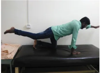
leg(quadruped position):
Week 1-2: Initially patient was asked to maintain quadruped position. Then he/ she was asked to lift one of the arms, hold this position for 10 seconds and relax.
Likewise the, patient was asked to lift one leg slowly, and hold the position for 10
seconds and relax
.
Progression of the exercise:
Week 3-4: Once after the quadruped position was maintained, patient was asked to extend alternate arm and leg and was asked to hold the position for 10 seconds and
relax.
Abdominal curl ups:
Week 1-2: Patient was asked to lie on their back with knees bent and feet flat on the floor. Then he/she was asked to lift the head and shoulders off the bed and try to touch
Progression of the exercise:
Week 3-4: lift the head and shoulders with hands across the chest.
GROUP B: MCKENZIE EXERCISE GROUP
Frequency: 2 times a day
Repetition: 15 times
Rest interval: 5 minutes after each exercise.
Week 1-2:
Lying on the stomach:
Patient was asked to lie on their stomach with arms beside the body and head
Extension in prone lying:
Patient was asked to lie on their stomach and support the upper body while
keeping their forearm flat on the bed. Then he / she was asked to lift the head as far as
possible and hold it for 10 seconds.
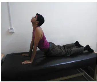
Extension in prone lying:
Patient was asked to push up their upper body with the palms of the hands on the
floor just in front of the shoulders and straighten the elbows elevating the upper part
of the body, while the hips and thigh remains relaxed and hold the position for 10
seconds.
Week 3-4: (along with all the above exercises)
Extension in standing:
The patient was asked to stand upright with feet slightly apart, hands placed at the
back so that fingers are pointed towards the floor and thumb forwards. The patient
bends backward at the waist as far as they can keeping the knees straight, maintaining
this position for 5 seconds and return to the starting position.
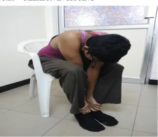
Flexion in sitting:
Patient was asked to sit on a chair, with knees and hips at 90 degrees, and
asked to bend the trunk forwards and hands close to the floor as possible. Then he /
she were asked to hold on to the ankle, bringing the trunk even close to the knees and
maintain the position for 5 seconds.
GROUP C: CONVENTIONAL EXERCISE GROUP
Frequency: 2 times a day
Rest period: 5 minutes after each exercise
Single knee to chest exercise:
Week 1-2: patient was asked to lie supine with their knees bent and feet flat on the floor. Then he /she was asked to clasp one of the knee with both the hands and pull it
towards their chest , hold this position for 5 seconds and relax.
Progression of the exercise:
Week 3-4: same exercise, hold it for 10 seconds. No of repetitions: 15 times for each leg
Lying trunk rotation:
Week 1-2: patient was asked to lie on their back with hips and knees bent, feet flat on the floor and arms straight beside the body. Then he / she was asked to
slowly rotate their both legs to one side and then to the opposite side.
No of repetitions: 10 times
Progression of the exercise:
Week 3-4: same exercise No of repetitions: 15 times
Hamstring stretches:
Week 1-2: patient was asked to lie on their back, clasp the hands under the thigh and to keep it vertically straight. Then he/she was asked to lift the leg up as far
as possible, hold the position for 10 seconds and relax.
Progression of the exercise:
Week 3-4: same exercise
No of repetitions: 10 times for each leg
Pelvic bridging:
Week 1-2: Patient was asked to lie on the back with the hip and knees bent and lift the buttocks up and away from the couch. He /she was asked to hold this position for
10 sec and relax.
No of repetitions: 10 times
Progression of the exercise:
Week 3-4: same exercise No of repetitions: 15 times
Prone straight leg raise:
Week 1-2: patient was asked to lie on their stomach, lift the leg up from the hip, with the knees straight and hold it for 10 seconds.
Progression of the exercise:
Week 3-4: same exercise
No of repetitions: 15 times for each leg
3.12.1 INTERVENTION DURATION
The intervention duration was 4 weeks in which the patient performed the exercises.
FREQUENCY: Once a day within 30-40 minutes.
REST INTERVAL: The patients were asked to perform deep breathing thrice during
the carryover from one exercise to another and also during the exercises, to avoid the
breath holding.
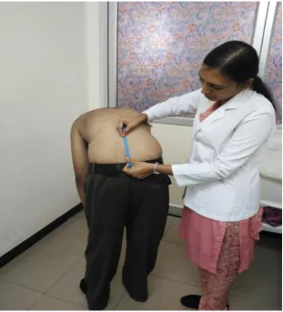
[image:34.595.161.473.167.511.2]
3.13
PHOTOGRAPHIC PRESENTATION
FIGURE NO : 3.13.1
EXPERIMENTAL GROUP-LUMBAR STABILISATION
[image:35.595.233.401.142.402.2] [image:35.595.143.489.449.734.2]EXERCISES
FIGURE NO: 3.1.2 -WALL SLIDES
FIGURE NO: 3.1.3 - PELVIC BRIDGING WITH LEG RAISE
FIGURE 3.1.4 -ALTERNATE ARM AND LEG (QUADRIPED)
FIGURE 3.1.5 - ABDOMINAL CURL UPS
[image:36.595.149.501.406.694.2][image:37.595.148.485.120.362.2]
EXPERIMENTAL GROUP: MCKENZIE EXERCISES
FIGURE 3.1.6 - EXTENSION IN PRONE LYING
[image:37.595.146.485.414.697.2]
FIGURE 3.1.8 - EXTENSION IN STANDING
[image:38.595.161.482.424.702.2]
3.14 STATISTICAL TOOLS
a) Paired ‘t’ Test
b)One way ANOVA
3.13.1 PAIRED
‘
t
’
TEST (within groups)
- Post-test values of the study are collected and assessed for variation in each group
and the results are analyzed using paired ‘t’test.
t =𝒅̅√𝒏
𝑺
where, S =√∑ 𝒅 −⌈𝒅̅⌉ ×𝒏
𝒏−
S = Combined standard deviation
d1 & d2 = difference between initial and final readings in group A & B
n1 & n2= number of patients in group A & group B X1 & X2 = mean of group A & group B
3.13.2 ONE WAY ANOVA
SOURCE OF VARIATION SQUARED VARIATION DEGREE OF FREEDOM MEAN SUM OF SQUARES F RATIO
SUM OF SQUARES
BETWEEN SAMPLE SSC C-1 MSC=SSC/C-1
F=MSC/MSE SUM OF SQUARES
WITHIN SAMPLE SSE N-C MSE=SSE/N-C
SSC = ∑ (𝑋̅̅̅̅
-𝑋̅̅̅̅ )2 + ∑(𝑋̅̅̅̅–𝑋̅̅̅̅)2+ ∑(𝑋̅̅̅̅–𝑋̅̅̅̅)2 SSC = ∑ (X1
-𝑋̅̅̅̅ )2 + ∑(X2–𝑋̅̅̅̅)2+ ∑(X3 –𝑋̅̅̅̅)2 C= number of sample
N= Total number of items in all sample groups
4.
DATA PRESENTATION
4.1 TABULAR PRESENTATION
4.1.1 PAIRED ‘t’ TEST: GROUP A- LUMBAR STABILISATION GROUP TABLE NO: 4.1.1.1- NUMERICAL PAIN RATING SCALE Outcome
measure
Mean value Calculated
‘t’ Value
Table ‘t’ Value
Level of Significance Pre-test Post-test
Numerical
Pain Rating
Scale 5.571 2.285 6.227 2.44
P < 0.05
Significant
TABLE NO: 4.1.1.2- ROLAND MORRIS DISABILITY QUESTIONNAIRE
Outcome measure Mean value Calculated
‘t’ value
Table
‘t’ value
Level of significance Pre-test Post-test
Roland Morris
Disability
Questionnaire
14.142 7 8.914 2.44 P < 0.05
Significant
TABLE NO: 4.1.1.3- LUMBAR FLEXION RANGE OF MOTION Outcome
measure
Mean value Calculated
‘t’ value
Table ‘t’ value
Level of significance Pre-test Post-test
Lumbar
Flexion
Range Of
Motion
4.18 5.285 9.537 2.44 P < 0.05
[image:41.595.89.545.79.594.2]
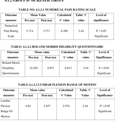
4.1.2 GROUP B- MCKENZIE GROUP
TABLE NO: 4.1.2.1 NUMERICAL PAIN RATING SCALE Outcome
measure
Mean Value Calculated
‘t’ value
Table ‘t’ value
Level of significance Pre-test Post-test
Numerical
Pain Rating
Scale
5.714 3.571 6.280 2.44 P < 0.05
Significant
TABLE: 4.1.2.2 ROLAND MORRIS DISABILITY QUESTIONNAIRE Outcome
measure
Mean value Calculated
‘t’ value
Table ‘t’ value
Level of Significance Pre-test Post-test
Roland Morris
Disability
Questionnaire
14.285 9.857 6.813 2.44 P < 0.05
Significant
TABLE 4.1.2.3 LUMBAR FLEXION RANGE OF MOTION Outcome
measure
Mean value Calculated
‘t’ Value
Table ‘t’ Value
Level of Significance Pre-test Post-test
Lumbar
Flexion
Range Of
Motion
4.84 5.457 2.974 2.44 P < 0.05
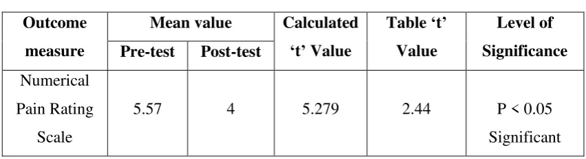
4.1.3 CONVENTIONAL GROUP
[image:42.595.101.527.114.230.2]
TABLE NO: 4.1.3.1- NUMERICAL PAIN RATING SCALE Outcome
measure
Mean value Calculated
‘t’ Value
Table ‘t’ Value
Level of Significance Pre-test Post-test
Numerical
Pain Rating
Scale
5.57 4 5.279 2.44 P < 0.05
Significant
TABLE NO: 4.1.3.2- ROLAND MORRIS DISABILITY QUESTIONNAIRE Outcome
measure
Mean value Calculated
‘t’ Value
Table ‘t’ Value
Level of Significance Pre-test Post-test
Roland Morris
Disability
Questionnaire
13.285 10.428 7.068 2.44 P < 0.05
Significant
TABLENO: 4.1.3.3- LUMBAR FLEXION RANGE OF MOTION Outcome
measure
Mean value Calculated
‘t’ Value
Table ‘t’ Value
Level of Significance Pre-test Post-test
Lumbar
Flexion
Range of
Motion
4.985 5.214 2.431 2.44 P >0.05
Not
[image:43.595.103.524.107.248.2]
4.1.4 ONE WAY ANOVA
TABLE NO: 4.1.4.1 PRE TEST-NUMERICAL PAIN RATING SCALE Source of
variation
Sum of Squares
Df Mean Square
[image:43.595.104.517.323.456.2]Calculated F value
Table f value
Level of significance
Between
Samples 0.094 2 0.047 0.050 3.55 p>5% Not
Significant Within
Samples 16.889 18 0.938
TABLE NO :4.1.4.2 POST TEST: NUMERICAL PAIN RATING SCALE
Source of variation
Sum of Squares
Df Mean Square
Calculated F value
Table f value
Level of significance
Between
Samples 11.142 2 5.571
5.851 3.55 p<5%
Significant Within
Samples 17.144 18 0.952
TABLE NO:4.1.2.3
PRE TEST:ROLAND MORRIS FUNCTIONAL DISABILITY QUESTIONNAIRE
Source of variation
Sum of Squares
Df Mean Square
Calculated F value
Table f value
Level of significance
Between
Samples 4.0 2 2.047
0.927 3.55 p>5% Not
Significant Within
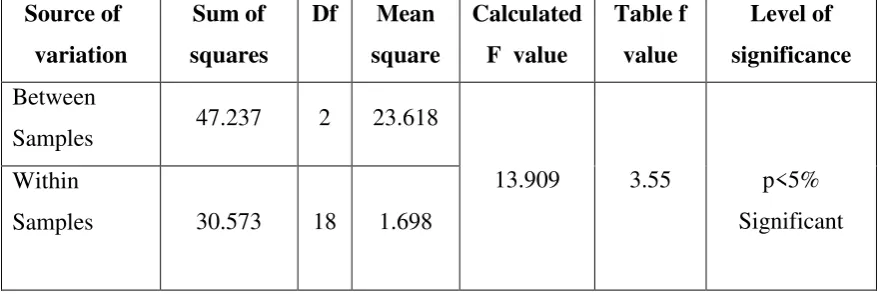
TABLE NO:4.1.2.4
POST TEST: ROLAND MORRIS FUNCTIONAL DISABILITY QUESTIONNAIRE
Source of variation
Sum of squares
Df Mean square
[image:44.595.99.539.116.723.2] [image:44.595.96.536.149.296.2]Calculated F value
Table f value
Level of significance
Between
Samples 47.237 2 23.618
13.909 3.55 p<5%
Significant Within
Samples 30.573 18 1.698
TABLE NO: 4.1.2.5
PRE TEST: LUMBAR FLEXION RANGE OF MOTION Source of
variation
Sum of squares
Df Mean square
[image:44.595.95.543.162.446.2]Calculated F value
Table f value
Level of significance
Between
Samples 4.095 2 2.047
0.927 3.55 p>5% Not
Significant Within
Samples 39.715 18 2.206
TABLE NO: 4.1.2.6
POST TEST: LUMBAR FLEXION RANGE OF MOTION
Source of variation
Sum of squares
Df mean square
[image:44.595.103.540.590.727.2]Calculated F value
Table F value
Level of significance
BETWEEN
SAMPLES 0.217 2 0.108
4.01 3.55 p<5%
Significant WITHIN
SAMPLES
4.2 GRAPHICAL REPRESENTATION
GROUP A: LUMBAR STABILISATION GROUP
4.2.1 NUMERICAL PAIN RATING SCALE
PAIRED
‘
t
’
TEST
4.2.2 ROLAND MORRIS DISABILITY QUESTIONNAIRE
PAIRED
‘
t
’
TEST
0 1 2 3 4 5 6
PRE TEST POST TEST
NUMERICAL PAIN RATING SCALE
PRE TEST POST TEST 5.571 2.285 0 2 4 6 8 10 12 14 16
PRE TEST POST TEST
ROLAND MORRIS DISABILITY QUESTIONNAIRE
PRE TEST POST TEST
14.142
4.2.3 LUMBAR FLEXION RANGE OF MOTION
PAIRED
‘
t
’
TEST
GROUP B: MCKENZIE GROUP
4.2.4 NUMERICAL PAIN RATING SCALE
PAIRED
‘
t
’
TEST
0 1 2 3 4 5 6
PRE TEST POST TEST
LUMBAR FLEXION RANGE OF MOTION
PRE TEST POST TEST
4.18
5.285
0 1 2 3 4 5 6
PRE TEST POST TEST
NUMERICAL PAIN RATING SCALE
PRE TEST POST TEST
5.714
4.2.5 ROLAND MORRIS DISABILITY QUESTIONNAIRE
PAIRED
‘
t
’
TEST
4.2.6 LUMBAR FLEXION RANGE OF MOTION
PAIRED
‘
t
’
TEST
0 2 4 6 8 10 12 14 16
PRE TEST POST TEST
ROLAND MORRIS DISABILITY QUESTIONNAIRE
PRE TEST POST TEST 9.857 14.285 4.5 4.6 4.7 4.8 4.9 5 5.1 5.2 5.3 5.4 5.5 5.6
PRE TEST POST TEST
LUMBAR FLEXION RANGE OF MOTION
PRE TEST POST TEST
4.84
GROUP C: CONVENTIONAL GROUP
4.2.7 NUMERICAL PAIN RATING SCALE
PAIRED
‘
t
’
TEST
4.2.8 ROLAND MORRIS DISABILITY QUESTIONNAIRE
PAIRED
‘
t
’
TEST
0 1 2 3 4 5 6
PRE TEST POST TEST
NUMERICAL PAIN RATING SCALE
PRE TEST POST TEST
5.57
4
0 2 4 6 8 10 12 14
PRE TEST POST TEST
ROLAND MORRIS DISABILITY QUESTIONNAIRE
PRE TEST POST TEST
13.285
4.2.9 LUMBAR FLEXION RANGE OF MOTION
PAIRED
‘
t
’
TEST
ONE WAY ANOVA
4.2.10 PRE TEST: NUMERICAL PAIN RATING SCALE
4.85 4.9 4.95 5 5.05 5.1 5.15 5.2 5.25
PRE TEST POST TEST
LUMBAR FLEXION RANGE OF MOTION
PRE TEST POST TEST 4.985 5.214 0 1 2 3 4 5 6 LUMBAR STABILISATION GROUP
MCKENZIE GROUP CONVENTIONAL GROUP
NUMERICAL PAIN RATING SCALE
LUMBAR STABILISATION GROUP MCKENZIE GROUP
CONVENTIONAL GROUP
5.571 5.714
4.2.11 POST TEST: NUMERICAL PAIN RATING SCALE
4.2.12 PRE TEST: ROLAND MORRIS DISABILITY
QUESTIONNAIRE
0 0.5 1 1.5 2 2.5 3 3.5 4 4.5 LUMBAR STABILISATION GROUPMCKENZIE GROUP CONVENTIONAL GROUP
NUMERICAL PAIN RATING SCALE
LUMBAR STABILISATION GROUP MCKENZIE GROUP CONVENTIONAL GROUP 4 3.571 2.285 12.6 12.8 13 13.2 13.4 13.6 13.8 14 14.2 14.4 LUMBAR STABILISATION GROUP
MCKENZIE GROUP CONVENTIONAL GROUP
ROLAND MORRIS DISABILITY QUESTIONNAIRE
LUMBAR STABILISATION GROUP MCKENZIE GROUP
CONVENTIONAL GROUP
14.28 14.14
4.2.13: POST TEST-ROLAND MORRIS DISABILITY
QUESTIONNAIRE
4.2.14 PRE TEST: LUMBAR FLEXION RANGE OF MOTION
0 2 4 6 8 10 12 LUMBAR STABILISATION GROUP
MCKENZIE GROUP CONVENTIONAL GROUP
ROLAND MORRIS DISABILITY QUESTIONNAIRE
LUMBAR STABILISATION GROUP MCKENZIE GROUP
CONVENTIONAL GROUP
7
9.85 10.42
3.6 3.8 4 4.2 4.4 4.6 4.8 5 5.2 LUMBAR STABILISATION GROUP
MCKENZIE GROUP CONVENTIONAL GROUP
LUMBAR FLEXION RANGE OF MOTION
LUMBAR STABILISATION GROUP MCKENZIE GROUP
CONVENTIONAL GROUP
4.18
4.84
4.2.15 POST TEST: LUMBAR FLEXION RANGE OF MOTION
5.05 5.1 5.15 5.2 5.25 5.3 5.35 5.4 5.45 5.5
LUMBAR STABILISATION
GROUP
MCKENZIE GROUP CONVENTIONAL GROUP
LUMBAR FLEXION RANGE OF MOTION
LUMBAR STABILISATION GROUP
MCKENZIE GROUP
CONVENTIONAL GROUP
5.28
5.45
5.
DATA ANALYSIS AND RESULTS
PAIRED
‘
t
’
TEST
5.1 GROUP A: LUMBAR STABILISATION GROUP
5.1.1 NUMERICAL PAIN RATING SCALE
For 6 degrees of freedom at 5% level of significance, the calculated ‘t’ value
was 6.227 and table ‘t’ value wass 2.44 for numerical pain rating scale in lumbar
stabilization group . Since calculated ‘t’ value was greater than the table ‘t’ value, the
null hypothesis was rejected. Hence there was a significant reduction in the pain of
group A.
5.1.2 ROLAND MORRIS FUNCTIONAL DISABILITY
QUESTIONNAIRE
For 6 degrees of freedom at 5% level of significance,the calculated ‘t’ value
was 8.914 and the table ‘t’ value was 2.44 for roland morris functional disability
questionnaire in lumbar stabilization group. Since calculated ‘t’ value was greater
than the table ‘t’ value, the null hypothesis was rejected. Hence, there was a
significant improvement in the functional ability of group A.
5.1.3 LUMBAR FLEXION RANGE OF MOTION
For 6 degrees of freedom at 5% level of significance, calculated ‘t’ was 9.537
and table ‘t’ value was 2.44 for lumbar flexion range of motion in lumbar stabilization
group. . Since calculated ‘t’ value was greater than the table ‘t’ value, the null
hypothesis was rejected. Hence, there was a significant increase in lumbar flexion
5.2
GROUP B: MCKENZIE GROUP
5.2.1
NUMERICAL PAIN RATING SCALE
For 6 degrees of freedom at 5% level of significance, calculated ‘t’ value was
6.280 and table ‘t’ value was 2.44 for numerical pain rating scale in Mckenzie group .
Since calculated ‘t’ value was greater than the table ‘t’ value, the null hypothesis was
rejected. Hence there was a significant reduction in the pain of group B.
5.2.2 ROLAND MORRIS FUNCTIONAL DISABILITY QUESTIONNAIRE
For 6 degrees of freedom at 5% level of significance, calculated ‘t’ value was
6.813 and table ‘t’ value was 2.44 for Roland Morris functional disability
questionnaire in Mckenzie group . Since calculated ‘t’ value was greater than the table
‘t’ value, the null hypothesis was rejected. Hence, there was a significant improvement in the functional ability of group B.
5.3.3 LUMBAR FLEXION RANGE OF MOTION
For 6 degrees of freedom at 5% level of significance, calculated ‘t’ value was
2.974 and table ‘t’ value was 2.44 for lumbar flexion range of motion in Mckenzie
group . Since calculated ‘t’ value was greater than the table ‘t’ value, the null
hypothesis was rejected. Hence, there was a significant increase in the lumbar flexion
range of motion.
5.3 GROUP C: CONVENTIONAL GROUP
5.3.1 NUMERICAL PAIN RATING SCALE
For 6 degrees of freedom at 5% level of significance, calculated ‘t’ value was
5.279 and table ‘t’ value was 2.44 for numerical pain rating scale in conventional
group . Since calculated ‘t’ value was greater than the table ‘t’ value, the null
5.3.2 ROLAND MORRIS FUNCTIONAL DISABILITY
QUESTIONNAIRE
For 6 degrees of freedom at 5% level of significance, calculated ‘t’ value was
7.068 and table ‘t’ value was 2.44 for Roland Morris functional disability
questionnaire in conventional group . Since calculated ‘t’ value was greater than the
table ‘t’ value, the null hypothesis was rejected. Hence there was a significant
improvement in the function of group C.
5.3.3 LUMBAR FLEXION RANGE OF MOTION
For 6 degrees of freedom at 5% level of significance, calculated ‘t’ value was
2.431 and table ‘t’ value was 2.44 for Roland Morris functional disability
questionnaire in conventional group . Since calculated ‘t’ value was lesser than the
table ‘t’ value, the null hypothesis was accepted. Hence there was no significant
increase in the lumbar flexion range of motion.
ONE WAY ANOVA
5.4
NUMERICAL PAIN RATING SCALE
5.4.1
PRE TEST OF NUMERICAL PAIN RATING SCALE
Pre test for experimental group I, experimental group II and control group
were analysed using one way ANOVA test. The calculated value was .0501. For 18
degrees of freedom at 5% level of significance, the table value was 3.55. Since the
calculated value was lesser than the table value, there was no significant difference
between pretest scores of experimental I, experimental II and control group. Hence
null hypothesis was accepted.
5.4.2
POST TEST OF NUMERICAL PAIN RATING SCALE
Post test for experimental group I, experimental group II, and control group
were analysed using oneway ANOVA test. The calculated value was 5.851. For 18
degrees of freedom at 5 % level of significance, the table value was 3.55. Since
post test score of experimental I, experimental II, and control group. Hence null
hypothesis was rejected.
5.5
ROLAND MORRIS FUNCTIONAL DISABILITY
QUESTIONNAIRE
5.5.1
PRE TEST OF ROLAND MORRIS DISABILITY
QUESTIONNAIRE
Pre test for experimental group I, experimental group II and control group
were analysed using one way ANOVA test. The calculated value was 0.927. For 18
degrees of freedom at 5% level of significance, the table value was 3.55. Since the
calculated value was lesser than the table value, there was no significant difference
between pretest scores of experimental I, experimental II and control group. Hence
null hypothesis was accepted.
5.5.2
POST TEST OF ROLAND MORRIS DISABILITY
QUESTIONNAIRE
Post test for experimental group I, experimental group II, and control group
were analysed using oneway ANOVA test. The calculated value was 13.909. For 18
degrees of freedom at 5 % level of significance, the table value was 3.55. Since
calculated value was greater than table value, there was significant difference between
post test score of experimental I, experimental II, and control group. Hence null
hypothesis was rejected.
5.6
LUMBAR FLEXION RANGE OF MOTION
5.6.1
PRE TEST OF LUMBAR FLEXION RANGE OF MOTION
Pre test for experimental group I, experimental group II and control group
were analysed using one way ANOVA test. The calculated value was 2.78. For 18
degrees of freedom at 5% level of significance, the table value was 3.55. Since the
between pretest scores of experimental I, experimental II and control group. Hence
null hypothesis was accepted.
5.6.2
POST TEST OF LUMBAR FLEXION RANGE OF MOTION
Post test for experimental group I, experimental group II, and control group
were analysed using oneway ANOVA test. The calculated value was 4.01. For 18
degrees of freedom at 5 % level of significance, the table value was 3.55. Since
calculated value was greater than table value, there was significant difference
between post test score of experimental I, experimental II, and control group. Hence
6.
DISCUSSION
Back pain is one of the most common medical problems, which seems to
occur at least once in 85% of adults less than 50 years of age. Among that, mechanical
low back pain is one of the major public health issues. In an attempt to prevent pain,
functional disabilities and most importantly to prevent transition towards the chronic
stage, various physiotherapeutic approaches have been emerged.
Indeed, it is one of the most common reasons for medical consultation and
second most common reason for absenteeism. Due to the high economic impact by
the disease on the society, cost efficient treatment approach is of the most essential
wanting. Exercise plays an important role in management of low back pain.
This study compared the effects of lumbar stabilization exercises, Mckenzie
exercises and conventional exercises on pain, function and range of motion in subjects
with mechanical low back pain.
In this study, 21 subjects who met the inclusion criteria were selected and
randomly allotted into 3 groups: group A, group B and group C.
Data was collected for pain using numerical pain rating scale , functional
ability using Roland Morris functional disability questionnaire and lumbar flexibility
using modified Schober’s test. The calculated data were analyzed using paired ‘t’ test
and one way ANOVA.
The result of the current study found that there was a significant decrease in
numerical pain rating scale , Roland Morris disability questionnaire and modified
Schober’s test in both the experimental groups(lumbar stabilization and Mckenzie
group) and the control group(conventional group). But the more percentage of
reduction in pain, improvement in the function and increase in the range of motion
was found in the lumbar stabilization group and the Mckenzie group than the control
group. Among the two experimental groups, it suggests that lumbar stabilization
group have a better effect in reducing pain, improving function and increasing lumbar
Also there was a significant improvement in the numerical pain rating scale,
Roland Morris functional disability questionnaire and the modified Schober’s test
between the three groups.
Mckenzie developed 3 major classifications of mechanical back pain:
postural, dysfunction and derangement syndromes. The definition of dysfunction
syndrome includes overstretching of soft tissues that have been shortened or contain
contracted scar tissue[34]. For dysfunction syndrome patients, symptom free movement
is accomplished until the end range of a shortened structure is realized, at which point
there is prohibition of further range accompanied by symptoms[38].
The goal is to remodel shortened tissue by frequently provoking the
discomfort of loading at the restricted end range. Dysfunction patients tend to avoid
their end range discomforts perpetuating the condition. Mckenzie maintains that once
the nuclear material has escaped from the annular wall, the inherent hydrostatic
mechanism is no longer intact.
Mckenzie exercise increases endorphins and alter perception of pain perhaps
by reducing anxiety and depression. It helps to centralize the pain in core back
structures rather than treat pain,that is localized in a specific area.
The overall goal of this Mckenzie exercise program is to reduce pain, develop
the muscle support of the trunk and spine and to diminish stress to the intervertebral
disc and other static stabilizers of the spine. Thus in the current study, Mckenzie
exercises are effective in reducing the pain, improving the function and increasing the
range of motion in low back pain patients.
The lumbar stabilization exercise programme concentrates on the local
muscle system that would be affected by the low back pain population[35,36]. Many
studies had shown the presence of dysfunction in multifidus and in the deep
abdominal muscles especially transverse abdominis muscle.It had a delayed reaction
in individuals with low back pain. Recent studies have found that all the muscles
In case of low back pain patients, they have altered slow motor unit
recruitment and this type of exercises would help in normal motor unit recruitment
pattern and thus reducing pain and functional ability. The stabilization exercises
concentrate on stable pain free positions without any movement. Thus lumbar
stabilization exercises are effective in reducing pain, improving the function and
7.
SUMMARY AND CONCLUSION
7.1 SUMMARY
To summarise this study, the aim of the study was to compare the effects of
lumbar stabilization exercises, Mckenzie exercises and conventional exercises on
pain, function and range of motion in patients with mechanical back pain.
21 patients diagnosed with mechanical low back pain who had met the
inclusion criteria were randomly allocated into 3 groups who received lumbar
stabilization exercises, Mckenzie exercises and conventional exercises
3 outcome measures were taken, numerical pain rating scale to assess the pain,
Roland Morris disability questionnaire to assess the functional ability and modified
Schober’s test to assess the lumbar flexion range of motion.
Results were analysed using paired ‘t’ test and one way ANOVA.
It suggests that lumbar stabilization exercises are more effective in reducing
the pain, and improving the function and increasing the lumbar flexion range of
motion than the Mckenzie group which is better than the control group. Also, there
was a significant increase in the three outcome measures when they were compared
between the groups.
7.2 CONCLUSION
Based on the results of the present study, it is concluded that lumbar
stabilization exercises are better than the Mckenzie exercises and the conventional
exercises in reducing the pain , increasing the range of motion and improving the
functional ability.
8.
LIMITATIONS AND SUGGESTIONS
8.1
LIMITATIONS
This study was done with small number of samples. Large sample is recommended.
This was a short term study and therefore long term study can be done to make the
result more valid.
It was not convenient for all patients to come for post test evaluation exactly after 4
weeks. This was overcome by scheduling their appointment with orthopaedician for
review along with the post test assessment.
8.2
SUGGESTIONS
Only pain, spinal mobility, and functional ability were stu