“COMPARATIVE STUDY OF FUNCTIONAL EFFICACY OF
TRANS FORAMINAL VS INTERLAMINAR EPIDURAL
STEROID INJECTION FOR LUMBAR DISC DISEASE”
Dissertation submitted for
M.S. Degree Examination
Branch II - ORTHOPAEDIC SURGERY
INSTITUTE OF ORTHOPAEDICS & TRAUMATOLOGY
MADRAS MEDICAL COLLEGE, CHENNAI –3
THE TAMIL NADU DR. M.G.R. MEDICAL UNIVERSITY
CHENNAI
CERTIFICATE
This is to certify that the dissertation entitled “COMPARATIVE STUDY OF FUNCTIONAL EFFICACY OF TRANS FORAMINAL
VS INTERLAMINAR EPIDURAL STEROID INJECTION FOR
LUMBAR DISC DISEASE” is a bonafide record of work done by Dr. SARAVANAN.B in the Institute of Orthopaedics and Traumatology,
Rajiv Gandhi Government General Hospital, Chennai, under the direct guidance of me.
Prof. R.VIMALA M.D Dean,
Madras Medical College & Rajiv Gandhi Government General Hospital,
Chennai - 600 003.
Prof. N.DEEN MUHAMMAD ISMAIL M.S.Ortho.,D.Ortho., Professor & Director I/C,
Institute of Orthopaedics and Traumatology, Madras Medical College &
DECLARATION
I hereby, declare the dissertation entitled “COMPARATIVE STUDY OF FUNCTIONAL EFFICACY OF TRANS FORAMINAL VS INTERLAMINAR EPIDURAL STEROID INJECTION FOR LUMBAR DISC DISEASE” submitted for the degree of M.S is the record work carried out by me during the period of March 2014 to September 2015 under the guidance of PROF.N.DEEN MUHAMMAD ISMAIL, M.S.Ortho., D.Ortho., Institute of Orthopaedics and Traumatology, Madras Medical College, Chennai. This dissertation is submitted to the Tamilnadu Dr.M.G.R. Medical University, Chennai, in partial fulfillment of the University regulations for the award of degree of M.S.ORTHOPAEDICS (BRANCH-II) examination to be held in April 2016. This work has not formed the basis for the award of any other degree or diploma to me previously from any other university.
Place: Chennai
Signature of the Candidate Date:
(Dr.B.SARAVANAN)
Signature of the Guide
Prof. N.DEEN MUHAMMAD ISMAIL M.S.Ortho., D.Ortho.,, Professor and Director I/C
ACKNOWLEDGEMENT
I am deeply indebted to my beloved chief and my teacher, Prof. Dr. N.Deen Muhammad Ismail, M.S.Ortho., D.Ortho.,
Professor of Orthopaedics and Director I/C , Institute of Orthopaedics
and Traumatology, Madras Medical College & Rajiv Gandhi Government General Hospital, Chennai for the able guidance, inspiration and encouragement he has rendered at every stage of this study.
I am grateful to my beloved teachers Prof.V. Singaravadivelu, Prof.A.Pandiaselvan, Prof.Nalli.R.Uvaraj ,Prof.M.Sudheer,
Prof.S.Karunakaran, Prof.K.P.Manimaran, for their invaluable help and guidance rendered to me in preparing this dissertation.
I express my heartfelt thanks to Dr.A. Saravanan, Assistant Professor, Institute of Orthopaedics and Traumatology, Madras Medical College for his excellent guidance and his valuable advice for preparing this study. Without his support and guidance I wouln’t have finished my work.
I express my heartfelt gratitude to Dr.P.Kannan, Dr.P. Kingsly, Dr.R.RajGanesh, Dr.Nalli.R.Gopinath, Dr.N.SarathBabu,
Dr.N.Muthalagan, Dr.G.Kaliraj, Dr.L.Muthukumar,
Madras Medical College, Chennai for their valuable advice and help in carrying out this study.
Also I thank Dr.N. Ashok kumar, Assitant Professor, Anesthesia Department for supporting and helping me in conducting my study.
I sincerely thank all my patients for their willingness to get enrolled in the study and for their kind co-operation during every follow up.
I extend my deep gratitude to my beloved teachers for their support and timely advice to complete my thesis.
Dr. SARAVANAN .B
TABLE OF CONTENTS
SLNO CONTENTS PAGE
NO
1 INTRODUCTION 1
2 REVIEW OF LITERATURE 5
3 AIM OF STUDY 11
4 MATERIALS & METHODS 39
5 RESULTS AND ANALYSIS 50
6 DISCUSSION 79
7 CONCLUSION 93
8 BIBLIOGRAPHY
1
INTRODUCTION
Currently variety of non operative therapies for back and leg pain are available. They are simple rest, exercises, massage, heat therapy, traction therapy.
“Epidural steroid injections (ESIs) have been used as an adjunct in the treatment of sciatica. Since the early reports, success rates ranging from 18% to 90% (average, 67%) have been documented. However, the efficacy of ESI has lasted, on the average, less than 3months.”38
In recent years, understanding of disc degeneration has undergone a significant transformation. Impairments of the back and spine are ranked as the most frequent cause of limitation of activity in individuals younger than 45 years old by the National Center for Health Statistics.1
2
without radiculopathy often is time limited, therapeutic injections help to manage pain and may alleviate or decrease the need for oral analgesics.
Epidural steroids are given by orthopaedicains, anesthetists, radiologists. Recent studies indicate that a high percentage of patients receiving the injections have significant pain relief and functional improvement. In a group of 70 patients with herniated discs in whom other conservative management had failed, epidural steroid injections provided significant pain relief and avoided surgery in 64% .Better outcomes were noted in patients older than 48 years and those who received the injections earlier (<100 days from diagnosis).
3
patients and is attributed to air injected into the epidural space, increased intrathecal pressure from fluid around the dural sac, and possibly an undetected dural puncture. Some minor, common complaints caused by corticosteroid injected into the epidural space include non-positional headaches, facial flushing, insomnia, low-grade fever, and transient increased back or lower extremity pain.
Epidural corticosteroid injections are contraindicated in the presence of infection at the injection site, systemic infection, bleeding diathesis, uncontrolled diabetes mellitus, and congestive heart failure. Epidural corticosteroid injections are performed in a fluoroscopy suite equipped with resuscitative and monitoring equipment.
It is recommended the use of fluoroscopy for diagnostic and therapeutic epidural injections for several reasons. Epidural injections performed without fluoroscopic guidance are not always made into the epidural space or the intended interspace.
4
5
REVIEW OF LITERATURE
Studies done two decades back did not reliably establish the efficacy of epidural injections, because of the lack of well-controlled studies.
Riew et al21 conducted a “prospective, randomized, controlled, double-blind study in: proceedings of the North American Spine Society, 14th Annual Meeting in 1999” showed early promising results
of transforaminal steroid injection (TFESI) in lumbar canal stenosis.
Only in 2013 North American Spine Society (NASS) forms a work group to address following issues - efficacy of lumbar transforaminal steroid injection (LTFESI) in radicular pain and lumbar disc herniation, complications of transforaminal steroid , patients likely to benefit from lumbar transforaminal epidural steroid injections (LTFESI), reasonable maximum number of therapeutic lumbar TFESI that a patient should receive within a six month period to treat lumbar radicular pain, the value (eg, cost per Quality Adjusted Life Years) of TFESI in the treatment of lumbar radicular pain.
Hospital For Special Surgery- Spine unit in their study in 2002 of
6
Radiculopathy,38 conducted a prospective RCT to assess the efficacy of LTFESI in patients with lumbar radiculopathy secondary to herniated intervertebral disc (HIVD). Of the 50 consecutively assigned patients, 25 received LTFESI and 25 received paravertebral trigger point injections. After an average follow-up period of 16 months, the group receiving LTFESI had a success rate of 84% as compared to 48% of the group receiving trigger-point injections. This study provides therapeutic evidence that for patients with herniated intervertebral disc (HIVD), LTFESI is more often effective (84%) than trigger point injections (48%) in providing at least 50% relief of radicular pain at 16 months”.
A retrospective study comparing Interlaminar epidural steroids (ILESI) to Transforaminal epidural injections (TFESI) for symptomatic lumbar intervertebral disc herniations found that transforaminal injections resulted in better short-term pain improvement and fewer long-term operative interventions.
7
these studies indicate that a high percentage of patients receiving the injections have significant pain relief and functional improvement.
According to Campbell - In a group of 70 patients with herniated cervical discs without myelopathy in whom conservative management had failed, cervical epidural steroid injections provided significant pain relief and avoided surgery in 63%.
North American Spine Society (NASS) states “patients with
lumbar scoliotic stenosis and radiculopathy experience significantly higher success rates after LTFESI if their symptoms were present for less than three months.”
North American Spine Society (NASS) also states that “in
patients treated with TFESI in the setting of disc herniation, effectiveness was more likely if the disc herniation was “contained” or abutted the nerve root and less likely if the nerve root was displaced or entrapped. The presence of stenosis, size of herniated intervertebral disc (HIVD), type of HIVD and hydration of HIVD do not predict outcome with LTFESI.”
8
A retrospective study by Schaufele et al30 conducted in year 2006 assessing pain improvement and surgical rates for managing lumbar disc herniation between IL and TF injection over 18 months, reported TF ESI’s superiority in short-term pain improvement and avoiding surgical interventions for long term.
Similarly Ackerman and Ahmad in 2007, “comparing efficacy of 3 fluoroscopically guided approaches (TF, IL, and caudal ESI) in their study demonstrated superior effect of transforaminal method of delivery.”31
An advantage of TFESI is that it can be performed in patients with failed back disease.
NASS (North American Spine Society) also states “patients with
radicular pain from an HIVD or central stenosis and/or lateral recess stenosis at the supra-adajacent intervertebral disc, obtain significant relief from a preganglionic LTFESI irrespective of age, gender, level of injection, symptom duration and pain intensity.”
9
[image:17.612.122.526.229.557.2]Traditional Interlaminar epidural steroids routinely given by anesthetists. But transforaminal approach commonly practiced by orthopedic surgeons and some well trained anesthetists, shows clear cut long term benefit also avoids surgical intervention.
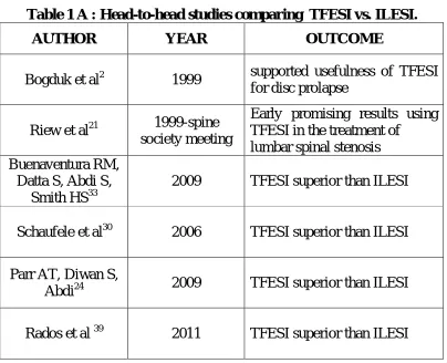
Table 1 A : Head-to-head studies comparing TFESI vs. ILESI.
AUTHOR YEAR OUTCOME
Bogduk et al2 1999 supported usefulness of TFESI for disc prolapse
Riew et al21 1999-spine society meeting
Early promising results using TFESI in the treatment of
lumbar spinal stenosis Buenaventura RM,
Datta S, Abdi S, Smith HS33
2009 TFESI superior than ILESI
Schaufele et al30 2006 TFESI superior than ILESI
Parr AT, Diwan S,
Abdi24 2009 TFESI superior than ILESI
Rados et al 39 2011 TFESI superior than ILESI
Bogduk et al2 study in year 1999 supported usefulness of TFESI for disc prolapse.
10
steroid directly over the inflamed nerve. Rados I, Sakic K, Fingler M et al, in their study in year 2011 at the end of 6 months found 28.3% in TFESI and 25% in ILESI as functional improvement.
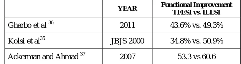
[image:18.612.114.523.281.390.2]However, some of the articles showing ILESI producing better results than TFESI. Such studies are,
Table 1B: Head-to-head studies comparing TFESI vs. ILESI
YEAR Functional Improvement
TFESI vs. ILESI
Gharbo et al 36 2011 43.6% vs. 49.3%
Kolsi et al35 JBJS 2000 34.8% vs. 50.9%
Ackerman and Ahmad 37 2007 53.3 vs 60.6
Gharbo et al36 study in year 2011, total duration of study was only 16 days.
Kolsi et al35 in their study of “Efficacy of nerve root versus interspinous injections of glucocorticoids in the treatment of disk related sciatica. A pilot, prospective, randomized, double-blind study, Joint Bone Spine 2000’ is also short duration study of only 28 days.
11
AIM OF STUDY
To compare efficacy of pain relief function of therapeutic transforaminal vs interlaminar epidural steroid injections for symptomatic lumbar disc disease patients.
To assess improvement in functional outcome in lumbar disc disease patents after treatment
To evaluate duration of relief and long term outcome after epidural steroid injections.
To assess the quality of improvement in pain relief.
12
RELEVANT ANATOMY
The human spinal column is an articulated segmental structure that serves dual purposes of protection and motion. The spinal columns functions include maintaining an upright posture, yet allowing for flexibility, while at the same time providing a conduit for neurological structures.
OSTEOLOGY:
LUMBAR SPINE
It consists of 5 vertebrae, which resemble each other structurally. The size gradually increases from L1 to L5. It consists of osseous and ligamentous structures
The block of bone on anterior part called vertebral bodyconsists of compact mass of spongy bone surrounded by cortical bone. Vertebral bodies and the intervening discs, along with anterior and posterior longitudinal ligaments constitute the anterior and middle column of Denis. This bears 80 % of load in uprightposition.
13
flattened laminae which completes the arch posteriorly. The articular processes are vertically arranged and consist of two superior and two inferior processes.
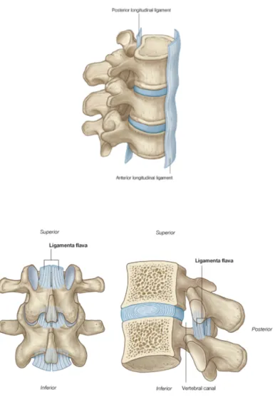
THE VERTEBRAL CANAL:
Boundaries
Anterior wall- Posterior longitudinal ligament, posterior surface of lumbar vertebrae
Posterior wall - Lamina, Ligamentum flavum
Lateral wall- pedicle
Between the pedicles of adjacent vertebrae, lies the intervertebral foramen.
14
INTERVERTEBRAL FORAMEN:
The vertebral notches are located on the superior and inferior aspects of the pedicles of all vertebrae. The inferior vertebral notch is the most prominent and together with the superior vertebral notch of the vertebra below forms an intervertebral foramen, which are the exit points for the spinal nerves that leave the vertebra.
THE INTERVERTEBRAL DISCS:
15
water. The intervertebral discs make up approximately 25 percent of the total length of the vertebral column above the sacrum. In the lumbar region, the disc material makes up 33 percent of the length of the column.
At birth, the disc has some direct blood supply contained within the cartilaginous endplates and the annulus. These vessels recede in the first years of life, and by adulthood there is no appreciable blood supply to the disc. Over time, for reasons not well understood, the water content of the gelatinous nucleus matrix decreases, with a decreased and altered proteoglycan composition. These changes lead to a more fibrous consistency of the nucleus, which ultimately fissures. Blood vessels grow into the disc through these outer fissures, with an increase in cellular proliferation and formation of cell clusters. Also, there is an increase in cell death, the mechanism of which is unknown. The cartilage endplates become thinned, with fissuring occurring with subsequent sclerosis of the subchondral endplates. Herniated discs have a greater number of senescent cells than non-herniated discs and have higher concentrations of matrix metalloproteinases.
16
more elongated and appear more like fibroblasts, whereas nucleus cells are oval and resemble chondrocytes. These two cell types behave differently and may be able to sense mechanical stresses. In culture, they respond differently to loads and produce different matrix proteins. The annulus cells produce predominantly type I collagen, whereas nucleus cells synthesize type II collagen. The characteristics of these cell types under normal and abnormal circumstances are beginning to be determined, and much is known.
17
18
BLOOD SUPPLY:
Paired lumbar arteries arise directly from the posterior aspect of the aorta, in front of the bodies of the lumbar vertebrae. During the adult phase of life, there is no active blood supply to the intervertebral discs. The vasculature of the nerve roots is formed by branches from the intermediate branch of the segmental artery distally and by branches from the vasa corona of the spinal cord proximally. The venous supply of the lumbar spine mirrors the arterial supply. The venous system is valveless, draining the internal and external venous system into the inferior vena cava .
NERVE SUPPLY:
19
LUMBAR DISC DISAESE
One theory of spinal degeneration assumes that all spines degenerate and that current methods of treatment are for symptomatic relief, not for a cure.
The degenerative process has been divided into three separate stages with relatively distinct findings.
The first stage is dysfunction, which is seen in individuals 15 to 45 years old.
It is characterized by circumferential and radial tears in the disc anulus and localized synovitis of the facet joints.
The next stage is instability. This stage, found in 35- to 70-year-old patients, is characterized by internal disruption of the disc, progressive disc resorption, degeneration of the facet joints with capsular laxity, subluxation, and joint erosion.
The final stage, present in patients older than 60 years, is stabilization. In this stage, the progressive development of hypertrophic bone around the disc and facet joints leads to segmental stiffening or frank ankylosis.
20
Disc herniation in this scheme is considered a complication of disc degeneration in the dysfunction and instability stages.
Spinal stenosis from degenerative arthritis in this scheme is a complication of bony overgrowth compromising neural tissue in the late instability and early stabilization stages.
Long-term follow-up studies of lumbar disc herniations have documented several principles, the foremost being that generally symptomatic lumbar disc herniation (which is only one of the consequences of disc degeneration) has a favorable outcome in most patients.
The primary benefit of surgery has been noted to occur early on in the first year after surgery, but with time the statistical significance of the improvement appears to be lost.
In general, the literature supports an active care approach, minimizing centrally acting medications. The judicious use of epidural steroids also is supported.
21
If surgery is necessary, it usually can be delayed 6 to 12 weeks to allow adequate opportunity for improvement. These principles are consistent with clinical findings and treatment practices at this clinic. Some patients are best treated surgically.
Similar principles are valid regarding cervical disc herniations, which also generally can be treated non operatively. The important exception is a patient with cervical myelopathy, who is best treated
surgically.
22
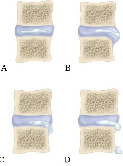
PATHOANATOMY OF INTERVERTEBRAL DISC PROLAPSE:
Weber expressed that, “disc herniation is a collective term, to describe a process with rupture of annulus fibrosus and subsequent displacement of the central mass of the disc into the intervertebral space, common to the dorsal or dorsolateral aspect of the disc. A herniation occurs in a lumbar intervertebral disc when a separate tissue fragment extrudes or sequestrates, through a tear of the annulus. Both a fissure and fragment appears to be required for prolapse to occur.”
A B
[image:34.612.217.421.335.609.2]C D
FIG 9; Types of disc herniation.
23
Sensory Symptoms:
The sensory symptoms appear with far more frequency than the motor symptoms. The most common symptom, following nerve irritation, is pain, in the form of paraesthesia, hyperesthesia.
Motor Symptoms:
24
Infrequently, the patient may present with lower extremity weakness which may be disabling. This is more likely to occur in disc lesions involving the fourth and fifth lumbar spinal nerve roots.
PHYSICAL EXAMINATION:
O’Connell classified the signs, in lumbar disc herniation as the spinal signs, nerve tension signs and neurological signs.
SPINAL SIGNS:
Loss of normal lumbar lordosis and paravertebral spasm are usually seen during the acute phase of disease. Occasionally in less acute situation the protective muscle spasm may be elicited only when the patient is stressed by prolonged standing or by forward flexion of the spine.
25
sagittal plane than in the frontal plane. Palpation of the patient either in erect or prone position, may evoke tenderness in the midline, at the level of the disc lesion and in Para vertebral areas on the side of a nuclear extrusion.
NERVE TENSION SIGNS:
Nerve irritation may be elicited by methods which increase the tension on the nerve root.
The straight leg raising test:
The passive straight leg raising test is the most commonly employed one. With the straight leg raising manoeuvre, the L5 and S1 nerve roots, move 2 to 6mm at the level of the foramina. In an analysis of the diagnosis of the straight leg raising test, it was noted that tension is realized within the nerve roots contributing to the sciatic nerve, at 35 to 70 degrees of elevation from the supine position. This test is performed with the patient supine and head flat or on a low pillow. Only when leg pain or reproduction of the patient’s radicular pain occurs, the test is considered positive.
Well Leg Raising Test:
26
distribution on the affected side, it is highly suggestive of disc prolapse compressing the exiting nerve root.
Bow String test:
Patient is asked to flex the hip with knee in full extension on the affected side till the pain is felt. At this point, the knee is flexed which instantaneously reduces the pain. On pressing the sciatic nerve in the popliteal fossa the painful radicular symptoms restarts which indicates tension on the nerve roots.
Sciatic nerve stretch test:
Patient is asked to lie supine and the foot is supported and gradually flexed at hip with knee in full extension; during this manoeuvre patient develops pain. When patient develops pain, flexion at hip is stopped, pressure is applied over the anterior aspect of ipsilateral knee in order to extend the knee. If there is sharp radicular pain, it indicates tension on the nerve root.
NEUROLOGIC SIGNS:
27
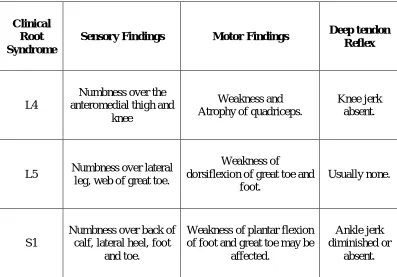
[image:39.612.123.520.263.540.2]vary. There may be no objective neurologic findings because the involved nerve often remains functional. Loss of Deep tendon reflex, motor weakness, muscle atrophy or sensory loss will be more suggestive of root compression. The neurological findings in the lumbosacral nerve root lesions are compiled in the following table.
Table 2 : Table showing the Clinical Root Syndrome
Clinical Root Syndrome
Sensory Findings Motor Findings Deep tendon
Reflex
L4
Numbness over the anteromedial thigh and
knee
Weakness and Atrophy of quadriceps.
Knee jerk absent.
L5 Numbness over lateral leg, web of great toe.
Weakness of
dorsiflexion of great toe and foot.
Usually none.
S1
Numbness over back of calf, lateral heel, foot
and toe.
Weakness of plantar flexion of foot and great toe may be
affected.
Ankle jerk diminished or
28
INVESTIGATIONS
X RAY LUMBO SACRAL SPINE:
The first line of investigations includes X-rays of lumbosacral spine in both anteroposterior and lateral views. There may be loss of lumbar lordosis with scoliosis depending on the location of disc prolapse and uniform reduction of disc space. In acute IVDP there may not be significant reduction in intervertebral disc space. Oblique views and flexion extension views should be taken to rule out instability of spine
MYELOGRAPHY:
29
COMPUTED TOMOGRAPHY (CT):
Major advantages of computed tomography over myelography are their ability to visualize the pathology, non-invasiveness and less radiation exposure for patients and radiologists. The importance of correlating, findings in the various imaging modalities with clinical symptoms has been emphasized in several studies. Wiesel and associates performed lumbar CT Scans in 52 asymptomatic subjects. The overall incidence of CT abnormalities was 37% and was more common in persons over 40 years of age.
MAGNETIC RESONANCE IMAGING (MRI):
30
has increased soft tissue delineation its advantage to the patient as well as the operating surgeon is obvious.
31
MANAGEMENT
1. NONOPERATIVE TREATMENT
2. OPERATIVE TREATMENT
1) NONOPERATIVE TREATMENT
I. CONSERVATIVE TREATMENT:
Currently variety of non-operative therapies for back and leg pain are available. They are are simple rest, massage, heat therapy, traction therapy. These sort of therapies are reported as producing “ miraculous cures”. But adequate scientific proof for these sort of therapies are very few.
32
A. BED REST AND TRACTION:
The simplest treatment for acute back pain is rest. Pain relief is usually experienced by a patient confined to bed. The optimal position is supine with knees and hips flexed.
B. MEDICATIONS:
Drug therapy may be directed to reduce nerve root inflammation, pain and for muscle relaxation. The sciatic pain is due to a perineural inflammatory response to the herniated disc material. In many instances this inflammatory change is decreased by anti-inflammatory drugs. Bed rest remains the best way to treat muscle spasm. Anti-depressants reduce the need for analgesics in patients with chronic pain.
C.PHYSIOTHERAPY:
33
IFT is a method of producing low frequency alternating currents around 4000 Hertz. Direct stimulation by interference current produces inhibition of sympathetic system resulting in vasodilatation and helps in removal of pain metabolites and exudates if present. It also reduces pain based on “gate theory of Melzack and Wall.”
TENS is the application of pulsed rectangular wave current forms through surface electrodes on the skin. It works on the principle of the pain gate theory and achieves pain relief by stimulating large afferent fibres preferentially, thus inhibiting transmission of pain impulses.
II. EPIDURAL STEROID INFILTRATION :
The epidural injection of a combination of a long acting steroid with an epidural anaesthetic is directed to reduce the inflammatory component of disc herniation. 60-70 percent of satisfactory results have been described in literature. Low pressure headaches, sciatic pain reproduced during injection and a transitory motor weakness lasting 15-20 minutes are some of the associated complications.
III. SURGICAL MANAGEMENT:
34
1. Chemonucleolysis
2. Standard laminectomy and discectomy 3. Microscope assisted lumbar discectomy 4. Percutaneous Discectomy
5. Discectomy and Spinal Fusion 6. Total Disc Replacement
INJECTION STUDIES
Whenever a diagnosis is in doubt, and the complaints seem real or the pathological condition is diffuse, identification of the source of pain is problematic. The use of local anesthetics or contrast media in various specific anatomical areas can be helpful. These agents are relatively simple, safe, and minimally painful.
35
The best choice of a contrast medium for documenting structures outside the subarachnoid space is an absorbable medium with low reactivity because it might be injected inadvertently into the subarachnoid space. Iohexol and metrizamide are the least reactive, most widely accepted, and best tolerated of the currently available contrast media.
Local anesthetics, such as lignocaine (Xylocaine), tetracaine and bupivacaine are used frequently epidurally and intradurally.
The use of bupivacaine should be limited to low concentrations and low volumes because of reports of death after epidural anesthesia using concentrations of 0.75% or higher.
36
triamcinolone . Isotonic saline is the only other injectable medium used frequently around the spine with no reported adverse reactions.
EPIDURAL CORTISONE INJECTIONS
Epidural injections in the cervical, thoracic, and lumbosacral spine were developed to diagnose and treat spinal pain.
Information obtained from epidural injections (ESI) can be helpful in confirming pain generators that are responsible for a patient’s discomfort. Structural abnormalities do not always cause pain, and diagnostic injections can help to correlate abnormalities seen on imaging studies with associated pain complaints.
In addition, epidural injections can provide pain relief during the recovery phase of disc or nerve root injuries and allow patients to increase their level of physical activity. Because severe pain from an acute disc injury with or without radiculopathy often is time limited, therapeutic injections help to manage pain and may alleviate or decrease the need for oral analgesics
LUMBAR EPIDURAL INJECTION
37
or lateral bony stenosis, most patients who received substantial relief of leg pain from a well-placed transforaminal injection, even if temporary, benefit from surgery for the radicular pain.
Table 3 Common Corticosteroids Used in Spinal Interventions
Compared with Hydrocortisone
H Y D R O C O R T IS O N E M E T H Y L P R E D N IS O L E (D E P O M E D R O L ) T R IA M C IN O L O N E B E T A M E T H A S O N E Relative Anti-inflammatory potency
1 5 5 25
pH 5.0-7.0 7-8 4.5-6.5 6.8-7.2
Onset Fast Slow Moderate Fast
Duration of
action Short Intermediate Intermediate Long
Concentration
(mg/mL) 50 40-80 20 6
Relative mineralocorticoid
activity
2+ 0 0 0
38
and leg pain of an acute nature (<3 months) respond better to epidural corticosteroids.
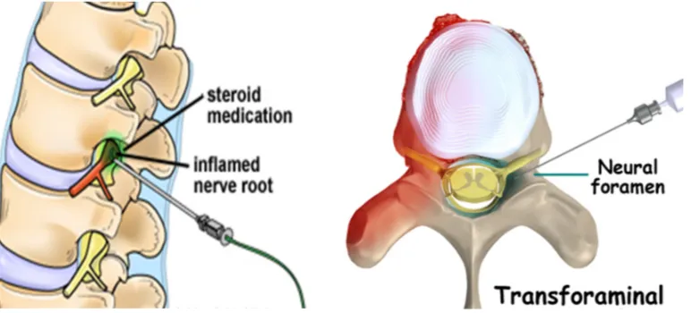
Figure 15A : Transforaminal Approach
39
MATERIALS AND METHODS
This study was conducted at Rajiv Gandhi Government General Hospital Chennai from March 2014 to Sep 2015.
One hundred patients with back pain documented with lumbar disc disease treated initially with rest, analgesics and physiotherapy for at least six weeks were included in the study and were treated with epidural steroid injection. The protocol was approved by ethical committee.
Patients to participate in this study were documented. Patients with lumbar disc disease were given transforaminal epidural or intralaminar epidural steroid injection since 1/3/2014 in Ortho OT of our institute & Pain clinic OT.
INCLUSION CRITERIA
1. Patients with duration of back pain and radiculopathy for more than 6 months with radiological evidence (MRI & X- RAY) of lumbar disc disease.
40
HNPs at various interspaces (L3–L4, L4–L5, L5–S1) and with differing axial presentations (e.g., far lateral, paracentral, and central protrusion) were examined.
3. Age group between 18 to 65 years. .
EXCLUSION CRITERIA
1) Patients with more than 2 level lumbar disc disease. 2) Patients with progressive neurological deficits. 3) Patients who underwent prior lumbar surgery.
4) Patients with a large herniation with severe central or foraminal stenosis on MRI,
5) Coagulation disorder.
6) Patients with a history of anaphylaxis to local anesthetics or corticosteroid.
Patients who met inclusion criteria were obtained informed consent after explaining all risks, benefits, objectives and outcomes of the study. They were all explained about nature of study.
Two groups were assigned
41
Each group con tains 50 patients. Pt having predominant unilateral symptoms were given transforaminal steroids. Other patients on random basis were equally divided. These patients were followed for one year. No patients were missed.
[image:58.612.160.489.236.403.2]The following methods were used to apply the epidural steroid.
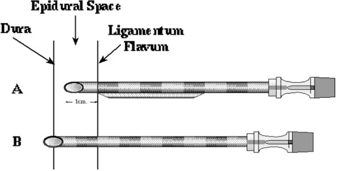
Figure 16A : Tuohy epidural needle
[image:58.612.165.482.478.625.2]42
TRANSFORAMINAL LUMBAR EPIDURAL INJECTION
• Place the patient prone on a fluoroscopy compatible surgical table. Under sterile aseptic precautions, the parts (fig 17) above and below the interspace to be injected were prepared and draped.
• A 22-gauge, 4 ¾ inch spinal needle is then inserted and advanced within the anesthetized soft tissue track under fluoroscopy guidance until contact is made near the junction of the superior articular process and lower edge of the superior transverse process.
• The spinal needle is retracted 2 to 3 mm, redirected towards the base of the appropriate pedicle and advanced it slowly to the 6-o’clock position of the pedicle under fluoroscopy. Adjusted the C-arm to a lateral projection to confirm the position, and then returned the C-arm to the anteroposterior view.
• Confirmed placement in safe triangle. Safe triangle roof is formed by pedicle, exiting nerve root forms tangential base and vertebral body forms lateral border.
43
• The stylet was removed and 1 mL of iohexol nonionic contrast agent was injected slowly to produce a perineurosheathogram observed adequate dye pattern.
At each level of nerve root, one ml of iohexol nonionic contrast agent was injected after positioning under C-ARM guidance. Needle position re-adjusted if fluoroscopy didn’t reveal flow to ipsilateral nerve root.
After documenting adequate flow of contrast to target site and no blood or cerebrospinal fluid was aspirated, 2ml of triamicinolone (each ml containing 40 mg) with 1ml of preservative free lignocaine were given. Injection were never given more than 2 levels to avoid systemic side effects of steroid.
The patients in Group I with the total of 50 patients (with average age of 47.1, with second dose of steroid in 21 poor responders after 4 weeks apart, total 72 injections of TFESI) received an average of 1.42 TFESI were followed for 12 months.
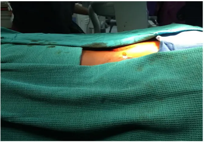
Figure 17A : Aseptic Prepartion
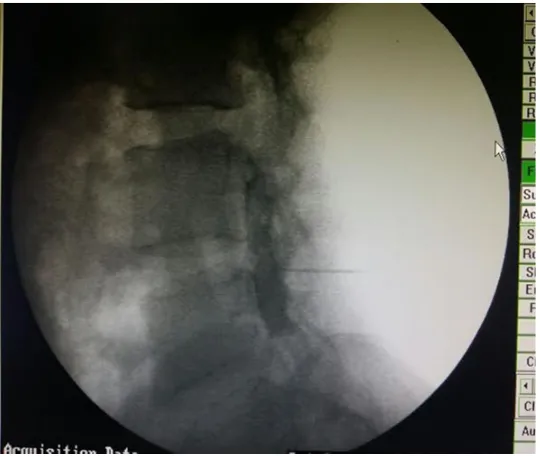
[image:61.612.214.439.405.602.2]Figure 18 : Injecting Contrast
Figure 20 : Lateral view of LS spine showing needle in position
[image:63.612.197.450.358.597.2]44
INTERLAMINAR LUMBAR EPIDURAL INJECTION
Patient prone position on a fluoroscopy compatible surgical table. Aseptically prepared the part with isopropyl alcohol and povidone-iodine several segments above and below the laminar interspace to be injected. Draped the part in a sterile fashion.
Under anteroposterior fluoroscopy view, identified the target interlaminar space. Using a 27-gauge, ¼ -inch needle, anesthetized the part over the target interspace on the side of the patient’s symptom with 1 mL of 1% preservative-free lignocaine without epinephrine.
22-gauge, 31/2 -inch spinal needle inserted vertically until contact is made with the upper edge of the inferior lamina at the target interspace, 1 to 2 cm lateral to the caudal tip of the inferior spinous process under fluoroscopy. Anesthetized the lamina with 1-2 mL of 1% preservative-free lidocaine without epinephrine. Anesthetized the soft tissue with 2 mL of 1% lidocaine as the spinal needle is withdrawn.
45
“Walk off” the lamina with the Tuohy needle onto the ligamentum flavum. Removed the stylet from the Tuohy needle, and attached a 10-mL syringe filled halfway with air and sterile saline to the Tuohy needle. Advanced the Tuohy needle into the epidural space using the loss-of-resistance technique. Avoided lateral needle placement to decrease the likelihood of encountering an epidural vein or adjacent nerve root. Removed the stylet when loss of resistance has been achieved. Aspirated to check for blood or CSF. If neither blood nor CSF is present, removed the syringe from the Tuohy needle and attached a 5-mL syringe containing 2 mL of nonionic contrast dye.
Epidural placement confirmed by an epidurogram with the iohexol. Placement documented with flouroscopy.
Removed the 5-mL syringe, and placed on the Tuohy needle. 10-mL syringe containing 1 10-mL of 1% preservative-free lidocaine and 2 10-mL of 40 mg/mL triamicinolone.
Figure 23A : Posteroanterior view of lumbar interlaminar epidurogram showing characteristic contrast flow pattern
[image:67.612.194.456.392.649.2]46
Self-directed lumbar stabilization programme were given for both groups. It consisted of
1) Abdominal strengthening exercises 2) Lumbar paraspinal strengthening 3) Hip flexibility
4) Hamstring flexibility
Started immediately after injection in both study groups.
Assessment and Follow-up
Compliance was ensured by therapeutic exercises reinforcement during follow-up visits.
Outcome of treatment was measured by
1) PATIENT SATISFACTION SCALE.
It had choice options as 0 (poor) 1 (fair) 2 (good) 3 (very good) and 4 (excellent).
2) ROLAND MORRIS LOW BACK PAIN DISABILITY
47
3) Measurement of FINGER-TO FLOOR DISTANCE. It is done with the patient in fully tolerated hip flexion;
4) VISUAL NUMERIC PAIN SCALE similar to the visual analog scale with a range of options from 0 (no pain) to 10 (worst pain). Outcome measures were assessed as follows:
1) Before and after treatment, then at 2 weeks, 1month, 3 month, 6 months and 12 months by a staff nurse blinded to programme. 2) Pt advised not to use Narcotics or NSAID medication during
study period. During follow up, patients were assessed for neurologic detoriation, worsening of pain, also new development of pain. Patients who failed to respond were given additional one dose of injection through same approach. Time interval between 2 doses were minimum of one month.
A successful outcome defined as
- improvement of a patient satisfaction score of 2 (good) or 3 (very good), 4 (excellent). (Improvement of at least 2 scores)
- improvement on the Roland-Morris Disability score of 5 or more,
48
The Roland-Morris Low Back Pain and Disability Questionnaire
I stay at home most of the time because of my back.
I change position frequently to try to get my back comfortable.
I walk more slowly than usual because of my back.
Because of my back, I am not doing any jobs that I usually do around the
house.
Because of my back, I use a handrail to get upstairs.
Because of my back, I lie down to rest more often.
Because of my back, I have to hold on to something to get out of an easy chair.
Because of my back, I try to get other people to do things for me.
I get dressed more slowly than usual because of my back.
I only stand up for short periods of time because of my back.
Because of my back, I try not to bend or kneel down.
I find it difficult to get out of a chair because of my back.
My back is painful almost all of the time.
I find it difficult to turn over in bed because of my back.
My appetite is not very good because of my back.
I have trouble putting on my sock because of the pain in my back.
I can only walk short distances because of my back pain.
I sleep less well because of my back.
49
I sit down for most of the day because of my back.
I avoid heavy jobs around the house because of my back.
Because of back pain, I am more irritable and bad tempered with people than
usual.
Because of my back, I go upstairs more slowly than usual.
I stay in bed most of the time because of my back.
50
RESULTS
In TFESI group, 36 patients out of 50 showed improvements at the end of one year where as in ILESI group, only 28 showed significant improvements at the end of year as per Patient Satisfaction Score study.
In TFESI Group, 8 patients didn’t show any improvement (6 patients absent response, 2 patients negligible improvement, among them 2 were females)
In ILESI Group, 20 patients didn’t show any improvement (13 patients absent response, 7 patients negligible improvement, among them 11 were females).
51
Table 4- Frequency table for sex in each sub cohort
TREATMENT TYPE Frequency Percent Valid Percent Cumulative
Percent
TFESI
Female 16 32.0 32.0 32.0 Male 34 68.0 68.0 100.0 Total 50 100.0 100.0
ILESI
Female 19 38.0 38.0 38.0 Male 31 62.0 62.0 100.0 Total 50 100.0 100.0
Chart Showing Sex Distribution.
Female (Blue Colour) And Male (Orange Colour)
52
Table 5A. Descriptives- Mean and Standard deviation in TFESI group.
Descriptive Statistics
TREATMENT TYPE N Minimum Maximum Mean Std. Deviation T R A N S F O R A M IN A L E P ID U R A L G R O U P
Roland@pre treatment 50 12 20 16.52 2.002 Roland@1month 50 6 17 11.04* 2.579 Roland@3month 50 6 17 11.70 2.558 Roland@6month 50 7 18 12.30 2.636 Roland@12month 50 7 19 13.14 2.828 Finger@Pre treatment 50 40 80 63.16 8.505 Finger@1month 50 15 69 32.52* 14.336 Finger@3month 50 16 69 34.94* 14.596 Finger@6month 50 18 75 37.14* 15.931 Finger@12month 50 19 75 40.18 16.768 Satisfication@Pre
treatment 50 0 1 .26 .443 Satisfication@1month 50 0 4 2.96* 1.195 Satisfication@3month 50 1 4 2.70* 1.147 Satisfication@6month 50 0 4 2.54* 1.199 Satisfication@12month 50 0 4 2.26* 1.175 Visual@Pre treatment 50 7 9 8.58 .538
Visual@0 50 1 8 3.30* 2.197 Visual@15th day 50 1 8 3.74* 2.284 Visual@1month 50 1 9 4.02* 2.369 Visual@3month 50 1 9 4.06* 2.469 Visual@6month 50 1 9 4.32* 2.543 Visual@1y 50 2 9 4.58 2.635
53
* Table 5B. Descriptives Mean and Standard deviation in ILESI group.
TREATMENT TYPE N Minimum Maximum Mean Std.
Deviation IN T E R L A M IN A R E P ID U R A L S T E R O ID G R O U P
Roland@Pre treatment 50 13 19 16.38 1.413 Roland@1month 50 10 16 13.38 1.817 Roland@3month 50 10 17 13.50 1.876 Roland@6month 50 10 17 13.58 1.939 Roland@12month 50 10 19 13.84 2.064 Finger@Pre treatment 50 50 70 63.84 5.250 Finger@1month 50 23 69 44.62 14.168 Finger@3month 50 23 69 45.18 14.512 Finger@6month 50 23 69 45.64 14.602 Finger@12month 50 23 69 46.16 14.829 Satisfication@Pretreatment
50 0 1 .02 .141
Satisfication@1month
50 0 4 1.94 1.316 Satisfication@3month
50 0 4 1.78 1.282 Satisfication@6month
50 0 4 1.76 1.287 Satisfication@12month
50 0 4 1.70 1.282 Visual@Pre treatment
50 8 9 8.84 .370 Visual@0
50 2 9 4.70 2.605 Visual@15th day
50 2 9 4.92 2.747 Visual@1month
50 2 9 5.06 2.810 Visual@3month
50 2 9 5.10 2.852 Visual@6month
50 2 9 5.20 2.814 Visual@1year
50 2 9 5.30 2.757
54
Table 6. Descriptives p value study
Treatment N Mean SD P value
Roland@Pre treatment TFESI 50 16.52 2.002 0.687 ILESI 50 16.38 1.413
Roland@1month TFESI 50 11.04 2.579 *<0.001** ILESI 50 13.38 1.817
Roland@3month TFESI 50 11.70 2.558 *<0.001** ILESI 50 13.50 1.876
Roland@6month TFESI 50 12.30 2.636 *0.007** ILESI 50 13.58 1.939
Roland@12month TFESI 50 13.14 2.828 0.161 ILESI 50 13.84 2.064
Finger@Pre treatment TFESI 50 63.16 8.505 0.632 ILESI 50 63.84 5.250
Finger@1month TFESI 50 32.52 14.336 *<0.001 ILESI 50 44.62 14.168
Finger@3month TFESI 50 34.94 14.596 *<0.001 ILESI 50 45.18 14.512
Finger@6month TFESI 50 37.14 15.931 *0.006 ILESI 50 45.64 14.602
Finger@12month TFESI 50 40.18 16.768 .062 ILESI 50 46.16 14.829
Satisfication@Pre treatment
TFESI 50 .26 .443
<0.001 ILESI 50 .02 .141
Satisfication@1month TFESI 50 2.96 1.195 *<0.001 ILESI 50 1.94 1.316
Satisfication@3month TFESI 50 2.70 1.147 *<0.001 ILESI 50 1.78 1.282
Satisfication@6month TFESI 50 2.54 1.199 *0.002 ILESI 50 1.76 1.287
55
Visual@Pre treatment TFESI 50 8.58 .538 *0.006 ILESI 50 8.84 .370
Visual@0 TFESI 50 3.30 2.197 *0.005 ILESI 50 4.70 2.605
Visual@15days TFESI 50 3.74 2.284 *0.022 ILESI 50 4.92 2.747
Visual@1month
TFESI 50 4.02 2.369
*0.048 ILESI 50 5.06 2.810
Visual@3month TFESI 50 4.06 2.469 0.054 ILESI 50 5.10 2.852
Visual@6month TFESI 50 4.32 2.543 0.104 ILESI 50 5.20 2.814
Visual@1year TFESI 50 4.58 2.635 0.185 ILESI 50 5.30 2.757
*means study significant with p value of <0.05
Table 7- T-Test study of standard error of mean.
Group Statistics
Treatment
type N Mean
Std. Deviation
Std. Error Mean Roland @Pre treatment
TFESI 50 16.52 2.002 .283 ILESI 50 16.38 1.413 .200 Roland@1month
TFESI 50 11.04 2.579 .365 ILESI 50 13.38 1.817 .257 Roland@3month
TFESI 50 11.70 2.558 .362 ILESI 50 13.50 1.876 .265 Roland@6month
TFESI 50 12.30 2.636 .373 ILESI 50 13.58 1.939 .274 Roland@12month
56
Finger @Pre treatment TFESI 50 63.16 8.505 1.203 ILESI 50 63.84 5.250 .743 Finger@1month TFESI 50 32.52 14.336 2.027
ILESI 50 44.62 14.168 2.004 Finger@3month TFESI 50 34.94 14.596 2.064 ILESI 50 45.18 14.512 2.052 Finger@6month TFESI 50 37.14 15.931 2.253 ILESI 50 45.64 14.602 2.065 Finger@12month TFESI 50 40.18 16.768 2.371 ILESI 50 46.16 14.829 2.097 Satisfication@Pretreatment TFESI 50 .26 .443 .063
57
[image:79.612.121.526.140.225.2]Table 8- ROLAND MORRIS DISABILITY - MEAN SCORE ANALYSIS
Table 8A- ROLAND MORRIS DISABILITY MEAN SCORE ANALYSIS in TFESI
Mean SD
Roland-Pre treatment 16.52 2.00
Roland-1month 11.04* 2.58
Roland-3month 11.70** 2.56
Roland-6month 12.30 2.64
Roland-12month 13.14 2.83
[image:79.612.126.518.278.653.2]*mean reduction of 5 scores of RMDQ significant at 1st month. ** mean reduction of almost 5 (4.82) by 3rd month
Table 8B- ROLAND MORRIS DISABILITY MEAN SCORE ANALYSIS in ILESI
Mean SD
Roland-Pre treatment 16.38 1.41
Roland-1month 13.38 1.82
Roland-3month 13.50 1.88
Roland-6month 13.58 1.94
Roland-12month 13.84 2.06
Type of Treatment
58
Pre procedure Roland Morris Disability mean score was 16.52 and it got reduced to 11.04 by end of one month, was 11.70 by 3rd month, by 6th month 12.30 and by the end of the study period, the mean Roland-Morris score in TFESI was 13.14
In ILESI group, pre procedure Roland –Morris Disability mean was 16.38 and it got reduced to 13.38 by end of one month, was 13.50 by 3rd month, by 6th month 13.580and by the end of the study period , the mean Roland-Morris score was 13.84
ROLAND MORRIS DISABILTY MEAN SCORE
Reduction of 5 score or more after procedure considered significant. Only TFESI group achieved significant reduction in disability by mean
Rol 1m Rol 3m Rol 6m Rol 12m
TFESI 5.48 4.82 4.22 3.38
ILESI 3 2.88 2.8 2.54
0 1 2 3 4 5 6 M e an im p ro ve m e n t o f R M D Q s co re Axis Title
59
score of 5.48 by 1st month. Mean reduction was 4.82 for 3rd month, the mean reduction was 4.22 by 6 months. By 12 months mean reduction of disability was 3.38.
ILESI group reduction in disability mean score was 3.00 by 1 month. Mean reduction was 2.88 for 3rd month . The mean reduction was 2.80 by 6 months. By 12 months mean reduction of disability was 2.54.
No of patients showing good response by Roland Morris Disability Improvement
37 16 28 14 24 13 20 9 0 5 10 15 20 25 30 35 40 TFESI 1M
ILESI IM TFESI 3M
ILESI 3M TFESI 6M
ILESI 6M TFESI 12M ILESI 12M No o f Pa ti en ts
Observation During Study Period
60
Roland_1m Roland_3m Roland_6m Roland_12m
TF 33.6 29.5 25.8 20.6
IL 18.0 17.3 16.8 15.2
0.0 5.0 10.0 15.0 20.0 25.0 30.0 35.0 40.0 Pe rc e n ta ge o f Im p ro ve d D is ab ili ty
61
VISUAL NUMERIC SCALE ASSESSMENT
Visual numeric scale 9,10- worst pain. 1,2 – mild pain
In TFESI, the Visual Numeric Pain pre procedure mean was 8.58 and after procedure it got reduced to 3.3 immediately, 4.02 by end of one month, was 4.06 by 3rd month, by 6th month 4.32 and by end of the year was 4.58
In ILESI, the Visual Numeric Pain pre procedure mean was 8.84 and after procedure it got reduced to 4.7 immediately, to 5.06 by end of one month, was 5.1 by 3rd month. By 6th month 5.2 and by end of the year was 5.3.
62
VISUAL NUMERIC SCALE -RESPONDERS OVER TIME PERIOD
Excellent response - Visual Numeric Scale of 1 Very Good response - Visual Numeric Scale of 2 Good response - Visual Numeric Scale of 3.
10 15
14
8 6
9
9 19
11
TFESI O ILESI 0 TFESI 12M ILESI 12M
No
o
f
Pa
ti
e
n
ts
63
In transforaminal group, 34 patients showed significant reduction in pain immediately, whereas in in ILESI group 27 patients showed significant reduction of pain immediately. But at the year end, 23 patients in TFESI and 17 patients in ILESI had a good pain relief. But excellent response of more than 80 percent reduction of pain noticed immediately in TFESI group vanished over the time period.
64
VISUAL NUMERIC SCALE - MEAN RESPONSE OVER TIME PERIOD
Diagram showing comparision of efficacy of TFESI & ILESI over a study period in pain relief. Chart showing immediate relief in pain and efficacy more in TFESI.
visual_pre visual_0 visual_15 visual_1m visual_3m visual_6m visual_1y
TF 8.58 3.3 3.74 4.02 4.06 4.32 4.58
IL 8.84 4.7 4.92 5.06 5.1 5.2 5.3
0 1 2 3 4 5 6 7 8 9 10 M e an V is u a l Nu m eri c Sc a le Axis Title
65
FINGER FLOOR DISTANCE ANALYSIS
Pre procedure Finger to floor distance mean was 63.36 cm and it got reduced to 32.52 cm by end of one month, was 34.94 cm by 3rd month, by 6th month 37.14 and by the end of the study period, the mean in TFESI was 40.18 cm.
Pre procedure Finger to floor distance mean was 63.84 cm and it got reduced to 44.62 cm by end of one month, was 45.18 cm by 3rd month, by 6th month 45.68 and by the end of the study period, the mean in ILESI was 46.16 cm.
Type of Treatment
ILESI TFESI
Me
a
n
70
60
50
40
30
20
10
0
Finger-Pre
Finger-1m
Finger-3m
Finger-6m
66
Mean improvent of 25 cm to floor distance noticed only in TFESI group. And improvement was seen up to six months. But by the end of study period it was 23 cm in TFESI which is also good improvement.
FINGER @PRE FINGER @1M FINGER @M FINGER @6M FINGER @12M
TFESI 63.16 32.52 34.94 37.14 40.18
ILESI 63.84 44.62 45.18 45.64 46.16
0 10 20 30 40 50 60 70 M e a n F in ge r Fl o o r D is ta n ce in c m Axis Title
FINGER FLOOR DISTANCE MEAN ANALYSIS
67
PATIENT SATISFACTION SCORE ANALYSIS
Patient satisfaction score - 0 (poor), 1 (fair), 2 (good), 3 (very good), and 4 (excellent)
Type of Treatment
ILESI TFESI
M
e
a
n
3.5
3.0
2.5
2.0
1.5
1.0
.5
0.0
Satisfication-Pre
Satisfication-1m
Satisfication-3m
Satisfication-6m
68
PATIENT SATISFACTION SCORE PRIOR TO TREATMENT
In TFESI group 37 and in ILESI group 49 patients were in poor response category prior to treatment.
PATIENT SATISFACTION SCORE RESPONSE I MONTH AFTER TREATMENT
At 1 month after treatment 24 patients in TFESI group and 6 patients in ILESI group showed excellent satisfaction (score 4).
Satisfication-Pre 1 0 C o u n t 60 50 40 30 20 10 0
Type of Treatment
TFESI ILESI Satisfication-1m 4 3 2 1 0 C o u n t 30 20 10 0
Type of Treatment
69
PATIENT SATISFACTION SCORE RESPONSE 3 MONTHS AFTER TREATMENT
At 3 month after treatment 15 patients in TFESI group and 2 patients in ILESI group showed excellent satisfaction (score 4). 17 patients in TFESI group and 17 patients in ILESI group showed very good satisfaction (score 3).
PATIENT SATISFACTION SCORE RESPONSE 6 MONTHS AFTER TREATMENT
At 6 months after treatment 13 patients in TFESI group and 2 patients in ILESI group showed excellent satisfaction. 15 patients in TFESI group and 17 patients in ILESI group showed very good satisfaction.
Satisfication-3m 4 3 2 1 0 C o u n t 20 10 0
Type of Treatment
TFESI ILESI Satisfication-6m 4 3 2 1 0 C o u n t 20 10 0
Type of Treatment
TFESI
70
PATIENT SATISFACTION SCORE RESPONSE I2 MONTHS AFTER TREATMENT
At 12 months after treatment 7 patients in TFESI group and 2 patients in ILESI group showed excellent satisfaction. 17 patients in TFESI group and 15 patients in ILESI group showed very good satisfaction.
The Patient Satisfaction mean pre procedure in TFESI was 0.26 and after procedure it got improved to 2.96 by end of one month, slightly decreased to 2.70 by 3rd month with further declinement to 2.54 by 6th month and bye end of the year it was 2.26
The Patient Satisfaction mean pre procedure in ILESI was 0.02 and after procedure it got improved to 1.94 by end of one month, slightly decreased to 1.78 by 3rd month with further declinement to 1.76 by 6th month and by end of the year it was 1.70
Satisfication-12m
4 3
2 1
0
C
o
u
n
t
20
10
0
Type of Treatment
71
Overall, 72% of the patients in Group 1 had a successful outcome, attaining maximal improvement within 4 weeks of treatment, overall improvement was 56% in ILESI group. Documented period of delay between final TFESI and improvement was 4 weeks.
The difference in outcomes between Groups 1 and 2 was statistically significant (P @ <0.001**). This difference was maintained throughout the duration of the study.
Factors associated with the unsuccessful outcome in Group 1 were presence of development of new degenerative spondylolisthesis in three patients and symptom
satisfication_pr e
satisfication_1 m
satisfication_3 m
satisfication_6 m
satisfication_12 m
TF 0.26 2.96 2.7 2.54 2.26
IL 0.02 1.94 1.78 1.76 1.7
0 0.5 1 1.5 2 2.5 3 3.5
Pt. Satisfication Score
72
duration exceeding 1 year in 6 patients. However, statistical significance cannot be determined because of the small sample for each subgroup of patients.
73
Table 9- Independent Samples Test for descriptives
Levene's Test for
Equality of Variances t-test for Equality of Means
F Sig. t df Sig. (2-tailed) Mean Difference Std. Error Difference 95% Confidence Interval of the
Difference Lower Upper
Roland@pre
Equal variances
assumed 5.730 .019 .404 98 .687 .140 .347 -.548 .828 Equal variances not
assumed .404 88.089 .687 .140 .347 -.549 .829
Roland@1month
Equal variances
assumed 4.707 .032 -5.245 98 .000* -2.340 .446 -3.225 -1.455 Equal variances not
assumed -5.245 88.029 .000* -2.340 .446 -3.227 -1.453
Roland@3month
Equal variances
assumed 5.084 .026 -4.013 98 .000* -1.800 .449 -2.690 -.910 Equal variances not
assumed -4.013 89.898 .000* -1.800 .449 -2.691 -.909
Roland@6month
Equal variances
assumed 4.998 .028 -2.766 98 .007* -1.280 .463 -2.198 -.362 Equal variances not
assumed -2.766 90.010 .007* -1.280 .463 -2.199 -.361
Roland@12month
Equal variances
assumed 6.060 .016 -1.414 98 .161 -.700 .495 -1.683 .283 Equal variances not
[image:95.792.68.732.92.504.2]74 Finger@Pre
Equal variances
assumed 8.095 .005 -.481 98 .632 -.680 1.414 -3.485 2.125 Equal variances not
assumed -.481 81.608 .632 -.680 1.414 -3.492 2.132
Finger@1month
Equal variances
assumed .437 .510 -4.245 98 .000* -12.100 2.850 -17.757 -6.443 Equal variances not
assumed -4.245 97.986 .000* -12.100 2.850 -17.757 -6.443
Finger@3month
Equal variances
assumed .282 .596 -3.518 98 .001* -10.240 2.911 -16.016 -4.464 Equal variances not
assumed -3.518 97.997 .001* -10.240 2.911 -16.016 -4.464
Finger@6month
Equal variances
assumed .110 .741 -2.781 98 .006* -8.500 3.056 -14.565 -2.435 Equal variances not
assumed -2.781 97.266 .007* -8.500 3.056 -14.565 -2.435
Finger@12month
Equal variances
assumed .634 .428 -1.889 98 .062 -5.980 3.166 -12.262 .302 Equal variances not
assumed -1.889 96.556 .062 -5.980 3.166 -12.263 .303
Satisfication@Pre
Equal variances
assumed 93.802 .000 3.649 98 .000 .240 .066 .109 .371 Equal variances not
assumed 3.649 58.881 .001 .240 .066 .108 .372 Satisfication@1month Equal variances
75 Equal variances not
assumed 4.059 97.100 .000* 1.020 .251 .521 1.519
Satisfication@3month
Equal variances
assumed 1.170 .282 3.781 98 .000* .920 .243 .437 1.403 Equal variances not
assumed 3.781 96.811 .000* .920 .243 .437 1.403
Satisfication@6month
Equal variances
assumed .614 .435 3.136 98 .002* .780 .249 .286 1.274 Equal variances not
assumed 3.136 97.514 .002* .780 .249 .286 1.274
Satisfication@12month
Equal variances
assumed 1.132 .290 2.278 98 .025* .560 .246 .072 1.048 Equal variances not
assumed 2.278 97.265 .025* .560 .246 .072 1.048
Visual@Pre
Equal variances
assumed 29.550 .000 -2.815 98 .006 -.260 .092 -.443 -.077 Equal variances not
assumed -2.815 86.925 .006 -.260 .092 -.444 -.076
Visual@0
Equal variances
assumed 4.707 .032 -2.905 98 .005* -1.400 .482 -2.356 -.444 Equal variances not
assumed -2.905 95.288 .005* -1.400 .482 -2.357 -.443
Visual@15
Equal variances
assumed 6.585 .012 -2.336 98 .022* -1.180 .505 -2.183 -.177 Equal variances not
76 Visual@1month
Equal variances
assumed 7.454 .008 -2.001 98 .048* -1.040 .520 -2.071 -.009 Equal variances not
assumed -2.001 95.279 .048* -1.040 .520 -2.072 -.008
Visual@3month
Equal variances
assumed 6.637 .011 -1.949 98 .054 -1.040 .533 -2.099 .019 Equal variances not
assumed -1.949 96.038 .054 -1.040 .533 -2.099 .019
Visual@6month
Equal variances
assumed 3.886 .052 -1.641 98 .104 -.880 .536 -1.944 .184 Equal variances not
assumed -1.641 97.012 .104 -.880 .536 -1.945 .185
Visual@1y
Equal variances
assumed 1.438 .233 -1.335 98 .185 -.720 .539 -1.790 .350 Equal variances not
assumed -1.335 97.799 .185 -.720 .539 -1.790 .350 Independent T test was performed to find the mean difference between two groups. (* means significant results). It is found that results are significant in RMDQ at 1month, 3rd month, 6th month and in Finger Floor Distance analysis at 1st month, 3rd month, 6th month and in Patient Satisfaction Score analysis at 1st month, 3rd month, 6th month, also in12th month and in Visual Numeric Scale Assessment in 1st month.