Simulating Drug-Eluting Stents
Progress Made and the Way Forward
Sean McGinty
1, Christopher McCormick
2, Sean McKee
1, Marcus Wheel
3, Simon Kennedy
4and
Keith Oldroyd
51Department of Mathematics and Statistics, University of Strathclyde, Glasgow, UK.
2Biomedical Engineering and Strathclyde Institute of Pharmacy and Biomedical Sciences, University of Strathclyde,
Glasgow, UK.
3Department of Mechanical and Aerospace Engineering, University of Strathclyde, Glasgow, UK. 4Institute of Cardiovascular and Medical Sciences, University of Glasgow, Glasgow, UK. 5West of Scotland Regional Heart and Lung Centre, Golden Jubilee National Hospital, Glasgow, UK.
{s.mcginty, christopher.mccormick, s.mckee, marcus.wheel}@strath.ac.uk, [email protected], [email protected]
Keywords: Drug-Eluting Stents, Mathematical Modelling, Drug Release, Binding.
Abstract: Drug-eluting stents have significantly improved the treatment of coronary artery disease. Compared with their bare metal predecessors, they offer reduced rates of restenosis and thus represent the current gold standard in percutaneous coronary interventions. Drug-eluting stents have been around for over a decade, and while progress is continually being made, they are not suitable in all patients and lesion types. Furthermore there are still real concerns over incomplete healing and late stent thrombosis. In this paper, some modelling approaches are reviewed and the future of modelling and simulation in this field is discussed.
1
INTRODUCTION
Coronary heart disease (CHD) is the main cause of death in developed countries (Murray and Lopez, 1997). Such is the extent of the condition, CHD is responsible for 18% of all deaths in the United States annually (Lloyde-Jones, 2010). In simple terms, CHD is caused by a blockage or interruption to blood flow and often results in heart attack. It is generally con-sidered that this problem is the result of fatty deposits accumulating and lining the arterial walls over a pe-riod of many years (Beers, 2004). The fatty deposits are called atheroma and the process during which the atheroma accumulates is termed atherosclerosis (Lu-sis, 2000). If left untreated, this leads to episodes of angina. As well as restricting blood flow, the atherosclerotic plaque is also vulnerable to rupture, ultimately resulting in a heart attack.
Traditionally, by-pass surgery was the only avail-able treatment option. However, in the majority of cases this has now been replaced by minimally in-vasive procedures such as the insertion of a small metallic cage called a stent into the occluded artery. When a stent is implanted into an artery, the endothe-lium is severely damaged. This provokes an
inflam-matory response which leads to excessive prolifera-tion and migraprolifera-tion of smooth muscle cells (SMCs) towards the lumen. The result is restenosis - the re-narrowing of the arterial wall. The introduction of drug-eluting stents (DESs) has significantly reduced the occurrence of in-stent restenosis (ISR), by releas-ing a drug to inhibit SMC proliferation.
2
THE CHICKEN OR THE EGG
DILEMMA
What is the logical sequence of events? Stent design and then modelling of drug release and uptake into the arterial wall? Or is it the other way round? Presently, the stent manufacturers are predominantly concerned with mechanical integrity of the device and as such the stent design is usually the first consideration. The stent must be flexible and expandable and stay in situ after deployment. During the expansion process the stent should undergo minimum shortening and after implantation should conform to the natural geome-try of the vessel without any unnatural straightening (Khan et al., 2012). Radial strength is another key component; without this the stent will collapse under the strain of the artery. Furthermore, the materials used must be biocompatible and must not fracture.
But it is no good having a stent which is mechan-ically sound but does not elute drug in a favourable fashion. The release of the drug must be controlled so that it elutes over a defined period of time and, fur-thermore, the drug concentration in the arterial wall should ideally be maintained between therapeutic and toxic levels over and beyond the period of release. Taking this into account, it would seem that the drug release and uptake is intrinsically linked to the stent design and so a fair argument could be made either way.
Ideally the stent should be optimised, both in terms of the mechanical design (material used, num-ber and pattern of struts) as well as drug loading (type and mass of drug, coating technique) so that the re-quired clinical outcomes are realised. This optimisa-tion is further complicated by the fact that every pa-tient is different; the lesions vary in size and compo-sition as well as location in the arterial tree. Further-more, some patients have other complications such as diabetes or hypertension. Thus a single optimised stent design is simply unrealistic, but it may well be possible to develop an optimised stent for a set of dif-ferent situations.
Realising the importance of stent design, a num-ber of authors have investigated various aspects. The influence of stent geometry on restenosis was inves-tigated (Garasic et al., 2000) while the distribution of the stent struts has been experimentally studied (Hwang et al., 2001), with the authors concluding that the mere proximity of delivery device to tissue does not ensure adequate drug targeting. The effect of the number of struts and the ratio between the coated area was researched by Delfour et al., and they attempted to optimize the effect of the dose (Delfour et al., 2005). A mathematical model for the study of the
me-chanical properties of endovascular stents in their ex-panded state has also been proposed (Tambaca et al., 2010). Three-dimensional models of stent expansion have been presented by, among others, (Zunino et al., 2009) and (Horner et al., 2010).
3
THE EVOLUTION OF THE
DRUG-ELUTING STENT
Over the past decade DESs have evolved, and al-ready third-generation DESs have been developed. Despite their differences and improvements, these de-vices typically all have three main components; the stent platform, the coating and the drug.
3.1
First generation DESs
The first-generation DESs Cypher (sirolimus-eluting stent; Cordis Corporation) and Taxus (paclitaxel-eluting stent; Boston Scientific Corporation) com-prised a stainless steel platform with a drug contain-ing polymer coatcontain-ing attached to the stent struts ((Ste-fanini and Holmes, 2013), (Tzafriri et al., 2012)). The philosophy behind this design was to allow the drug to be released gradually so as to avoid toxic levels of drug initially, but also to permit sustained deliv-ery over many weeks. The Cypher stent actually con-sists of three distinct layers; a base coat, a drug-filled middle layer and a drug-free topcoat. This design en-hances the controlled nature of the release. While the polymers used in these first generation DESs are dif-ferent, they are both non-erodible. The drugs used (sirolimus and paclitaxel) are both lipophilic and are able to inhibit SMC proliferation and migration.
3.2
Second generation DESs
3.3
Third generation DESs
Since the polymer coating in the earlier DES has been associated with local vascular inflammatory reaction and potentially inducing late stent thrombosis, newer generation stents have focussed on biodegradable polymers (BioMatrix, Biosensors Inc and NEVO, Cordis Corporation, Johnson & Johnson), where the polymer carries and controls the drug release and then erodes or vanishes, and also coatings which do not contain any polymer at all (Yukon, Translumina and BioFreedom, Biosensors Inc), with the drug being contained on a modified surface of the stent.
4
MODELLING THE RELEASE
OF DRUG FROM STENTS
Most of the modelling of drug release from stents in the literature has thus far been concerned with first generation DESs. Drug release from these stents has been modelled as a diffusion dominated process (see for example (McGinty et al., 2011) and (Pontrelli and de Monte, 2010)), with the drug concentration in the polymer Cpsatisfying a diffusion equation with drug
diffusion coefficient Dp. In one dimension this is
sim-ply
∂Cp
∂t =Dp
∂2C
p
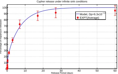
∂x2 . (1) For the case of in-vitro drug release a zero flux bound-ary condition is normally assumed at the impermeable stent and either an infinite sink or Robin type bound-ary condition at the interface with the release medium. Models such as these can admit analytical solutions and have shown favourable results when compared with in-vitro experimental data. Figure 1 displays a comparison between the model predicted cumulative percentage of drug released and the average of four experimental in-vitro release profiles from the Cypher stent (for more details see Section 6). These analyti-cal solutions also allow the drug diffusion coefficient to be estimated via a best fitting process.
Several simplifying assumptions are usually made. Firstly, it is assumed that the device geometry is that of a thin film with no edge effects so that the modelling may be restricted to one dimension. The diffusion of the drug in the polymer is thus consid-ered to be isotropic and it is usually assumed that the diffusion coefficient is independent of time, space and concentration. Furthermore, the initial drug concen-tration is usually taken to be uniform. However, in reality this is not always the case. For example, the polymer coating on the Cypher stent consists of three distinct layers; a base coat, a drug-filled middle layer
0 10 20 30 40 50 60
0 10 20 30 40 50 60 70 80 90 100
Cypher release under infinite sink conditions
Release Period (days)
Percentage of Rapamycin released
Model, Dp=6.3x10⁻¹⁷
[image:3.612.327.516.47.166.2]EXPT(Average)
Figure 1: Comparison between in-vitro experimental data and model of Cypher release
and a drug-free topcoat. However, the good fit be-tween the experimental data and the simple model de-scribed above suggests that these modelling assump-tions are reasonable and capture the release of drug from the Cypher stent, albeit with a layer averaged, or effective diffusion coefficient. A tighter fit is antici-pated if the polymer is modelled as a tri-layer system with the drug contained only within the middle layer initially. Actually, it has been shown that the drug can be found in the top coat prior to implantation, sug-gesting that the intended drug-free top coat may not always be realised. However it is less clear how the in-vivo situation may be modelled. This is considered in the following section.
NEVO stents:
Mstent(0)−Mstent(t) =Mf0
1−e−Kf0t+Qsus
√
t,
(2) where Mstent(0), Mf0, Kf0 and Qsus denote, respec-tively, the initial load of drug, the initial pool of first order eluting drug, rate constant and Higuchi rate con-stant. This equation, however, does not satisfy mass conservation principles: the mass of drug released eventually tends to infinity as time increases. As a re-sult, while their data is well-fitted to this equation for this set of experiments, it is unlikely that their model may be used in a predictive capacity. A further warn-ing must be issued when attemptwarn-ing to identify the re-lease mechanism: it may be possible to fit experimen-tal data to a given model with a variety of different parameter combinations, even if the values of some of the parameters are unlikely (Sirianni et al., 2010).
5
MODELLING UPTAKE OF
DRUG INTO THE ARTERIAL
WALL
While the stent manufacturer may be primarily con-cerned with stent design and the drug release profile, clinicians are more interested in the drug concentra-tion profile across the arterial wall, and in particular in the target SMCs, as well as the therapeutic dura-tion. Thus an understanding of what happens to the drug in the arterial wall is essential. The arterial wall is porous and plasma flows through the extracellular matrix as a result of the transmural pressure gradi-ent. This pressure gradient induces convection so that drug transport across the arterial wall is governed by a combination of convection, molecular diffusion and binding. A drug with anti-proliferative properties is usually chosen so that when it is uptaken by SMCs it suppresses their proliferation. Depending on the par-ticular properties of the drug, it may also bind to sites within the extracellular matrix.
5.1
One-dimensional Models
Early models of drug transport through the arterial wall tended to include only convection and diffusion ((Pontrelli and de Monte, 2007), (Zunino, 2004)) and neglected drug uptake/binding. Furthermore, they re-stricted the number of dimensions and reduced the problem to a single layer in the arterial wall. One of the first models which encompassed convection, dif-fusion and uptake into SMC within the porous media
was presented by McGinty et al.:
φ∂CE
∂t +v
∂CE
∂x = Dm
∂2C
E
∂x2 −α
CE−
CI
K
(3)
(1−φ)∂CI
∂t = α
CE−
CI
K
, (4)
where CE and CI denote the volume averaged
con-centration of drug in the extracellular and cellular re-gions, respectively (McGinty et al., 2011). The pa-rametersφ, v, Dm,αand K denote the porosity,
mag-nitude of transmural convection, drug diffusion co-efficient in the media, drug uptake rate constant and partition coefficient. Equation 4 expresses the rate of uptake of drug by the cells: it is initially propor-tional to CE but that proportionality diminishes with
increasing CIuntil the carrying capacity (or partition
coefficient) of the drug is reached at which point the uptake becomes zero. This system of equations al-lows for an exchange of drug between the extracel-lular phase and the cells which is dependent on the concentration in the extracellular phase. They con-sidered the coupled polymer/media system (equations 1,3 and 4) with continuity of drug concentration and continuity of the relative fluxes expressed as bound-ary conditions at the interface:
Cp = CE (5)
−Dp
∂Cp
∂x = −Dm
∂CE
∂x +vCE. (6) They assumed that the flux of drug out of the media was proportional to the concentration at the interface between the media and adventitia to provide the final boundary condition. Their model was also extended to include the adventitia region (where fibroblast cells were modelled in a similar way to SMCs), a topcoat of polymer to slow the release of the drug, and one of the first models of atherosclerotic plaque (modelled using an equilibrium model in the same way as SMCs uptake).
concentrations. The McGinty et al. model did, how-ever, neglect the intimal region of the arterial wall and the endothelium layer of cells. Their justification for this is that the endothelium is severely damaged when a stent is inserted and in some cases is completely re-moved; and indeed the properties of the intimal may not be too different from those in the media.
Despite considering two layers and accounting for convection, diffusion and uptake/binding in each, their model makes several simplifying assumptions. Firstly, the geometry is restricted to one dimension and as such cannot account for the anisotropic nature of diffusion within the tissue ((Levin et al., 2004), (Hwang et al., 2001)). Furthermore, the 1D model clearly has its limitations in approximating 3D geom-etry. The strut is considered to be in contact with the arterial wall, when in reality it is likely embed-ded within it. Finally, the flow interaction problem between the blood and the struts is not accounted for, leading to the potential for under-prediction of drug lost to the lumen, an aspect which has also been in-vestigated in the literature (Zunino, 2004).
Pontrelli and de Monte (Pontrelli and de Monte, 2010) proposed a similar model to that of McGinty et al. which allowed for a diffusion controlled re-lease from the stent as well as convection-diffusion-reaction in the arterial wall. Their most sophisticated model has the benefit of being multi-layered, but is unable to model the drug concentration in the SMCs. They do, however, model drug consumption via a lin-ear reaction:
φ∂Ci
∂t +2γi
∂Ci
∂x =Di
∂2C
i
∂x2 −βiCi, (7)
where the subscript i indicates the ith layer and 2γi
represents a constant characteristic convection param-eter. Pontrelli and de Monte’s model has the advan-tage of admitting an analytical solution.
The convective and diffusive element of the drug transport is well established, as evidenced by the above models and countless others. However, the is-sue of uptake/binding is more controversial. Some authors have assumed equilibrium models ((McGinty et al., 2011), (Horner et al., 2010), (Abraham et al., 2013)), while others have considered simple loss terms (Pontrelli and de Monte, 2010). More recently, a second order reaction model which allows for sat-urable reversible binding of sirolimus to specific re-ceptors and general extracellular matrix (ECM) sites has been proposed (Tzafriri et al., 2012). The equa-tions for the rate of uptake of sirolimus to ECM sites
and receptors are: ∂bECM
∂t = k
ECM
on c(bECM,max−bECM)
−kECMon kECMd bECM, (8)
∂bREC
∂t = k
REC
on c(bREC,max−bREC)
−kRECon kRECd bREC. (9)
Here, c is the molar concentration of free drug per unit tissue volume, bECMand bRECare the molar
con-centrations of ECM-bound and receptor-bound drug, respectively. The parameters bECM,max and bREC,max denote the local molar concentration of ECM and re-ceptor drug binding sites, kECMon and kRECon are the re-spective binding on-rate constants and kECMd and kRECd are the respective equilibrium dissociation constants. It is certainly true that the drug will bind to bind-ing sites in the tissue and in the cells ((Levin et al., 2005), (Tzafriri et al., 2012), (Bierer et al., 1990)) although the strength of the affinity will likely vary substantially with the particular drug under consider-ation. Furthermore, it is not clear how the density of the binding sites could easily be determined. Thus it may be that this binding model is specific to a partic-ular class of drugs and not suitable for more general compounds. A greater understanding of the binding process would undoubtedly assist with model devel-opment.
5.2
Higher-dimensional Models
Despite providing useful, and in some cases, counter-intuitive physiological insights, one-dimensional models are inadequate for accurately resolving quan-titative aspects. When the dimension of the model is increased, numerical approaches are necessarily re-quired.
Two-dimensional models in simplified geometries were computed by (Hwang et al., 2001), (Grassi et al., 2009) and (Zunino, 2004) among others. A number of three-dimensional models have also been devised. (Weiler et al., 2012) provided a broad generalization of the works of (Mongrain et al., 2007), (Zunino et al., 2009) and (Vairo et al., 2010); a three-dimensional model of drug transport in the lumen and the arterial wall. Laminar steady flow was assumed in the lumen and the steady diffusion equation (no convection) in the arterial wall. Through numerical simulation using commercial finite element software, they found that the highest rates of mass transfer occurred at the for-ward portion of the stent and the rate of drug delivery to the lumen was greater than that to the tissue.
reaction-diffusion-convection model in a realistic geometry. They stress the importance of considering two phases of the drug (bound and unbound) and use a first or-der reaction kinetics model to describe the transfer of drug between the two phases. They utilise ABAQUS to obtain a realistic geometry of a deformed stent and vessel wall and then utilise FLUENT to solve their transport equations. Their three-dimensional setting allows for the consideration of anisotropic diffusion in the arterial wall. They do, however, make three significant simplifications. Firstly, they model the ar-terial wall as a linear homogeneous solid and do not distinguish between the intima, media and adventi-tia. Secondly, despite calculating the transmural ve-locity field, they assume this is fixed when solving the transport equations. Perhaps the most unrealistic assumption is that the drug concentration on the stent remains constant and does not deplete. They find that deposition patterns tend to follow the pattern of the stent struts and that the drug is able to penetrate deep into the arterial wall. The pattern of bound drug be-comes less uniform as the Peclet number is increased, eventually becoming restricted to areas adjacent to the struts, as convection dominates over diffusion.
6
EXPERIMENTAL VALIDATION
An important aspect of modelling and simulation is validation. The accuracy of the model results can only ever be as good as the quality of the inputs, espe-cially when the model is sensitive to changes in one or more of the parameters. At present, before a DES is approved for use in humans it must undergo in-vivo testing in an animal model (for example porcine coro-nary artery); this is a very costly exercise and also raises ethical questions. However, a series of in-vitro and ex-vivo experiments can be carried out at various stages of the modelling process to verify certain as-pects and to suggest improvements/modifications to the model.
One such example of this is in the estimation of the model parameters. In our laboratory we have been performing in-vitro DES release experiments which have allowed us to estimate the diffusion co-efficient of the drug in polymer-coated stents based on a least squares analysis and utilising analytical so-lutions (Figure 1). The experiments consisted of plac-ing Cypher DESs in a sealed glass vial containplac-ing physiological release medium (phosphate buffered saline:ethanol (90:10)). At several time points up to 60 days, the stent was removed and placed in a sep-arate vial containing fresh release medium, with the mass of drug in the original solution subsequently
quantified using UV-spectroscopy. These simple ex-periments give confidence in the modelling and pro-vide reliable estimates of the drug diffusion coeffi-cient in the polymer, which can feed into more sophis-ticated models. Alternatively, a diffusion cell contain-ing a membrane made from the polymer under study can be used. The diffusion coefficients in each layer of the arterial wall can be measured in a similar way. The porosity of each layer of the arterial wall may be measured from histological sections by quantitative microscopy. Drug uptake/binding parameters, such as the partition coefficient K and uptake rate constant αin equations (3-4) may be estimated by quantifying drug uptaken by cells grown in culture plates at dif-ferent time points. For estimation of the parameters in the second order reaction model (equations 8-9), the reader is referred to (Tzafriri et al., 2012) and ref-erences therein.
But the use of experiments to inform the mod-elling is not a one-way process. Indeed, our group have utilised the modelling to design experiments which in turn have fed back into the model. For ex-ample, recognising that one of the important features of in-vivo drug release from DESs is transport by con-vection, in our laboratory we are developing ex-vivo perfusion circuit experiments which will allow us to control the intraluminal pressure (and thus vary con-vection) across the arterial wall. Thus, experimental validation should not be seen as the final step, only to be performed once the model has been built and the results simulated. Instead, the modelling and exper-imentation should go hand-in-hand, complementing each other.
7
MODELLING
CONSIDERATIONS FOR THE
FUTURE
Whilst significant progress has been made in simulat-ing various aspects of drug-elutsimulat-ing stents, there is still a need to better understand the drug elution process and the drug transport in the lumen and arterial wall. Here we indicate some possible modelling consider-ations for future research in this field so that the aim of achieving an ‘optimal’ drug-eluting stent may be realised.
7.1
Simulating Drug Release from New
Generation DESs
biodegrad-able polymer coated stents and polymer-free surface-modified stents. It may be that the dominant release mechanism in these stents is not diffusion and so models which assume purely diffusion may not cap-ture the release kinetics. Any new models should be experimentally validated in-vitro to verify the phys-ical processes governing the release have been cap-tured. It is anticipated that for most DESs the in-vivo release profile may be significantly different due to the complex biological processes involved, but if the in-vitro release can be well modelled then this should shed some light as to how to model the in-vivo situa-tion. Comparison of the in-vitro drug release profile of different stent platforms may indicate the release mechanism(s) which give rise to the most favourable release profile. This, in turn, may allow for better de-sign of DESs in the future.
7.2
Inclusion of the Endothelium,
Intima and Plaque
The inner layer of the the arterial wall, the intima, is a thin layer comprising the endothelium as well as the elastic lamina. It is well established that the endothe-lium is damaged during the stent insertion process and in some cases even removed. As a result, many cur-rent models neglect the intima region.
A more complete model of the arterial wall should include the intima region as well as the endothelium and internal elastic lamina. The endothelium is im-portant in vasoconstriction and vasodilation and reg-ulates the uptake of plasma into the arterial wall. While diseased endothelium is dysfunctional (this is the starting point of atherosclerosis) it will neverthe-less have an effect on the relative importance of con-vection to diffusion in the arterial wall. It is antici-pated that drug transport through the initma will oc-cur via similar processes to that in the media, namely diffusion (albeit with a different diffusion coefficient from the media), convection and possibly binding.
Another possible modelling consideration is atherosclerotic plaque; the presence of the plaque is the very reason that the stent is inserted and yet it has received very little attention in the literature. McGinty et al. seem to be the only authors who have attempted to model the plaque to date, although (Tzafriri et al., 2010) have experimentally examined the effect of the plaque. The plaque is known to con-tain a fibrous cap of variable thickness as well as a necrotic core made up of cellular debris, cholesterol cleft and cell membranes. Furthermore, the plaque also contains macrophages and SMCs as well as a lipid pool containing lipid dispersed in a collagen matrix. A More sophisticated model of plaque,
tak-ing into account its various components, may provide more insight into the effect of plaque on tissue drug concentrations.
7.3
Modelling Lumenal Blood Flow and
Stent Interaction
In simulations where the blood flow is taken into ac-count, it is common for the blood flow to be modelled as steady Poiseuille flow. Of course, in reality blood flow near the heart is pulsatile and the artery is contin-ually contracting and expanding. The presence of the stent interrupts the flow (Peacock et al., 1995) and the effect this has on the drug transport should be simu-lated.
In addition to simply considering some of the pa-rameters to be time and space dependent, it may be necessary to consider the proliferation of SMCs and neointima growth as a wound healing problem.
7.4
Inclusion of Complex
Three-Dimensional Geometry
While simplified one-dimensional models can pro-vide useful insights into this problem, ultimately three-dimensional models which capture the full com-plex geometry of the stent and the arterial wall are required. The idea of numerically simulating such a complex problem may have seemed impossible not so long ago, but with the accelerating advances in computational power and numerical techniques it is now possible. The existing three-dimensional models in the literature all make certain simplifying assump-tions, whether it be in idealising the stent geometry, or in neglecting convection, diffusion or binding, or in considering only single or bi-layer arterial walls. Thus there is an opportunity to increase the sophisti-cation of the three-dimensional models, whether it be incrementally or in one fell swoop. However, caution must be exercised to ensure that the results of the sim-ulations are not subject to high uncertainty, in which case the fidelity of the results may be called into ques-tion.
8
CONCLUSIONS
complex in-vivo situation where flowing blood, pul-satility, wound healing, proliferation and migration of SMCs and complex uptake/binding no doubt all play some part. Future research should include the modelling of drug release from biodegradable and polymer-free modified surface stents, more accurately modelling lumenal blood flow and stent interaction, including the endothelium, the intima and atheroscle-rotic plaque. In order to be able to use simulations in a predictive capacity, three-dimensional models which encompass the full complex geometry are necessarily required. But care should be taken to verify the cor-rectness of the numerical results. Finally, the mod-elling should be complemented by appropriate exper-iments to validate the resulting simulations and im-prove on the model.
ACKNOWLEDGEMENTS
We would like to acknowledge the funding provided by EPSRC under grant number EP/J007242/1. The first author would also like to acknowledge the receipt of a Carnegie Scholarship.
REFERENCES
Abraham, J. P., Gorman, J. M., Sparrow, E. M., Stark, J. R., and Kohler, R. E. (2013). A mass transfer model of temporal drug deposition in artery walls. Int. J. Heat
Mass Trans., 58:632–638.
Beers, M. H. (2004). The Merck Manual of Health & Aging. Elsevier Health Sciences, London.
Bierer, B. E., Patilla, P. S., Standaert, R. F., Herzenberg, L. A., Burakoff, S. J., Crabtree, G., and Schreiber, S. (1990). Two distinct signal transmission pathways in t lymphocytes are inhibited by complexes formed between an immunophilin and either fk605 or ra-pamycin. Proc. Natl. Acad. Sci. USA, 87:9231–9235. Delfour, M. C., Garon, A., and Longo, V. (2005). Modeling
and design of coated stents to optimize the effect of the dose. SIAM J. Appl. Math.., 65(3):858–881. Fredenberg, S., Wahlgren, M., Reslow, M., and
Axels-son, A. (2011). The mechanisms of drug release in poly(lactic-co-glycolic acid)-based drug delivery sys-tems - a review. Int. J. Pharmaceutics, 415:34–52. Garasic, J. M., Edelman, E. R., Squire, J. C., Seifert, P.,
Williams, M. S., and Rogers, C. (2000). Stent and artery geometry determine intimal thickening inde-pendent of arterial injury. Circulation, 101(7):812– 818.
Grassi, M., Pontrelli, G., Teresi, L., Grassi, G., Comel, L., Ferluga, A., and Galasso, L. (2009). Novel design of drug delivery in stented arteries: a numerical compar-ative study. Math. Biosci. Eng., 6(3):493–508. Horner, M., Joshi, S., Dhruva, V., Sett, S., and Stewart, S.
F. C. (2010). A two-species drug delivery model is re-quired to predict deposition from drug-eluting stents.
Cardiovasc. Eng. Technol., 1(3):225–234.
Hwang, W., Wu, D., and Edelman, E. R. (2001). Physiolog-ical transport forces govern drug-distribution for stent based delivery. Circulation, 104(7):600–605. Khan, W., Farah, S., and Domb, A. J. (2012). Drug eluting
stents: Developments and current status. J. Controlled
Release., 161:703–712.
Levin, A. D., Jonas, M., Hwang, C. W., and Edelman, E. R. (2005). Local and systemic drug competition in drug-eluting stent tissue deposition properties. J.
Controlled Release, 109:236–243.
Levin, A. D., Vukmirovic, N., Hwang, C. W., and Edel-man, E. R. (2004). Specific binding to intracellular proteins determines arterial transport properties for ra-pamycin and paclitaxel. Proc. Natl. Acad. Sci. USA, 101(25):9463–9467.
Lloyde-Jones, D. (2010). Heart disease and stroke statistics-2010 update: A report from the american heart asso-ciation. Circulation, 121:e46–e215.
Lusis, A. (2000). Atherosclerosis. Nature, 407:233–241. McGinty, S., McKee, S., Wadsworth, R. M., and
Mc-Cormick, C. (2011). Modelling drug-eluting stents.
Math. Med. Biol., 28:1–29.
simu-lations for drug eluting coronary stents. J. Biomech.
Eng., 129:733–742.
Murray, C. and Lopez, A. (1997). Alternative projections of mortality and disability by cause 1990-2020: Global burden of disease study. The Lancet, 349(9064):1498– 1504.
Peacock, J., Hankins, S., Jones, T., and Lutz, R. (1995). Flow instabilities induced by coronary artery stents: Assessment with an in vitro pulse duplicator. J. Biomech, 28:17–26.
Pontrelli, G. and de Monte, F. (2007). Mass diffusion through two-layer porous media: an application to the drug-eluting stent. Int J. Heat Mass Trans., 50:3658– 3669.
Pontrelli, G. and de Monte, F. (2010). A multi-layer porous wall model for coronary drug-eluting stents. Int J.
Heat Mass Trans., 53:13629–3627.
Siepmann, J. and Siepmann, F. (2008). Mathematical modelling of drug delivery. Int. J. Pharmaceutics,
364:328–343.
Sirianni, R. W., Jang, E.-H., Miller, K. M., and Saltzman, W. M. (2010). Parameter estimation methodology in a model of hydrophobic drug release from a polymer coating. SIAM J. Appl. Math., 142):474–482. Stefanini, G. G. and Holmes, D. R. (2013). Drug-eluting
coronary artery stents. N. Engl. J. Med., 368:254–265. Tambaca, J., Kosor, M., Canic, S., and Paniagua, D. (2010). Mathematical modeling of vascular stents. SIAM J.
Appl. Math., 70(6):1922–1952.
Tzafriri, A., Vukmirovic, N., Kolachalama, V., Astafieve, I., and Edelman, E. R. (2010). Lesion complexity deter-mines arterial drug distribution after local drug deliv-ery. J. Controlled Release, 142(3):332–338.
Tzafriri, A. R., Groothuis, A., Price, G. S., and Edelman, E. R. (2012). Stent elution rate determines drug de-position and receptor-mediated effects. J. Controlled
Release, 161:918–926.
Vairo, G., Cioffi, M., Cottone, R., Dubini, G., and Migli-avacca, F. (2010). Drug release from coronary artery stents: a multidomain approach. J. Biomech., 43:1580–1589.
Weiler, J. M., Sparrow, E. M., and Ramazani, R. (2012). Mass transfer by advection and diffusion from a drug-eluting stent. J. Heat Mass Transfer, 55:1–7. Zunino, P. (2004). Multidimensional pharmacokinetic
mod-els applied to the deign of drug-eluting stents.
Car-diov. Eng.: Int. J., 4(2):181–191.
Zunino, P., D’Angelo, C., Petrini, L., Vergara, C., Capelli, C., and Migliavacca, F. (2009). Numerical simula-tion of drug eluting coronary stents: mechanics, fluid dynamics and drug release. Comput. Methods Appl.