ERYTHRASMA - A CLINICAL STUDY
Dissertation Submitted in
partial fulfillment of the university regulations for
MD DEGREE IN
DERMATOLOGY, VENEREOLOGY AND LEPROSY
(BRANCH XII A)
THE TAMILNADU DR.M.G.R. MEDICAL
UNIVERSITY
CHENNAI.
CERTIFICATE
Certified that this dissertation entitled "ERYTHRASMA - A CLINICAL
STUDY” is a bonafide work done by Dr.M.Nandhini, Post Graduate Student of
Department of Dermatology and Leprosy and Institute of STD, Madras Medical College,
Chennai - 600 003, during the academic year 2003 - 2006. This work has not previously
formed the basis for the award of any degree or diploma.
Prof. Dr. B.Parveen, M.D.,D.D.,
Professor and Head
Department of Dermatology & Leprosy Madras Medical College,
Chennai - 600 003.
Prof. Dr. Kalavathi Ponniraivan B.Sc., M.D.,
The Dean,
SPECIAL ACKNOWLEDGEMENT
My sincere thanks to Prof. Dr. Kalavathi Ponniraivan, B.Sc., M.D., the Dean,
Madras Medical College for allowing me to do this dissertation and utilize the institutional
ACKNOWLEDGEMENT
I am gratefully indebted to Dr.B.Parveen,M.D., D.D., Professor and Head of Department of Dermatology for her invaluable guidance motivation and help throughout the study. I would like to express my sincere and heartfelt gratitude to Dr.N.Gomathy,M.D.,D.D., former Head of Department of Dermatology who was instrumental in initiating the study and for her constant support.
My sincere and heartfelt thanks to Dr.C.Janaki, M.D.,D.D., Reader of Dermatology (Mycology) for her immense support and guidance throughout the study.
I express my earnest gratitude to Dr.D.Prabavathy, M.D.,D.D., Professor and Head of Department of Occupational Dermatology and Contact Dermatitis for her constant motivation and guidance. I thank Dr.V.Somasundaram, M.D.,D.D., Additional Professor, Department of Occupational Dermatology and Contact dermatitis for his kind help and support.
I am very grateful to Dr.V.S.Dorairaj, M.D.,D.V., Director, Institute of STD for his cooperation and help. I thank Dr.N.Usman M.D.,D.V., Ph.D., former Director, Institute of STD for his support. I wish to thank Dr.S.Mohan, M.D., D.V., Registrar, Institute of STD for his kind help.
I express my sincere gratitude to Dr.K.Rathinavelu, M.D.,D.D., Professor of Leprosy and
Dr.R.Arunadevi, M.D.,D.D., Lecturer / Registrar, Department of Dermatology for their support.
I thank Dr.A.Hameedullah, M.D., D.D., Dr.Kumaravelu, M.D., D.D., and
Dr.J.Manjula, M.D., D.N.B (Derm) Assistant Professors for their support and help.
I wish to thank Dr.S.Venkateshwaran, M.D.,D.V., Dr.Ilangovan, M.D.,D.V., Dr.Thilagavathy,M.D., Dr.Thirunavukkarasu,M.D.,D.V., Dr.Ramachandra Reddy, M.D.,D.V., Dr.P.Mohan, M.D.D.V., and Dr.S.Arunkumar, M.D.,D.V., Assistant Professor, Institute of STD, for their help and suggestions.
I am also thankful to Dr.Senthamilselvi, M.D.,D.D., MNAMS, Ph.D., Dr.K.Manoharan, M.D.,D.D., and Dr.V.Sampath, M.D.,D.D., for their continuing guidance and support.
I gratefully acknowledge Mr.Kannan M.Sc., Microbiology for his help throughout the study.
I duly acknowledge the paramedical staff and my colleagues for their help and favours.
CONTENTS
Sl. No. Title Page
No.
I INTRODUCTION 1
II REVIEW OF LITERATURE 2
III AIMS OF THE STUDY 30
IV MATERIALS AND METHODS 31
V OBSERVATIONS 33
VI DISCUSSION 45
VII CONCLUSION 51
REFERENCE
MASTER CHART
KEY TO MASTER CHART
INTRODUCTION
The normal human skin is colonized by huge numbers of bacteria that live
harmlessly as commensals on its surface and within its follicles. At times, overgrowth of
some of these resident organisms may cause minor disease of skin or its appendages. If
the skin is damaged or the immune status of the subject is impaired, bacteria usually
regarded nonpathogenic on body surface may assume the role of opportunist pathogens.
Erythrasma is a chronic superficial infection of the skin widely prevalent all over
the world. The causative agent is an aerobic diphtheroid called Corynebacterium
minutissimum. Extensive work has been carried out by scientists regarding its
microbiology, biochemical characters and pathogenicity which has expanded our
knowledge about the condition. These organisms contribute to cutaneous ecosystem
normally and their behaviour as pathogens requires local or systemic devitalising
REVIEW OF LITERATURE
HISTORICAL ASPECTS
The disease was first described by Burchardt, a German scientist in 1859, who
suggested that the delicate filaments and granules found in the scales were of fungal
origin and were the cause of the disease1.
The term `erythrasma' was coined in 1862 by Von Baren Sprung, Burchardts'
teacher. He named the causative organism as Microsporum minutissimum.
Most of subsequent workers had no doubt that erythrasma was a separate entity
(Kobner 1884; Balzer and Dubrevilh 1883; Unna 1896). Some including Weyl (1884)
considered that various transitional forms existed between erythrasma and pityriasis
versicolor. Gougerot (1936) recognised the disseminated and subacute forms of
erythrasma and pointed out that the condition would be complicated by eczematisation
or associated with bacterial or fungal infections. Kobner in 1884 succeeded in
reproducing the disease for the first time by applying the scales infected with erythrasma
to the skin of one of his pupils2.
The numerous names under which the causative organism of erythrasma has
appeared in the literature includes Microsporum minutissimum, Nocardia minutissima,
Sporotrichum minutissimum3.
In discussing the etiological factors Poehlmann (1928) considered local factors
sweating, a delicate integument and systemic illness like diabetes mellitus for the
causation of erythrasma.
Robean and Guerra (1936) gave an account of 16 patients with the condition
affecting the toe-webs and added that some of the cases had an associated fungal
infection.
Nikolowski and Stable (1949) reported erythrasma in unusual sites including a
case in which the lesions were confined to the forearms. The generalised form is
characterised by well defined scaly lamellated plaques in the trunks and limbs was
reported by Goncalves and Mangeon (1960).
The bacterial cause for erythrasma was first suggested by Lagana (1960).
However Sarkany, Taplin and Blank (1961) isolated consistently a diphtheroid which
they named Corynebacterium minutissimum from the lesions of erythrasma and this had
given a new outlook on the condition for subsequent scientists to carry out their work4.
Now various studies are being carried out relating erythrasma to obesity, diabetes and its
EPIDEMIOLOGY OF ERYTHRASMA
The overall incidence of erythrasma is 4%. It is prevalent all over the world. Mild
forms of erythrasma of axilla, groins and toe-webs are relatively common in temperate
climates; the less common generalised form is predominantly seen in obese subjects
particularly in middle aged Negro women.
Incidence is higher in closed communities for example the armed forces and
institution. In some institutions, the infection appears to be endemic, the incidence
among the patients remaining relatively constant over a period of years. Sarkany, Taplin
and Blank in 1962 found that in tropical climates the incidence is higher and generalised
form is frequent5. The incidence also increases with age, though a case has been
recorded in one year old child. This increasing incidence was studied by Laube S
(2004)6.
The incidence of erythrasma does not appear to be greater in males than females,
though Board man found toe webs of male students being more commonly affected than
those of females.
Erythrasma is not significantly contagious. Holdiness (2003) found that factors
such as warm climate, poor hygiene, obesity, hyperhidrosis, advanced age, and diabetes
mellitus play a role in the occurrence of the disease7.
Bowyer A (1971) has shown that erythrasma can be associated with pruritus ani8.
Schlappner OL, Rosenblum GA in 1979 reported the association of erythrasma
with dermatophytosis of groin9. Variable data has been recorded in this regard.
Association with pityriasis versicolor has been recorded in groin and axillae10.
The incidence and severity of the disease is apparently greater amongst diabetics.
Somerville and Lancaster in 1973 found that even mild cases of erythrasma were
common among diabetics and carriage of fluorescent diphtheroid was greater11. It is
possible that diabetics are as susceptible to erythrasma as candidiasis and this is perhaps
related to the high levels of cutaneous free glucose as shown in a review by Haroon TS
(1974)12. Montes LF and Dobson H et al have also studied the association of erythrasma
and diabetes mellitus in 196913. Schein-feld NS (2004) reported that obesity is a
predisposing factor for the development of erythrasma. The use of deodorants does not
appear to affect the incidence of erythrasma14.
NORMAL CUTANEOUS FLORA AND DIPHTHEROIDS15
It is important to mention a few words about the normal flora of skin for a better
understanding of the disease.
Skin bacteria are found to be of 2 types transients and residents. Transients which
are relatively scarce on clean unexposed skin are most abundantly present on exposed
skin. The resident flora is a relatively stable population both in size and composition.
The resident aerobic flora consists of Gram positive cocci of Staphylococcus
species, Micrococcus species and a variety of Gram positive rods, the coryneforms
negative residents are Acinetobacter species previously known as Mima and Herellea.
The coryneform (or diphtheroid) organisms are aerobic, Gram positive
non-sporing pleomorphic rods. The coryneform bacteria are difficult to separate by
conventional taxonomic methods and chemotaxonomic methods. A recently proposed
scheme in which the aerobic coryneforms are divided into four corynebacterium species
complexes, C.bovis, C.minutissimum, C.xerosus and C.hofmani with Brevibacterium
epidermis and Propionibacterium spp. making up a total of six groups.
Cutaneous diphtheroids were assigned to different groups depending initially on
their O2 requirements. The anerobic under the term C.acnes and aerobic diphtheroids
according to their origin, lipophilicity and porphyrin production. C.acnes has now been
shown to be a propionibacteria which are anerobic Gram positive rods. P.acnes are
particularly associated with follicles that have large pilosebaceous glands over face and
upper trunk. It is associated with acne lesions in which its role is a matter of
considerable interest.
CORYNEFORM BACTERIA
Human commensals or pathogens Aerobic
Corynebacterium diphtheriae C.haemolyticum
C.pyogenes C.xerosis
Primarily throat
C.hofmanii C.minutissimum
Brevibacterium epidermis
Anerobic
Propionibacterium acnes P.granulosum
P.avidum
Pathogens of other vertebrates sometimes infecting humans
Listeria monocytogenes Erysipelothrix rusiopathiae
Aerobic diphtheroids are commonly found in large numbers on the skin especially
in adult life. Two characteristics features of these aerobic diphtheroids are lipophilia
and porphyrin production.
Sebum is an important source of metabolites for both the aerobic and anerobic
diphtheroids. Palmitate is the most common fatty acid formed in human surface lipids
and all lipid dependent strains hydrolyse this substance.
Porphyrin production can be demonstrated by examining under UV light, the
cultures grown on suitable media when, the typical coral red fluorescence is seen. In
1961, Sarkany, Taplin & Blank detected porphyrin production by the diphtheroid
responsible for erythrasma16. However this characteristics is more prevalent amongst
corynebacteria than expected. In addition to C.minutissimum, C.xerosis, C.bovis,
C.ulcerans, C.renale also show fluorescence. C.diphtheriae is known to produce
coproporphyrin III under conditions of iron deficiency and the red fluorescence of the
comedones of face has been shown to be due to the production of coproporphyrin III and
Protoporphyrin IX by P.acnes.
Somerville in 1973 used nine simple tests (lipophilia, glucose fermentation, nitrate
reduction etc) to arrange the aerobic cutaneous diphtheroids into 15 groups seven of
which produce porphyrins and eight non porphyrin producers11.
Phages have been isolated from various species of corynebacteria other than
C.diphtheriae. Montes and his colleagues demonstrated by electron microscope an
apparent bacteriophage cycle in diphtheroids on human skin.
The aerobic diphtheroids may be isolated from the skin in virtually all parts of the
body in most people. They are particularly common in axilla, groin and toe webs. Age
plays an important role in determining the incidence of the organisms. In newborn, at
first there are relatively few organisms though an increase in number occurs within a
matter of few hours. Numbers and incidence increase in childhood but are highest in
young adults. It is suggested that with the increase in the amount of sebum secreted on
to the skin at puberty, conditions favour the survival and growth of these organisms
source of which are lipolytic and some lipophilic. In the axilla diphtheroids with the
micrococci contribute to the development of axillary odour.
So much for the role of diphtheroids as normal flora, among which few are
MICROBIOLOGY OF CORYNEBACTERIUM MINUTISSIMUM
C.minutissimum belonging to the aerobic diphtheroids has common morphologic
features of diphtheroids. On staining with Gram stain, the isolates from the skin lesion of
fluorescent scales of erythrasma show rod like organisms, filaments and coccoid forms.
The filaments are tortuous and measure about 4µ to 10µ by 1µ. The longer filaments
have segmented or beaded appearance. The bacillary forms are 1µ-3µ long and 0.5µ in
diameter. Some of them reveal subterminal granules.
The shorter bacillary forms are more profuse in the erythrasma lesions of toes,
however filamentous forms and chains of bacilli are also found1. The lesions often
contain higher proportion of filaments but invariably also shorter bacillary forms.
Under electron microscope17 numerous bacteria will be seen at different levels of
stratum corneum. Proliferating over the skin surface lying freely between the superficial
cornified cells, penetrating these cells from the intercellular space or less frequently,
directly from the skin surface and intracellularly within the keratinised cells.
Most of the organisms observed to be on the skin surface are characterised by
homogenous fine structures. Bacteria within the stratum corneum are quite pleomorphic.
Furthermore, dividing organisms are more common on the surface than within the
stratum corneum.
The stratum corneum itself becomes hyperkeratotic with superficial layers widely
separated and the cell boundaries disrupted at the sites of bacterial penetration. In the
observed around the intracellular bacteria probably suggesting a keratolytic process.
Electron micrographs of the full stratum corneum thickness have shown that the
bacteria in erythrasma can penetrate as deeply as one-half of the thickness of that layers.
Bacteriophages apparently exist within the bacterial cell18.
R.Marks and N.D.Ramnarain (1972) studied erythrasma patients using skin
surface biopsy technique3. The skin surface biopsies were tested for enzyme
histochemical reactions and were also subjected to scanning electron microscope
studies.
Skin surface biopsies from the affected sites stained with PAS reagent or Gram
stain demonstrated the presence of numerous fusiform microorganisms which are
arranged in clusters, chains or singly and scattered over variously sized areas.
The chains that were seen usually comprised three, four or occasionally more,
individual bacterial cells. The microorganisms appeared approximately three to four
times as long as they were broad.
With PAS stain there was noticeable diffuse staining of the sites containing the
micro organisms which was visible macroscopically. There did not appear to be a
particular accentuation around hair follicles or sweat gland openings.
The enzyme histochemical studies show that corynebacterium minutissimum
possess a wide range of enzyme activities. Mitochondrial enzyme activities seemed
reaction products from the tests performed were not distributed diffusely within the
individual bacterial cells but appeared aggregated in well defined foci within each cell.
With the scanning electron microscope single rod shaped and oval structures and
chains were seen lying on, and embedded in, the surface of the individual cornified cells.
The surface of the bacterial cells appeared regularly smooth. The chain of bacterial cells
were frequently seen to be penetrating the cornified cells. At the site of penetration there
were, in several scales openings that were two or three times larger than the diameter of
the microorganisms.
The involved cells in the stratum corneum had an irregular ridge pattern, mainly
composed of low, broken, undulating ridges which sometimes branched. The
surrounding uninvolved cells often possessed prominent villi resembling the surface
structure of the scale in psoriasis and some other parakeratotic disorders (Dawber,
Marks and Swift, 1972).
This skin surface biopsy study showed that there was a remarkable disparity
between the numbers of bacteria seen in stained preparations with light microscope and
the number seen by scanning electron microscope. This is probably a reflection of the
fact that the surface scanned by scanning microscope is five or six cell layers down into
stratum corneum, while with the light microscope the whole thickness of the specimen
removed is examined.
Montes, Black and McBride (1967) demonstrated by conventional electron
further than the superficial part of the stratum corneum19.
The surfaces of the horn cells into which the micro organisms had penetrated
possessed a disorganized ridge pattern suggesting a possibility of disruption of the
tonofilament desmosome complexes. At the site of penetration the opening was wider
than the micro organisms. This suggests that penetration was accomplished by a
chemical dissolution rather than by purely physical pressure.
Montes et al. (1967) noted disorganisation of keratin fibrils in horn cells invaded
by C.minutissimum.
The enzyme reaction products were aggregated in well defined areas confirming a
complex subcellular arrangement akin to mammalian cells. A similar arrangement has
been noted with enzyme reactions in the dermatophyte fungi (Meinhof, 1968).
The smooth exterior of the organism observed by scanning electron microscope
with only a slight constriction at the site of the junctions between microorganisms in a
chain and the PAS reactivity of the microorganisms reflects the secretion of a
mucopolysaccharide sheath by the organism in some situations.
Somerville (1972) discussed the microbiology of cutaneous diphtheroids and
stated that the diphtheroids responsible for trichomycosis axillaris produced secretions
that stick them together and to the hair, and cause, in addition, destruction of the hair
keratin. A similar material may well be produced by C.minutissimum11.
organisms under specific conditions4. This medium consist of 78% tissue culture
medium without bicarbonate, 20% fetal bovine serum and 2% agar. The prepared
medium is autoclaved for 10 mts at 15 lbs pressure per square inch. Plates are poured by
decanting the hot liquid from the coagulated proteins which are discarded. On
inoculation at 380C the fluorescent scales of erythrasma lesions, within 24-48 hrs, small
shiny, round, translucent, colorless, slightly elevated convex colonies appear. There is
no pigment production in visible light, but red fluorescence diffusing into the
surrounding medium can be observed under Woods light. Some batches of fetal bovine
serum have an inhibitory effect on the growth of the organisms. Autoclaving the
medium or using a dialysate of fetal bovine serum helps to promote growth and
fluorescence. Inhibition of growth also takes places, when autoclaved medium 199 is
used with human or horse serum. Certain batches of Tissue Culture Medium 199 are
marketed with the addition of antibiotics such as penicillin or streptomycin. Such media
suppress the growth of the organisms and are unsuitable. A pink fluorescence in and
around the colonies is also seen on sheep blood agar, chocolate agar and yeast extract
casein agar, but the fluorescence assumes striking proportions only on the tissue culture
medium. Subculture of the organisms grow on a finally large number of bacteriological
media. They also thrive on the chorioallantoic membrane of ten day old embryonated
eggs at 380C.
Gram stain of a smear from a colony shows Gram positive rods which become
pleomorphic in older cultures. These organisms contain metachromatic granules and are
non-motile, aerobic or microaerophilic, catalase positive, indole negative, non
field microscopy shows typical bacilli with well rounded ends and a marked thick outer
wall.
In vitro sensitivity tests carried out on solid and liquid media show that the
organisms are maximally sensitive to erythromycin.
Sarkany and his colleagues applied pure culture to stripped or scarified areas of
skin of forearms and kept them occluded for 72 hrs. Scaling and fluorescence occurred
in three out of five inoculations. These were of relatively short duration and did not
develop into permanent erythrasma. Fluorescent cultures were reobtained from these
experimentally induced lesions. Hence Sarkany et al. showed that this organism fulfilled
Koch's postulates1.
The grouping scheme proposed by Somerville shows that any of the seven groups
of fluorescent diphtheroid may be isolated from the lesions of erythrasma. Using this
grouping scheme Somerville and her colleagues found that in their community, one
particular group was more commonly associated with erythrasma than with healthy skin
and this represents an endemic strain. The initial isolate described by Sarkany, Taplin
and Blank and used as type strain in the National collection of type culture belongs to
Group 24.
It is clear that fluorescent diphtheroids associated with erythrasma are members of
normal skin flora which multiply under certain conditions like chafing and maceration in
axilla and groins to become predominant members of the skin flora and eventually
diphtheroids also appear in greater members in these lesions rather than on healthy, non
scaling, non fluorescing areas. So conditions favour the multiplication of all the skin
PATHOGENESIS 20,21
The skin with mucous membrane forms the first line of defense against infection.
Both dryness and keratinisation limit proliferation of microorganisms. This is supported
by low pH of skin surface. These characteristics also limit its colonisation with
commensal organisms.
Microbes that are able to cause infections need to overcome the above defense
mechanisms. These get through an intact epidermis either mechanically or by lysing the
keratinocytes. Some organisms are continuously on look for an opportunity to get under
the epithelium. This occurs when a break usually traumatic occurs in epidermis.
The stratum corneum consists of avital keratin mixed with lipids secreted by
different skin glands. The organisms that parasitise on the skin mostly skin commensals
use these lipids as substrate for metabolism. Often their presence goes unnoticed but if
there is enough substrate available they grow out in numbers, that evoke an
inflammatory response of underlying tissue. This results in itching with or without
erythema. Because secretory glands of skin have highest concentration in skin folds,
such disease originate almost exclusively from one of these, of which erythrasma is
one22.
Thus the causative organism corynebacterium minutissimum which is a
commensal outgrows in number due to excessive sweating and maceration and being
lipophilic produces lesions predominantly involving flexures.
and life threatening infection however, there have been no previous reports of deep
tissue invasion by Corynebacterium minutissimum.
Stephen A Berger et al. has studied recurrent breast abscesses caused by
Corynebacterium minutissimum in a patient23. There was no clinical evidence of
erythrasma in the patient but had skin disruptions. Local trauma may have served as the
portal of entry leading to invasive infections.
Clinical features
The common sites of involvement are groin, upper thigh scrotum, pubis, axillae,
inguinal folds, inframammary areas, toe webs. In toe webs, infection is chronic and most
commonly between 4th & 5th toe followed by 3rd and 4th toes24. Scaling, fissuring and
maceration occur.
Cabo H et al (1983) have described a case of generalised erythrasma in a patient
with Type II diabetes25. In generalised forms well defined scaly, lamellar plaques in
larger areas of trunk, proximal parts of limbs, breast folds may occur. In discoid form26
of erythrasma circular scaling patches which are easily confused with pityriasis
versicolor, pityriasis rotunda and psoriasis. This form is also called tropical
erythrasma27. A case of disciform erythrasma was presented by Tschen JA and Ramsdell
WM in 1983. It was characterised by an atrophic appearing surface and diagnoses was
made with help of Wood's light examination and Gram stain.
The lesion of classic form morphologically appear as punctate, well
lesions are associated with fine scaling. The advancing ends are serpiginous. The lesion
has no tendency for vascularisation. The color of lesion depends upon i) age of lesion ii)
underlying skin pigmentation. It is initially pink color later brown and has no central
cleaning. Occasionally vesiculation of lesion was reported by Grigoria and J.Delacretaz
in interdigitoplantar type of erythrasma28. Vesiculation in the lesions were preceeded by
pruritus. According to them, the lesions showed initial vesiculation and erythema
followed by large bullae which were initially yellow and later became opalescent.
Rarely lesions may become eczematous.
Many of the lesions were asymptomatic and patients seek medical attention for
cosmetic disability. Occasionally these lesions are pruritic. Chronicity and recurrence
are noted in many cases. Occasional association of erythrasma with pruritus ani was
descirbed by A.Bowyer & McColl8. The pruritus was longstanding and cure was
achieved with erythromycin in such cases.
Toe nails may be involved in erythrasma in the form of subungual hyperkeratosis
and onycholysis as reported by Negroni P29 in 1976. Shelley WB and Shelley ED (1982)
studied the corynebacterial triad of coexistent erythrasma, trichomycosis axillaris and
pitted keratolysis. They showed that pitted keratolysis caused by Corynebacterium taplin
and trichomycosis axillaris caused by Corynebacterium tenuis are associated with
erythrasma in few patients30.
Few systemic infections occurs due to Corynebacterium minutissimum. Guarderas
J and A. Karnad (1986) reported Corynebacterium minitissimum bacteremia in a patient
C.minutissimum endocarditis was reported in 1985 by Brian H.J. and Brucker A.J32.
In 1984 Stephen A Berger and Alfred Gorea reported recurrent breast abscesses
caused by C.minutissimum. The systemic infections that can occur due to
C.minutissimum are i) septicemia in neutropenic patients ii) infective endocarditis in
valvular heart disease patients iii) post surgical wound infections iv) recurrent abscesses.
INVESTIGATIONS
i) Wood's lamp examination26:
Wood's lamp is a high pressure mercury lamp with a special filter that allows the
emission of a largely monochromatic UV light with a wavelength of 365 nm. It is
especially useful in the diagnosis of certain fungal and bacterial disease and in the
assessment of pigmentary disorders.
For examination, the room has to be completely darkened and the patient should
be totally undressed. Lesions caused by Microsporum audouinii, M.canis and
M.ferrugineum are identified by their blue-green fluorescence. Pityriasis versicolor
lesions gives a golden yellow fluorescence. Pseudomonas aeruginosa colonisation gives
rise to a blue color.
Hypopigmentation or depigmentation are more easily detected with the aid of
Wood's lamp. Urine, stool and red blood cells of patients suspected of having porphyria
In erythrasma the suspected lesions are examined with Wood's light and coral red
fluorescence occurs due to a porphyrin production by the bacteria corynebacterium
minitissimum. Wigger Alberti N and Elsner P in 1997 did an extensive work on
fluorescence with Wood's light and has shown that if the lesions are washed with
antibiotic soaps before Wood's lamp examination, fluorescence may be transiently
absent33.
Sometimes fluorescence may persist even after treatment of erythrasma.
Mattox TF and Rutgers J (1993) have described a case of non fluorescent
erythrasma of the vulva. It was diagnosed by Gram stain and culture34.
Other lesions that give pink fluorescence under Wood's lamp examination are.
i. Necrotic tumors
ii. Normal tongue
iii. Follicular openings of face and upper trunk
iv. Acanthosis nigricans of groin and axilla
2. Direct Examination:
a) 10% KOH preparation:
To the scrapings from the lesion 10% KOH solution is added and examined under
b) Gram stain:
The scales from the lesion are scraped and fixed to the slide using egg albumin.
Alternatively Padilha (1996) described a single method to stain Malassezia furfur and
C.minutissimum in scales collected on scotch tape with lactophenol cotton blue35.
On staining Gram positive filamentous and coccoid forms were seen.
c) Culture
Culture is difficult and rarely necessary but may be accomplished with a special
medium the ideal one being Tissue culture medium 199. Stephen N Cohen and Dorothy
Nicholai in 1969 described simple mediums for pigment production by erythrasma
diphtheroid36.
On inoculation at 380C the scales of erythrasma lesion, in 24-48 hours smooth
small shiny translucent convex colonies appear. These colonies fluoresce under Wood's
lamp for upto 4 days. Gram stain of smear from a colony shows Gram positive rods
which become pleomorphic in older cultures.
Skin biopsy37:
Biopsy of erythrasma lesion often appears normal. It is described as an example of
invisible dermatoses. Small coccobacilli may be seen in the superficial layers of stratum
corneum. Though difficult to visualise under Hematoxylin and Eosin stain, special stains
like Periodic Acid Schiff and Gomoris Methanamine silver stain usually reveal these
e) Others:
Investigations for underlying factors such as obesity, diabetes mellitus,
hypothyroidism are carried out.
DIFFERENTIAL DIAGNOSIS38
1. Pityriasis versicolor39
The primary lesion is a sharply demarcated macule characterised essentially by
fine branny scaling. Typically the eruption shows large confluent areas, scattered oval
patches and outlying macules. Most commonly upper trunk is affected and often spreads
to upper arms, abdomen, axillae and groins. Under the Wood's lamp, the scaly lesions
may show pale yellow fluorescence.
2. Candidal intertrigo
Most cases of cutaneous candidosis occur in skin folds where occlusion by
clothing or shoes produce abnormally moist conditions. Typically presents with
erythema, cracking and maceration. Lesions have an irregular margin with surrounding
satellite papules and pustules. 10% KOH preparation reveals pseudohyphae.
3. Bacterial Intertrigo
It is an inflammatory condition of skin folds characterized by erythema and
weeping leading on to maceration and crusting. Pustules and vesicles may herald the
stool, urine or topical agents. KOH examination, Gram stain and culture help to identify
the organism.
4. Tinea cruris
It manifests as large patches of erythema with central clearing centered on the
inguinal creases and extend distally down the medial aspect of the thighs with scaling at
the periphery. 10% KOH examination reveals septate branching hyphae with
arthrospores. Woods lamp examination is negative unlike erythrasma.
5. Inverse pattern psoriasis
Posriasis is a chronic, relapsing inflammatory skin disorder with a strong genetic
basis characterized by circular to oval red plaques over extensor body surfaces and the
scalp. Inverse psoriasis is a variant of psoriasis that spares the typical extensor surfaces
and affects intertriginous areas with minimal scaling. Skin biopsy confirms the
diagnosis.
6. Contact Dermatitis
Allergic and irritant contact dermatitis may mimic erythrasma. Allergic contact
dermatitis presents as pruritic papules and vesicles on an erythematous base whereas
irritant contact dermatitis manifests as erythema and hyperkeratosis. A bacterial culture
and 10% KOH examination are done. Patch testing is done to diagnose contact allergies.
Seborrheic dermatitis is a papulosquamous disorder occurring on sebum rich areas
of scalp, face, upper trunk characterized by greasy scaling over erythematous skin. The
flexural variants are non scaly lesions occurring in axillae, inframammary and inguinal
folds, perineum or anogenital crease.
8. Acanthosis nigricans
It is characterised by symmetrical hyperpigmented velvety plaques most
commonly on the intertriginous areas of the axillae groin and neck. Skin biopsy helps in
the diagnosis.
9. Pityriasis rotunda
Gupta S (2001) reported a case of pityriasis rotunda mimicking erythrasma40.
Pityriasis rotunda is a rare disease characterized by perfectly round to oval sharply
defined, scaly, hypo or hyperpigmented patches over trunk and extremities.
Treatment41
The organism involved in this disease are sensitive to many antibiotics.
Erythromycin was first suggested for treatment of erythrasma by Sarkany et al.
Erythrasma responds to antimicrobials and keratolytic preparations, though toe web
infection is particularly difficult to cure.
Whitfield's ointment, 1 to 2% clotrimazole, miconazole, 10 - 20% aluminium chloride
and 2% clindamycin hydrochloride. Ayres and Mehan (1968) achieved clinical
clearance and disappearance of red fluorescence after 2- 3 weeks in eight patients treated
with tolnaftate with no relapses occurring. Seville and Somerville found that Whitfield's
ointment was more or less equally effective. In 1970 Macmillan and Sarkany proved
sodium fusidate to be effective particularly in the removal of fluorescent diphtheroids
though it was not more effective than Whitfield ointment in producing resolution of
clinical lesions clotrimazole and Whitfield's ointment were shown to equally effective
by Gaylon and Lonnor B.C in 1973.
Topical erythromycin has not been consistently effective. Topical tolnaftate and
clotrimazole lead to resolution of lesions associated with dermatophyte infections. Also
topical nadifloxacin and clarithromycin can be used.
Koorshad found that an antibacterial soap was effective in the prophylaxis and
control of erythrasma of toe webs in an adult male population. But Somerville, Seville
and Noble (1970) found that vigorous use of any soap under trial conditions reduced the
incidence of erythrasma, though the antibacterial soaps are more effective and better in
reducing the amount of scaling. Benzoyl peroxide or povidone iodine soaps when
showering and powders as drying agents are also effective.
Systemic erythromycin in consistently effective in doses of 250 mg qid for 5 days
by some and for 10 - 14 days by some others. Holdiness MR (2002) showed that
systemic erythromycin when compared with tetracycline has greater efficacy in patients
Wharton JR and Wilson PL (1998) showed that erythrasma can be treated with
1gm single doses of clarithromycin43.
Prognosis: Excellent, however recurs if predisposing factors are not eliminated.
Prevention: Prevention can be achieved by maintaining good hygiene and healthy
body weight, keeping skin dry, wearing clean absorbent clothing and by avoiding excess
AIMS OF THE STUDY
The aims of the study are
1. To study the age and sex distribution of the patient.
2. To study the clinical profile of erythrasma.
3. To study the predisposing conditions.
4. To study the association of other corynebacterial skin infections and fungal
infections.
MATERIALS AND METHODS
60 cases of erythrasma were collected at random from the outpatients of
Mycology section, Department of Dermatology, GGH, Chennai over a period of 2 years
from Dec 2003 to Dec 2005 for the study.
There was no exclusion criteria and with informed consent the cases were chosen
for the study with a clinical diagnosis.
Subsequently careful history was elicited with particular reference to the
following:
(i) Age, Sex of patient
(ii) Symptoms and duration of disease.
(iii) Symptoms related to predisposing conditions.
A detailed systemic and dermatological examination were done.
Routine analysis of hemoglobin, urine, blood sugar, serum cholesterol, blood
grouping and typing were done in all patients.
In appropriate cases thyroid function tests, ELISA for HIV, USG abdomen and
pelvis were done.
The skin lesions erythrasma were subjected to clinical examination and Wood's
lamp visualisation.
For Wood's lamp examination the patient was asked to come without having bath.
lesions were examined under Wood's lamp.
The scales from the lesion were collected by scraping. Using egg albumin the
scales were fixed on to the slide. The smears were then stained by Gram method and
visualised under light microscopy.
In all cases culture was done. The site of the lesion was thoroughly cleaned and
scraping was done. The scales collected were inoculated using a sterile platinum loop
into a medium containing Mueller - Hinton Agar enriched with blood. The inoculated
plates were incubated at room temperature for 48 hours. After 48 hours the colonies
which appeared were subjected to Woods lamp examination and observed for coral red
OBSERVATIONS
Sex distribution
Of the 60 cases of erythrasma studied 24 were males and 36 were females. The
incidence of males was 40% and females was 60%.
Following bar diagram shows sex distribution
60
40
0 10 20 30 40 50 60 70
Female Male
P
e
rc
e
n
ta
g
Age distribution
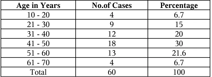
[image:37.612.82.452.186.320.2]Age distribution in the study varied from 17 yrs to 70 yrs.
Table - I Age distribution
Age in Years No.of Cases Percentage
10 - 20 4 6.7
21 - 30 9 15
31 - 40 12 20
41 - 50 18 30
51 - 60 13 21.6
61 - 70 4 6.7
Total 60 100
30% of cases in the study were between the age of 40 - 50 years.
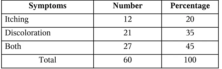
Symptoms
Itching and discoloration of the flexures were the predominant symptoms of
[image:38.612.81.450.215.330.2]erythrasma.
Table - II Symptoms
Symptoms Number Percentage
Itching 12 20
Discoloration 21 35
Both 27 45
Total 60 100
45% of patients had presented with both itching and discoloration.
Whereas in 35% the disease was more of a cosmetic compliant with only
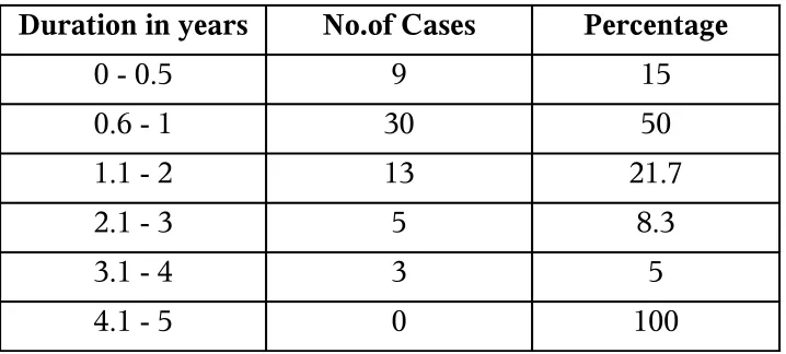
Duration of the disease
[image:39.612.88.447.184.346.2]In 72% of cases the duration of disease varied between 6 months and 2 years.
Table - III Duration
Duration in years No.of Cases Percentage
0 - 0.5 9 15
0.6 - 1 30 50
1.1 - 2 13 21.7
2.1 - 3 5 8.3
3.1 - 4 3 5
4.1 - 5 0 100
The longest duration observed was 4 years and shortest was one month.
There were remissions and relapses in 23 patients. The relapses occured with
increase in environmental temperature, sweating and maceration.
Presence of disease in family members
11 out of 60 patients had similar complaints in family members.
Dermatological Lesions
The skin lesions of erythrasma patients studied varied in their morphology as well
Morphology of lesion
The various types of erythrasma lesions observed were macular, maculopapular,
[image:40.612.119.411.215.358.2]lamellar, intertriginous, follicular, eczematous and disciform types.
Table - IV Various morphological patterns observed.
Lesions No.of Cases
Maculopapular 51
Macular 5
Lamellar 4
Follicular 3
Intertriginous 3
Disciform 1
Eczematous 1
Of these maculopapular (Fig. 1) type was the commonest. Satellite macules (Fig.
2) around larger patches were frequent and characteristic. The lamellar type (Fig. 3) was
seen in patients with generalised erythrasma. Intertriginous forms existed alone or along
with dermatophytosis and candidiasis. A case of disciform erythrasma was also
observed.
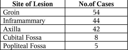
Distribution of lesion
Table V The distribution of lesions
Site of Lesion No.of Cases
Groin 54
Inframammary 44
Axilla 42
[image:40.612.146.400.629.728.2]Groins (Fig. 4) were the most common site to be involved followed by axillae
(Fig.5). In both these sites the lesions were bilateral and symmetrical. The scales were
dry and brown and not erythematous.
Other sites involved were inframammary regions (Fig.6), abdominal folds,
popliteal (Fig.7) and cubital fossa (Fig.8). In few cases the periumbilical area and
perianal area (Fig.9) was also involved.
Interestingly the lesions were seen over the left side of neck in female patients
(Fig.10) which might be due to the friction and occlusion by the clothes. In a patient
undergone colostomy erythrasma occured under the colostomy belt due to occlusion
(Fig.11). 44 54 42 8 5 0 10 20 30 40 50 60 N o . o f C a se s
Inframamm ary Groin Axilla Cubital Fossa Popliteal Fossa
Sites
Associated dermatological infections
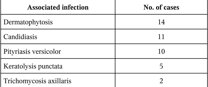
Table VI Associated dermatological infections
Associated infection No. of cases
Dermatophytosis 14
Candidiasis 11
Pityriasis versicolor 10
Keratolysis punctata 5
Trichomycosis axillaris 2
Dermatophytosis (Fig.6) was the most common fungal infection associated.
Candidiasis (Fig.12) and pityriasis versicolor were next commonly associated.
Other corynebacterial infections like keratolysis punctata (Fig.13) and
Trichomycosis axillaris and pubis (Fig.14) were also seen in association with
erythrasma.
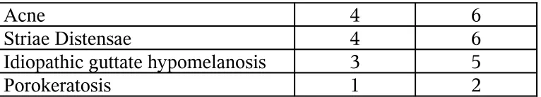
Associated dermatological disorders
Table VII Dermatological disorders associated
Associated Lesion Cases Percentage
Melasma 12 20
Acanthosis Nigricans 11 18
Vitiligo 9 15
Traumatic Fissures 5 8
[image:42.612.69.456.631.733.2]Acne 4 6
Striae Distensae 4 6
Idiopathic guttate hypomelanosis 3 5
Porokeratosis 1 2
Melasma was the most common association seen in 12 cases (20%). Acanthosis
nigricans (Fig.15) was next common seen in 11 cases (18%). It was probably due to
obesity or underlying hypothyroidism. Vitiligo (Fig.16) was associated with erythrasma
in 9 cases (15%).
Traumatic fissures of feet, ichthyosis, striae distensae (Fig.7) were the other
associated disorders. In 3 cases idiopathic guttate hypomelanosis (Fig.17) and in one
case porokeratosis were observed.
Associated systemic conditions
Among systemic conditions obesity was commonest occurring in 18 cases (30%).
Obesity was simple or associated with diabetes mellitus or hypothyroidism. Next
common association was diabetes mellitus which was seen in 15 cases (25%).
[image:43.612.73.456.55.124.2]Hypothyroidism occurred in 12 cases (20%).
Table VIII Systemic associations
Systemic Association Cases Percentage
Obesity 18 30
Diabetes Mellitus 15 25 Hypothyroidism 12 20 Hypercholesterolemia 10 16 Rheumatoid Arthritis 2 3
Hypercholesterolemia was present in 10 cases (16%) Two patients had
rheumatoid arthritis. ELISA was positive in 2 patients one male and one female and both
patients presented with generalised erythrasma.
Among the 36 females patients 10 had menstrual disturbances who also had
INVESTIGATIONS
Complete Hemogram
In 60 cases recorded, anemia was seen in 6 cases leukocytosis was seen in 4
patients.
Blood Sugar : 15 cases showed hyperglycemia which were later confirmed by glucose
tolerance test.
Serum Cholesterol : Hypercholesterolemia was observed in 10 cases.
Blood grouping and typing : Most common blood group observed was O +ve followed
by A +ve.
Thyroid Function test: Revealed hypothyroid status in 12 cases.
ELISA for HIV : Two cases showed HIV seropositivity by ELISA test.
Wood's Lamp Examination : Portable wood's lamp was used. The patient was advised
to come without having bath. Coral red fluorescence (Fig 18) was observed in all except
one case.
Gram Stain: The scales were scraped and fixed on to slide using egg albumin and
stained by Gram method. Gram positive coccobacilli were seen in all cases (Fig.19).
Histologically there was hyperkeratosis with basket weave stratum corneum,
acanthosis and papillomatosis. There was increased pigment in the basal cell layer.
Sparse inflammatory infiltrate was present in the upper dermis and around blood vessels.
The coccobacilli were seen in stratum corneum by GMS stain (Fig.20) and also by H&E
method (Fig.21).
Culture
The scales were collected and inoculated into a medium of Mueller Hinton Agar
enriched with blood. Small pale grey convex colonies (Fig.22) appeared after 48 hours
in only 4 cases out of 60 cases inoculated. Few of the colonies showed coral red
fluorescence on Wood's lamp visualisation (Fig.23).
Treatment:
All the cases were treated with T.Erythromycin 250 mg qid for 14 days along with
topical application of 2% clotrimazole cream. In many patients there was a good
response but recurrences were not prevented unless the predisposing factors were
DISCUSSION
Sex Distribution
In literature erythrasma was reported to be more common in men by Peter K Lee
et al38., and equal sex incidence was reported by Gary L Darmstadt et al27. In this study
there were 36 female and 24 male cases showing that the incidence of erythrasma is
more in females than in males. This increased incidence may be due to the obesity
commonly observed in females more than forty years.
Age Distribution
The most common age group to be affected were between 40 - 50 years in which
one third of cases occured. 43 cases (75%) occured in the 30 - 60 years age group.
The youngest age group observed was 17 yeas and oldest was 70 years.
This correlates with the literature that incidence of erythrasma increase with age
as reported by Gary L Darmstadt et al27 and Laube S6.
The increased prevalence of metabolic derangements like diabetes mellitus,
obesity and hypothyroidism in middle age group might be the reason for the occurrence
of erythrasma in them.
Also the presence of lipid in apocrine secretion after puberty may also influence
the above finding.
RJ Hay and BM Adrians15 have reported that in temperate climates the erythrasma
lesions were symptomless but in tropics, irritation of lesions occur leading to scratching.
Similarly in this study 27 patients (45%) had itching and discoloration of flexures as
their predominant complaint. It was asymptomatic in 21 cases (35%) who reported for
the cosmetic inconvenience.
The associated candidal intertrigo and increased sweating and maceration in the
flexures due to obesity might be the reason for itching in majority of cases.
Duration of disease
It is shown in this study that erythrasma is a chronic disease with remissions and
relapses. These remissions were associated with the hot and humid tropical climate due
to increased sweating and thereby maceration.
Presence of disease in family members
11 patients (18%) had family members who were also diagnosed as erythrasma.
This indicates that the disease is not significantly contagious. The incidence of
erythrasma among family members might be related to the common living conditions
and similar predisposing factors in families.
Dermatological lesions
In literature the most common type reported was the maculopapular type which
was also the commonest in this study. It was seen in 51 patients (85%). In patients with
generalised erythrasma lamellar type was seen. One case of disciform type and
Distribution of lesions
In literature by RJ Hay and BM Adrians erythrasma was described to occur most
commonly in the groins15.
In this study also groins were the most common site to be affected followed by
axilla bilaterally and symmetrically. All the intertriginous areas were affected.
Apart from intertriginous areas few cases showed involvement of left side of neck
due to friction by clothes in females and in one case erythrasma occured under
colostomy belt which may be due to occlusion.
The lesions were not erythematous as reported in literature. They were brown to
Associated dermatological disorders
The coexistence of erythrasma with pitted keratolysis and trichomycosis axillaris
was described by Shelley et al30. 1982. Concomitant erythrasma and dermatophytosis of
groin was reported by Schlappner et al9. in 1979. In 2004 Karakatsanis G described
coexistence of pityriasis versicolor and erythrasma10.
The most common skin infections associated with erythrasma in this study were
dermatophytosis, candidiasis, pityriasis versicolor, keratolysis punctata and
trichomycosis axillaris. The coexistence of the above infections with erythrasma
indicates that common predisposing factors of moisture and obesity are involved in these
conditions.
The high incidence of melasma and vitiligo and few cases of idiopathic guttate
hypomelanosis observed in these cases of erythrasma suggests a possible common factor
related to pigmentation is involved in them.
The coexistence of acanthosis nigricans and striae distensae might be due to
Associated systemic disorders
The increased frequency of obesity and diabetes in the patients studied concurs
with the literature reports of Scheinfeld NS14 & Haroon TS12 that erythrasma is
commonly associated with both of the conditions.
This may be related to the high levels of cutaneous free glucose occuring in
diabetes mellitus. The association of hypercholesterolemia in 10 patients (16%) in the
study group suggests that possibly an increase in cutaneous lipid may promote the
growth of lipophilic Corynebacterium minutissimum.
10 female patients (35%) in the study had menstrual irregularities and 2 had
polycystic ovarian syndrome on ultrasonogram. Hypothyroidism, obesity, polycystic
ovarian syndrome may be the underlying cause in them thereby predisposing them to
erythrasma.
The association of thyroid disorders, ovarian dysfunction, diabetes, rheumatoid
arthritis and vitiligo suggests that a possible autoimmune mechanism may play a role in
predisposing them to erythrasma.
Investigations
Anemia was recorded in 6 cases and it indicates that it does not predispose to
erythrasma. Leucocytosis was seen in 4 cases. There was no reactive leucocytosis as this
bacteria remains mainly in stratum corneum and is usually not invasive.
As in general population, in the study group the most common blood group
The literature states that erythrasma prevalence is not increased in
immunosuppressed patients41. Similarly only 2 cases were positive for HIV by ELISA in
this study.
Wood's lamp and Gram stain smear were the procedures which proved the
etiological agent Corynebacterium minutissimum by demonstrating its coproporphyrin
production by coral red fluorescence and visualisation of gram positive coccobacilli.
These two tests gave consistently positive results in almost all cases and hence
CONCLUSION
The following conclusions were drawn from this study
1. More common in females.
2. Mean age of occurrence was 40 - 50 years.
3. Itching and cosmetic disability were the predominant complaints.
4. Maculopapular form in groins was the most common presentation.
5. The disease was chronic with remissions and relapses related to the tropical
climate.
6. Obesity, diabetes, hypothyroidism were the common predisposing
conditions.
7. Trichomycosis axillaris and keratolysis punctata were the other
corynebacterial infections associated with it.
8. Dermatophytosis and candidiasis were the most common fungal infections
associated.
9. Melasma, acanthosis nigricans and vitiligo were the most common skin
disorders seen in association.
10.Woods lamp examination and Gram stain were useful procedures to
REFERENCE
1. Maibach HI, Raza A : Bacterial infections of skin in Moschella SL, Hurley JH (eds): Dermatology, Ed 3. Philadelphia, WB Saunders Co, 1992, pp 731 - 732.
2. Kamalam. A, Thambiah. A.S., Bagavan das. M. and Govindaraju. S Mycoses in India-Study in Madras. Transactions of the Royal Society of Tropical Medicine and Hygeine 1981, Vol.75, 92-100.
3. Marks. R, N.D. Ramnarain, B.Bhogal and N.T.Moore. The erythrasma micro organism insitu-studies using the skin surface biopsy technique. J. Clin. Pathol. 1972 September, 25(9): 799-803.
4. Sarkany I, Taplin D, Blank H. The etiology and treatment of erythrasma. J.Invest Dermatol. 1961; Vol.37, pp.283-290.
5. Sarkany I, Taplin D, Blank H. Incidence and Bacteriology of erythrasma. Arch Dermatol 1962 May; 85: 578-82.
6. Laube S. Skin Infections and ageing. Ageing Res Rev 2004 Jan; 3(1): 69-89.
7. Holdiness MR. Erythrasma and common bacterial skin infections. Am Fam Physicians 2003 Jan 15; 67(2): 254.
8. Bowyer A, Mc Coll I. Erythrasma and pruritusani. Acta Derm Venereol 1971; 51(6) 444-7.
10. Karakatsanis G, Vakirlis E, Kastoridou C, Devliotou-Pangiotidou D Coexistence of pityriasis versicolor and Erythrasma. Mycoses 2004 Aug; 47(7):343-5.
11. Somerville DA, Lancaster-Smith M. The anerobic cutaneous microflora of diabetic subjects. Br. J. Dermatol 1973, Vol.89; 395-400.
12. Haroon TS. Diabetes and Skin-a review. Scott Med J. 1974 Nov. 19; 257-67.
13. Montes LF, Dobson H, Dodge BG, Knowles WR. Erythrasma and diabetes mellitus. Archieves of Dermatology, 1969, Vol.99, 674-680.
14. Scheinfeld NS. Obesity and dermatology. Clin Dermatol 2004 Jul-Aug; 22(4): 303-9.
15. Textbook of Dermatology Rook edited by Tony Burns, Stephen Breathnach, Neil Cox, Christopher Griffiths. 7th edition, Vol.2 Bacterial infections. Author RJ Hay
and BM Adrians.
16. Sarkany I, Taplin D, Blank H. Erythrasma-Common bacterial infection of skin JAMA 1961 Jul 15; 177; 130-2.
17. Montes LF, McBride ME, Johnson WP, Owens DN, Knox JM. Ultrastructural study of host bacterium relationship in erythrasma. J. Bacteriol 1965 Nov; 90(5): 1489-91.
18. Marks R. Keio. Seeing through the stratum corneum. J. Med. 2000 Jun. 49:80-3.
19. Montes LF, Black SH, McBride ME. Bacterial invasion of stratum corneum in erythrasma, ultrastructural evidence of keratolytic action exerted by corynebacterium minutissimum. J. Invest Dermatol. 1967, Nov; 49; 474-85.
20. Hartmann AA. The influence of various factors on the human resident skin flora. Sem in Dermatol 1990 Dec. 9 : 305 - 8.
22. Golledge CL, Phillips G. Corynebacterium minutissimum infection. J. Infect 1991, Jul 23: 73-6.
23. Stephen A Berger, Alfred Gorea, Jona Stadler, Michael Dan. Recurrent breast abscesses caused by C.minutissimum. Journal of clinical. Microbiology, Dec 1984, P.1219 - 1220.
24. Andrews' diseases of skin-clinical dermatology; 9th edition Edited by Richard B.Odom, William. D.James, Timothy G.Berger. Bacterial infections Page 326.
25. Cabo H, Franco de Montes de Oca N, Tzovanis Mc, Gallardo H. Generalised erythrasma. Med Cutan Ibera Lat Am 1983; 11(2): 129-32.
26. Tschen JA, Ramsdell WM. Disciform erythrasma. Cutis 1983 May; 31(5): 541-2, 547.
27. Pediatric Dermatology 3rd edition: Edited by Lawrence A.Schachner and Ronald C Hansen. Bacterial infections by Gary L. Darmstadt, Wesley king Galen and Gayle Fischer. Page 1036.
28. Grigoriu D, Delacretaz J. Vesiculobullous erythrasma of feet. Dermatologica 1976; 152(1): 1-7.
29. Negroni P. Erythrasma of the nails. Med Cutan Ibera Lat Am 1976, 4(5): 349-57.
30. Shelley WB, Shelley ED. Coexistent erythrasma, trichomycosis axillaris and pitted keratolysis-an overlooked corynebacterial triad? J Am Acad Dermatol 1982 Dec; 7(6): 752-7.
31. Guarderas J; A.Karnad, S.Alavarez, S.L.Berk. Corynebacterium minutissimum bacteremia in a patient with CML in blast crisis. Diag. Microbiol. Infect. Dis. 1986, Vol.5, pp.327-330.
32. Brian H.J; A.J.Brucker; 1985. Embolic retinopathy due to corynebacterium minutissimum endocarditis. Br. J. Ophthalmol, Vol.69, pp.29-31.
in dermatologic diagnosis, therapy, follow-up and prevention. Hautarzt 1997, Aug 48: 523-7.
34. Mattox TF, Rutgers J, Yoshimori RN, Bhatia NN. Nonfluorescent erythrasma of vulva. Obstet Gynecol 1993 May: 81(5): 862-4.
35. Padilha - Goncalves A. A single method to stain Malassezia furfur and corynebacterium minutissimum in Scales. Rev Inst Med. Trop. Sao Paulo 1996 Jul - Aug 38 : 299 - 302.
36. Stephen N Cohen, Dorothy Nickolai. Simple medium for pigment production by the erythrasma diphtheroid. Appl. Microbiol 1969 March; 17(3): 479-480.
37. Skin pathology David Weedon 2nd edition. Bacterial and rickettsial infection. Page 622.
38. Fitz patricks Dermatology in General Medicine-6th Edition: Edited by Irwin M Freedberg (et al) Vol.2, Chap. 194, Pyodermas. Author Peter KLee, Mathew T. Zipolli, Arnold N Weinberg, Morton Swartz and Richard A.Johnson Pg. 1876.
39. Aste N, Pau M. Pityriasis versicolor on groin mimicking erythrasma. Mycoses 2004; June; 47(5-6): 249-51.
40. Gupta S. Pityriasis rotunda mimicking tinea cruris/corporis and erythrasma in an Indian patient. J. Dermatol 2001 Jan; 28(1): 50-3.
41. Treatment of skin disease. 1st edition. Edited by Mark Lebwohl, Warren R Heymann, John Berth Jones, Ian Loulson. Erythrasma by Andrew G. Smith page 203.
42. Holdiness MR. Management of cutaneous erythrasma. Drugs 2002; 62(8): 1131-41.
43.Wharton JR, Wilson PL, Kincannon JM, Erythrasma treated with single dose clarithromycin. Arch Dermatol 1998 Jun; 134(6): 671-2.
ERYTHRASMA - A CLINICAL STUDY
Name: Address:
Age: Marital Status:
Sex : Occupation :
Case No: Hospital No:
HISTORY
Dark discoloration of flexures: Yes No
Duration : Yes No
Itching: Yes No
Past History
Similar illness: Yes No
DM HT TB RA Others
Family History
Similar illness Partner Parents Children Others
Drug History
Steroids: Yes No
Menstrual History
Menarche:
Periods: Regular Irregular
Pelvic Surgeries: Yes No
EXAMINATION
General Examination
Built: Well Moderate Poor
Height: (m)
Weight: (kg)
BMI:
Anaemia: Yes No
Lymphadenopathy: Yes No
Systemic Examination
CVS RS ABDOMEN CNS
Dermatological Examination
Clinical type of lesion: Macular Papular Intertriginous
Eczematous Lamellar Disciform
Others:
Distribution of lesion: Genitocrural Axilla Inframammary
Toe web Neck Periumbilical
Generalised Others:
Colour of lesion: Reddish brown Brown
Black Hypopigmented
Extent of lesion:
Mucosa: Normal
Associated Skin Disorders
Other Corynebacterium causing disorders: Trichomycosis axillaries pitted keratolysis Acne vulgaris
Fungal infections: Dermatophytosis Candidiasis
Tinea versicolor White Piedra
Other skin disorders: Vitiligo Melasma Acanthosis nigricans
Others:
INVESTIGATIONS
Blood: Hb Urine: Albumin Sugar Deposits
Blood sugar: GTT
Serum cholesterol: Blood grouping: Skin Surface pH: Optional
Thyroid function tests: ELISA:
X-ray chest: USG:
Specific
Wood's Lamp Examination: Gram's stain smear:
Culture: Biopsy:
FOLLOW UP
KEY TO MASTER CHART
Symptoms
D - Duration I - Itching
Sites
G - Groin A - Axilla
I - Inframammary N - Neck
C - Cubital fossa P - Popliteal fossa U - Periumbilical
Lesion
M - Macular
MP - Maculopapular L - Lamellar
F - Follicular I - Intertriginous E - Eczematous D - Disciform
Associated Skin Infections
Association Skin Disorders
M - Melasma
AN - Acanthosis nigricans T - Traumatic Fissures V - Vitiligo
A - Acne
S - Striae distensae Ic - Ichthyosis
I - Idiopathic guttate hypomelanosis P - Porokeratosis
Associated Systemic Disorders
O - Obesity
D - Diabetes mellitus H - Hypothyroidism RA - Rheumatoid arthritis
Others
TFT - Thyroid function Test H - Hypothyroid picture