JOURNALOFVIROLOGY,Apr. 1994, p.2272-2285 Vol. 68, No.4 0022-538X/94/$04.00+0
Copyright© 1994,AmericanSocietyforMicrobiology
Membrane
Anchoring
Domain of
Herpes
Simplex
Virus
Glycoprotein gB
Is
Sufficient
for
Nuclear
Envelope Localization
RENALD GILBERT,t KAKOLI GHOSH, LEN RASILE, AND HARA P. GHOSH* Department ofBiochemistry, McMasterUniversity, Hamilton, Ontario L8N3Z5, Canada
Received 16September1993/Accepted 27 December 1993
We have usedtheglycoprotein gBofherpes simplexvirustype1 (gB-1),which buds from the innernuclear membrane, as a model protein to study localization of membrane proteins in the nuclear
envelope.
To determinewhetherspecificdomains ofgB-1glycoproteinareinvolved in localization in the nuclearenvelope, we have used deletion mutants ofgB-1 protein as well as chimeric proteins constructed by replacing the domains of the cellsurfaceglycoproteinG ofvesicular stomatitisvirus with thecorresponding
domains ofgB. Mutant and chimeric proteins expressed in COS cells were localized by immunoelectron microscopy. A chimericprotein (gB-G)containingtheectodomainofgBand the transmembrane andcytoplasmicdomainsof G did not localize in the nuclearenvelope.When the ectodomain of G was fused to the transmembraneand cytoplasmic domains of gB, however, the resulting chimeric protein (G-gB) was localized in the nuclear envelope. Substitution ofthetransmembrane domainof G with the 69hydrophobicamino acidscontainingthe membrane anchoring domain of gB allowed the hybrid protein (G-tmgB) to be localized in the nuclear envelope, suggestingthatresidues721 to795ofgBcanpromoteretention ofproteinsin the nuclearenvelope. Deletion mutations in the hydrophobic region further showed that a transmembrane segment of 21 hydrophobic amino acids, residues 774 to795 ofgB, was sufficient for localization in the nuclearenvelope. Sincewild-type gBandthe mutant andchimericproteinsthat were localized in the nuclearenvelopewerealso retained in theendoplasmic reticulum,the membranespanning segment of gB could also influenceretention in the endoplasmic reticulum.The mechanism of sorting and transport of proteins to specific subcellular organelle membranes in eukaryotes has been of major recent interest. Glycoproteins of enveloped
animalviruses have been extensively used asmodelsto study the sorting of integral membrane proteins to cell surface,
endoplasmic reticulum (ER), and Golgi complex membranes
(16, 17, 19, 45, 51). In eukaryotic cells the nuclear envelope
(NE)physically separates the nucleus from thecytoplasm and has amajor role in nuclear import and export of macromole-culesaswellasintheorganization of the nucleararchitecture (5, 14, 15, 18). The nuclear envelope has a complex organiza-tion consisting of an outer and an inner nuclear membrane, nuclear lamina, and the nuclear pore complex(5, 14, 15, 18). Although excellent progress has been made in understanding the import of proteins into the nucleus (5, 15), information on thebiogenesisofthe NE, and in particular the mechanism of
targeting ofintegral membrane proteins to the inner nuclear membrane, remains to be clarified. Recently the NE localiza-tion determinants of two integral membrane proteins, the nuclear pore complex protein gp210 (63) and the lamin B receptor (LBR) (53, 55), have been studied by using indirect
immunofluorescenceof transfected cells. In the case of gp210, the transmembrane and cytoplasmic domains could target a cellsurface protein to the NE (63). However, in the case of the LBR,different results were reported; thus, in one case the first of the eight membrane anchoring segments of the LBR was
*Corresponding author.Mailing address: Department of
Biochem-istry,McMaster University, Health Science Centre, Room 4H24, 1200 Main St. West, Hamilton, Ontario L8N 3Z5, Canada. Phone: (905) 525-9140,ext.22451. Fax: (905)522-9033.
tPresent address: Departmentof Cell Biology,CornellUniversity MedicalSchool, New York, New York.
sufficient forlocalization to the NE (53), while the otherstudy showed therequirement of the amino-terminal nucleoplasmic
tail of theLBR for NE localization (55).
We have used the envelope glycoproteinofherpessimplex virus type 1 (HSV-1) as a model to study the targeting of integral membrane proteins to the nuclear membrane (2, 20,
46, 47). Since HSV acquires itsenvelope bybuddingthrough theinner membrane of the nuclear envelope (12, 21, 37,39,56, 59, 60), it may be assumed that the HSV envelope
glycopro-teins aretargeted to and localized in the NE. Theglycoprotein gB-1 of HSV-1 is essential for viral infectivity (33) and is known to be involved in virus entry and cellfusion (7, 9,56). Fromthe deduced amino acid sequence of gB-1, the presence of a large cytoplasmic domain of 109 amino acids and a hydrophobic stretch of 69 amino acids which could serve as the membrane anchor domain can be predicted (8, 44). We have previously expressed the gB-1 gene in COS-1 cells withcloned DNA. Theexpressedglycoprotein waslocalized both in the NE and intheplasmamembrane (2).Recentstudiesusing immu-noelectronmicroscopy of HSV-1-infected cells confirmed that gB-1 was indeed localized in the inner membrane of the nucleus (21, 60). Earlier, using indirect immunofluorescence andbiochemical cellfractionation,weshowed that the deletion of the cytoplasmic domain of gB-1 did not affect its NE localization(47). Recently, using mutants with deletions in the predicted membrane anchoring sequence of 69 hydrophobic amino acids, we have shown that the segment containing amino acids 774 to 795 of gB-1 specifies the membrane
anchoringdomain and that mutants containing this segment of
hydrophobicamino acids were localized in the NE (46). Inthisstudywehave, as an extension of our previous work
(46, 47), attempted to define the NE localization determi-nant(s) of gB-1 byimmunoelectron microscopic localization of
2272
on November 9, 2019 by guest
http://jvi.asm.org/
NUCLEAR ENVELOPE LOCALIZATION SIGNAL OF gB-1 2273 mutantgB-1 proteins containing shortened cytoplasmic tailsor
deletions in the predicted hydrophobic membrane anchoring domain aswell as chimeric proteinsconstructed byswapping thedomains of the cell surface proteinGof vesicular stomatitis virus(VSV) withcorresponding domains of gB-1 proteins.The results obtained with deletion mutants as well as with the
chimerasshowthatthe69-amino-acidhydrophobicsegmentof gB-1 specifies NE localization and that the transmembrane segmentencoding residues 774to795of gB-1wassufficientfor
localization of proteins in the NE. The results also indicated thatproteins thatwerelocalized in theNEwerealsoretained
inthe ER,suggesting that the membraneanchoring domainof gB-1 mayalso influence retention in theER.
MATERIALS ANDMETHODS
Materials. Restriction and DNA-modifying enzymes were
purchased from Bethesda Research Laboratories, Pharmacia Canada, or New England Biolabs. Endoglycosidase H (endo H) was from ICN-Immunobiologicals. Fluorescein
isthiocya-nate-conjugatedgoatanti-rabbitimmunoglobulin G (IgG)was
from Dako Corporation. Escherichia coli BW 313 (dut ung) cells(31)wereobtainedfromN.Skipper, AllelixLaboratories, Mississauga, Ontario, Canada. Mutagenic oligonucleotides
weresynthesizedonanApplied Biosystems model 381-A DNA
synthesizerwith 3-cyanoethyl phosphoramidites orpurchased
fromtheCentral Facility of the Institute for Molecular Biology and Biotechnology, McMaster University. COS-1 cells were
grown in high-glucose Dulbecco's modified Eagle's medium
supplementedwith 7% fetal calfserum. Recombinant
adeno-viruses expressing eitherglycoprotein B of HSV-1 (AdgB-1) (36) or glycoprotein G of VSV (AdG12) (52) were obtained
fromD.Johnsonand L. Prevec, respectively.
Antibodypreparation. Rabbitswereinjectedsubcutaneously
three times at weekly intervals with 0.5 ml of 108 PFU of
AdgB-1orAdG12 dilutedinphosphate-bufferedsaline (PBS)
containing 2% rabbitserum.One week after the lastinjection, the rabbitswerekilled and bled, and the IgG fraction of the
serum was isolated by ammonium sulfate precipitation as
describedpreviously (24).
Anti-gB antibodieswereaffinitypurified by usingacolumn madewithamixtureofglycoproteinsofHSV-1-infectedcells. Theglycoproteinswereisolatedfrom BHK cellsinfected with
HSV-1 by using a lentil lectin-Sepharose 4B column as
de-scribed byPachletal.(42).Theglycoproteinswerecoupled to CNBr-activated Sepharose 4B (Sigma), and the beads were
used foraffinitypurificationofanti-gB antibody as described previously (24). Anti-G antibodies were affinity purified by
usingacolumn of Gprotein coupledtoSepharose4B beads. G proteinwas isolated from gradient-purified VSV Indiana
se-rotype asdescribedpreviously (30).
Plasmid construction and mutagenesis. The 3.4-kb BglII-EcoRI fragment of p9gB (2) and the 1.7-kb fragment of pSVGL (48) were cloned into the vector pXM (64) for expression ofwild-type gB and G proteins, respectively.
Oli-gonucleotide-directedmutagenesis bythe method of Kunkel et al. (31)was used to construct gB mutants
gBCT799,
gBA12, andgBA23asdescribedpreviously (46, 47).ChimeragB-GwasconstructedbyjoiningaBglII-Tth1111fragmentofgBwith the
TaqI-EcoRIfragment of G containingthetransmembraneand cytoplasmicdomains. To construct the G-gBhybrid, anXhoI sitewasintroducedat nucleotideposition 1416ofG (32),and
aTthlllI-EcoRI fragment containingthetransmembraneand cytoplasmic domains ofgBwas clonedin theXhoI site of G.
Thechimera G-tmgBwas constructedby fusing aBglII-MluI
fragmentofG-gBintotheBglII-XhoIsite ofpXMGandthen
deletingtheectodomainof gB and thetransmembrane domain
of G by using the primer
5'-GCCTTCTfl7CGCCThICGAG
TTGGTATCCATCIT1-3'.
The two hybrids G-tmgBAl andG-tmgBA12wereconstructed from G-tmgB by using primers used for
gBA1
andgBA12 mutants, respectively (46).Transfection, labeling, and immunoprecipitation.
Subcon-fluent monolayers of COS-1 cells were transfected by the
Ca3(PO4)2 precipitation protocol (22, 46). The transfected cellswerelabeled with
[35S]methionine
at24to40 h posttrans-fection andprocessedforimmunoprecipitationandanalysis by sodium dodecyl sulfate-polyacrylamide gel electrophoresis (SDS-PAGE) asdescribed earlier (46).Intracellular transport.Transfected COS-1 cells were fixed with 2% paraformaldehyde for surface immunofluorescence. For internal immunofluorescence cells were fixed with 2%
paraformaldehyde,treated with 1% Triton X-100, and reacted with anti-gBor anti-Gantibody and stained with fluorescein
isothiocyanate-conjugated goat anti-rabbit IgG as described
previously (46). Transport of the mutant gB proteins to the
Golgi complexwas determined by the acquisition of endo H resistance (29) asdescribedpreviously (46).
Immunoelectron microscopy. COS-1 cells at 24 to 40 h
posttransfectionwere fixed for 2 hat room temperaturewith
2% glutaraldehyde in 0.03 M PIPES
[piperazine-N,N'-bis(2-ethanesulfonic acid)] (pH 7.4). The cells were then washed with 0.03 M PIPES (pH 7.4), scraped with a razor blade,
centrifuged,and embedded in2% Noble agar. Thespecimens were cut intosmall cubes(1
mm3),
dehydrated with ethanol, andembedded inLowicrylK4M(Polysciences)
at -20°C(21, 50). Ultrathin sectionson nickelgridscoated with a Formvar film were labeled by incubation with the affinity-purified antibodiesat room temperature. Before the antibodyincuba-tion, the grids were washed with PBS and
preincubated
with PBScontaining 1%bovineserumalbumin and5%goatserum (buffer A). The gridswere incubated with anti-gB or anti-G diluted in buffer A for 1 h and then with a goat anti-rabbit antibody coupled to 1-nm gold(Amersham International)
dilutedinbufferAfor 30 min. Thegridswerewashed with PBS and distilledwaterand dried. The 1-nmgoldwasvisualizedbythe silver enhancement method of Danscherg and
Norgaard
(11).Thespecimenswereobserved withaJEOL JEM 1200EX electronmicroscope, usinganacceleration
potential
of 80 kV. Forstatisticalanalysis,
randompictures
of cellsweretaken. Foreachcell,the average numberof silvergrains
per microme-terofNE, ER,orplasmamembranewasdetermined.Thedata were analyzed asdescribed earlier (21,62).
RESULTS
Wehave
previously expressed
the HSV-1glycoprotein
gB-1
gene inCOS cells
by using
clonedDNA. Theexpressed
gB-1
proteinistransportedto both the nuclear membrane and the cellsurface(2).
Truncation of the 109-amino-acidcytoplasmic
tail showed that this domain did not affect the nuclear local-ization
(47).
Deletions in thepredicted
membraneanchoring
sequenceof the
carboxy-terminal
69hydrophobic
amino acidsof gB-1
(residues
727 to795)
showed that ahydrophobic
sequencepresent in segment 3
(amino
acids 774to795 ofgB-1
protein)
wassufficientfor stablemembraneanchoring
and thusmay
specify
the transmembrane domain ofgB-1
glycoprotein.
Also, all mutants
containing
segment3were localizedin theNE
(46).
To examine whether aspecific
domain ofgB-1
glycoprotein
isrequired
for localization in theNE,
we haveused a number of deletion mutants
(46)
as well as chimericproteins.The
cytoplasmic
tailmutantCT799
wasgenerated
by
creating
achain-terminating
codonat amino acid residue 799VOL.68, 1994
on November 9, 2019 by guest
http://jvi.asm.org/
2274 GILBERT ET AL.
720
gB-l: DTVIHAD ANAAMFAGLGAFFEGMGDLGRAVG
KVVMSIVGGVVSAVSGVSSFMS 800 NPFGALAVGLLVLAGLAAAFFAF RYVMR 452
VSVG:ELVEGWFSSWK SAFFIGIGFV
31
EC TM CT
7j7j ,6 994
m r
£7 4 1
3,1
721
452 511l
17~~~~~
46726
72172 ,7795
17 4 1
727/2 7 95
17 4 1
3,1 -
~798
3, 7_
31 7 76
gB VSVG gB-G G-gB G-tmgB G-tmgBAl G-tmgBA12 CT799 gBA12 gBA3
FIG. 1. Schematic diagram of gBmutants and chimeras used. (a) Aminoacidsequencesof thehydrophobic carboxy-terminal regionof gB-1 and the transmembranedomain of Gprotein. (b)Numbers above and below the boxes are amino acid residues, starting from the N
terminus, for gB (solid boxes) and G (open boxes), respectively. Deletionsareshownbygaps.Thelargerbox in the middle represents thepredicted transmembrane domain. EC, ectodomain; TM,
trans-membraneanchor; CT, cytoplasmicdomain.
(47); thus this mutant contained only 3 amino acids in the cytoplasmic tail, compared with 106 residues present in the wild-type gB-1 proteins (8, 44).ThemutantsgBzv12andgBA3
were constructed by deleting amino acid residues 727to 772 and 774to795, respectively, from the 69-hydrophobic-amino-acid segment ofgB-1 (46). The chimeric proteins were
con-structedby swapping domains ofgB-1 proteinwith the
corre-sponding domains of a reporter protein G of VSV. Glycoprotein G is a type I membrane protein containing a
transmembrane domainof20hydrophobicamino acids and a
cytoplasmic tail of 29 amino acids (49a) and has been used extensively as a model for cell surface protein as well as a
reporterproteintostudy intracellularproteintrafficking(1, 13, 16, 17, 19, 49, 51, 57).Aschematicdiagram of themutantsand the chimeric proteins is shown in Fig. 1. The chimeras G-gB and gB-Gwere constructed by replacing the ectodomains of
thegB-1andGproteinswiththe ectodomains of G and gB-1, respectively. The chimera G-tmgBwasconstructed by
replac-ing the transmembranedomain of G protein with the predicted membrane anchoring domain of thecarboxy-terminal 69 hy-drophobic amino acids ofgB-1. The chimeras G-tmgBAl and G-tmgBA12 were obtained by replacing the transmembrane
domainof Gprotein withthehydrophobicamino acids 747to
795 and 773 to 795 of gB-1, respectively. The mutant genes
were expressed transiently, as before (2, 20, 46, 47), in COS
cellsby usingthehigh-efficiencyvectorpXM(27, 64), and the
agB
m n
m
m > U m(O O
aG
02c
m en am DE
m; > U cm tO (
GtmgB A12 Al
in
m
0C) 0)
C)dd 205K-116K- _ 97K- I 66K-
45K-FIG. 2. Expression of wild-type and mutant proteins in COS-1 cells.Untransfected cellsorcells transfectedwithgB,G, CT799,gB-G, G-gB, G-tmgB,G-tmgBA12,or
G-tmgBRM
werelabeled with [35S]me-thionineat24to40 hposttransfectionfor 2 h andimmunoprecipitated withanti-gBoranti-Gantibodies. Theproteinswerethenanalyzed by SDS-PAGE followed by fluorography (46). Numbers at left aremolecularweights (K, thousands).
intracellulartransportand localizationofthe
expressed
glyco-proteins
were studied. Intracellular localization wasdeter-mined
by
indirect immunofluorescence as well as at the ultrastructural levelby
immunoelectronmicroscopy.
Expression
and intracellulartransportofchimericproteins.
COS-1 cellswere transfected with
pXM,
encoding
the chime-rasand the wild-typegB
andGglycoproteins.
Thetransfected cellsweremetabolically
labeled with[3S]methionine,and theproteins
from the celllysate
immunoprecipitatedwith rabbitpolyclonal
anti-gB or anti-G antiserum were analyzed onSDS-polyacrylamide gels.
It can be seen fromFig.
2 that thehybrid gB-G
wasimmunoprecipitated
specifically
withanti-gB
antibody,
while the chimerasG-gB,
G-tmgB, G-tmgBA1,
andG-tmgBA12
wererecognized only by
anti-Gantibody.
Theprotein G-gB
wasalsoimmunoprecipitated
withanantipeptide
antibody
(anti-gB CT)
raisedagainst
thecarboxy-terminal
16amino acids of
gB-1
(data
notshown).
Acquisition
of endo H-resistantoligosaccharides by
glyco-proteins
has beenusedasa measureoftherateoftransport
of the protein from the ER to theGolgi
complex
(29).
To examine the rate of intracellular transport of thehybrid
glycoproteins
compared
with that ofwild-type
gB-1orG,
the immunoprecipitates from apulse-chase
experiment
weredi-gested
withendoH andanalyzed
onSDS-polyacrylamide gels.
Previously it was shown that a fraction of
gB-1 protein
remained endoH sensitive even after along period
ofchase(10,
26).
In agreement, we have found that thewild-type gB
becomes almost
totally
endo H resistant after a 1-h chaseperiod,
while the mutantsgBCT799,
gBA12,
andgBA3
wereonly
partially
resistanttoendo Hevenaftera3-hchaseperiod
(46,
47).
Acalculationof the ratesofloss ofendoHsensitivity showed thatwhile the wild-typegB-1
and themutantgBA3
hadhalf-lives
cpta2)
of 27 and 135mi,respectively,
fortransporttothe
Golgi
complex,
the mutantsgBCT799
gBon2
tSr2
and hadofmore than 3 h
(46, 47)
andwerethusnottransported
fromER to Golgi
complex efficiently. Figure
3 shows that the maturation of theoligosaccharides
ofthehybrid glycoproteins
occurred atdifferentrates.
Following
a15-mmpulse,
all of the hybrid proteins werecompletely digested
withendoH,
indi-cating
thatthey
areglycosylated
with N-linkedoligosaccha-a
b
J. VIROL.
on November 9, 2019 by guest
http://jvi.asm.org/
[image:3.612.50.282.72.372.2] [image:3.612.307.548.75.237.2]NUCLEAR ENVELOPE LOCALIZATION SIGNAL OF gB-1 2275
GtmgB Al A12 Al A1 2
TIME(MIN) 0 30 ENDOH - + - + - + - +
GtmgB
al al2 Al a12
ENDO
FIG. 3. Intracellular transport of mutant gB-1 glycoproteins as
determined by endo H digestion. COS-1 cells transfected with the
mutant constructswerepulse-labeledwith [35S]methionineat24or40
hposttransfection for 15 min and then chased with methionine-rich medium for 0min,30min, 1h, or2 h. Celllysateswere
immunopre-cipitatedwithanti-gBoranti-Gantiserum and resuspended insodium
phosphate buffer, andanaliquotwassubjectedtodigestionwith endo H as described in the text. Only the results for the chimeras are
presented.The quantitation of the bands by densitometrywasdoneby
usingexposuresinthe linearrangeofthefilms.
rides. Therateoftransport ofthe hybrid gB-G from theERto
the Golgi complexwas slower
(tQ12
of 52min)than thatofthewild-type gB. The hybrid G-gB protein, in contrast, was
transported extremely slowly and remained endo H sensitive afterachase of2h.Inagreementwith the published data(48), wild-type G protein showedarapidrateoftransportfromER to Golgi complex
(tQ12
of 22 min). The chimeras G-tmgB and G-tmgBA12showedrates of acquisition of endo H resistance(t112of 33 and27min,respectively)comparabletothat ofgB-1. Incontrast,the hybridG-tmgBAi was veryslowly transported
from the ER tothe Golgi complex. Moreover, the amount of the endo H-resistant form ofG-tmgB decreasedwith increas-ing chasetime, andverylittle G-tmgBwasobserved aftera2-h
chase, suggesting degradation of the chimera at the cell surface. Similar to the casewith gB and gB-G, a fraction of
G-tmgB was sensitive to endo H even after 2 h ofchase. It appears that when the ectodomain ofgBis replaced with the
ectodomain of G, the hybrid protein is not transported from the ER to the Golgi complex, but replacement of the trans-membrane domain of G with either the 69 hydrophobic residues (G-tmgB) or the 21-residue membrane anchoring
segment(G-tmgBA12) ofgB-1 restoresintracellulartransport.
Surprisingly,mutantG-tmgBAl,whichcontainsthe
transmem-brane domain, including segments 2 and 3 (residues 745 to
795),was retained inthe ER.
Intracellular localization ofmutant gB-1 glycoproteins by
immunofluorescencestaining.ThegB-1 glycoprotein has been shown to be localized in the ER, Golgi complex, and cell surface aswell asinthe NE by cell fractionation, immunoflu-orescence, orimmunoelectronmicroscopy(2, 10, 21,28, 46,47,
59, 60). G protein, on the other hand, is localized in the cell
surface, ER, and Golgicomplex (1, 13, 16, 17, 45, 48, 49, 51, 57). COScells transfected with the wild-type gB-1 or G gene
and the mutant gB-1 as well as the chimeric genes were
analyzed for intracellular localization ofthe proteins by
indi-rect immunofluorescence. Cells fixed with paraformaldehyde weretreatedwithTriton X-100 and were reacted sequentially
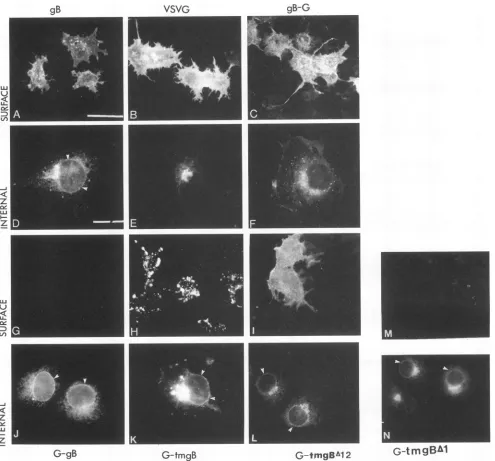
withanti-gB-1 or anti-G rabbit IgG and fluorescein isothiocya-nate-labeled goat anti-rabbit IgG. Fluorescence microscopic examination showed that the wild-type gB was located in intracellular structures which appear to be ER and NE as well asin theperinuclear region (Fig. 4D), which is in agreement with earlier reports (2, 28). As shown earlier (48), glycoprotein G was localized predominantly in a compact juxtanuclear
region consistent with the Golgi complex and perinuclear structures. Nostainingwasobserved in the NE rim (Fig. 4E). Theinternalstaining pattern observed with the gB-G chimera wasverysimilar tothat observed with G (Fig. 4F). No bright
nuclear rim staining was observed. In contrast, the chimeras
G-gB, G-tmgB, G-tmgBA12, and G-tmgBAl all showed inter-nal immunofluorescence patterns similar to that observed in
thecaseofgB-1 protein (Fig. 4J,
K,
L, and N,respectively). All four of the chimeric proteins containing the membrane an-choringdomains of gB-1 showed strong nuclear rim staining as well as perinuclear labeling, indicating NE and ER localiza-tion. As reported earlier (46, 47), the staining patterns for the mutantsgBCT799 andgBA12 were similar to that of wild-type gB-1, while the mutant gBA3 showed weak staining of the nuclear rim. However, when cells fixed with paraformaldehyde were examined for cell surface immunofluorescence, both wild-type gB-1 and G (Fig. 4A and B, respectively) and chimeras gB-G, G-tmgB, andG-tmgBAl12 (Fig. 4C, H, and I, respectively) showed cell surface labeling. As reported previ-ously, mutant gBC`T799 showed very faint surface labeling (47), while mutant gBA3 showed cell surface labeling similar to that of gB(46). Hybrids G-gB and G-tmgBAi (Fig. 4G and M, respectively), similar to the mutantgBA12 (46), failed to show anysurfacelabeling. The patterns of the surface immunofluo-rescence of the two hybrids G-tmgB (Fig.4H) andG-tmgBA12 (Fig. 41) were strikingly different. The surface fluorescence of G-tmgBA12 was similar to that observed with wild-type gB (Fig. 4A) and G (Fig. 4B). In contrast, G-tmgB showed a punctate pattern of distribution on theplasma membrane. The punctate distribution of G-tmgB on cell surface could be due to its degradation in the cell surface. The decrease in the amountof the endo H-resistant form ofG-tmgB with increas-ing chase time (Fig. 3) is in agreement with this possibility.Ultrastructural localization of membrane glycoproteins. Im-munofluorescence analyses of cells expressing wild-type gB and proteinscontaining themembrane spanning domain of gB showed localization in the NE as well as in the ER. However, the immunofluorescence data are not precise because of overlapping of membrane organelles in theperinuclear region and cannot be quantitated easily (23). We therefore used immunoelectron microscopy to determine the localization of the proteins at a more detailed resolutionatthe ultrastructure level of the intracellular membranes. Griffiths et al. (23) have recently critically assessed the use of immunofluorescence microscopy to study localization of intracellular membrane compartments in cells and emphasized that a complete and accuratelocalization of the membrane compartments requires analyses using bothimmunofluorescence and electron micros-copy.
To study thedistribution ofwild-typegB and Gaswell asof the mutants of gB and the chimeras in the intracellular membrane locations at the ultrastructure level, transfected COS cells were fixed withglutaraldehydeand embeddedatlow temperature in Lowicryl K4M. Ultrathin sections were then incubated sequentially with the affinity-purified rabbit
anti-gB
or anti-GIgG and goat anti-rabbit IgG coupledto 1-nm gold.
The goldcomplex was visualizedby silver enhancement.Using TIME (MIN) 0 30 60 120
ENDO H
-G-tmgB
VOL.68, 1994
on November 9, 2019 by guest
http://jvi.asm.org/
[image:4.612.69.304.74.257.2]J.VIROL. 2276 GILBERT ET AL.
VSVG
gB-G
[image:5.612.58.553.75.536.2]G-gB
G-tmgBG-tmgBA12
Gtmg2U
FIG. 4. Cellsurfaceandintracellularlocalization ofwild-typeandmutantproteins byimmunofluorescence. COS-1cellstransfected withgB (A andD),G(BandE),gB-G (CandF), G-gB (GandJ), G-tmgB (HandK),G-tmgBA12 (IandL),orG-tmgBAl (MandN)werefixedwith
paraformaldehyde and processed forcellsurface(A, B, C, G, H,I,andM)orinternal(D, E, F, J, K, L,andN)immunofluorescence.For internal
immunofluorescence, thecellswerefixed withparaformaldehydeand then treated with 1% Tritontopermeabilizethecells.Bars,60 pLmforpanels A,B, C, G, H, I, L, M, andNand 20,umforpanels D, E, F, J,and K.Small arrowheads indicate NE rim.
thispostembedding immunolabeling technique,we previously
confirmed thatinHSV-1-infected cells gB-1 is localized in the inner membrane of the NE (21). Similar results were also
obtainedby using freeze-fracture labeling(60). Strong labeling
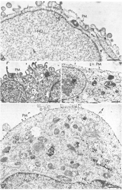
ontheNEofCOS cells transfected withgBplasmids (Fig. 5A)
indicates that the localizationof gBproteinonthe NE didnot require the expression of other HSV genes. The absence of
silvergrains in the sections ofCOS cells not expressing gB-1 and treatedwith anti-gB antibody(Fig. 5B) or in sections of
cellsexpressing gB-1 andreactedwith anonspecific antibody,
namely, purified anti-G antibody (Fig. 5C), established the specificity of the labeling method. Also, no labeling was
observed when sections of nontransfected cellswereincubated
withaffinity-purified anti-G antibodyorwhensections of cells expressing Gwere reacted with anti-gB-1 antibody (data not shown). The ER of cells transfected with gB-1 was also
intenselylabeled.Expressed gB-1 proteinwasalso localized in the plasma membrane and incytoplasmic vesicles of various sizes (Fig. 5A). In contrast, when COS cells expressing G protein were analyzed, the plasma membrane was intensely
labeled and small cytoplasmic vesicles and Golgi-like struc-tureswere labeled with anti-G antibody,butonlyafewsilver
grainswereobservedontheER,while the NE showedveryfew grains of silver (Fig. 5D). The distribution of grains on the
gB
LU
U--J
z
u.
I-z
I
on November 9, 2019 by guest
http://jvi.asm.org/
VOL.68, 1994 NUCLEAR ENVELOPE
LOCALIZATION
SIGNAL OF gB-1 2277P,
PM {
[image:6.612.115.521.71.703.2]is
'.'
FIG. 5. GlycoproteingBbutnotG is localized in the NE. COS-1 cells transfected with
gB
(A
andC)
orwith G(D)
ornottransfected(B)
wereprocessed for electronmicroscopyand labeledwithanti-gB (Aand
B)
oranti-G(C
andD)
antibodies asdescribed in the text.Arrowheads in panelsBandCcorrespondtononspecificlabeling. Go,Golgi
complex;
NU, nucleus; PM,plasma
membrane.Bars, 1p.m.on November 9, 2019 by guest
http://jvi.asm.org/
2278 GILBERT ET AL.
TABLE 1. Labeling density of silver particles onintracellular membranes of COS cells transfectedwith gB mutants andchimeras
No. of silver particles (mean ± SEM)/,urm of
Transfecting membrane measured No.of cells
plasmid Plasma counted NE ER membrane
None 0.15 ± 0.05 0.08 ± 0.047 0.13 ± 0.04 11 gB 6.0 ± 0.77 7.0 ± 0.9 3.8 ± 0.50 33
G 1.4 ± 0.2 2.2 ± 0.25 8.4 ± 0.9 24
gB-G 1.1 ± 0.2 1.9 ± 0.25 6.8 ± 0.99 24 G-gB 11 ± 1.1 16 ± 2.2 0.18 ± 0.03 15 G-tmgB 7.4 ± 0.9 6.9 ± 1.1 2.7 ± 0.7 24 gBCT799 3.3 ± 0.3 4.1 ± 0.6 1.4 ± 0.2 19 gBA12 5.9 ± 0.9 6.8 ± 1.0 0.15 ± 0.03 13 gBA3 2.1 ± 0.3 2.7 ± 0.4 2.5 ± 0.5 15 G-tmgBA12 10.1 ± 1.1 6.2 ± 0.9 7.1 ± 1.1 15
plasma
membranewasfairly
uniform. Thelabeling intensity
of the NEof cellsexpressing
Gwashigher
than thebackground
(Table 1),
which indicates that a small fraction of Gprotein
waslocalized in theNE. Asimilar distribution of G
protein
in this compartment was also observedpreviously
in VSV-in-fected cells(4).
Quantitation
of theintracellular distribution of silvergrains
showed thatonly
6%of the total number of silvergrains
were localized in the NE of cellsexpressing G,
com-pared
with 25% in cellsexpressing gB-1 (Table 2).
The outer and the inner membranes of theNEwerenotwellseparated
in most of the cases, and therefore no attempts were made todistinguish
the localizations ofproteins
in these two mem-branes.However,
in a fewspecimens
the outer and inner membraneswereseparated.
The distributions of silvergrains
in the outer and inner membranes in these cases were
quite
similar(data
notshown).
Thelabeling density
(grains
per micrometer ofmembrane)
ofgB-1
on theNEwasalso about four timeshigher
thanthatobserved with G(Table 1).
Takentogether,
these data suggest thatgB-1
wasspecifically
localized intheNE.Thedensity
oflabeling
ofgB-1
intheERmembranewassimilartothat observedfor the
NE,
showing
thatafraction of thegB-1
protein
was retained in the ER. Thelabeling
density
of G in theplasma
membranewasveryhigh
andwas two timeshigher
than observedwith thegB-1 protein (Table
1).
When the ratios of thelabeling
densities in theplasma
membrane and in theNEwere
compared,
values of0.5and4wereobserved for the
gB-1
and Gproteins, respectively.
TheseTABLE 2. Distributionofsilvergrainsinintracellularmembranes of COS cells transfected withgBmutantsandchimeras
% ( SEM)of silvergrains
Transfecting associated with: No. of Total no.
plasmid cells ofgrains
NE Cytoplasmic Plasma examined counted
membrane' membrane
gB
25 ± 2.9 45 ± 3.0 30 ± 2.4 14 4,893 G 6.3 ±0.87 31 ± 5.6 62 ± 5.6 11 3,917 gBCT799 25 ± 3.2 61 ± 4.7 13 ± 3.2 15 3,802 gB-G 7.1 ± 0.99 53 ± 5.0 40 ± 4.8 15 4,122 G-gB 33 ±3.8 67± 4.3 0.87 ± 0.23 11 3,902G-tmgB
24± 4.7 67 ± 4.2 7.7 ± 2.9 13 6,558gBA12
38 ±5.2 60 ± 5.4 2.0 ± 0.53 8 2,446gBA3 16 ±3.3 64 ± 5.7 20± 4.7 11 2,829
G-tmgBA12
33 ±4.5 46± 5.2 21 ± 5.1 8 3,617"Cytoplasmic membrane includes the ER, Golgi, and other membrane vesicles.
data suggest that G protein is preferentiallytransportedtothe plasma membrane and hencecanbe usedas areporterprotein
to study NE localization. Gprotein has also been used as a reporterprotein to determine the Golgi retention signal (35,
58).
The ectodomain ofgB-1 does not specify NE localization. To determine the role of the ectodomain ofgB-1 in NE localiza-tion, two chimeras, gB-G and G-gB, were constructed by switching the membrane anchoring domain and the
cytoplas-mic tail or the ectodomain of gB with the corresponding
domains of Gprotein. The two hybrids showed clearly distinct patterns of labeling. Cells expressing gB-G were strongly labeled on theplasma membrane and on cytoplasmicvesicles,
while very few silvergrainswerelocatedonthe NE and on the ER(Fig. 6A and Table 1). The distribution of gB-Gwasvery similar to the distribution of G, and only 7% of gB-G was associated with the NE(Table 2). These data and results from theimmunofluorescence analysissuggestthat the ectodomain of gB-1 is not sufficient for NE localization. When cells
expressing G-gBwereprocessed for immunoelectron micros-copywith anti-G antibody, both the NE and the ER showed intenselabeling (Fig. 6B and C and Table 1), but the plasma membrane and Golgi-like vesicles were not labeled. About 33% of the labeling in cells expressing G-gB was associated
withthe NE(Table 2).These results show that the ectodomain ofgB-1cannotdirect the chimeragB-Gto theNE, while the
carboxy-terminaltransmembrane andcytoplasmicdomains of gB-1 can target the chimeraG-gB to the NE, suggesting that thecarboxy-terminal regionofgB may specify NE localization. The transmembrane domain ofgB specifies NE localization. Results obtained from the two chimeras gB-G and G-gB suggest that the determinants for NE localization ofgB must be present either in the transmembrane domain or in the
cytoplasmic tail or in both. Our previous studies using gB-1 mutantswith shortened cytoplasmic tails have shown that the
cytoplasmictail maynotberequired for NE localization (47).
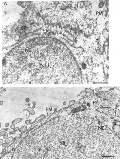
However, those results were obtained with immunofluores-cence and biochemical cell fractionation studies. To obtain moredefinitiveresults,westudied theintracellular localization of the cytoplasmic tail mutant gBCT799 by immunoelectron
microscopy. TheresultspresentedinFig.7Ashow labeling of theNEand the ER ofcellsexpressing gBCT799.About 25% ofthelabelingwasassociated with the NE(Table2),
suggest-ing thatthe deletion of thecytoplasmictail did not affect the NE localization. In agreement with the immunofluorescence data (47), theplasmamembrane was labeled poorly (Fig. 7A and Table 1). The amount of labeling for cells expressing
gBCT799 was generally lower than that observed for cells
expressing gB-1.This could be due to the fact that expression vectorp91023wasusedforexpressing gBCT799 (47), while the vector pXM was used to express the gB-1 gene. To prove
directly
that the predicted transmembrane domain of thecarboxy-terminal 69 hydrophobic amino acids of gB-1 were sufficient for NE localization, the chimera G-tmgB, in which the transmembrane segment of G was replaced with the 69-amino-acid predicted membrane anchoring segment of
gB-1, was constructed. COS cells transfected with G-tmgB
plasmid were analyzed by postlabeling immunoelectron mi-croscopy. Cells expressingG-tmgB showed strong labeling of the NEand theER (Fig.7Band Table 1). About 24% of the silver grains were localized in the NE of cells expressing
G-tmgB (Table
2), indicating that the predicted membraneanchoring sequence of gB-1 was sufficient to specify NE localization. The labeling density of G-tmgB in the NE was similartothat ofgB-1 andwas aboutfour- to fivefold higher than that observed in the cases of G and gB-G (Table 1). J. VIROL.
on November 9, 2019 by guest
http://jvi.asm.org/
[image:7.612.54.294.104.243.2]CD CD*0w@*:*7* 0<t=AWF
CD T' ,
gL
-r:oOSo
<#/>vetisX+vs@-
ki*s;;
§yAo,f;
weehX
h#4*w
4j
CDJ*'
CD $ t<9
= 1; sw
5-D4P<@~~~~~~~~~~~~~~~~~~w f~~~~~~~~~ ~ ~ ~ ~ ~ ~ ~~~~~~ ~~~~~~ 4 @~~Ar
(DsLP:Ws,'-$t*Ss-aX'- *' -* *4~~~~~~~Vq
CA!-. .: v< * .-i + 0- ;s. t ,
* -> - ---- E - ; wV4
-sCD
Ai, ,J'i'''s-v-Y~~~~~~~~~~~~A
U)D G ,D
CD3; , - R -'v'- > +
-°
crt > ; g t s N *
r>: ;. - :-1 Pl,i
2279
on November 9, 2019 by guest
http://jvi.asm.org/
2280 GILBERT ET AL.
FIG. 7. Thehydrophobicdomain of gB issufficienttospecifyNElocalization. COS-1cellstransfected withgBCT799(A)orG-tmgB(B)were processed for electron microscopy and labeled withanti-gBandanti-G antibody,respectively.Bars, 1 p.m.Abbreviationsare asdescribed in the legendtoFig. 5.
Plasma membrane labeling of cells expressing G-tmgB was strong,but thegrainswere notuniformly distributed (Fig. 7B);
rather, theywerepresentinpatches of the plasma membrane. Thiswasin agreement with the punctuate surface immunoflu-orescencelabeling pattern observed withG-tmgB.
Hydrophobic segment 3 is sufficient for NElocalization of
gB-Iprotein.Using deletionmutantslackingone, two,orthree
segments ofhydrophobic amino acids in thepredicted mem-branespanningdomains of 69residues in gB-1,wepreviously
showed that segment 3 represents the membrane anchoring
domain of gB-1 (46). To determine whether segment 3 was sufficient for NE localization ofgB-1, we used the deletion mutants
gBA12
andgBA3.
Earlier (46) we showed that theprotein gBA12, which contains only segment 3, can stably
J. VIROL.
on November 9, 2019 by guest
http://jvi.asm.org/
[image:9.612.104.504.80.606.2]&i
tt ai S
t
4~~~~~
t*wi,4 008;S>.,+t0SsW ,x <r
;r
_ t> 't >+ >*pt-t&A't .. 4' 9;
* v rrwt E
9
;
tC:49X
*4 &, 4 2',o,
'4-a
4Ft<,-ER~
¼
*~~~~~~~j #.- *#
PM
.*.;-A
6t.".t;
t -K. th.X."
[image:10.612.133.510.52.693.2]U
O~~~~~~
.VI,;.,,-- W4-s,t#,!FIG. 8. Thehydrophobic membrane anchoring segment3(residues 774 to 795) is sufficient to specify NE localization.COS-1 cells transfected with mutantgBA12, gBA3,G-tmgBA1,orG-tmgBA12(A, B, C, and D, respectively) were processed for electron microscopy. Cells transfected with gBA12 orgBA3werelabeled withanti-gB antibody, while cells transfected with G-tmgBAI or G-tmgBA12 were labeled with anti-G antibody. Bars, 1 p.rm. Themagnifications forG-tmgBAl andG-tmgBA12 were x 17,000 and x16,000,respectively. Abbreviations are as described in the legend to Fig. 5.
2281
I
on November 9, 2019 by guest
http://jvi.asm.org/
2282 GILBERT ET AL.
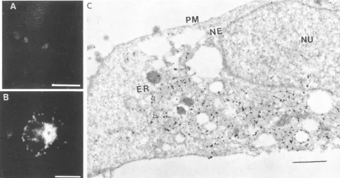
FIG. 9. Mutant G protein retained in the ER is not localized in the NE. COS-1 cells transfected with mutant GH6 were processed for
immunofluorescence and immunoelectronmicroscopywith anti-Gantibody.(A)Surfaceimmunofluorescence; (B)internalimmunofluorescence; (C) immunoelectronmicroscopyof cells labeled with anti-Gantibody.Bars,60, 20,and 1 p.mforpanels A, B,andC,respectively.Abbreviations are asdescribed in thelegendtoFig.5.
anchortothemembrane.Inthecaseof themutant
gBA3,
most of theproteinwassecreted,butasmallfractionwasanchored to intracellular membranes (46). When cellsexpressing
thegBA12 or gBA3 mutant were processed for immunoelectron microscopy, the NE and ER of cells expressing
gBA12
were strongly labeled(Fig. 8A).Theoverall distribution ofgBA12in thecellwas similartothat ofG-gB; 38% of the total number ofgrainswerepresent intheNE (Table 2).The mutantgBA3showed labelingofthe NE, ER, andplasma membrane
(Fig.
8B andTable 1),butthe distribution of silvergrainsin theNE was weaker (16%, compared with 25 and 38% for gB and gBA12,respectively) (Table 2). In agreementwith the immu-nofluorescence data (46), the immunoelectron microscopy
resultsshowed that gBA3 was also localized in the cellsurface, while mutant gBA12 failed to be transported to the plasma membrane(Fig.8Band A,respectively). Thus, segment3may be sufficient forNE localization of gB but isnot essentialfor NEretention, becauseNElocalization at areducedefficiency was observed with the mutant gB protein lacking segment 3
(gBA3) (Fig. 8B and Table 2). The labeling densityofgBA3 wasessentially similar in the NE, ER, and plasma membrane
(Table 1).
To test whether segment 3 alone was sufficient for NE localization, chimeric glycoproteins G-tmgBAl and G-tmg
BA12wereconstructed. In chimeraG-tmgBAl,the transmem-brane segment of G protein was replaced with segments 2 and 3ofgB-1, while the hybrid
G-tmgBzv12
contained segment 3 of gB-1 inplace of the transmembrane segment of G. Results ofimmunoelectron microscopic analyses of cells expressing
G-tmgBAM
and G-tmgBA12 show that the NE and the ER were strongly labeled in both cases (Fig. 8C and D, respectively). However, only the plasma membrane of the cells expressingG-tmgBA12
showed strong labeling (Fig. 8D). The twomu-tantsG-tmgBAl and
G-tmgBAl12,
however,showed amarkeddifference in the distribution of theproteins (Fig. 8C and D).
Thus, deletion of segment 1 leads to an increased accumula-tion in thecytoplasmic membranes (Fig.8C), while deletion of segments 1 and 2causes increasedlocalization in the plasma
membrane (Fig. 8D).
Itmay be noted thatgBaswellas mutantsofgBorchimeras that were localized in the NE also accumulated in the ER. The labeling densities of proteins in the NE and ER of cells expressing these proteins were also similar (Table 1), suggest-ing that the membraneanchoring segment of gB-1notonly can promote localization of proteins in the NE but also may influence the retention of proteins in the ER.
Thepossibility that localization ofproteins in the NE could be duetoretention inthe ER was tested byusing a mutant of G protein. We have previously shown that the mutant GH6, constructedby insertion of three amino acidsatposition 244 of G protein, did not acquire endo H resistance and failed to reach the cell surface (32) andthus was retained in the ER.
Immunofluorescenceanalyses of cellsexpressing GH6 showed staining of perinuclearregions similar tothat seenwith G or gB-G (Fig. 9B). No staining of the cell surfacewas observed (Fig. 9A). When cells expressing GH6 were examined by immunoelectron microscopy, the labeling was associated al-most exclusively with a cytoplasmic membrane structure re-sembling the ER network. No labeling was observed in the cell
surface,andveryfewgrainswereassociated with theNE(Fig. 9C). These results suggest that retention in the ER alone may not besufficient for localization of the protein in the NE.
DISCUSSION
The results presented in this report show that the carboxy-terminal sequence of69 hydrophobic residues of gB-1 glyco-protein can specify its localization in the NE. The fact that J.VIROL.
I.f- -, i-,
on November 9, 2019 by guest
http://jvi.asm.org/
[image:11.612.60.545.82.336.2]NUCLEAR ENVELOPE LOCALIZATION SIGNAL OF gB-1 2283
mutantgBA12 and the chimera G-tmgBA12, both containing only segment 3 as the membrane spanning sequence, were present in the NE also suggests that segment 3 is sufficient for NE localization. Mutant gBA3 lacking segment 3 was also located in the NE, but thelabeling in the NE in the case of
gBA3 was much less than that observed with gB mutants or chimeras containing segment 3. It may also be noted that the absence of segments 1 and 2 in the mutant gBA12 and in the chimeraG-tmgBA12 resulted in increased accumulation in the NE. Although they showed very similar NE localizations, gBA12 andG-tmgBA12 had markedly different distributions in the cytoplasmic and plasma membranes. The absence of segment 1 (G-tmgBAl) resulted inincreased accumulation in thecytoplasmicmembrane, whereas the absence of segments 1 and 2 (G-tmgBA12) resulted in enhanced localization in the plasma membrane. Taken together, these results suggest that in the case of gB-1 protein, regions other than segment 3 may also be contributing, albeit weakly, to NE localization. Thus, although segment 3 issufficient, segments 1 and 2 and possibly the cytoplasmic domain may augment localization of gB-1 protein in the NE. Studies with chimeric proteins, however, showed that the cytoplasmic domain and segments 1 and 2 of the transmembrane domain of gB-1 protein were not necessary for NE localization, but their presence in the hybrid protein resulted in a higher retention in the cytoplasmic compartments
(67% for both G-gB and G-tmgB compared with 46% for
G-tmgBA12). Furthermore,deletion of segments 1 and 2 from G-tmgB resulted ina threefold increase in the localization of
G-tmgBA12inthe plasma membrane. The sequence present in segment 3 is very hydrophobic and lacks uncharged polar aminoacids but shows a high degree of sequence conservation with similar regions of glycoproteins of other herpes viruses which are homologous to gB of HSV. In fact, within the
ct-herpesvirus
family, segment 3 of gB homologs is almosttotally conserved. Inall of these cases, segment 3 contains an invariantproline precedingthe first hydrophobicresidue and thuscould be part ofa,B-turnthatlinks segments 2 and 3 (44). Studies with mutants with deletions in thehydrophobicregion further showed that these sequences also played an important role inintracellular transport of theglycoprotein and in virus
infectivity(46).Thehighlyconservedsegment 3 maytherefore notonlyfunction in membrane anchoring and viral entry but also promote localizationin the NE.
Transmembranedomainsofanumber of integral membrane
proteins targeted to intracellular membranes have recently beenshowntospecify retentionorsorting signals(3, 6, 35, 38, 41, 43, 53, 63). Membrane spanning segments of the spike glycoproteinMof infectiousbronchitisvirus, which buds in the
Golgi complex, wasshown tocontainaGolgi retention signal
(35, 58). Membrane anchoring sequences of a number of enzymes localized in the Golgimembrane were also sufficient for targetingreporter
proteins
totheGolgi complex (3, 6, 38,40, 41, 43). The transmembrane domain of the nuclear pore
complex protein gp210 was also sufficient for sorting to the poremembraneof the NE
(63).
Similarly,the first of theeight transmembrane segmentsofthe LBR was shown to be suffi-cient for targeting to the innernuclear membrane (53). Our results thusprovidefurther evidence that membraneanchoringdomains of proteins may contain information
required
for localization of theproteinsinorganellemembranes suchastheGolgi complexorNE.However,noobvioushomologybetween the transmembrane domains of gp210, the LBR, and
gB-1
glycoprotein wasobserved.The three distinct NE domains, i.e., the inner nuclear membrane, outernuclearmembrane, and nuclear pore com-plex,areincontinuitywith theERmembrane
(14,
15, 18),andtherefore protein synthesized in the ER or in the outer nuclear membrane may reach all of these domains of the NE by lateral diffusion. However, unique integral membrane proteins have been identified in the inner nuclear membrane and in the nuclear pore complex, suggesting the existence of specific NE targeting signals (14, 15, 18). The NE targeting signal may specifically retain proteins synthesized in the outer nuclear membrane following lateral diffusion along the continuous membrane to the inner membrane or the pore complex. Such amechanism was proposed to explain NE targeting signals of the LBR (53) and the inner membrane protein gp210 (63). HSVglycoproteins such asgB-1 that aretargeted to the inner nuclearmembrane for virus assembly may also follow the same pathway. Alternatively, gB-1 may be targeted to the NE by yet-uncharacterized transport vesicles originating from the ER or by aspecific sortingreceptor which transports it to the inner membrane. Interactions of the membrane spanning domain of gB-1 with the specific transport vesicle or with the transmem-brane domain of the sorting receptor may mediate the NE sortingprocess. Thetransmembrane domain ofgB-1 may also interact with the membrane spanning regions of proteins resident in the inner nuclear membrane to form oligomeric structures which are retained in the NE. Such a mechanism involving "kin recognition" mediated by the transmembrane domains of Golgi resident proteins has been proposed as a mechanism for retention of proteins in the Golgi membrane (35, 41). In HSV-infected cells, viral envelope glycoproteins mayalso formhetero-oligomers by interacting with each other via the membrane anchoring domain and be retained in the inner nuclearmembrane where virus budding occurs (45).
The fact that
gB-1
as well as mutants of gB and chimeras that arelocalized in the NE alsoaccumulate in the ER suggests that the transmembrane domain of gB may contain both ER retention and NE localization signals. However, studies with ER transmembrane proteins have shown that a cytoplasmic signal consisting of the C-terminal sequence Lys-Lys-X-X or Lys-X-Lys-X-X (where X can correspond to any amino acid) is required for retention or retrieval of proteins in the ER (25,34,
43).The observed accumulation in the ER could be due to overexpression of the protein in the HSV-infected cells (54) or in cellstransfected with a high-level expression vector, such as the pXM vector used in this study (27, 64). Indeed, overex-pression has been shown to cause retention in the ER of proteins targeted to the Golgi complex (38, 40, 41), nuclear pore complex (63), or inner nuclear membrane (53). Our results with the mutant GH6, which is retained in the ER but is notlocalized in the NE, also suggest thataccumulation in the ER alone may not be sufficient for localization in the NE. Mutagenesis of thetransmembrane segment 3 of gB-1, which is in progress, may help to further elucidate the mechanism of how this domainfunctions as an NElocalization signal.ACKNOWLEDGMENTS
We thank D. Andrews and J. Capone for critical review of the manuscript and B. Murphy and D.Tomlinson for help in preparing the manuscript.
This work was supported by the Medical Research Council of Canada.
REFERENCES
1. Adams, G. A., and J. K. Rose. 1985. Structural requirementsofa
membrane-spanning domain for protein anchoring and cell sur-facetransport. Cell41:1007-1015.
2. Ali, M. A., M.Butcher, and H. P. Ghosh. 1987. Expression and nuclear envelope localization ofbiologically active fusion glyco-protein gB of herpes simplex virus in mammalian cells using VOL.68,1994
on November 9, 2019 by guest
http://jvi.asm.org/
2284 GILBERT ET AL.
cloned DNA. Proc.Natl. Acad. Sci. USA 84:5675-5679. 3. Aoki, D., N.Lee, N.Yamaguchi, C. Dubois,and P. M. Fukudo.
1992. Golgiretention ofatrans-Golgimembrane protein,
galac-tosyltransterase, requires cysteineand histidine residues within the membrane anchoring domain. Proc. Natl. Acad. Sci. USA 89: 4319-4323.
4. Bergmann, J.E.,andS.J.Singer. 1983. Immunoelectron
micro-scopic studies of the intracellular transport of the membrane
glycoprotein (G)of vesicularstomatitisvirus in infected chinese hamster ovary cells. J. Cell Biol. 97:1777-1787.
5. Burke,B.1990. The nuclearenvelopeand nucleartransport.Curr.
Opin.Cell Biol. 2:514-520.
6. Burke,J.,J.M.Petitt,H.Schachter,M.Sarkar,andP. A. Gleeson. 1992. The transmembrane and flanking sequences of
P1,2-N-acetylglucosamincyltransferaseIspecifymedial-Golgilocalization. J.Biol. Chem. 267:24433-24440.
7. Butcher, M., K. Raviprakash, and H. P. Ghosh. 1990. AcidpH induced fusion of cellsby herpes simplexvirusglycoproteinsgB andgD.J.Biol. Chem. 265:5862-5868.
8. Bzik, D. J., B. A. Fox, N. A. DeLuca, and S. Person. 1984. Nucleotide sequence specifying the glycoprotein gene gB, of
herpes simplexvirustype 1.Virology133:301-314.
9. Cai,W., B. Gu, and S. Person. 1988. Role ofglycoproteinB of
herpes simplexvirustype 1 inviral entry and cell fusion. J. Virol. 62:2596-2604.
10. Compton, T.,and R. J. Courtney. 1984. Virus-specific glycopro-teins associated with the nuclear fraction ofherpes simplexvirus type 1-infectedcells. J. Virol. 49:594-597.
11. Danscherg, G., andR. Norgaard. 1983.Light microscopic visual-ization of colloidalgoldonresin-embeddedtissue. J. Histochem.
Cytochem.31:1394-1398.
12. Darlington, R. W., and L. H.Moss. 1968. Herpesvirus
envelop-ment.J.Virol. 2:48-55.
13. Doms, R. W., R. A. Lamb, J. K. Rose, and A. Helenius. 1993.
Foldingand assemblyof viral membraneproteins. Virology 193: 545-562.
14. Dessev,G. N. 1992. Nuclearenvelopestructure.Curr. Opin.Cell Biol. 4:430-435.
15. Dingwall, C., and R. Laskey. 1992. The nuclear membrane. Science258:942-947.
16. Einfeld, D.,andE. Hunter.1991.Transportofmembraneproteins
tocellsurface. Curr.Top. Microbiol. Immunol. 170:107-139. 17. Garoff, H. 1985. Using recombinant DNA techniques to study
protein targeting in the eukaryotic cell. Annu. Rev. Cell Biol. 1:403-445.
18. Gerace, L., and B. Burke. 1988. Functional organization of the nuclearenvelope.Annu. Rev.Cell Biol. 4:335-374.
19. Ghosh,H. P. 1980. Synthesisandmaturation ofglycoproteinsof
envelopedanimalviruses. Rev. Infect. Dis. 2:26-39.
20. Ghosh-Choudhury, N., M. Butcher, and H. P. Ghosh. 1990.
Expressionfrom clonedDNAofbiologicallyactiveglycoproteinC ofherpes simplexvirus type 1 in mammalian cells. J. Gen. Virol. 71:689-699.
21. Gilbert,R.,andH. P.Ghosh. 1993. Immunoelectronmicroscopic localizationofherpes simplexvirusglycoproteingBin the nuclear
envelopeofinfected cells. Virus Res. 28:217-231.
22. Graham,F.L.,and A.J.vander Eb.1973. Anewtechniquefor the assay of infectivity of human advenovirus 5 DNA. Virology 52:456-467.
23. Griffiths,G., R. C. Parton,J. Lucocq, B. van Deurs, D. Brown, J.W. Slot,and H.J.Geuze. 1993. Theimmunofluorescenteraof membrane traffic. Trends Cell Biol. 3:214-219.
24. Harlow, E. D., and D. Lane. 1988. Antibodies, a laboratory manual. Cold Spring Harbor Laboratory, Cold Spring Harbor, N.Y.
25. Jackson,M.R.,T.Nilsson,andP. A.Peterson. 1990.Identification ofaconsensusmotif for retention of transmembrane proteinsin the endoplasmicreticulum.EMBO J. 9:3153-3162.
26. Johnson,D.C.,andP. G.Spear. 1983. 0-linkedoligosaccharides
are acquired by herpes simplexvirus glycoproteins in the Golgi apparatus. Cell32:987-997.
27. Kaufman,R.J. 1990.Vectors used forexpressionin mammalian cells. MethodsEnzymol.185:487-51 1.
28. Koga, J., S. Chatterjee, and R.J.Whitley.1986. Studies onherpes simplex virustype 1 glycoproteins using mononclonalantibodies. Virology151:385-389.
29. Kornfeld, R., and S. Kornfeld. 1985. Assembly of asparagine-linkedoligosaccharides.Annu. Rev. Biochem.54:631-664. 30. Kotwal, G. J., J. Capone, R. A. Irving, S. H. Rhee, P.Bilan, F.
Toneguzzo,T.Hofmann, and H. P. Ghosh. 1983. Viral membrane glycoproteins: comparison of the amino terminal amino acid sequences of the precursor and mature glycoproteins of three serotypesofvesicular stomatitisvirus.Virology129:1-11. 31. Kunkel, T., J. D. Roberts,and R. A. Zakour. 1987. Rapid and
efficient site-specific mutagenesis without phenotypic selection. MethodsEnzymol. 154:367-382.
32. Li, Y., C. Drone, E. Sat, and H. P. Ghosh. 1993. Mutational analysis of the vesicular stomatitis virus glycoprotein G for mem-brane fusion domains. J. Virol.67:4070-4077.
33. Little, S. P., J. T. Jofre, R. J. Courtney, and P. A. Schaffer. 1981. A virion-associatedglycoprotein essential for infectivity of herpes simplexvirus type-1.Virology114:149-160.
34. Luzio, J. P., and G.Banting. 1993. Eukaryotic membrane traffic: retrieval and retention mechanismtoachieveorganelleresidence. Trends Bicohem. Sci. 18:395-398.
35. Machamer, C. 1991. Golgi retention signals: do membraneshold the key?Trends Cell Biol. 1:141-144.
36. McDermott,M.R., F. L. Graham, T. Hanke, and D. C.Johnson. 1989. Protection of mice against lethal challenge with herpes simplex virus by vaccination withanadenovirusvectorexpressing HSVglycoproteinB.Virology169:244-247.
37. Morgan, C., H. M. Rose, M. Holden, and E. P. Jones. 1959. Electron microscopic observationon the development of herpes simplexvirus. J. Exp.Med. 110:643-656.
38. Munro, S. 1991.Sequenceswithin andadjacenttothe transmem-branesegment of(-2,6 sialyltransferase specifyGolgi retention. EMBO J. 10:3577-3588.
39. Nii, S.,C.Morgan,and H. M. Rose. 1968. Electronmicroscopyof
herpes simplex virus. II. Sequence of development. J. Virol. 2:517-536.
40. Nilsson, T., J.M.Cucocq,D.MacKay,and G. Warren. 1991. The membranespanningdomain of 3-1,4-galactosyltransferase speci-fiestransGolgilocalization. EMBO J. 10:3567-3575.
41. Nilsson, T.,P.Slusarewicz,M.H.Hoe, and G. Warren. 1993. Kin recognition: a model for the retention ofGolgienzymes. FEBS Lett. 330:1-4.
42. Pachl,C.,R. L.Burke,L.L.Stuve,L.Sanchez-Pescador, G. Van Nest,F.Masiarz,and D.Dina. 1987.Expressionofcell-associated and secretedforms ofherpes simplex virustype 1glycoproteingB inmammalian cells. J. Virol.61:315-325.
43. Pelham, H. R. B., and S. Munro. 1993. Sorting of membrane
proteinsin thesecretorypathway.Cell75:603-605.
44. Pellet,P.E.,K. G.Kousoulas,L.Pereira,and B.Roizman. 1985. Anatomy of the herpes simplexvirus 1 strain F glycoprotein B gene:primarysequenceandpredicted proteinstructureof the wild type and of monoclonal antibody-resistant mutants. J. Virol. 53:243-253.
45. Pettersson,R. F. 1991.Proteinlocalizationandvirusassemblyat intracellularmembranes. Curr.Top. Microbiol.Immunol. 170:67-106.
46. Rasile, L., K. Ghosh, K. Raviprakash, and H. P. Ghosh. 1993. Effectsof deletions in thecarboxy-terminalhydrophobic regionof herpes simplexvirusglycoproteingBonintracellulartransport and membraneanchoring.J.Virol.67:4856-4866.
47. Raviprakash, K., L. Rasile, K. Ghosh, and H. P. Ghosh. 1990. Shortened cytoplasmicdomain affects intracellular transport but
not nuclear localization of a viral glycoprotein. J. Biol. Chem. 265:1777-1782.
48. Rose, J. K., and J. E. Bergmann. 1983. Altered cytoplasmic domains affectintracellular transport ofthe vesicularstomatitis virusglycoprotein.Cell 34:513-534.
49. Rose,J.K.,and R. Doms.1988.Regulationofproteinexportfrom theendoplasmicreticulum. Annu. Rev. Cell Biol. 4:257-288. 49a.Rose,J. K.,andC.J.Gallione. 1981.Nucleotidesequencesof the
mRNA'sencodingthevesicular stomatitis virus G and M proteins determined from cDNA clones containing the complete coding
J.VlIROL.
on November 9, 2019 by guest
http://jvi.asm.org/
NUCLEAR ENVELOPE LOCALIZATION SIGNAL OF gB-1 2285 regions. J. Virol.39:519-528.
50. Roth, J., M. Bendayan, E. Carlemaim, W. Villiger, and M. Garavito. 1981. Enhancement ofstructural preservation and im-munocytochemical staining in lowtemperatureembedded
pancre-atictissue.J.Histochem.Cytochem.29:663-671.
51. Schlesinger, M. J., and S. Schlesinger. 1987. Domains of virus glycoproteins.Adv. Virus Res. 33:1-44.
52. Schneider, M., F. L. Graham, and L. Prevec. 1989.Expression of theglycoproteinofvesicular stomatitisvirusby infectious adeno-virusvectors.J. Gen. Virol. 70:417-427.
53. Smith, S., and G. Blobel. 1993. The first membrane spanning region of lamin B receptor is sufficient for sorting to the inner nuclear membrane. J.Cell Biol. 120:631-637.
54. Sommer, M., and R. J. Courtney. 1991. Differential rates of processing and transportof herpes simplex virustype I
glycopro-teins gBandgC.J. Virol. 65:520-525.
55. Soullam, B.,andH.J.Worman.1993. The amino terminaldomain ofthe lamin B receptorisanuclearenvelope targetingsignal. J. Cell Biol. 120:1093-1100.
56. Spear,P. G. 1993. Membrane fusion induced byherpes simplex
virus, p.201-232.InJ.Bentz(ed.),Viral fusionmechanisms.CRC Press, Boca Raton, Fla.
57. Stephens, E. B., and R. W.Compans. 1988. Assemblyofanimal virusesatcellularmembranes. Annu. Rev. Microbiol. 42:489-516.
58. Swift, A. M., and C. Machamer. 1991. A Golgi retention signal in
amembrane-spanning domain ofcoronavirusEl protein. J. Cell
Biol. 115:19-30.
59. Torrisi, M. R., M. Cirone, A. Pavan, C. Zompetta, G. Barile, L. Frati, and A. Faggioni. 1989. Localization of Epstein-Barrvirus envelope glycoproteins on theinner nuclearmembraneof
virus-producing cells. J. Virol.63:828-832.
60. Torrisi, M. R., C. DiLazzaro, A. Pavan, L. Pereira, and G. Campadelli-Fiume. 1992. Herpes simplexvirus envelopment and maturationstudied by fracturelabel. J.Virol.66:554-561. 61. Torrisi, M. R., L. V. Lotti, A. Pavan,G. Migliaccio,andS.Bonatti.
1987. Free diffusiontoandfrom theinner nuclear membraneof
newlysynthesized plasma membraneglycoproteins. J. Cell Biol. 104:733-737.
62. Woolson, R. F. 1987. Statistical methods for the analysis of biomedical data. John Wiley & Sons, New York.
63. Wozniak,R. W., and G. Blobel. 1992. The single transmembrane
segment ofgp210issufficient for sortingtothe poremembrane domainof the nuclearenvelope. J. Cell Biol. 119:1441-1449. 64. Yang, Y.-C., A. B. Ciarletta, P. A. Temple, M. P. Chung, S.
Kovacic, J. S. Witek-Giannoti, A. C. Leary, R. Kriz, R. E. Donahue, G. G. Wong, and S. C. Clark. 1986. Human IL-3 (multi-CSF): identification by expression cloningofanovel hema-topoietic growthfactorrelatedtomurine IL-3. Cell 47:3-10. VOL. 68, 1994