A STUDY OF
THYROID PROFILE IN CHRONIC KIDNEY
DISEASE
DISSERTATION SUBMITTED FOR M.D.DEGREE
EXAMINATION
BRANCH I – GENERAL MEDICINE
APRIL 2011
TIRUNELVELI MEDICAL COLLEGE
THE TAMILNADU DR. M.G.R. MEDICAL UNIVERSITY
CERTIFICATE
This is to certify that this dissertation entitled “Thyroid profile in chronic kidney disease” submitted by Dr.S.RAJINI appearing for M.D. Branch I General Medicine Degree examination in April -2011 is a bonafide record of work done by him under my direct guidance and supervision in partial fulfillment of regulations of the Tamil Nadu M.G.R. Medical University, Chennai. I forward this to the Tamil Nadu Dr. M.G.R. Medical University, Chennai, Tamil Nadu, India
Dr.R.Geetha Rani M.D Dr.J.Kaniraj Peter M.D
Professor &chief Professor & Head of the Department
Department of Medicine Department of Medicine
Tirunelveli medical college hospital Tirunelveli medical college hospital
Tirunelveli Tirunelveli
The Dean
Tirunelveli medical college
DECLARATION
I solemnly declare that the dissertation titled “Thyroid Profile in chronic kidney disease” is done by me at Tirunelveli Medical College hospital, Tirunelveli under the guidance and supervision of
Prof.Dr.R.Geetha Rani, M.D., The dissertation is submitted to The
Tamilnadu Dr. M.G.R. Medical University towards the partial fulfilment
of requirements for the award of M.D. Degree (Branch I) in General
Medicine.
Place: Tirunelveli
Date:
Dr.S.RAJINI
Postgraduate Student
M.D. General Medicine
Department of Medicine
Tirunelveli Medical College
Tirunelveli
ACKNOWLEDGEMENT
I am extremely thankful to our beloved
Dean, Dr.N.Palaniappan.M.D., for granting me permission to carry out this study in Tirunelveli medical college hospital.
It is an immense pleasure to acknowledge Dr.J.Kaniraj Peter.M.D., Professor and Head of the department, Department of
medicine, who has given the moral support, philosophical guidance and
ever available help to carry out this study.
With deepest appreciation and gratitude I thank Dr.R.Geetha Rani M.D., my unit chief and Associate Professor of medicine and
Dr.V.Ramasubramanian, M.D., D.M., Professor and Head of Nephrology, Tirunelveli Medical College, my guide and mentor to carry
out this study.
I thank Professor and the staff belonging to the department of
biochemistry and radiology.
I also thank Dr.S.S.Nazar M.D., and Dr.M.Mohamed Rafi M.D.,
assistant professors for their moral support.
Finally with Almighty God and with the cooperation of the
ABBREVIATIONS
CKD - Chronic Kidney Disease CRF - Chronic Renal Failure ESRD - End stage renal disease FT4 - Free Thyroxine
GFR - Glomerular Filtration Rate PTH - Parathyroid hormone T3 - Triiodothyronine T4 - Thyroxine
TRH - Thyrotropin Releasing Hormone TSH - Thyroid Stimulating Hormone TBG - Thyroxine Binding Globulin HD - Hemodialysis
PD - Peritoneal Dialysis TTR - Transthyretin
SES - Sick Euthyroid Syndrome TH - Thyroid Hormone
NS - Nephrotic Syndrome
CONTENTS
Sl.No. TITLE PAGE. No
1. INTRODUCTION 1
2. AIM OF THE STUDY 3
3. REVIEW OF LITERATURE 4
4. MATERIALS AND METHODS 31
5. OBSERVATIONS AND RESULTS 34
6. DISCUSSION 50
7. CONCLUSION 54
8. REFERENCES
9. PROFORMA
1
INTRODUCTION
Patients with chronic renal failure often have signs & symptoms
suggestive of thyroid dysfunction. These findings include dry skin, sallow
complexion, low temperature, cold intolerance, decreased basal metabolic
rate, lethargy, fatigue, edema & hyporeflexia. Various studies of thyroid
functions in uremic patients have been carried out which have shown
conflicting results. Hyperthyroidism, hypothyroidism & euthyroid state have
all been reported by various workers.
Serum triiodothyronine (T3) levels were consistently found to be low
without any regard to treatment of CRF. Serum total & free thyroxine (T4)
concentrations have been reported as low, normal or high. Serum thyroid
stimulating hormone (TSH) levels were found to be normal in most patients
of CRF even in those whose CRF is complicated by low T3 concentration.
A reduction in total T3, but not in free T3 concentrations
wasassociated with an increased all-cause and cardiovascular mortalityin
euthyroid CKD patients45. Total and free T3 behave assurvival markers in
patients with CKD both in HD and inPD.
Prevalence of hypothyroidism in end stage renal disease (ESRD) have
been estimated between 0 and 9%. There is also increased prevalence of
goiter in patients with ESRD. Though there are multiple factors which
predicts the overall mortality and severity of renal disease, one among the
2
treating physician to be aware of thyroid dysfunction so that early
intervention can be instituted to improve the outcome. In view of variability
of thyroid function test in patients with CRF in previous studies, a
cross-sectional study on thyroid function in CRF patient in Department of
3
AIM OF THE STUDY
1. To study the prevalence of thyroid dysfunction in patients with
chronic renal failure.
2. To study the correlation between thyroid dysfunction and severity
of renal failure.
3. To differentiate primary thyroid diseases from thyroid
dysfunction due to chronic renal failure.
4
REVIEW OF LITERATURE
Chronic kidney disease (CKD)1 encompasses a spectrum of different
pathophysiologic processes associated with abnormal kidney function, and a
progressive decline in glomerular filtration rate (GFR).
DEFINITION CRITERIA:
Chronic Kidney Disease is defined according to the presence or
absence of markers of kidney damage and the level of kidney function
(GFR), irrespective of kidney disease (the specific diagnosis).
1. Kidney damage for ≥3months,as defined by structural or functional
abnormalities of the kidney, with or without decreased GFR, manifest by
either,
a) Pathological abnormalities or
b) Markers of kidney damage, including abnormalities in the
5
CLASSIFICATION:
A widely accepted classification, based on recent guidelines of the
National Kidney Foundation [Kidney Dialysis Outcomes Quality Initiative
(KDOQI)]2, in which stages of CKD are defined according to the estimated
GFR.
Stage GFR ml/min/1.73m2
0 >90a
1 >90b
2 60-89 3 30-59 4 15-29 5 <15
a. with risk factors for CKD
b. with demonstrated kidney damage (persistent protienuria, abnormal
urine sediment, abnormal blood and urine chemistry, abnormal
imaging studies)
PATHOPHYSIOLOGY:
The pathophysiology of CKD involves two broad sets of mechanisms of
damage:
1. Initiating mechanisms specific to the underlying etiology (e.g:
6
glomerulonephritis or toxin exposure in certain diseases of the renal
tubules and interstitium),
2. Set of progressive mechanisms, involving hyperfiltration and
hypertrophy of the remaining viable nephrons, that are a common
consequence following long-term reduction of renal mass, irrespective
of underlying etiology. The responses to reduction in nephron number
are mediated by vasoactive hormones, cytokines, and growth factors.
Eventually, these short-term adaptations of hypertrophy and
hyperfiltration become maladaptive as the increased pressure and flow
predisposes to sclerosis and dropout of the remaining nephrons.
UREMIA:
Hundreds of toxins that accumulate in renal failure have been
implicated in the uremic syndrome. These include water-soluble,
hydrophobic, protein-bound, charged and uncharged compounds. Additional
categories of nitrogenous excretory products include guanido compounds,
urates and hippurates, products of nucleic acid metabolism, polyamines,
myoinositol, phenols, benzoates, and indoles. Compounds with a molecular
mass between 500 and 1500 Da, the so-called middle molecules, are also
retained and contribute to morbidity and mortality. The pathophysiology of
the uremic syndrome can be divided into manifestations in three spheres of
dysfunction: (1) those consequent to the accumulation of toxins normally
7
(2) those consequent to the loss of other renal functions, such as fluid and
electrolyte homeostasis and hormone regulation and (3) progressive systemic
inflammation and its vascular and nutritional consequences.
MANIFESTATIONS OF CHRONIC KIDNEY DISEASE
Stages 1 and 2 CKD are usually not associated with any symptoms
arising from the decrement in GFR. However, there may be symptoms from
the underlying renal disease itself, such as edema in patients with nephrotic
syndrome or signs of hypertension secondary to the renal parenchymal
disease in patients with polycystic kidney disease, some forms of
glomerulonephritis and many other parenchymal and vascular renal diseases,
even with well-preserved GFR. If the decline in GFR progresses to stages 3
and 4, clinical and laboratory complications of CKD become more
prominent. Virtually all organ systems are affected, but the most evident
complications include anemia and associated easy fatiguability, decreasing
appetite with progressive malnutrition, abnormalities in calcium, phosphorus
and mineral-regulating hormones, such as 1,25(OH)2D3 (calcitriol) and
parathyroid hormone (PTH) and abnormalities in sodium, potassium, water
and acid-base homeostasis. If the patient progresses to stage 5 CKD, toxins
accumulate such that patients usually experience a marked disturbance in
their activities of daily living, well-being, nutritional status and water and
8
PHYSIOLOGY OF THYROID HORMONES:
The thyroid gland produces two related hormones, thyroxine (T4) and
triiodothyronine (T3). Acting through nuclear receptors, these hormones play
a critical role in cell differentiation during development and help to maintain
thermogenic and metabolic homeostasis in the adult. Autoimmune disorders
of the thyroid gland can either stimulate the overproduction of thyroid
hormones (thyrotoxicosis) or cause glandular destruction and hormone
deficiency (hypothyroidism).
Iodide uptake is a critical first step in thyroid hormone synthesis.
Ingested iodine is bound to serum proteins, particularly albumin. Unbound
iodine is excreted in the urine. The thyroid gland extracts iodine from the
circulation in a highly efficient manner.
ORGANIFICATION, COUPLING, STORAGE AND RELEASE
After iodide enters the thyroid, it is trapped and transported to the
apical membrane of thyroid follicular cells, where it is oxidized in an
organification reaction that involves TPO and hydrogen peroxide. The
iodotyrosines in Tg are then coupled via an ether linkage in a reaction that is
also catalyzed by TPO. Either T4 or T3 can be produced by this reaction,
depending on the number of iodine atoms present in the iodotyrosines. After
coupling, Tg is taken back into the thyroid cell, where it is processed in
9
deiodinated by the enzyme dehalogenase, thereby recycling any iodide that
is not converted into thyroid hormones.
REGULATION OF THYROID AXIS
TSH, secreted by the thyrotrope cells of the anterior pituitary, plays a
pivotal role in control of the thyroid axis and serves as the most useful
physiologic marker of thyroid hormone action.
Hypothalamic TRH stimulates pituitary production of TSH, which in
turn, stimulates thyroid hormone synthesis and secretion. Thyroid hormones
feed back to inhibit TRH and TSH production. The "set-point" in this axis is
established by TSH. TRH is the major positive regulator of TSH synthesis
and secretion. Peak TSH secretion occurs ~15 min after administration of
exogenous TRH. Dopamine, glucocorticoids and somatostatin suppress TSH
but are not of major physiologic importance except when these agents are
administered in pharmacologic doses.
TSH is released in a pulsatile manner and exhibits a diurnal rhythm,
its highest levels occur at night. TSH has a relatively long plasma half-life
(50 min).
FACTORS INFLUENCE THYROID HORMONE SYNTHESIS AND RELEASE:
Although TSH is the dominant hormonal regulator of thyroid gland
growth and function, a variety of growth factors, most produced locally in
10
These include insulin-like growth factor I (IGF-I), epidermal growth
factor, transforming growth factor (TGF), endothelins, and various
cytokines. Excess iodide transiently inhibits thyroid iodide organification, a
phenomenon known as the Wolff-Chaikoff effect. In individuals with a
normal thyroid, the gland escapes from this inhibitory effect and iodide
organification resumes. The suppressive action of high iodide may persist, in
patients with underlying autoimmune thyroid disease.
CHARACTERISTICS OF CIRCULATING T4 AND T3
Hormone Property T4 T3
Serum concentrations
Total hormone 8ug/dL 0.14 ug/dL
Fraction of total hormone in the free form 0.02% 0.3%
Free (unbound) hormone 21 x 10-12 M 6 x 10-12 M
Serum half-life 7 d 0.75 d
Fraction directly from the thyroid 100% 20%
Production rate, including peripheral conversion
90 ug/d 32 ug/d
Intracellular hormone fraction
20% 70%
Relative metabolic potency 0.3 1
11
ABNORMALITIES OF THYROID HORMONE BINDING PROTEINS:
Mutations in TBG, TTR and albumin may increase the binding
affinity for T4 and/or T3 and cause disorders known as euthyroid
hyperthyroxinemia or familial dysalbuminemic hyperthyroxinemia (FDH).
These disorders result in increased total T4 and/or T3, but unbound hormone
levels are normal.
Certain medications, such as salicylates and salsalate, can displace
thyroid hormones from circulating binding proteins. Although these drugs
transiently perturb the thyroid axis by increasing free thyroid hormone
levels, TSH is suppressed until a new steady state is reached, thereby
restoring euthyroidism. Circulating factors associated with acute illness may
also displace thyroid hormone from binding proteins.
DEIODINASES:
T4 may be thought of as a precursor for the more potent T3. T4 is
converted to T3 by the deiodinase enzymes. Type I deiodinase, which is
located primarily in thyroid, liver and kidney, has a relatively low affinity for
T4. Type II deiodinase has a higher affinity for T4 and is found primarily in
the pituitary gland, brain, brown fat, and thyroid gland. Type II deiodinase is
also regulated by thyroid hormone. Hypothyroidism induces the enzyme,
resulting in enhanced T4 to T3 conversion in tissues such as brain and
12
acute trauma, oral contrast agents and a variety of medications
(propylthiouracil, propranolol, amiodarone, glucocorticoids). Type III
deiodinase inactivates T4 and T3 and is the most important source of reverse
T3 (rT3).
HYPOTHYROIDISM:
Hypothyroidism is a clinical syndrome caused by decreased level of
thyroid hormones. It can be primary in which there is intrinsic disorder of
thyroid gland or it may be secondary in which there is pituitary or
hypothalamic defect.
Florid hypothyroidism can be diagnosed clinically. The symptoms of
hypothyroidism in descending order of frequency are:
• Tiredness, weakness
• Dry Skin
• Feeling Cold
• Hair Loss
• Difficulty in concentrating and poor memory
• Constipation
• Weight gain with poor appetite
• Dyspnea
• Hoarse voice
• Menorrhagia (Later amenorrhea)
• Paraesthesia
13
The signs of hypothyroidism in descending order of frequency are as
follows:
• Dry coarse skin
• Cool peripheral extremities
• Puffy face, hands and feet (myxedema)
• Diffuse alopecia
• Bradycardia
• Peripheral edema
• Delayed tendon reflex relaxation
• Carpal tunnel syndrome
• Serous cavity effusions
In biochemical studies, TSH is the single most important parameter
for screening hypothyroidism. A normal TSH level rules out primary
hypothyroidism, but not secondary. To diagnose primary hypothyroidism,
TSH level should be above 20 μIU/ml or at least above 10 μIU/ml if clinical
features strongly suggest.
In the presence of elevated TSH, low T4 especially free T4 is
necessary to confirm hypothyroidism. Circulating free T3 is usually reduced.
But it may be normal in 25% of hypothyroid patients. So, T3 measurements
14
HYPERTHYROIDISM:
Hyperthyroidism is a clinical syndrome which results from exposure
of the body tissues to excess circulating levels of free thyroid hormones.
The symptoms of hyperthyroidism in descending order of frequency
are as follows:
• Hyper activity, irritability, dysphonia
• Heat intolerance and sweating
• Palpitations
• Fatigue and weakness
• Weight Loss with increased appetite
• Diarrhoea
• Polyuria
• Oligomenorrhea, loss of libido
The signs of hyperthyroidism in descending order of frequency are as
follows:
• Tachycardia; atrial fibrillation in elderly
• Tremor
• Goiter
• Warm, moist skin
• Muscle weakness, proximal myopathy
• Lid retraction or lag
15
Laboratory investigation shows TSH below normal level. Free and
total thyroid hormone levels are increased.
In 2 to 5% of patients, only T3 is increased, a condition called T3
thyrotoxicosis. Occasionally, total and free T4 will be increased with normal
T3 level. This condition is called T4 thyrotoxicosis.
SICK EUTHYROID SYNDROME:
Any acute, severe illness can cause abnormalities of circulating TSH
or thyroid hormone levels in the absence of underlying thyroid disease,
making these measurements potentially misleading. The major cause of these
hormonal changes is the release of cytokines such as IL-6. Unless a thyroid
disorder is strongly suspected, the routine testing of thyroid function should
be avoided in acutely ill patients.
The most common hormone pattern in sick euthyroid syndrome
(SES) is a decrease in total and unbound T3 levels (low T3 syndrome) with normal levels of T4 and TSH. The magnitude of the fall in T3 correlates with
the severity of the illness. T4 conversion to T3 via peripheral deiodination is
impaired, leading to increased reverse T3 (rT3). Despite this effect,
decreased clearance rather than increased production is the major basis for
increased rT3.
Very sick patients may exhibit a dramatic fall in total T4 and T3 levels
(low T4 syndrome). This state has a poor prognosis. A key factor in the fall
16
normal unbound T4 level in such patients, depending on the assay method
used. Fluctuation in TSH levels also creates challenges in the interpretation
of thyroid function in sick patients, levels may range from <0.1 to >20
mU/L. The exact mechanisms underlying the subnormal TSH seen in 10% of
sick patients and the increased TSH seen in 5% remain unclear but may be
mediated by cytokines including IL-12 and IL-18.
Any severe illness can induce changes in thyroid hormone levels, but
certain disorders exhibit a distinctive pattern of abnormalities. Acute liver
disease is associated with an initial rise in total (but not unbound) T3 and T4
levels, due to TBG release. The levels become subnormal with progression
to liver failure. A transient increase in total and unbound T4 levels, usually
with a normal T3 level, is seen in 5–30% of acutely ill psychiatric patients.
TSH values may be transiently low, normal or high in these patients. In the
early stage of HIV infection, T3 and T4 levels rise, even if there is weight
loss. T3 levels fall with progression to AIDS (Acquired immune Deficiency
Syndrome), but TSH usually remains normal. Renal disease is often
accompanied by low T3 concentrations, but with normal rather than
increased rT3 levels, due to an unknown factor that increases uptake of rT3
into the liver.
Treatment of SES with thyroid hormone (T4 and/or T3) is
controversial, but most authorities recommend monitoring the patient's
17
hormone, unless there is historic or clinical evidence suggestive of
hypothyroidism.
RELATION BETWEEN THYROID HORMONES AND KIDNEY: Thyroid hormones (TH) are necessaryfor growth and development of
the kidney and for the maintenanceof water and electrolyte homeostasis. On
the other hand, kidneyis involved in the metabolism and elimination of
Thyroid Hormone. From aclinical practice viewpoint, it should be
mentioned that bothhypothyroidism and hyperthyroidism are accompanied
by remarkablealterations in the metabolism of water and electrolyte, as
wellas in cardiovascular function. All these effects generate changesin water
and electrolyte kidney management 11. Moreover,the decline of kidney
function is accompanied by changes inthe synthesis, secretion, metabolism
and elimination of Thyroid Hormone.Thyroid dysfunction acquires special
characteristics in thosepatients with advanced kidney disease.
EFFECT OF THYROID HORMONES ON RENAL PHYSIOLOGY: Thyroid Hormone plays an important role in growth, development and
physiologyof the kidney12. It is known that hypothyroidismreduces and
hyperthyroidism increases the kidney-to-body weightratio by a not fully
18
Thyroid Hormone have a hold upon tubular transport of sodium, via
their actionson the sodium–potassium ATP pump (Na+/K+ ATPase) and
onthe potassium permeability in the membrane of proximal tubules 14, 15, 16.
Asit occurs with Na+, the reduction of TH activity at kidney levelis
accompanied by a decrease in the absorption of calcium attubular level
without affecting magnesium18. Thyroid Hormone stimulates renin release
by the juxtaglomerular cells througha mechanism independent of the
ouabain-sensitive sodium pumpand protein synthesis19 and influence kidney
19
EFFECTS OF THRYOID DYSFUNCTION ON THE KIDNEY:
Thyroid dysfunction causes significant changes in kidney function.
Both hypothyroidism and hyperthyroidism affect renalblood flow, GFR,
tubular function, electrolytes homeostasis,electrolyte pump functions and
kidney structure21.
Hypothyroidism Thyrotoxicosis
Increased serum creatinine Decreased serum creatinine
Decreased glomerular filtration Increased glomerular filtration
Decreased renal plasma flow Increased renal plasma flow
Decreased sodium reabsorption Increased tubular reabsorption
Decreased renal ability to dilute urine Resistance to rhEPO action?
Hyponatremia
KIDNEY DISEASE ASSOCIATED WITH THYROID DYSFUNCTION:
Different types of kidney diseases can be associated withvarious
disorders of thyroid function 22.
Glomerular disease:
Thyroid disease may be linked to different forms of
glomerulonephritis 23. Both hypothyroidism and hyperthyroidism can
coincide with different formsof glomerular disease. The more frequent form
20
Thyroid dysfunction has been reported tobe associated with IgA
glomerulonephritis25, mesangiocapillaryor membranoproliferative
glomerulonephritis andminimal change glomerulonephritis .
Several mechanisms have been involved in these
associations.Proteinuria may promote the development of primary
hypothyroidism and the immune activation of the thyroid or kidney
disorderscould induce the formation of immune complexes 26. The presence
of immune complexes is common in patientswith thyroid disease.
33–55% patients with anautoimmune process have correlation with the
presence of thyroidperoxidase antibodies, but not with the titre of these
antibodies27. Several data support the autoimmunepathogenesis for the
association: i) the association of kidneyand thyroid diseases of autoimmune
origin, ii) its associationwith other autoimmune diseases such as type 1
diabetes and iii) the presence of deposits of immunoglobulins
andthyroglobulin in the glomeruli of some patients28 . Although autoimmune
thyroid disease has occasionally been reportedin patients with
glomerulonephritis, no causal relationshipbetween the two disorders has
been proved so far. Glomerulardisease in general is associated and
occasionally caused byautoimmune disease (e.g. lupus nephritis,
antineutrophil cytoplasmicantibodies (ANCA) associated vasculitis) that can
21 Tubular disease
Although less frequent than glomerular disease, tubular or
tubulointerstitial damage has also been reported to be associatedwith thyroid
dysfunction 29. Isolated casesof hyperthyroidism have been reported in
association with tubulointerstitialnephritis and uveitis, a self-limited
syndrome of unknown etiologythat responds to glucocorticoids30. In
thesecases, the etiology of hyperthyroidism was not Grave’sdisease, but
rather a destructive thyroiditis with the absenceof thyroid autoimmunity, low
uptake in thyroid scintigraphy,and adequate response to steroid therapy.
Tubulointerstitialnephritis and hyperthyroidism has been reported to be
associatedin patients under treatment with rifampicin 31.
Nephrotic syndrome
Nephrotic Syndrome is associated with changes in serum Thyroid
Hormone levels32. Urinary losses of binding proteins such as thyroxine
bindingglobulin (TBG), transthyretin or pre-albumin, albumin and Thyroid
Hormone bound to them, result in a reduction in serum total thyroxine(T4)
and sometimes, in total T3 levels. These hormonal changesare related both
to the degree of proteinuria and to serum albuminlevels 33. However,
patients often remain euthyroid, becausefree T4 and T3 levels are usually
normal. This suggeststhat thyroid is able to compensate for hormonal
urinary losseskeeping the patient euthyroid. However, in patients with
22
Syndrome may increase the exogenous levothyroxine needs in patientswith
hypothyroidism.
CHRONIC KIDNEY DISEASE:
CKD affects both hypothalamus–pituitary–thyroidaxis and TH peripheral
metabolism.Uremia influences the function and size of the thyroid. Uremic
patients have an increased thyroidvolume compared with subjects with
normal renal function anda higher prevalence of goiter, mainly in
women36.Also, thyroid nodules and thyroid carcinoma are more commonin
uremic patients than in the general population.
23
Serum TSH concentrations are usually normal or elevated in CKD,but
its response to its releasing hormone (TRH) is generallylow. These findings
suggest the presenceof intrathyroidal and pituitary disturbances associated
withuremia 37. Also, both TSH circadian rhythm and TSH glycosylationare
altered in CKD. The latter may compromise TSH bioactivity.
Free and total T3 and T4 concentrations are usually normal orlow in
patients with CKD. The reductionin T3 levels (low T3 syndrome) is the
most frequently observedthyroid alteration in these patients. The reduction
in T3 concentrations has been linked toa decrease in the peripheral synthesis
of T3 from T4. Chronicmetabolic acidosis associated with the CKD may
contribute inthis effect 38. Although free and total T4 concentrationsmay be
normal or slightly reduced, sometimes free T4 may behigh due to the effect
of heparin used in anticoagulation duringhemodialysis (HD), which inhibits
T4 binding to its bindingproteins.
In CKD patients, the sick euthyroid state is characterized by the
absence oftotal rT3 rising, a typical feature in other patients with
non-thyroidaldisease39. Despite the fact that the total rT3 clearancein CKD
patients is diminished, there is a redistribution of rT3 from the vascular to
the extra vascular space and an increasein rT3 cellular uptake. However, free
rT3 concentrations arehigh due to a reduction in its renal clearance.
CKD is associated with a higher prevalence of primary
24
In fact, the prevalence of primary hypothyroidism,mainly in the subclinical
form, increases as GFR decreases 40.A recent study has shown a prevalence
of subclinical hypothyroidismof 7% in patients with estimated GFR 90
ml/min per 1.73 m2 thatincreased to 17.9% in subjects with GFR <60
ml/min per 1.73m2 . The prevalence of hypothyroidism is higher in
womenand is associated with an increased frequency of high titersof
anti-thyroid antibodies.
The prevalence of hyperthyroidism in CKD is similar to thatfound in
general population ( 1%), in areas with inadequate intakeof iodine. On the
other hand, uremic patients undergoingdialysis with hyperthyroidism due to
either Grave’s diseaseor toxic multinodular goiter can be adequately treated
withtherapeutic doses of I131. Moreover, hyperthyroidismhas been
considered as one of the many causes of anemia resistantto recombinant
human erythropoietin (rh-EPO) in CKD patientson HD with an adequate
response to antithyroid treatment.
The kidney contributes to the iodine clearance primarily
throughglomerular filtration. Serum iodine concentrations are highin CKD
but are not correlated with the degree of kidney failure. This iodine excess
has been linked to increased prevalenceof goiter and hypothyroidism
reported in CKD. A highexposure to iodine facilitates the development of
hypothyroidismin CKD patients. Some authors have reported that a
25
thehypothyroidism avoiding the need for hormone replacement
withlevothyroxine 41.
EFFECTS OF DIALYSIS ON THYROID FUNCTION: Hemodialysis
Most HD patients are euthyroid. Hypothyroidism is not infrequentin
these patients. However, a diagnosis of hypothyroidism inHD patients
should not be made solely on the basis of reducedT4 and T3 levels but
requires documentation of substantial TSHelevation (TSH>5 mIU/l but <20
mIU/l may occur in 20% ofuremic patients and are more indicative of
non-thyroidal illnessthan hypothyroidism). HD is associated with alterations in
theconcentration of circulating Thyroid Hormone, usually to a reduction in
serumtotal and free T3 concentrations. This reduction is associatedwith
systemic acidosis, time on dialysis, and some markers ofendothelial damage
and inflammation. Low Thyroid Hormone may be a protectiveadaptation for
nitrogen conservation and therefore inappropriateThyroid Hormone
supplementation can result in excessive protein nitrogenwasting in these
patients. HD influences the cellular transportof Thyroid Hormone. This
effect could act as a compensatory mechanism toneutralize the thyroid
dysfunction in order to maintain euthyroidstatus 42.
Treatment with ablative dose of I131 has been successfully usedin the
26
Peritoneal dialysis:
The most common thyroid dysfunction in peritoneal dialysis
(PD)patients is primary hypothyroidism, especially subclinical
hypothyroidism(27.5%) 43. This entity might be implicated in cardiac
dysfunctionin PD patients due to the fact that these patients show lowerleft
ventricular ejection fractions and fractional shorteningat endocardial levels
compared with those with normal TSH levels. Other common alteration in
thyroid function tests islow T3 syndrome (16%). The high protein loss
induced bythis type of dialysis could be related to an increased incidenceof
thyroid dysfunction. One of the important issues inPD patients is the
continuous loss (due to the continuous natureof the method) of substantial
amounts of proteins in the peritonealcavity. Nevertheless, TBG
concentrations remain within normallimits in these patients. When
hypothyroidism develops, leftventricular function can be compromised but
this is not specificto PD patients.
Thyroid function and renal transplantation
Kidney transplantation is associated with abnormalities in
thyroidfunction, mainly a reduction in T3 concentrations. An independent
relationship between T3with different markers of endothelial dysfunction
has been reported. Both thyroid volume and serum concentration of free
T3are correlated with the graft function.A positive correlation between
27
diminished values of T3before transplantation are at increased risk of graft
failure,thus suggesting that T3 quantification might be a potentialmarker for
this risk. However, treatmentwith T3 does not appear to prolong the half-life
and functionof the graft 44.
MANAGEMENT
Several studies have been conducted in patients with the T3 syndrome
in order to correct the thyroid profile by treating with Levothyroxine and
Triiodothyronine.
Gregory Brent et al.47 conducted study in non-thyroidal illness patients by treating all the patients with serum total T4 less than 5 μg/dl with 1.5 μg/Kg
of Levothyroxine for 2 weeks. Thyroxine level increased significantly in
treated patients. Serum T3 levels were also raised. But mortality was
increased in treatment group on day 5 – 17.
Carter et al.48 studied effects of Triiodothyronine administration in patients with chronic renal failure. Study showed serum T3 level did not
change over a period of 12 weeks. But the mean serum T4 and TSH levels
were affected significantly. There was no subjective improvement in these
patients.
Based on this observation, it has been suggested that low serum T3
level in patients with severe renal failure is metabolically protective and it is
28
and to conserve energy in an adverse environment. Hence, this condition has
been renamed as “Thyroid hormone adaptation syndrome”
Administration of T4 or T3 causes suppression of TSH and increases
the catabolism. So, administration of thyroid hormone is not beneficial.
Study also showed increased morality with the treatment. Therefore, thyroid
hormone should not be given in CRF unless true hypothyroidism can be
documented.
Thyroid function, morbidity, and mortality in kidney disease
There is a relationship between plasma levels of T3 and
variousmarkers of inflammation, nutrition, and endothelial activationin
patients with CKD 45. These patients show an associationbetween low serum
values of T3 with inflammation markers (elevatedlevels of high sensitivity
C-reactive protein, interleukin6(IL-6) and vascular adhesion molecule-1)
and nutrition(decrease of albumin and IGF-1), and cardiac function. The
lowerthe concentration of T3, the greater the degree of inflammation and
poorer the nutritional status and cardiac function. Therefore,low T3 is
associated with a survival disadvantage. The relationshipbetween survival
and T4 is less defined.
A reduction in total T3, but not in free T3 concentrations
wasassociated with an increased all-cause and cardiovascular mortalityin
euthyroid CKD patients. Total and free T3 behave assurvival markers in
29
have recommended measuringT3 levels to assess the relationship between
thyroid dysfunctionand risk of mortality in this population. Finally, it has
beenrecently reported that low levels of T3 before renal transplantationare
associated with decreased survival of the graft.
Several factors, including malnutrition and intercurrent processes,may
be involved in the reduction of serum T3 in uremic patients.Fasting and
disease alter iodothyronine deiodination, thus reducingperipheral production
of T3. The presence of chronic proteinmalnutrition is associated with a
reduction of binding proteinsynthesis and could reduce plasma total
T3 concentration. TNF-α and interleukin-1 inhibit the expression of type 1
5'-deiodinase,the enzyme responsible for T4 to T3 conversion in
peripheraltissues. This would explain how chronic inflammation and
vasculardamage associated to CKD, interfere with the normal process of
T3 synthesis from T4 46.
SUMMARY:
Kidney and thyroid function and dysfunction areinterrelated through
several mechanisms. From a clinical perspective, in patients with
kidneydisease, it is generally sufficient to use thyroid functiontests
commonly used in the clinic. However, to avoid mistakesin diagnosis, it is
important to know the effects of hypothyroidismand hyperthyroidism on
renal function, as well as the changesin thyroid function tests induced by
30
kidneydiseases may induce changes in renal and thyroid physiology
respectively. Treatment of CKD by HD, PD or renal transplantationis also
accompanied by specific changes in thyroid physiology.In patients with
differentiated thyroid carcinoma, some modificationsin the usual therapies
may be necessary, especially in the doseof I131, in the presence of a decline
in renal function. Onthe other hand, recent investigations have shown
interestingrelationships in neoplastic diseases affecting the thyroid andthe
kidney. A relationship between T3 levels and mortality hasbeen proven in
uremic patients. The relationship betweenTSH and survival, well established
in other population groups,has not been reported in patients with different
31
MATERIALS AND METHODS
Study group
Patients admitted to the Medical Ward in TIRUNELVELI MEDICAL
COLLEGE HOSPITAL with chronic renal failure who are on conservative
management.
Study design
Single Centre, Cross-sectional study
Study period
Study was conducted between September 2009- September 2010 for
a period of 12 months.
Sample size
In the study period of 12 months, among patients admitted in Medical
Ward after applying inclusion and exclusion criteria, 50 patients were
included in this study. Patients who fulfill the criteria for CRF and who are
on conservative management were taken for the study. Thyroid profile is
done in all patients who fulfill the criteria.
Informed consent was Obtained from all patients
Inclusion Criteria for Chronic Renal Failure
1. Symptoms of uremia for 3 months or more.
2. Elevated blood urea, serum creatinine and decreased creatinine
32
3. Ultra sound evidence of chronic renal failure:
a) Bilateral contracted kidneys – size less than 8 cm in male
and female.
b) Poor corticomedullary differentiation.
4. Supportive laboratory evidence of CRF like anemia, low
specific gravity, changes in serum electrolytes, etc.,
5. Radiological evidence of renal osteodystrophy.
Exclusion criteria
1. Patients underwent peritoneal dialysis or hemodialysis.
2. Nephrotic range of proteinuria.
3. Low serum protein especially albumin.
4. Other conditions like:
a. Acute illness
b. Recent surgery, trauma or burns
c. Diabetes mellitus
d. Liver diseases
e. Drugs altering thyroid profile like amiodarone, steroids,
dopamine, phenytoin, beta-blocker, estrogen pills and
iodine-containing drugs.
Detailed clinical history and examination is undertaken with
33
The following investigations are performed:
∗ Urine for specific gravity and broad cast
∗ Renal parameters like blood urea, serum Creatinine and
creatinine clearance (using Modified diet and renal disease )
∗ Serum calcium
∗ Serum cholesterol for hypothyroidism
∗ 24 hours urine protein and serum protein to rule out nephrotic
syndrome and hypoproteinemia respectively
∗ ECG and chest X ray to look for features for hypothyroidism and renal
failure like pleural effusion, pericardial effusion
∗ X ray wrist, forearm and spine for evidence of renal osteodystrophy
∗ USG abdomen for evidence of chronic renal failure.
After selecting the patients, fulfilling the above criteria, about 5 ml of
blood sample is collected in non-heparinised serum bottle and sent for
thyroid profile.
Components of thyroid profile in this study
∗ Serum triiodothyronine(T3)-70-200 ng/dl.
∗ Serum thyroxine(T4)-4.5-12.5 µg/dl.
∗ Serum free T3-1.4-4.0 pg/ml.
∗ Serum free T4-0.80-2.0 ng/dl.
34
OBSERVATIONS AND RESULTS
50 patients with chronic kidney disease who were on conservative
management were studied. Among 50 patients, 13 patients were female and
37 patients were male.
The duration of CRF in this study varied from 3 months to 1 year. The
creatinine clearance varied from 3ml/min to 64ml/min. 29 patients had
creatinine clearance of <15ml/min accounting for 58%, 16 patients had
creatinine clearance of 15ml – 29ml / min accounting for 32%, 5 patients
had creatinine clearance of ≥30ml/min accounting for 10%.
24hrs urine protein excretion was <1gm/day in all patients in this
study group. Serum calcium was normal in all patients. 70% of the patients
had anemia with peripheral smear revealing normocytic normochromic
anemia in 50% and hypochromic anemia in 20% of the patients. 4 patients in
this study had pleural effusion, 1 patient showed evidence of osteodystrophy.
STATISTICAL ANALYSIS
The prevalence of thyroid dysfunction in chronic kidney disease trial
was described and analyzed in terms of percentages and averages. The
analytical data was interpreted by students unpaired and students
proportion‘t’ test. The relations between the related biochemical variable in
CKD were analyzed and interpreted by the point biserial correlation
coefficient (rp1bis). The correlation between the thyroid indices were analyzed
and interpreted by the Karl Pearson’s coefficient(r). The above analysis and
interpretation of statistical procedures were performed by the statistical
35
RESULTS
The clinical trial was described and analyzed according to their age,
[image:42.595.84.530.266.634.2]sex and prevalence of thyroid dysfunction.
TABLE 1: SEX WISE AGE DISTRIBUTION OF PATIENTS TAKEN FOR STUDY
Age group in years
Male Female Total
frequency % frequency % frequency %
30-39 8 21.6 0 Nil 8 16
40-49 4 10.8 5 38.4 9 18
50-59 10 27 6 46.2 16 32
60-69 15 40.5 2 15.4 17 34
total 37 100 13 100 50 100
Median age 55(35-69) 55(40-62) 55(35-69)
mean±S.D. 53.8±11.8 51.5±7.2 52.7±10.3
‘t’ 0.486
Significance d.f.=48 P>0.05
The sex wise age distribution shown in the table 1 reveals that the
median age of the males and females and total clinical trials was 55 years.
36
male and female patients was 53.8±11.8 and 51.5±7.2 years respectively.
The difference between the mean age of the male and female was
statistically not significant P>0.05. The male participation was 74% and the
female participation was 26%.
0 5 10 15 20 25 30 35 40 45 50
30‐39 40‐49 50‐59 60‐69
Percentage
of
cases
Age Group
SEX
WISE
AGE
DISTRIBUTION
OF
PATIENTS
TAKEN
FOR
STUDY
37
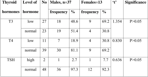
TABLE 2 : SEX WISE PREVALANCE OF THYROID DYSFUNCTION IN CKD PATIENTS
Thyroid hormones
Level of hormone
No Males, n=37 Females=13 ‘t’ Significance frequency % frequency %
T3 low 27 18 48.6 9 69.2 1.354 P>0.05
normal 23 19 51.4 4 30.8
T4 low 11 7 18.9 4 30.8 0.830 P>0.05
normal 39 30 81.1 9 69.2
TSH high 2 1 2.7 1 7.7 0.636 P>0.05
normal 48 36 97.3 12 92.3
The prevalence of thyroid dysfunction among the sexes was shown in
the above table 2. The prevalence of low T3 syndrome was 54% (27 cases)
and the low T4 syndrome was 22 % (11 cases). The prevalence of TSH in
hypothyroidism range was 4 %( 2 cases). Among the males 48.6% of
patients had low T3 syndrome. And among the females was 62.2%. The
difference was not statistically significant P>0.05. The prevalence of low T4
among the males was 18.9 % and among the females was 30.8%. The
difference among the sexes was not statistically significant i.e. P>0.05. The
prevalence of TSH in clinical hypothyroidism range among males was 2.7%.
And among the females was 7.7%. The prevalence between the sexes was
38 51.4 30.8 48.6 69.2 0 10 20 30 40 50 60 70 80 Male Female Percentage of cases
Total T3
Sexwise
Distribution
of
Total
T3
Level
normal low 81.1 69.2 18.9 30.8 0 10 20 30 40 50 60 70 80 90 Male Female Percentage of cases
Free T4
Sexwise
Distribution
of
Free
T4
Level
39
TABLE 3: AGE WISE THYROID HORMONES AND TSH DISTRIBUTION TAKEN FOR STUDY
Age group Frequency Mean Total T3
Mean Free T4
Mean TSH
30-39 8 75.8±23.8 0.8±0.2 11±25.9
40-49 9 70.4±33.5 0.9±0.2 3±3.2
50-59 16 71.4±26.3 1.1±0.2 1.3±1.2
60-69 17 95.3±33.6 1.1±0.3 1.3±1
Total 50 80±31.1 1±0.3 3.2±10.5
The above table 3 reveals that the mean level of thyroid biomarkers
does not show significant difference in the various age groups.
TABLE 4: DISTRIBUTION OF TOTAL T3 FREE T4 AND TSH IN VARIOUS STAGES OF CKD
Stages of CKD
Frequency Mean Total T3
Mean free T4
Mean TSH
1-3 5 103.4±30.7 1.25±0.1 1.8±1.9
4 16 91±36.6 1.1±0.2 1.2±0.8
5 29 68.8±24 0.9±0.3 4.5±13.7
The above table 4 reveals the mean T3, free T4 and TSH levels in
various stages of CKD. The mean T3 is decreased significantly with reduced
creatinine clearance. The free T4 is also significantly decreased in stage 5
[image:46.595.83.534.471.597.2]0 20 40 60 80 100 120 Mean T3 ng/dl
R
0 0.2 0.4 0.6 0.8 1 1.2 1.4 Mean Free T4 ng/dlR
1‐3
ELATION
1‐3
RELATIO
Sta
N
OF
TO
STA
Stage
ON
OF
FR
STA
40
4 ages of CKD
OTAL
T3
AGES
OF
4 es of CKD
REE
T4
L
AGES
OF
5
LEVEL
I
F
CKD
5
LEVEL
IN
F
CKD
IN
VARI
N
VARIO
OUS
M
OUS
Mean f ean T3
41
TABLE 5 : RELATIONSHIP BETWEEN CREATININE CLEARANCE WITH TOTAL T3, FREE T4 AND TSH Relation with Cr.
Clearance
r Significance
Total T3 0.320 P<0.05
Free T4 0.381 P<0.01
TSH -0.133 P>0.05
The above table shows positive correlation between total T3 and
Creatinine clearance and it is statistically significant. The free T4 and
creatinine clearance shows positive correlation and it is statistically
significant. The above table shows negative correlation of TSH with
42
CORRELATION OF TOTAL T3 WITH CREATININE CLEARANCE
CORRELATION OF FREE T4 WITH CREATININE CLEARANCE 0
20 40 60 80 100 120 140 160 180
0 10 20 30 40 50 60 70
total
t3(ng/ml)
creatinine clearence(ml/min)
0 0.2 0.4 0.6 0.8 1 1.2 1.4 1.6 1.8
0 10 20 30 40 50 60 70
free
t4(ng/ml)
43
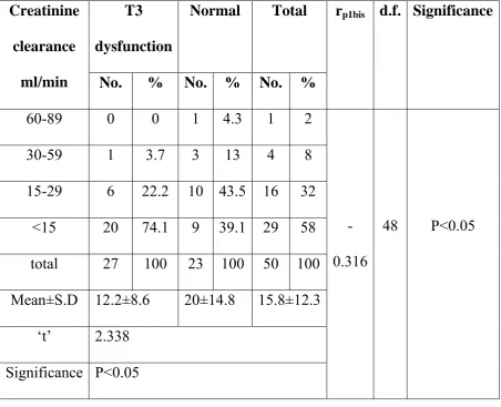
TABLE 6: CORRELATION OF TOTAL T3 WITH CREATININE CLEARANCE
Creatinine clearance
ml/min
T3 dysfunction
Normal Total rp1bis d.f. Significance
No. % No. % No. %
60-89 0 0 1 4.3 1 2
-0.316
48 P<0.05
30-59 1 3.7 3 13 4 8
15-29 6 22.2 10 43.5 16 32
<15 20 74.1 9 39.1 29 58
total 27 100 23 100 50 100
Mean±S.D 12.2±8.6 20±14.8 15.8±12.3
‘t’ 2.338
Significance P<0.05
The table 6 explains the relation between the creatinine clearance with
total T3. The mean creatinine clearance in low T3 syndrome was 12.2±8.6
ml and in normal patients was 20±14.8 ml. The difference between the
patients was statistically significant i.e. P<0.05. The rp1bis determines the
direction between the creatinine clearance with low total T3 patients. The
dysfunction with creatinine clearance was negatively correlated i.e. rp1bis =
-0.316. Statistically explain the negative relationship between them
44
TABLE 7 : RELATIONSHIP BETWEEN CREATININE CLEARANCE WITH FREE T4
Creatinine clearance
ml/min
FREE T4 dysfunction
Normal Total rp1bis d.f. Significance
No. % No. % No. %
60-89 0 0 1 2.6 1 2
-0.340 48 P<0.05
30-59 0 0 4 10.3 4 8
15-29 1 9.71 15 38.5 16 32
<15 10 90.4 19 48.6 29 58
Total 11 100 39 100 50 100
Mean±S.D 7.9±4 18±13 15.8±12.3
‘t’ 2.518
Significance P<0.05
The table 7 describes the correlation between the creatinine clearance
with free T4. The mean creatinine clearance with low free T4 dysfunction
and normal patients was 7.9±4 and 18±3 ml respectively. The difference
between the mean was statistically significant i.e. P<0.05. The point biserial
TA Cr cle m Me Sig
ABLE 8: R
reatinine earance ml/min 60-89 30-59 15-29 <15 total ean±S.D ‘t’ gnificance 0 10 20 30 40 50 60 70 80 90 100 Percentage of Cases
FR
RELATIO High T No. 0 0 0 2 2 5±1.4 1.271 P>0.05 60‐89C
REQUEN
WI
ONSHIP B
TSH N
% N
0 1
0 4
0 1
100 2
100 4
16 30‐59 Creatinine Cle
NCY
OF
ITH
CRE
45 BETWEE WITH T Normal o. % 1 2.1 4 8.3 6 33.3 7 56.3 48 100 .2±12.4 15‐29 earance ml/m
LOW
T3
ATININE
EN CREA TSH Tota No. 1 4 16 29 50 15.8±12 <15 in3
AND
L
E
CLEAR
ATININE al rp1 % 2.1 -0.1 8 32 58 100 2.3
OW
FRE
RANCE
E CLEARA
1bis d.f.
178 48
EE
T4
T3 dysfunctio FREE T4 dysf
46
The relationship displayed in the table 8 reveals the mean creatinine
clearance in TSH dysfunction and normal were 5±1.4 and 16.2±12.4 ml
respectively. The difference between the mean was not statistically
[image:53.595.97.529.296.462.2]significant i.e.P>0.05.
TABLE 9: PREVALANCE OF GOITER IN CHRONIC KIDNEY DISEASE PATIENTS
S.No. Disease No. %
1 Goiter 2 4
2
Goiter with pleural
effusion 1 2
3 No evidence 47 94
The above table 9 describes the prevalence of goiter in CKD patients.
Among the 50 patients 47(94%) patients had no evidence of goiter. With
remaining 3patients 2(4%) patients had exclusively goiter , 1 (2%)patients
had goiter with pleural effusion . The total prevalence of goiter in our study
47
6
94
PREVALENCE
OF
GOITER
IN
CHRONIC
KIDNEY
DISEASE
PATIENTS
48
Table-10 : RELATIONSHIP BETWEEN TOTAL T3 WITH TSH EXCLUDING HYPOTHYROIDISM
S.NO. LOW T3 (ng/dl) TSH (µIU/ml)
1 64 2.55
2 61 3.06
3 60 0.40
4 64 0.97
5 70.4 0.87
6 62 0.49
7 48 0.49
8 69 0.96
9 54 0.46
10 53 1.61
11 70 4.35
12 59 2.13
13 64 2.71
14 55 4.85
15 50 1.52
16 50 1.80
17 64 0.64
18 50 1.20
19 50 0.74
20 57 2.12
21 63 1.01
22 60 3.17
23 70 0.30
24 68 0.99
hyp
Acc
TSH
This t
pothyroidi
cording to
H in vario
[image:56.595.94.527.232.501.2]DI
table show
ism. The m
o our study
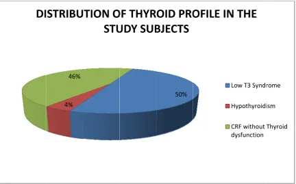
ous stages 4% 46%
ISTRIBU
ws relation mean valuy, in patie
of renal fa
UTION
O
STU
49
nship bet
ue of TSH
ents with l
failure are
OF
THYR
UDY
SUB
tween low
in low T3
low T3 syn
within no
50%
OID
PRO
BJECTS
w T3 and
3 syndrom
ndrome th
ormal rang
OFILE
IN
TSH exc
me is 1.6±1
he mean v
ge.
N
THE
50
DISCUSSION
A large number of hormonal systems are affected by CRF, yet it
remains unclear to what extent these changes are responsible for
manifestations of uremic syndrome. Patients with CRF often have signs &
symptoms suggestive of thyroid dysfunction & hence the diagnosis of
thyroid disease in these patients has obvious prognostic implications. The
data reported deals primarily with the biochemical parameters. In uremia the
mean values of both serum T3 & T4 were significantly low. This is
comparable to Ramiraz et al.3
and Lim VS et al.17study. In our study, out of 50 patients 27 patients (54%) had low T3 syndrome. The prevalence of
low T3 in stage 1- 3 is 20 %, for stage 4 is 38%, and stage 5 is 70%. This
observation is consistent with Sang Heon Song et al.4 in which the
prevalence of low T3 will be increased according to the increase in stage of
CKD. In our study there is a positive correlation between Total T3 and
creatinine clearance and it is statistically significant P<0.05.This shows
serum T3 levels were associated with severity of CKD even in the normal
TSH level.
There was higher frequency of reduced free T4 values in our study
(22%) which is consistent with Kaptein et al.5
and Avasthi et al.6 study but it is not statistically significant. In our study there is a positive correlation
between Free T4 and creatinine clearance and it is statistically significant
51
The high serum TSH level is > 75 µIU/ml. Both these patients had very low
serum T3 concentration which can be explained by the normal feedback
regulation of the pituitary thyroid axis. One patient is having both goiter and
pleural effusion. This observation is consistent with Joseph et al.50 who
studied 175 patients of CRF who had low T3, T4, fT4 but had high TSH
levels suggested maintenance of pituitary thyroid axis. 48 patients i.e. 96%
reported normal level of serum TSH ≤5 µIU/ml. Out of 48 patients 25
patients had low T3 and 9 patients had low total T3 and free T4. So these 25
patients had normal level of serum TSH in spite of low serum T3 level. They
demonstrated abnormality in the hypophyseal mechanism of TSH release in
uremic patients as the TSH response to TRH was blunted. These results are
consistent with study of Spector et al.49 and Ramirez et al. reported normal
level of serum TSH in patients of CRF in spite of low serum T3 levels.
In Mehta H.J. Joseph et al.7 study low TT3, FT3 and TT4 values is seen in clinically euthyroid CKD patients. However finding of normal T4
values and TSH would indicate functional euthyroid status. It can be
presumed that free T4 values would fall if these patients develop
hypothyroidism and TSH values would rise simultaneously. Thus Free T4
and TSH levels combined can be used for the diagnosis of hypothyroidism in
52
Subjects with TSH > 10 µIU/ml and free T4 below the reference range
have overt primary hypothyroidism and should be treated with thyroid
hormone replacement.
The prevalence of primary hypothyroidism in CKD ranges from
0-9.5% as evidenced in previous studies. The prevalence of hypothyroidism in
our study is 4%. This is consistent with results of Kaptein et al.5
Ramirez et al.3 reported high prevalence of goiter in CRF patients especially those on chronic dialysis. Incidences were increased in end stage
renal disease. The possible explanation is due to accumulation of iodides in
thyroid gland due to decreased renal clearance in CRF patients. Study
conducted by Hegedus et al.8 showed thyroid gland volume was
significantly increased in patients with CRF.
In our study, 3(6%) patients had evidence of goiter. Out of 3 patients,
1(2%) had clinical and biochemical features of hypothyroidism. Remaining 2
patients had low T3 level with normal TSH and T4.
Dialysis
As stated previously, Hemodialysis and continuous ambulatory
peritoneal dialysis have shown to affect the thyroid profile independently of
CRF. Also drugs like heparin, furosemide used during dialysis will affect the
thyroid profile.
Kayima et al.9 and Giordano et al.10 have conducted studies regarding
53
This study showed no significant improvement in thyroid profile after
repeated hemodialysis.
But in the patients who have undergone renal transplant surgery,most
of the thyroid function parameters returned to normal with TSH below
54
CONCLUSION
1. The prevalence of thyroid dysfunction in patients with CKD is 54%.
2. Number of patients with low T3 and T4 syndrome progressively
increases with severity of renal failure.
3. Serum level of total T3 and free T4 is directly proportional to
creatinine clearance level.
4. Total T3 and free T4 had correlation with the severity of renal failure.
5. TSH values will be useful to differentiate hypothyroidism from
non-thyroidal illness due to CKD.
6. Only 6% of the study population had evidence of goiter.
7. Alteration in the values of T3 and T4 occurs as a part of body
REFERENCE
1. Harrison’s principle of Internal medicine Volume II Seventeenth edition
2. National Kidney Foundation [Kidney Dialysis Outcomes Quality
Initiative (KDOQI)
3. Thyroid dysfunction in uremia: evidence for thyroid and hypophyseal
abnormalities. Ramirez G, O'Neill W Jr, Jubiz W, Bloomer
HA.Nephron. 1985;40(2):171-4
4. The prevalence of low triiodothyronine according to the stage of
chronic kidney disease in subjects with a normal thyroid-stimulating
hormone Sang Heon Song1,2, Ihm Soo Kwak1,2, Dong Won Lee1,2,
Yang Ho Kang1,2, Eun Young Seong1,2 and Jin Sup Park1,2 Oxford
Journals Medicine Nephrology Dialysis Transplantation
Volume24, Issue5Pp. 1534-1538
5. Kaptein E et al, (1988). The Thyroid in end stage renal diseases,
Medicine 67:187 – 97.
6. Avasthi G et al, (2001) Study of Thyroid function in patients of chronic
renal failure. Indian Journal of Nephrology, 11:165 – 170
7. Total and free thyroid hormone levels in chronic renal failure. Mehta
HJ, Joseph LJ, Desai KB, Mehta MN, Samuel AM, Almeida AF,
Acharya VN Department of Nephrology, K. E. M. Hospital, Bombay,
8. Hegedus L et al,(1985). Thyroid gland volume and serum
concentrations of thyroid hormone in chronic renal failure. Nephron,
40:171 – 4.
9. Kayima JK et al, (1992). Thyroid hormones profile in patients with
chronic renal failure on conservative management and regular
hemodialysis. East Afr Med J, 69:333 – 6.
10. Giordano C et al, (1984). Thyroid Status and nephron loss – a study in
patients with chronic renal faialure, end stage renal disease and/or on
hemodialysis. Int J Artif organs 7;119 – 22.
11. Katz AI & Lindheimer MD. Actions of hormones on the kidney.Annual
Review of Physiology 1977 39 97–133.
12. Braunlich H. Thyroid hormones influencing renal electrolyte excretion
in saline loaded rats of different ages. Physiologia Bohemoslovaca 1984
33 303–308.
13. Vargas F, Moreno JM, Rodrı´guez-Go´mez I, Wangensteen R,Osuna A,
Alvarez-Guerra M & Garcı´a-Estan˜ J. Vascular and renal function in
experimental thyroid disorders. European Journal of Endocrinology
2006 154 197–212.
14. Li Bok N, Fekete F & Ha´rsing L. Renal structural and functional
changes and sodium balance in hypothyroid rats. Acta Medica
15. Katz AI & Lindheimer MD. Renal sodium- and potassiumactivated
adenosine triphosphatase and sodium reabsorption in the hypothyroid
rat. Journal of Clinical Investigation 1973 52 796–804.
16. Capasso G, Kinne R, De Santo NG & Giordano C. The use of
micropuncture, isolated tubule, and vesicle technique in the study of the
action of thyroid hormones on the proximal tubule function. Uremia
Investigation 1985 9 151–157.
17. Thyroid dysfunction in chronic renal failure. A study of the pituitary-
thyroid axis and peripheral turnover kinetics of thyroxine and
triiodothyronine. Lim VS, Fang VS, Katz AI, Refetoff S. Kidney
International (2005) 67, 1047–1052; doi:10.1111/j.1523-1755.2005.00169.x
18. McCaffrey C & Quamme GA. Effects of thyroid status on renal calcium
and magnesium handling. Canadian Journal of Comparative Medicine
1984 48 51–57.
19. Vaamonde CA, Sebastianelli MJ, Vaamonde LS, Pellegrini EL, Watts
RS, Klingler EL Jr & Papper S. Impaired renal tubular reabsorption of
sodium in hypothyroid man. Journal of Laboratory and Clinical
Medicine 1975 85 451–466.
Alba F, de Gasparo M & Prieto I. Influence of thyroid disorders on
kidney angiotensinase activity. Hormone and Metabolic Research 2006
38 48–52.
21. den Hollander JG, Wulkan RW, Mantel MJ & Berghout A. Correlation
between severity of thyroid dysfunction and renal function. Clinical
Endocrinology 2005 62 423–427.
22. Kaptein EM, Quion-Verde H, Chooljian CJ, Tang WW, Friedman PE,
Rodriquez HJ & Massry SG. The thyroid in endstage renal disease.
Medicine 1988 67 187–197.
23. Enrı´quez R, Sirvent AE, Amoro´s F, Andrada E, Cabezuelo JB &
Reyes A. IgA nephropathy and autoimmune thyroiditis. Clinical
Nephrology 2002 57 406–407.
24. Akikusa B, Kondo Y, Iemoto Y, Iesato K & Wakashin M. Hashimoto’s
thyroiditis and membranous nephropathy developed in progressive
systemic sclerosis (PSS). American Journal of Clinical Pathology 1984
81 260–263.
25. Ikeda K, Maruyama Y, Yokoyama M, Kato N, Yamanoto H, Kaguchi
Y, Nakayama M, Shimada T, Tojo K, Kawamura T & Hosoya T.
Association of Graves’ disease with Evans’ syndrome in a patient with
IgA nephropathy. Internal Medicine 2001 40 1004–1010.
26. Horikoshi T, Tamura J, Kaneko Y, Maezawa A, Kaji T, Matsushima T,
Membranous nephropathy associated with chronic thyroiditis. Nephron
1993 63 246.
27. Brohee D, Delespesse G, Debisschop MJ & Bonnyns M. Circulating
immune complexes in various thyroid diseases. Clinical and
Experimental Immunology 1979 36 379–383.
28. Calder EA, Penhale WJ, Barnes EW & Irvine WJ. Evidence for
circulating immune complexes in thyroid disease. BMJ 1974 6 30–31.
29. Sasaki H, Joh K, Ohtsuka I, Ohta H, Ohhashi T, Hoashi S, Takahashi T,
Tokuda T, Koyama K & Isogai Y. Interstitial nephritis associated with
glomerulonephritis in a patient with Hashimoto’s disease and idiopathic
portal hypertension. Internal Medicine 1992 31 641–648.
30. Hudde T, Heinz C, Neudorf U, Hoef S, Heiligenhaus A & Steuhl KP.
Tubulointerstitial nephritis and uveitis (TINU syndrome) – co morbidity
and complications in four patients. Klinische Monatsbla¨tter fu¨r
Augenheilkunde 2007 219 528–532.
31. Paydas S, Balal M, Karayaylali I & Seyrek N. Severe acute renal failure
due to tubulointerstitial nephritis, pancreatitis, and hyperthyroidism in a
patient during rifampicin therapy. Advances in Therapy 2005 22 241–
243
32. Junglee NA, Scanlon MF & Rees DA. Increasing thyroxine
requirements in primary hypothyroidism: don’t forget the urinalysis
33. Feinstein EI, Kaptein EM, Nicoloff JT & Massry SG. Thyroid function
in patients with nephrotic syndrome and normal renal function.
American Journal of Nephrology 1982 2 70–76.
34. Chadha V & Alon US. Bilateral nephrectomy reverses hypothyroidism
in congenital nephrotic syndrome. Pediatric Nephrology 1999 13 209–
211
35. Holmberg C, Antikainen M, Ro¨nnholm K, Ala Houhala M & Jalanko
H. Management of congenital nephrotic syndrome of the Finnish type.
Pediatric Nephrology 1995 9 87–93.
36. Kutlay S, Atli T, Koseogullari O, Nergizoglu G, Duman N & Gullu S.
Thyroid disorders in hemodialysis patients in an iodinedeficient
community. Ar