Copyright © 1999, American Society for Microbiology. All Rights Reserved.
Association with the Cellular Export Receptor CRM 1 Mediates
Function and Intracellular Localization of Epstein-Barr Virus
SM Protein, a Regulator of Gene Expression
SARAH M. BOYLE,1VIVIAN RUVOLO,1ASHISH K. GUPTA,1ANDSANKAR SWAMINATHAN1,2*
Sealy Center for Oncology and Hematology and Division of Infectious Diseases, Department of Internal Medicine,1and
Department of Microbiology and Immunology,2University of Texas Medical Branch, Galveston, Texas 77555-1048
Received 25 February 1999/Accepted 10 May 1999
Splicing and posttranscriptional processing of eukaryotic gene transcripts are linked to their nuclear export and cytoplasmic expression. Unspliced pre-mRNAs and intronless transcripts are thus inherently poorly expressed. Nevertheless, human and animal viruses encode essential genes as single open reading frames or in the intervening sequences of other genes. Many retroviruses have evolved mechanisms to facilitate nuclear export of their unspliced mRNAs. For example, the human immunodeficiency virus RNA-binding protein Rev associates with the soluble cellular export receptor CRM 1 (exportin 1), which mediates nucleocytoplasmic translocation of Rev-HIV RNA complexes through the nuclear pore. The transforming human herpesvirus Epstein-Barr virus (EBV) expresses a nuclear protein, SM, early in its lytic cycle; SM binds RNA and posttranscriptionally activates expression of certain intronless lytic EBV genes. Here we show that both the
trans-activation function and cytoplasmic translocation of SM are dependent on association with CRM 1 in
vivo. SM is also shown to be associated in vivo with other components of the CRM 1 export pathway, including the small GTPase Ran and the nucleoporin CAN/Nup214. SM is shown to be present in the cytoplasm, nucleoplasm, and nuclear envelope of transfected cells. Mutation of a leucine-rich region (LRR) of SM inhibited CRM 1-mediated cytoplasmic translocation and SM activity, as did leptomycin B, an inhibitor of CRM 1 complex formation. Surprisingly, however, leptomycin B treatment and mutation of the LRR both led to SM becoming more tightly attached to intranuclear structures. These findings suggest a model in which SM is not merely a soluble carrier protein for RNA but rather is bound directly to intranuclear proteins, possibly including the nuclear pore complex.
The Epstein-Barr virus (EBV) protein SM posttranscrip-tionally activates intronless genes and inhibits expression of intron-containing genes (23, 24, 27, 39). In contrast to the majority of cellular genes, many EBV genes expressed during lytic replication are intronless (2, 26), and SM may therefore be important in enhancing expression of other lytic EBV genes. Activation of intronless genes by SM appears to be exerted both at the level of pre-mRNA stability and nucleocy-toplasmic mRNA export (39). SM binds RNA in vitro and is capable of shuttling from nucleus to cytoplasm in a hetero-karyon assay (39, 41). It is therefore probable that SM, like the human immunodeficiency virus (HIV) Rev protein, is an RNA transport protein.
It has been shown that several proteins involved in nucleo-cytoplasmic transport of RNA or protein bind to exportins, such as the recently characterized cellular export receptor CRM 1 (exportin 1) (for a review, see reference 43). Various proteins, including HIV Rev and cyclic AMP-dependent pro-tein kinase inhibitor (PKI), contain leucine-rich regions (LRR) which serve as nuclear export signals (NESs) that are required for nuclear export (12, 45). The mechanism of NES-dependent export has been elucidated by the finding that CRM 1 forms a physical complex with NES-containing peptides and conjugates (14, 15, 33). Complex formation may require the presence of a small GTPase, Ran, associated with GTP (Ran-GTP), result-ing in a tripartite complex of CRM 1 with NES-containresult-ing
proteins and Ran-GTP (14, 38). Current models for NES-dependent nuclear export postulate a gradient of Ran-GTP across the nuclear envelope, with more Ran-GDP present in the cytoplasm. Thus, the CRM 1–Ran-GTP–NES complex is thought to be formed in the nucleus and translocated to the cytoplasm where Ran-GTP is converted to Ran-GDP, accom-panied by release of CRM 1 and the NES protein. CRM 1 also associates with the nuclear pore complex (NPC) via CAN/ Nup214 and other nucleoporins (13). Thus, CRM 1 is postu-lated to direct movement of its cargo NES protein to the NPC and dock with components of the NPC. The exact details of these interactions and the mechanism of directional transloca-tion of the CRM 1-NES protein complex through the NPC remain to be delineated.
SM facilitates the expression of intronless mRNAs, as op-posed to the unspliced retroviral RNAs transported by Rev-like proteins. Nevertheless, it was considered possible that SM-mediated RNA export occurs via a CRM 1-dependent pathway. We therefore performed immunoprecipitation and immunofluorescence experiments to determine whether SM interacts physically with CRM 1 in vivo. The specificity of CRM 1-mediated effects was confirmed with a specific inhibi-tor of CRM 1 complex formation, leptomycin B (LMB). The role of CRM 1 in SM function was investigated in reporter assays that measuretransactivation of gene expression by SM. These studies revealed an in vivo association of SM with CRM 1 that mediates both nuclear export and functional activity of SM. The subcellular distribution of SM was also analyzed, by cellular fractionation studies. These studies revealed an effect of CRM 1 not only on nuclear export of SM but also on the degree to which SM is bound to structural elements of the
* Corresponding author. Mailing address: Sealy Center for Oncol-ogy and HematolOncol-ogy, MRB 9.104, University of Texas Medical Branch, Galveston, TX 77555-1048. Phone: (409) 1935. Fax: (409) 747-1938. E-mail: [email protected].
6872
on November 9, 2019 by guest
http://jvi.asm.org/
nucleus. Finally, the functional role of a putative leucine-rich SM NES was analyzed by site-directed mutagenesis. These experiments demonstrate that the LRR is important in medi-ating interaction with CRM 1 and suggest a novel mechanism for SM function.
MATERIALS AND METHODS
Immunofluorescence assays.Cells were grown on glass coverslips prior to washing in phosphate-buffered saline (PBS) and fixation with ice-cold acetone. Fixed cells were incubated in polyclonal rabbit anti-SM antisera at a 1:500 dilution for 1 h at room temperature, washed three times in PBS, and incubated for 1 h with rhodamine-conjugated affinity-purified F(ab⬘)2goat anti-rabbit an-tibodies (Rockland, Gilbertsville, Pa.) at a 1:1,000 dilution. Cells were washed and overlaid with glycerol, and immunofluorescent microscopy was performed with a Nikon Optiphot 2 microscope. Deconvoluted fluoromicrographs were acquired with a DeltaVision deconvolution fluorescent microscope system (Ap-plied Precision, Issaquah, Wash.). Individual optical sections of 200-nm thickness were obtained from slides prepared as described above. Nuclei were counter-stained with 0.5g of DAPI (4⬘,6-diamidino-2-phenylindole) per ml, and slides were overlaid with Faramount aqueous mounting medium (DAKO Corporation, Carpinteria, Calif.) prior to microscopy.
Cell lines, plasmids, and antibodies.SM, antisense control, and CMV-CAT plasmids have been previously described (39). SM mutants were generated by oligonucleotide-directed site-specific mutagenesis (8). CRM 1 cDNA, the influ-enza virus hemagglutinin (HA)-tagged carboxy-terminal amino acid fragment of CAN/Nup214 (amino acids 1864 to 2090), and polyclonal rabbit anti-CRM 1 (13) were kind gifts of G. Grosveld (St. Jude Children’s Research Hospital, Memphis, Tenn.). CRM 1 cDNA and the HA-CAN/Nup214 fragment were cloned in the pCDNA3 expression vector (Invitrogen Corp.). Polyclonal anti-SM antibodies were generated by injecting rabbits with gel-purified SM–glutathioneS -trans-ferase fusion proteins (39). Polyclonal anti-Ran antibodies were purchased from Covance Laboratories (Richmond, Calif.). BJAB, an EBV-negative B lymphoma cell line, and Cos 7 cells have been previously described (16, 31). Anti-FLAG monoclonal antibody was purchased from Sigma (St. Louis, Mo.).
Transfections and reporter assays.BJAB cells were electroporated with ex-pression constructs, and chloramphenicol acetyltransferase (CAT) assays were performed exactly as previously described (39). Each data point represents pooled results from at least three independent transfections. LMB (kind gift of B. Wolff; Novartis AG) treatment was begun immediately after transfection at a concentration of 10 nM and continued until harvest 16 h later. Cos 7 cells were transfected with LipofectaminePlus, per the manufacturer’s protocol (Gibco Life Sciences).
Immunoprecipitation and immunoblotting.Cells were lysed 48 h after trans-fection in immunoprecipitation buffer (Tris-buffered saline [pH 7.4], 1% Triton X-100, 1 mM dithiothreitol, 100M GTP-␥S, and a mixture of protease inhib-itors [Sigma protease inhibitor cocktail no. P2714]). One hundred fifty microli-ters of each lysate was cleared with preimmune serum, incubated with 1l of undiluted polyclonal antibody and 20l of protein A-conjugated agarose beads (Sigma) for 90 min at 4°C, and washed four times in immunoprecipitation buffer. Precipitation with anti-HA monoclonal antibodies was performed with 150l of 12CA5 hybridoma supernatant (10). Precipitates were boiled in protein loading buffer, electrophoresed, and immunoblotted as previously described (44). Im-munoblotting was performed with a 1:400 dilution of polyclonal SM anti-body and a 1:7,500 dilution of horseradish peroxidase-linked donkey anti-rabbit immunoglobulin or a 1:4,000 dilution of horseradish peroxidase-linked goat anti-mouse immunoglobulin (Amersham), and immunoreactive proteins were detected by enhanced chemiluminescence.
Cell fractionation and nuclear extractions.Cos 7 cells were harvested 48 h after transfection by scraping into ice-cold PBS, washed in PBS, and lysed in 0.5% Nonidet P-40, 50 mM Tris–5 mM MgSO4. Under these conditions the nuclei remain intact, as confirmed by light microscopy. Nuclei were separated by centrifugation at 950⫻gfor 10 min. Nuclei were resuspended in a solution of 250 mM sucrose, 50 mM Tris (pH 7.4), and 5 mM MgSO4and treated with DNase (250g/ml) and RNase A (1 mg/ml) for 2 h at 4°C, followed by washing and resuspension in 50 mM Tris (pH 7.4)–5 mM MgSO4. High-salt extractions were performed by dropwise addition of 2 M NaCl–50 mM Tris (pH 7.4) with constant mixing to a final concentration of 1.6 M NaCl and incubation on ice for 30 min. High-salt extractions with-mercaptoethanol were performed identi-cally, with the inclusion of-mercaptoethanol at 1% (vol/vol). The remaining nuclear envelopes were sedimented by centrifugation at 13,000⫻gfor 30 min. The salt-extracted fraction was desalted and concentrated with a Microcon 10 filter apparatus (Amicon, Beverly, Mass.). Protease inhibitors, as described above, were included at all steps of the isolation process. Equal fractions from each step of the fractionation procedure were prepared for sodium dodecyl sulfate-polyacrylamide gel electrophoresis (SDS-PAGE) and immunoblotted as described above. In experiments with LMB, cells were treated with LMB for 16 h after transfection. Treated and untreated cells were harvested at 16 h posttrans-fection and fractionated exactly as described above.
RESULTS
SM activity is dependent on CRM 1 (exportin 1) function.
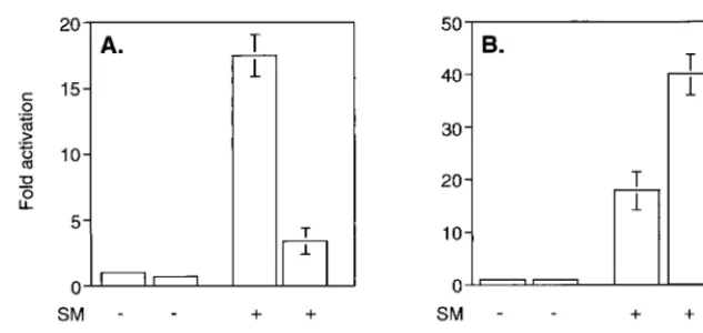
SM-mediated gene activation is correlated with enhanced cy-toplasmic accumulation of intronless target gene mRNAs (7, 39). SM contains an LRR resembling an NES found in certain proteins which shuttle from nucleus to cytoplasm (45). Such proteins, notably the HIV Rev protein, bind CRM 1 (exportin 1) and the small GTPase Ran in the nucleus (14). Transloca-tion to the cytoplasm is thought to be followed by hydrolysis of Ran-associated GTP and complex dissociation. Thus, it was considered possible that SM chaperones intronless EBV mRNAs to the cytoplasm via a CRM 1-dependent pathway. We therefore wished to determine whether SM-mediated gene activation is dependent on an interaction with CRM 1. We first examined the effect of a specific inhibitor of CRM 1 complex formation, LMB (47), on SM function. BJAB cells were trans-fected with an intronless CAT reporter plasmid (CMV-CAT) and an expression vector encoding either SM or antisense SM as a control. Immediately after transfection, cells were incu-bated in growth medium containing LMB or in control me-dium without LMB. Cells were harvested after 16 h, at which time viability was not affected by LMB treatment (data not shown) and CAT activity was measured. As previously re-ported, SM led to activation of CAT expression (approximately 16-fold). LMB itself did not affect baseline expression of CAT activity from the reporter plasmid. However, LMB treatment led to a marked reduction in activation by SM (to four times that of the control) (Fig. 1A).
These experiments indicated that an interaction with CRM 1 was involved in some aspects of gene activation by SM. We therefore attempted to determine whether overexpression of CRM 1 could augment SM-mediated gene activation. A CRM 1 expression plasmid or control vector was cotransfected with CMV-CAT and either SM or antisense SM into BJAB cells, and CAT activity was measured 48 h after transfection. As shown in Fig. 1B, CRM 1 overexpression stimulated SM acti-vation. Activation by SM alone was 17-fold and increased to 40-fold over the control when CRM 1 was cotransfected. CRM 1 overexpression did not increase CAT activity in the absence of SM, demonstrating that CRM 1 does not have a nonspecific stimulatory effect on CAT gene expression.
CRM 1 expression affects intracellular localization of SM.
The preceding experiments suggested that CRM 1 may be involved in nucleocytoplasmic translocation of SM. While it has been shown that SM can shuttle from nucleus to cytoplasm in a heterokaryon assay (41), immunofluorescence studies have demonstrated exclusively nuclear localization of SM (6, 48). SM-transfected cells typically display a speckled nuclear fluo-rescence with nucleolar sparing when stained with anti-SM antibodies (Fig. 2). Such apparently exclusive nuclear localiza-tion of other known shuttling proteins has been reported, pre-sumably because the concentration of cytoplasmic proteins is below the limits of detection of conventional indirect immu-nofluorescence microscopy (34). We reasoned that if cytoplas-mic transport of SM is CRM 1 dependent, overexpression of CRM 1 might lead to visible cytoplasmic accumulation of SM. We therefore transfected Cos 7 cells, in which nuclei and cytoplasm are easily differentiated, with SM and either CRM 1 expression vector or control plasmid and examined the trans-fected cells by indirect immunofluorescence microscopy. As shown in Fig. 2, overexpression of CRM 1 led to dramatic intracellular relocalization of SM. The nuclei of CRM 1 co-transfected cells were relatively depleted of SM, and the cyto-plasm became diffusely stained by the anti-SM antibodies.
To confirm the role of CRM 1 in nucleocytoplasmic export
on November 9, 2019 by guest
http://jvi.asm.org/
of SM, we studied the effect of LMB on cytoplasmic translo-cation of SM. Cos 7 cells were transfected with SM and CRM 1 expression vectors, as described above, and incubated in growth medium in the presence or absence of LMB. Sixteen hours after transfection, the cells were fixed and stained with anti-SM antibodies and examined by indirect immunofluores-cence microscopy. LMB treatment resulted in exclusively nu-clear localization of SM despite overexpression of CRM 1 (Fig. 2). These data thus confirm that cytoplasmic translocation of SM is directly or indirectly dependent on CRM 1 complex formation.
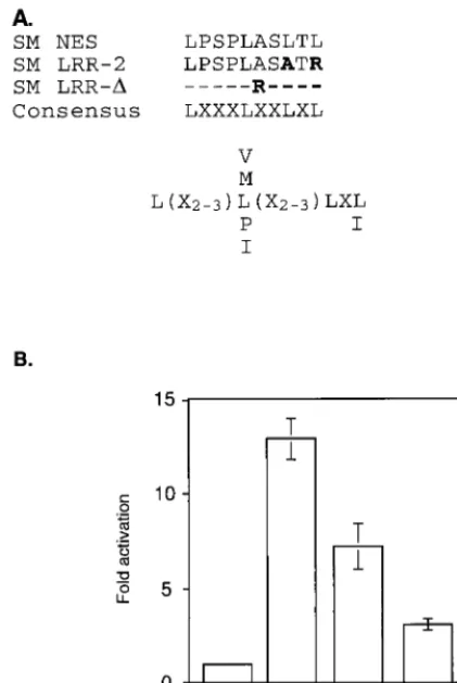
Mutation of a putative NES impairs SM function.The pre-dicted amino acid sequence of SM contains an LRR which satisfies the consensus requirements (LX2–3LX2–3LXL) for an NES, as found in other proteins known to interact with CRM 1 (4) (Fig. 3A). It should be noted that although there is not strong selection for a particular amino acid at positions de-noted by “X”, some amino acids at these positions lead to nonfunctional NESs (4). Thus, not all regions meeting these broad criteria are functional NESs. In order to determine if the putative SM NES (amino acids 227 to 236) is required for SM function, we examined the effect of mutating this region on SM-mediated gene activation. Site-directed mutagenesis was used to generate SM mutants altered in the relevant region. The mutant LRR-2 has leucines 234 and 236 replaced by alanine and arginine, respectively, whereas all 11 amino acids
are deleted in the LRR-⌬mutant. We then compared these mutants with wild-type SM in the ability to activate gene ex-pression in the CAT reporter assay (Fig. 3B). Both mutants were impaired in activation function, with LRR-⌬ being the least active. We have previously demonstrated that in addition to its putative RNA transport function, SM stabilizes and leads to increased accumulation of target gene RNAs in the nucleus as well as the cytoplasm (39). Therefore, the residual activating function of the SM mutants was not unexpected despite a potential defect in the ability to interact with CRM 1 and hence in the ability to translocate to the cytoplasm.
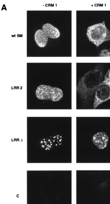
[image:3.612.145.462.76.226.2]Deletion of the LRR abolishes CRM 1-mediated nuclear export of SM.We next wished to determine whether the pu-tative SM NES was important for CRM 1-mediated cytoplas-mic localization of SM. Cos cells were transfected with wild-type or LRR mutant plasmids and either CRM 1 plasmid or control vector and examined by immunofluorescence micros-copy. In the absence of CRM 1 overexpression, both LRR mutants were detectable only in the nucleus, as expected (Fig. 4). However, both mutants exhibited a more punctate nuclear distribution than wild-type SM. This difference was most ob-vious with the LRR-⌬ mutant, where the fluorescence was most prominent in large nuclear dots. It should be noted that a similar distribution of wild-type SM, albeit not as marked, was observed when SM-transfected cells were treated with LMB (Fig. 2), suggesting that the more diffuse nuclear distri-FIG. 1. Effects of modulating CRM 1 activity on SM function. (A) Effect of LMB ontrans-activation by SM. CAT activity was measured in lysates of BJAB cells transfected with SM or antisense control plasmid and CAT reporter plasmid CMV-CAT. Cells were treated with either LMB (10 nM) or control medium immediately after transfection. Results are means of at least three independent transfections. (B) Effect of CRM 1 overexpression ontrans-activation by SM. CAT activity was measured in lysates of BJAB cells transfected with SM or antisense control plasmid, CAT reporter plasmid CMV-CAT, and either control or CRM 1 expression vector.
FIG. 2. Effects of CRM 1 overexpression and LMB treatment on cellular distribution of SM. Cos 7 cells were grown on coverslips prior to transfection, and immunofluorescence microscopy was performed with anti-SM antibodies. Cells were transfected with SM plasmid alone or with SM and CRM 1 expression plasmids. Cells were also transfected with SM and CRM 1 expression plasmid and treated with LMB immediately after transfection.
on November 9, 2019 by guest
http://jvi.asm.org/
[image:3.612.126.476.575.701.2]bution normally seen with wild-type SM is dependent on in-teraction with CRM 1.
The effect of CRM 1 overexpression on cytoplasmic trans-location of SM in the case of LRR-⌬was also markedly dif-ferent from wild-type SM or LRR-2. When CRM 1 was over-expressed by cotransfection, the LRR-2 mutant retained the ability to translocate to the cytoplasm (Fig. 4). However, the LRR-⌬mutant displayed a different immunofluorescence pat-tern, could not be detected in the cytoplasm despite overex-pression of CRM 1, and remained confined to large nuclear foci. These data indicate that LRR-2 retains, at least partially, the ability to be exported and interact with CRM 1, whereas deletion of the LRR results in a more severe export defect. These findings thus correlate well with the data on LRR mu-tant function which revealed that LRR-2 retained more acti-vating capability than LRR-⌬.
In order to confirm these findings and further examine the unusual distribution of the LRR-⌬mutant in the nucleus, SM-and LRR-⌬-transfected cells were examined by immunofluo-rescence deconvolution microscopy (1). As shown in Fig. 4B, cotransfection of CRM 1 had the expected effect on wild-type SM, causing cytoplasmic translocation, whereas LRR-⌬ re-mained confined to the nucleus. The LRR-⌬mutant was
lo-calized to numerous large circular foci (from 10 to 20 per cell) which appeared to be distributed throughout the nucleus and were not confined to the nuclear rim. These foci were less intensely stained in the center and were approximately 1m in diameter. The size and distribution of these foci were not affected by overexpression of CRM 1. Whether these foci cor-respond to areas of pre-mRNA processing or other intranu-clear functions remains to be determined.
SM associates with CRM 1 in vivo.In order to determine whether SM could be found complexed to CRM 1 in vivo, we attempted to immunoprecipitate SM from cell lysates with anti-CRM 1 antibodies. Since CRM 1-NES complexes exist as ternary complexes with the GTP-bound form of Ran (14, 38), immunoprecipitations were performed in the presence of the nonhydrolyzable GTP derivative GTP-␥S to maximize the like-lihood of detecting SM-CRM 1 complex formation. Proteins immunoprecipitated from SM-transfected Cos 7 cells with an-ti-CRM 1 antibodies were separated by SDS-PAGE and im-munoblotted with anti-SM antibodies. Anti-CRM 1 antibodies precipitated a protein of the appropriate molecular weight (detected by anti-SM antibodies) from SM-transfected cells but not from control-transfected cells (Fig. 5A). Comparison with unprecipitated lysate indicates that approximately 30% of the SM in transfected cells is precipitable by anti-CRM 1 antibodies under these conditions. However, anti-SM antibod-ies did not precipitate CRM 1 from SM-transfected cells (data not shown), suggesting that SM antibodies may block complex formation or, less likely, that the CRM 1-bound form of SM is not reactive with our antibody. Similar results were obtained with SM-transfected BJAB cells (Fig. 5B).
Since it is known that proteins that are exported by CRM 1 may cooperatively associate with both CRM 1 and the small GTPase Ran (14), we asked whether we could also demon-strate an in vivo association of SM with Ran. Such an associ-ation would confirm the functional importance of the CRM 1-SM interaction and suggest that directional export of SM is dependent on the nuclear to cytoplasmic gradient of Ran-GTP. SM-transfected or control-transfected cells were there-fore lysed and immunoprecipitated with anti-Ran antibodies. As shown in Fig. 5C, anti-Ran antibodies also immunoprecipi-tated SM, suggesting that SM forms a tripartite complex with CRM 1 and Ran GTPase in vivo.
A third line of evidence that supports the existence of an SM-CRM 1 interaction was provided by experiments in which the association of SM with a nucleoporin known to interact with CRM 1 was investigated. CRM 1 binds the nucleoporin CAN/Nup214 via a series of FXFG repeats in the carboxy-terminal portion of CAN (13). Expression of a carboxy-termi-nal fragment of CAN (amino acids 1864 to 2090 [⌬CAN]) containing these repeats has been shown to competitively in-hibit CRM 1 function (3, 49). We therefore asked whether a potential indirect association of SM with the carboxy-terminal portion of CAN could be detected by coimmunoprecipitation. Cells were transfected with SM and HA-tagged⌬CAN, lysed, and immunoprecipitated with anti-HA monoclonal antibodies. Immunoprecipitates were analyzed for the presence of SM by SDS-PAGE and immunoblotting. As shown in Fig. 5D, an-ti-HA antibodies precipitated SM from cells cotransfected with HA-⌬CAN. (Multiple forms of SM, as previously described [7], were visualized in this experiment, as electrophoresis was performed at a higher polyacrylamide concentration). These experiments indicate that SM interacts with at least one component of the NPC, CAN/Nup214, possibly indirectly via CRM 1.
[image:4.612.68.279.77.392.2]Mutation of the LRR alters intranuclear compartmentaliza-tion of SM.Based on the above findings, we expected that the FIG. 3. Effect of mutation of a leucine-rich putative SM NES on
SM-medi-ated activation. (A) Amino acids 227 to 236 of SM, with four leucines separSM-medi-ated by three, two, and one amino acid, fits the broad consensus sequence described for an NES (3). LRR-2 and LRR-⌬are SM mutants with two leucines altered or the entire LRR deleted and replaced with an arginine, respectively. Amino acid substitutions are shown in bold. “X” represents no selection for a particular amino acid at that site. Preferred amino acids at particular sites are shown by their one-letter codes. (B) BJAB cells were transfected with a CAT reporter plasmid and either SM or a mutant SM plasmid, and CAT activity was measured as described in the text.
on November 9, 2019 by guest
http://jvi.asm.org/
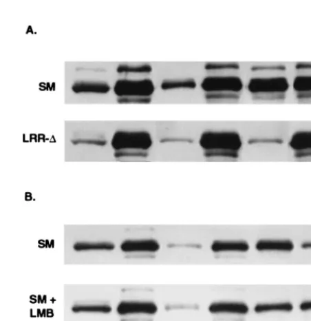
LRR mutants, particularly LRR-⌬, might not bind to CRM 1 and therefore would not be precipitable with anti-CRM 1 antibodies. Parallel immunoprecipitation experiments were therefore performed with lysates from cells transfected with wild-type SM, LRR-2, and LRR-⌬. As expected, little or no mutant protein was precipitated with anti-CRM 1 antibodies (Fig. 6A, lanes 2 and 3). Surprisingly, however, the total amount of mutant SM protein in unprecipitated cell lysates was also less than that of wild-type SM (Fig. 6, lanes 4, 5, and 6). This was in spite of there being no obvious difference in the amounts of mutant and wild-type SM protein when assessed by immunofluorescence (Fig. 4) and our previous finding of equal amounts of mutant and wild-type SM in immunoblots of whole-cell lysates of similarly transfected cells (data not shown). We therefore further analyzed the amounts and intra-cellular distribution of mutant and wild-type SM proteins in transfected cells. Cells transfected with mutant or wild-type SM plasmids were lysed in immunoprecipitation buffer con-taining 1% Triton X-100, and both the lysate and the remain-ing nuclear pellet were analyzed by immunoblottremain-ing. As shown in Fig. 6B, whereas approximately 50% of wild-type SM was found in the soluble lysate, which is expected to contain soluble cytoplasmic and nucleoplasmic SM, only 10 and 5% of LRR-2 and LRR-⌬, respectively, were present in this fraction. Con-versely, correspondingly greater amounts of the mutant SM proteins were found in the nuclear pellet fraction.
This effect of LRR mutation on intracellular distribution of SM, resulting in a tighter association with nuclear structures, was thus consistent with the immunofluorescence studies de-scribed previously which showed that LRR-⌬did not translo-cate to the cytoplasm. In addition, it was apparent that a large proportion of even wild-type SM remained associated with the nucleus despite detergent treatment. In order to further ana-lyze the nature of the association of SM with nuclear struc-tures, transfected cells were subjected to a further series of fractionation steps. Cos 7 cells transfected with wild-type SM or LRR-⌬plasmid were washed and rapidly lysed in hypotonic buffer containing 0.5% Nonidet P-40. At this detergent con-centration, the nuclei remain physically intact and adherent cytoplasm is minimal, as confirmed by light microscopy (data not shown). The lysates were reserved and the nuclei were nuclease treated and extracted with high-salt buffer with or without-mercaptoethanol. Extraction with -mercaptoetha-nol reduces and releases proteins which are oxidatively bound to the nuclear matrix, in addition to the soluble matrix-associ-ated fraction extracted with high salt alone (11, 22). Each fraction and the remaining nuclear envelopes were then sub-jected to SDS-PAGE and immunoblotted with SM anti-bodies (Fig. 7A). Approximately 20% of wild-type SM was found in the detergent-soluble fraction, compared to 5% of LRR-⌬. Approximately 10% of the remaining nuclear wild-type SM was extracted with high salt, and an additional 30% was extracted with the inclusion of-mercaptoethanol, indi-cating that the latter fraction was also associated with the nuclear matrix. The remaining 60% was associated with the nuclear envelope fraction, which includes the nuclear pore complexes (35). In contrast, the majority of LRR-⌬remained tightly associated with the nuclear envelope fraction and was resistant to the high-salt extraction steps (Fig. 7A). These data indicate that under normal conditions, wild-type SM is found in both a soluble and a tightly nucleus-bound fraction. Further, while SM is also present in an extractable matrix-bound form, a substantial proportion of SM remains tightly associated with the nuclear envelope. In contrast, the majority of LRR-⌬ pro-tein is found in the nuclear fractions and particularly in the nuclear envelope fraction.
[image:5.612.326.550.72.484.2]These results were somewhat surprising in the context of the conventional model for CRM 1-mediated export of NES teins, in which NES proteins are diffusible nucleoplasmic pro-teins which are bound and transported to the NPC by CRM 1. Further, if CRM 1 mediates docking and interaction with nucleoporins, one might have expected that an inability to interact with CRM 1 would result in less rather than more association with the nuclear envelope. To further test the hy-pothesis that interaction with CRM 1 modulates the intranu-clear attachment of SM, we examined the effect of LMB on the intranuclear compartmentalization of SM. If the increased af-finity of LRR-⌬ for the nuclear envelope and matrix were FIG. 4. Effects of CRM 1 overexpression on cellular distribution of SM and SM LRR mutants. (A) Immunofluorescence microscopy was performed on SM-or SM mutant-transfected cells with anti-SM antibodies as fSM-or Fig. 2. Cos 7 cells were transfected with either wild-type SM (wt SM), SM mutant plasmid LRR-2 or LRR-⌬, or vector plasmid (C), as indicated. Cells were also cotransfected with either control vector (⫺CRM 1) or CRM 1 expression plasmid (⫹CRM 1). (B) Cos 7 cells transfected with wt SM or LRR-⌬plasmid and cotransfected with either CRM 1 or control plasmid were stained with anti-SM antibodies. Nuclei were visualized by staining with DAPI. Immunofluorescence images were ac-quired with a deconvolution fluorescence microscope system. SM staining ap-pears red, and nuclei are purple. Bar, 15m.
on November 9, 2019 by guest
http://jvi.asm.org/
indeed due to decreased CRM 1 binding, LMB treatment should result in similar changes in distribution of wild-type SM. SM-transfected cells were treated with LMB and harvested 16 h posttransfection to minimize toxicity. The cells were frac-tionated, and the distribution of SM in each fraction was com-pared to those from non-LMB-treated cells by immunoblot-ting. SM was present in both the soluble and nuclear fractions of untreated cells, as expected, and could be extracted from the nuclei with high salt plus -mercaptoethanol (Fig. 7B). The proportion of SM that was extractable with high salt and -mercaptoethanol in non-LMB-treated cells was greater than in the previous experiments and may be a reflection of the
[image:6.612.128.535.73.267.2]shorter time interval between transfection and harvest. Never-theless, as can be seen by the relative amounts in each fraction, LMB treatment led to a decrease in the amount of SM that was extractable with high salt, particularly in the presence of -mercaptoethanol, and to a corresponding increase in the amount that remained attached to the nuclear envelope frac-tion. These data indicate that LMB, by inhibiting the associa-tion of SM with CRM 1, alters its intranuclear compartmen-talization in a manner that correlates with the effect of LRR mutation. The LRR therefore appears to be required not only for CRM 1-mediated transport to the cytoplasm but also for the proper association of SM with intranuclear structures. FIG. 4—Continued.
on November 9, 2019 by guest
http://jvi.asm.org/
DISCUSSION
In this study, we demonstrate that the SM protein of EBV binds the export receptor CRM 1 and that CRM 1 binding is important for activity and cytoplasmic localization of SM. The role of CRM 1 binding in both aspects of SM function is shown to be specific by the use of an inhibitor of CRM 1 complex formation, LMB. SM is also shown to associate in vivo with the CRM 1-binding GTPase Ran and the carboxy-terminal portion of a nucleoporin, CAN/Nup214, two components of CRM 1-linked nuclear export pathways. Further, we demonstrate that an LRR of SM is required for function and proper in-tranuclear distribution of SM.
SM is an EBV protein which enhances expression of intron-less genes in a gene-dependent manner (23, 30). Although SM clearly has multiple mechanisms of action, including stabiliza-tion of RNA and enhancement of posttranscripstabiliza-tional process-ing (25, 39), it is likely that SM is involved in nucleocytoplasmic export of lytic EBV mRNAs. Several lines of evidence support a role for SM as a carrier protein for RNA. SM has been shown to bind RNA in vitro and to shuttle from cytoplasm to nucleus
in a heterokaryon assay (39, 41). SM also enhances cytoplasmic accumulation of target mRNAs (7, 39). Further, SM is homol-ogous to the herpes simplex virus ICP27 protein, which has been shown to shuttle from nucleus to cytoplasm and bind RNA in both locations in vivo (40).
[image:7.612.326.533.70.214.2]Our finding that there is a functionally important association of SM with CRM 1 in vivo indicates that EBV utilizes a cellular FIG. 5. Coimmunoprecipitation of CRM 1 and CRM 1-associated proteins
with SM. (A) Lysates of Cos 7 cells transfected with SM or control vector (SM⫹
[image:7.612.73.270.71.356.2]and⫺) were immunoprecipitated with anti CRM 1 (anti-CRM 1 IP) or anti-SM (anti-SM IP) antibodies and immunoblotted to detect SM. A control immuno-precipitation of SM-transfected cell lysate with preimmune rabbit serum (PI) is also shown. (B) Lysates of BJAB cells transfected with SM or control vector were immunoprecipitated with anti-CRM 1 antibodies and immunoblotted as in panel A. (C) Lysates of Cos 7 cells transfected with SM and CRM 1 were immuno-precipitated with anti-Ran antibodies (anti-RAN IP) and immunoblotted to detect SM. Control immunoprecipitations with preimmune rabbit serum (PI) are also shown. (D) Cells were transfected with a plasmid expressing an HA-tagged carboxy-terminal fragment of CAN/Nup214 (HA-⌬CAN) and SM or control plasmid. Lysates were immunoprecipitated with anti-HA monoclonal antibody CA125 (anti-HA IP) and immunoblotted with anti-SM antibodies. A control immunoprecipitation (C) performed with an irrelevant monoclonal antibody (anti-FLAG) is also shown. In all panels, lanes containing an equivalent amount of unimmunoprecipitated lysate are indicated.
FIG. 6. Effect of LRR mutation on intracellular compartmentalization of SM. (A) Detergent-solubilized lysates of cells transfected with SM, LRR-2, or LRR-⌬were immunoprecipitated with anti-CRM 1 antibodies (anti-CRM 1 IP) or electrophoresed directly (non IP lysate) and immunoblotted with anti-SM antibodies. (B) Distribution of SM between detergent-soluble and insoluble fractions. Detergent-soluble (C) and insoluble nuclear pellet (N) fractions of cells transfected with wt SM, LRR-2, or LRR-⌬were analyzed by immunoblot-ting with anti-SM.
FIG. 7. Effects of LRR mutation and LMB treatment on intranuclear com-partmentalization of SM. Cells were lysed and separated into soluble (C) and nuclear (N) fractions. Intact nuclei were nuclease treated and extracted with high salt (HS) or high salt plus 2-mercaptoethanol (HSM). Nuclear envelopes re-maining after HS extraction (HS NE) or HSM extraction (HSM NE) were collected by centrifugation. Equivalent amounts of each fraction were analyzed by immunoblotting with anti-SM antibodies. (A) Cells were transfected with SM plasmid or LRR-⌬plasmid, as shown, and harvested for fractionation after 48 h. (B) Cells were transfected with SM plasmid and incubated in growth medium alone (SM) or growth medium with LMB (SM⫹LMB), harvested, and frac-tionated 16 h posttransfection.
on November 9, 2019 by guest
http://jvi.asm.org/
[image:7.612.318.544.403.636.2]export pathway normally utilized for snRNA and 5S RNA export (21) to facilitate lytic EBV gene expression. The need for such mechanisms to enhance viral RNA expression may be a reflection of the inherent inefficiency with which intronless RNAs are expressed (19). It has been known for some time that addition of exogenous intron sequences to cDNA expres-sion constructs enhances their expresexpres-sion (5). The exact rea-sons for such a stimulatory effect of intervening sequences on gene expression are poorly understood. The presence of intron sequences facilitates 3⬘processing and polyadenylation of pre-mRNA, and a direct interaction between U1 snRNP protein U1A and polyadenylation factors has been shown (29, 32). Engagement of mRNA by the polyadenylation machinery also facilitates nucleocytoplasmic export, possibly due to an inter-action of polyadenylation factors with the NPC (9, 20). Lack of assembly of spliceosomes on genes encoded as single open reading frames may thus be a relative barrier to entry of in-tronless mRNAs into a pathway that culminates in nuclear export. SM may allow direct targeting of intronless EBV mRNAs for signal-mediated export via CRM 1. Utilization of the CRM 1 pathway also provides another potential advantage for EBV gene expression since, in mammalian cells, the path-ways for mRNA export and CRM 1-mediated export appear to be independent (3, 14, 47). Thus, inhibition of host cell gene expression during EBV replication could occur without neces-sarily affecting EBV lytic gene expression. In this context, it is relevant that SM and its homologs in herpes simplex virus and
herpesvirus saimiri also inhibit expression of spliced host genes (17, 18, 46).
We have shown that the LRR of SM is required for CRM 1-mediated cytoplasmic translocation of SM and for full SM activity. In the case of HIV Rev, several lines of genetic and in vitro evidence indicate that the major function of the NES is CRM 1 binding (14, 15, 33, 42, 47). Mutational analysis of the Rev NES has demonstrated a good correlation between the ability to bind CRM 1 and Rev function (3). It is likely that the SM LRR performs a similar function for the reasons outlined above. Further, LMB treatment produces the same effects as LRR mutation on cytoplasmic localization and SM activity, indicating that the processes are dependent on CRM 1-NES complex formation. However, the additional effects of LMB treatment and LRR mutation on intranuclear distribution of SM suggest that SM may be mechanistically quite different from HIV Rev.
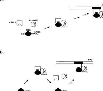
[image:8.612.135.487.86.395.2]The results of LMB treatment and deletion of the LRR suggest that in the absence of CRM 1 binding, SM remains more tightly associated with nuclear structures and particularly with the nuclear envelope. Such a finding is somewhat surpris-ing in the context of current models for CRM 1-NES protein export, in which SM plays the role of a soluble transport sub-strate for CRM 1 (Fig. 8A). According to such a model, SM might become at least transiently tethered to the NPC via CRM 1. Inhibition of CRM complex formation by mutation of the NES or LMB treatment, while leading to nuclear retention FIG. 8. Models for intranuclear translocation of SM. (A) Conventional model for CRM 1-mediated export of an NES-containing protein. CRM 1 is shown binding to soluble SM via its NES and transporting it to the NPC. CRM 1 docks at the NPC by binding to an FXFG nucleoporin-binding site (shown in gray). (B) Alternatively, CRM 1 binding detaches SM from its sites on the nuclear matrix or nuclear envelope (diagonal bars). Successive rounds of SM release and binding by CRM 1 constitute a possible mechanism of translocation along the nuclear matrix and through the NPC.
on November 9, 2019 by guest
http://jvi.asm.org/
of SM, would not be expected to increase the attachment of SM to macromolecular nuclear structures. It is unlikely that the LRR mutations described have merely resulted in an over-all decrease in solubility and thus intranuclear aggregation of the mutant proteins for several reasons. First, the SM mutants are properly imported to the nucleus and retain partialtrans -activating function. Second, a large fraction of wild-type SM is normally associated with the nuclear envelope. Finally, LMB treatment has effects on the intranuclear distribution and at-tachment of SM that are similar to mutation of the LRR.
The finding of SM in the nuclear matrix and nuclear enve-lope fractions suggests an alternative model for SM transport in which SM interacts directly with nuclear matrix and enve-lope proteins. In such a scenario, SM with its RNA cargo binds directly to one or more nuclear matrix sites. CRM 1 binding to SM in the presence of Ran-GTP would detach SM from its stationary binding site. Binding of SM to another site on the nuclear matrix accompanied by release from CRM 1 could then occur. Successive rounds of CRM 1 binding, release, and matrix binding could thus result in physical translocation of SM to the cytoplasmic face of the NPC. Such interactions would be expected to increase the efficiency of SM complex movement to the NPC, providing a track along the nuclear matrix. It should be noted that this model is similar to one proposed to explain directional translocation of import substrates into the nucleus (36, 37). In that model, the importin␣/-Ran-GDP complex dissociates from its cargo nuclear localization signal (NLS) when it binds NPC components. The import receptor is then released from the NPC on binding Ran-GTP, GTP hy-drolysis occurs, NLS cargo is bound, and the cycle is repeated, leading to a “saltatory movement” of the importin-NLS com-plex across the NPC. Our proposed model is similar in that it postulates a successive series of CRM 1-SM binding and re-lease reactions. However, SM is predicted to be also capable of interacting with nucleoporins or other structural nuclear pro-teins directly and does not invoke GTP hydrolysis, which does not appear to be required for the export process itself (28). Such a model does not preclude CRM 1-nucleoporin interac-tions and can therefore involve both SM and CRM 1 as active participants in translocation through the NPC. The compo-nents and location of the intranuclear foci of SM accumula-tion, especially those seen with the LRR-⌬mutant, remain to be determined. Based on the known effects of SM on nuclear mRNA, it is likely that some of these sites are involved in mRNA processing.
In summary, our data demonstrate that SM is a transport protein that functionally interacts with CRM 1 but that it may not be merely a soluble carrier of EBV RNA that acts as a link between the RNA, CRM 1, and the NPC. Rather, it is possible that SM exists in a dynamic structural association with the nuclear matrix and other intranuclear structures. Determina-tion of the exact intranuclear sites of SM accumulaDetermina-tion and whether SM associates directly with structural components of the NPC is likely to yield further insights into the mechanism of viral and cellular gene regulation at the level of mRNA transport.
ACKNOWLEDGMENTS
This work was supported by a recruitment grant to S.S. from the John Sealy Memorial Endowment Fund for Biomedical Research.
We express our appreciation to Barbara Wolff of Novartis AG for providing leptomycin B and to Gerard Grosveld for CRM 1 and CAN/ Nup214 cDNA and anti-CRM 1 antibodies. We also thank C. Patter-son, N. Murray, and A. Fields for many helpful discussions and review of the manuscript.
REFERENCES
1.Agard, D. A., Y. Hiraoka, P. Shaw, and J. W. Sedat.1989. Fluorescence microscopy in three dimensions. Methods Cell Biol.30:353–377. 2.Baer, R., A. T. Bankier, and M. D. Biggin.1984. DNA sequence and
expres-sion of the B95-8 Epstein-Barr virus genome. Nature310:207–211. 3.Bogerd, H. P., A. Echarri, T. M. Ross, and B. R. Cullen.1998. Inhibition of
human immunodeficiency virus Rev and human T-cell leukemia virus Rex function, but not Mason-Pfizer monkey virus constitutive transport element activity, by a mutant human nucleoporin targeted to Crm1. J. Virol.72:8627– 8635.
4.Bogerd, H. P., R. A. Fridell, R. E. Benson, J. Hua, and B. R. Cullen.1996. Protein sequence requirements for function of the human T-cell leukemia virus type 1 Rex nuclear export signal delineated by a novel in vivo random-ization-selection assay. Mol. Cell. Biol.16:4207–4214.
5.Callis, J., M. Fromm, and V. Walbot.1987. Introns increase gene expression in cultured maize cells. Genes Dev.1:1183–1200.
6.Cho, M. S., K. T. Jeang, and S. D. Hayward.1985. Localization of the coding region for an Epstein-Barr virus early antigen and inducible expression of this 60-kilodalton nuclear protein in transfected fibroblast cell lines. J. Virol. 56:852–859.
7.Cook, I. D., F. Shanahan, and P. J. Farrell.1994. Epstein-Barr virus SM protein. Virology205:217–227.
8.Deng, W. P., and J. A. Nickoloff.1992. Site-directed mutagenesis of virtually any plasmid by eliminating a unique site. Anal. Biochem.200:81–88. 9.Eckner, R., W. Ellmeier, and M. L. Birnstiel.1991. Mature mRNA 3⬘end
formation stimulates RNA export from the nucleus. EMBO J.10:3513–3522. 10. Field, J., J. Nikawa, D. Broek, B. MacDonald, L. Rodgers, I. A. Wilson, R. A. Lerner, and M. Wigler.1988. Purification of a RAS-responsive adenylyl cyclase complex fromSaccharomyces cerevisiaeby use of an epitope addition method. Mol. Cell. Biol.8:2159–2165.
11. Fields, A. P., S. H. Kaufmann, and J. H. Shaper.1986. Analysis of the internal nuclear matrix. Oligomers of a 38 kD nucleolar polypeptide stabi-lized by disulfide bonds. Exp. Cell Res.164:139–153.
12. Fischer, U., J. Huber, W. C. Boelens, I. W. Mattaj, and R. Luhrmann.1995. The HIV-1 rev activation domain is a nuclear export signal that accesses an export pathway used by specific cellular RNAs. Cell82:475–483. 13. Fornerod, M., J. van Deursen, S. van Baal, A. Reynolds, D. Davis, K. G.
Murti, J. Fransen, and G. Grosveld.1997. The human homologue of yeast CRM1 is in a dynamic subcomplex with CAN/Nup214 and a novel nuclear pore component Nup88. EMBO J.16:807–816.
14. Fornerod, M., M. Ohno, and M. Yoshida.1997. CRM1 is an export receptor for leucine-rich nuclear export signals. Cell90:1051–1060.
15. Fukuda, M., S. Asano, T. Nakamura, M. Adachi, M. Yoshida, M. Yanagida, and E. Nishida.1997. CRM1 is responsible for intracellular transport me-diated by the nuclear export signal. Nature390:308–311.
16. Gluzman, Y.1981. SV40-transformed simian cells support the replication of early SV40 mutants. Cell23:175–182.
17. Hardwicke, M. A., and R. M. Sandri-Goldin.1994. The herpes simplex virus regulatory protein ICP27 contributes to the decrease in cellular mRNA levels during infection. J. Virol.68:4797–4810.
18. Hardy, W. R., and R. M. Sandri-Goldin.1994. Herpes simplex virus inhibits host cell splicing, and regulatory protein ICP27 is required for this effect. J. Virol.68:7790–7799.
19. Huang, M. T. F., and C. M. Gorman.1990. Intervening sequences increase efficiency of RNA 3⬘ processing and accumulation of cytoplasmic RNA. Nucleic Acids Res.18:937–947.
20. Huang, Y., and G. G. Carmichael.1996. Role of polyadenylation in nucle-ocytoplasmic transport of mRNA. Mol. Cell. Biol.16:1534–1542. 21. Jarmolowski, A., W. C. Boelens, E. Izaurralde, and I. W. Mattaj.1994.
Nuclear export of different classes of RNA is mediated by specific factors. J. Cell Biol.124:627–635.
22. Kaufmann, S. H., W. Gibson, and J. H. Shaper.1983. Characterization of the major polypeptides of the rat liver nuclear envelope. J. Biol. Chem.258: 2710–2719.
23. Kenney, S., J. Kamine, E. Holley-Guthrie, E. C. Mar, J. C. Lin, D. Marko-vitz, and J. Pagano.1989. The Epstein-Barr virus immediate-early gene product, BMLF1, acts intransby a posttranscriptional mechanism which is reporter gene dependent. J. Virol.63:3870–3877.
24. Kenney, S., J. Kamine, D. Markovitz, R. Fenrick, and J. Pagano.1988. An Epstein-Barr virus immediate-early gene product trans-activates gene ex-pression from the human immunodeficiency virus long terminal repeat. Proc. Natl. Acad. Sci. USA85:1652–1656.
25. Key, S. C., T. Yoshizaki, and J. S. Pagano.1998. The Epstein-Barr virus (EBV) SM protein enhances pre-mRNA processing of the EBV DNA poly-merase transcript. J. Virol.72:8485–8492.
26. Kieff, E.1996. Epstein-Barr virus and its replication, p. 2343–2396.InB. N. Fields, D. M. Knipe, and P. M. Howley (ed.), Virology. Lippincott-Raven, Philadelphia, Pa.
27. Lieberman, P. M., P. O’Hare, G. S. Hayward, and S. D. Hayward.1986. Promiscuoustransactivation of gene expression by an Epstein-Barr virus-encoded early nuclear protein. J. Virol.60:140–148.
28. Love, D. C., T. D. Sweitzer, and J. A. Hanover.1998. Reconstitution of HIV-1
on November 9, 2019 by guest
http://jvi.asm.org/
rev nuclear export: independent requirements for nuclear import and export. Proc. Natl. Acad. Sci. USA95:10608–10613.
29. Lutz, C. S., K. G. K. Murthy, N. Schek, J. P. O’Conner, J. L. Manley, and J. C. Alwine.1996. Interaction between the U1 snRNP-A protein and the 160-kD subunit of cleavage-polyadenylation specificity factor increases poly-adenylation efficiency in vitro. Genes Dev.10:325–337.
30. Markovitz, D. M., S. Kenney, J. Kamine, M. S. Smith, M. Davis, and E.-S. Huang.1989. Disparate effects of two herpesvirus immediate-early gene trans-activators on the HIV-1 LTR. Virology173:750–754.
31. Menezes, J., W. Liebold, G. Klein, and G. Clements.1975. Establishment and characterization of an Epstein-Barr virus (EBV)-negative lymphoblastoid B cell line (BJA-B) from an exceptional EBV-genome-negative African Bur-kitt’s lymphoma. Biomedicine22:276–284.
32. Niwa, M., S. D. Rose, and S. M. Berget.1990. In vitro polyadenylation is stimulated by the presence of an upstream intron. Genes Dev.4:1552–1559. 33. Ossareh-Nazari, B., F. Bachelerie, and C. Dargemont.1997. Evidence for a role of CRM1 in signal-mediated nuclear protein export. Science278:141– 144.
34. Pinol-Roma, S., and G. Dreyfuss.1992. Shuttling of pre-mRNA binding proteins between nucleus and cytoplasm. Nature355:730–732.
35. Radu, A., G. Blobel, and R. W. Wozniak.1993. Nup155 is a novel nuclear pore complex protein that contains neither repetitive sequence motifs nor reacts with WGA. J. Cell Biol.121:1–9.
36. Radu, A., M. S. Moore, and G. Blobel.1995. The peptide repeat domain of nucleoporin Nup98 functions as a docking site in transport across the nuclear pore complex. Cell81:215–222.
37. Rexach, M., and G. Blobel.1995. Protein import into nuclei: association and dissociation reactions involving transport substrate, transport factors, and nucleoporins. Cell83:683–692.
38. Richards, S. A., K. L. Carey, and I. G. Macara.1997. Requirement of guanosine triphosphate-bound ran for signal-mediated nuclear protein ex-port. Science276:1842–1844.
39. Ruvolo, V., E. Wang, S. Boyle, and S. Swaminathan.1998. The Epstein-Barr
virus nuclear protein SM is both a post-transcriptional inhibitor and activator of gene expression. Proc. Natl. Acad. Sci. USA95:8852–8857.
40. Sandri-Goldin, R. M.1998. ICP27 mediates HSV RNA export by shuttling through a leucine-rich nuclear export signal and binding viral intronless RNAs through an RGG motif. Genes Dev.12:868–879.
41. Semmes, O. J., L. Chen, R. T. Sarisky, Z. Gao, L. Zhong, and S. D. Hayward. 1998. Mta has properties of an RNA export protein and increases cytoplas-mic accumulation of Epstein-Barr virus replication gene mRNA. J. Virol. 72:9526–9534.
42. Stade, K., C. S. Ford, C. Guthrie, and K. Weis.1997. Exportin 1 (Crm1p) is an essential nuclear export factor. Cell90:1041–1050.
43. Stutz, F., and M. Rosbash.1998. Nuclear RNA export. Genes Dev.12:3303– 3319.
44. Swaminathan, S., B. Tomkinson, and E. Kieff.1991. Recombinant Epstein-Barr virus with small RNA (EBER) genes deleted transforms lymphocytes and replicates in vitro. Proc. Natl. Acad. Sci. USA88:1546–1550. 45. Wen, W., J. L. Meinkoth, R. Y. Tsien, and S. S. Taylor.1995. Identification
of a signal for rapid export of proteins from the nucleus. Cell82:463–473. 46. Whitehouse, A., M. Cooper, and D. M. Meredith.1998. The immediate-early
gene product encoded by open reading frame 57 of herpesvirus saimiri modulates gene expression at a posttranscriptional level. J. Virol.72:857– 861.
47. Wolff, B., J. J. Sanglier, and Y. Wang.1997. Leptomycin B is an inhibitor of nuclear export: inhibition of nucleo-cytoplasmic translocation of the human immunodeficiency virus type 1 (HIV-1) Rev protein and Rev-dependent mRNA. Chem. Biol.4:139–147.
48. Wong, K.-M., and A. J. Levine.1986. Identification and mapping of Epstein-Barr virus early antigens and demonstration of a viral gene activator that functions intrans. J. Virol.60:149–156.
49. Zolotukhin, A. S., and B. K. Felber.1999. Nucleoporins Nup98 and Nup214 participate in nuclear export of human immunodeficiency virus type 1 Rev. J. Virol.73:120–127.