STUDY ON CHRONIC PANCREATITIS
AT
GOVERNMENT RAJAJI HOSPITAL, MADURAI
DISSERTATION SUBMITTED FOR
BRANCH - I M.S., (GENERAL SURGERY)
MARCH 2009
THE TAMILNADU
BONAFIDE CERTIFICATE
This is to certify that the dissertation entitled “ STUDY ON
CHRONIC PANCREATITIS AT GOVT. RAJAJI HOSPITAL, MADURAI ”
submitted by Dr. M. SIVAKUMAR to the Tamil Nadu Dr. M.G.R.
Medical University, Chennai in partial fulfillment of the requirement for the award
of M.S Degree Branch – I (General Surgery) is a bonafide research work were
carried out by him under direct supervision & guidance.
Prof.Dr.V. Seetharaman, M.S., Prof.Dr.M.Gopinath, M.S., Unit Chief, Head of the Department, Department of Surgery, Department of Surgery, Madurai Medical College, MaduraiMedical College,
Madurai. Madurai.
Prof.Dr.S.M. Sivakumar, M.S., The Dean,
Madurai Medical College, Govt. Rajaji Hospital,
DECLARATION
I Dr. M. SIVAKUMAR declare that, I carried out this work on, “STUDY ON
CHRONIC PANCREATITIS AT GOVT. RAJAJI HOSPITAL, MADURAI”
at the Department of Surgery, Govt. Rajaji Hospital during the period of June 2006 to August 2008. I also declare that this bonafide work or a part of this work was not submitted by me or any others for any award, degree, diploma to any other University, Board either in India or abroad.
This is submitted to The Tamilnadu Dr. M. G. R. Medical University, Chennai in partial fulfillment of the rules and regulations for the M.S degree examination in General Surgery.
Place : Madurai Dr. M. SIVAKUMAR
ACKNOWLEDGEMENT
I am greatly indebted to my unit Chief Prof.Dr.V.Seetharaman, M.S., and Prof. Dr.M.Gobinath, M.S., Professor and Head of the Department of General Surgery, Government Rajaji Hospital, Madurai and Prof.Dr.Muthukrishnan, Mch., HOD Surgical Gastroenteroloy for their excellent guidance in conducting this study.
I am grateful to my unit Assistant Professors, Dr.M.Sekaran, M.S., Dr.S.Chitra,M.S.,Dr.S.Kalirathnam,M.S.,Dr.Ashokachakaravarthy,M.S., for their help and guidance throughout this study.
I express my gratitude to Dr.S.M.Sivakumar, M.S., Dean, Government Rajaji Hospital, Madurai Medical College, Madurai for permitting me to use the clinical material for the study.
CONTENTS
S.No.
Title
Page
1.
Introduction
1
2.
Aim of the Study
2
3.
Review of Literature
3
4.
Anatomy, Embryology, Histology, and
Physiology of Pancreas
10
5.
Chronic Pancreatitis
22
6.
Materials and Methods
33
7.
Analysis
35
8.
Discussion
38
9.
Conclusion
52
INTRODUCTION
Chronic pancreatitis is a relentlessly progressive fibroinflammatory
process, resulting in various amounts of destruction of endocrine and exocrine
elements, which may eventually lead to pancreatic insufficiency.
Abdominal pain which is excruciating and recurrent is dominant feature of
chronic pancreatitis that initially brings most of the patients to physician’s
attention. The pathogensis of pancreatic pain is often multifactorial and explains
why not all patients respond to same mode of therapy.
In contrast to the quantitatively huge interest stands. The fact that basic
problem concerning the disease, the initial steps, the propagation, the mechanisms
are still unsolved. This obvious defect in knowledge and understanding of what
really going on, when individual get a chronic pancreatitis bears a profound
AIM OF THE STUDY
1. To study the epidemiological patterns in relation to age, sex and
place.
2. To study the different etiological factors and pattern of clinical
presentations.
3. To study the incidence of the Chronic pancreatitis at GRH,
Madurai
4. To know the outcome and response of the surgical drainage
procedure.
REVIEW OF LITERATURE
patient. Br.J. Surg 1988.
Mannell A ; Adson MA ; Mcllrath DC ; IIstrup DM ; Department of
Surgery Mayo Clinic Rochester, Minnesota
• 141 patient were operated for chronic pancreatitis at Mayo clinic. The main
indication was pancreatic pain and choice of operation was based on
anatomical abnormalities in the gland.
• Mean follow up period – 8.5 years
• Number of patient operated – 141 patients
• Conclusion
– 77% of operated patient had lasting pain relief
– Longitudinal pancreatico jejunostomy in those with dilated duct and
whipple operation for disease of pancreatic head gave good results.
2. Local resection of the head of the pancreas combined with longitudinal
pancreatico-jejunostomy in the management of patients with chronic
pancreatitis.
Ann Surg 1994, Frey CF : Amikura K, Department of Surgery, University
of California, Davis Medical Centre, Sacramento.
Material -The operation was performed on 50 patients, pain relief, endocrine and
Conclusion - The LR – LPJ provides good pain relief with modest increase in
endocrine and exocrine insufficiency and a significant increase in weight. Even
when relieved of pain, patients seldom return to the work force.
3. Pain relapse in the first 10 years of chronic pancreatitis, An. J Surg 1996.
Talamani G ; Bassi C, Falconi ; Sartonin ; Salvia R ; DiFrancesco V ;
Frulloni L ; Vaona B ; Bovo P ; Vantin I ; Pederzoli P ; Cavallini G.
Department of Medicine University of Verora, Italy.
To evaluate whether the annual number of pain relapse of chronic
pancreatitis correlated with sex, type of pancreatitis, drinking and smoking and
type of surgery .
Length of follow up - 10 yrs
Number of patients - 2,034 / year
Conclusion - Regardless of surgical treatment patients should be advised to
reduce both their alcohol intake and cigarette smoking.
4. Chronic Pancreatitis, Paul Georg Lankisch MD, sur Opin
Gastroenterology 2007.
Abstract : Purpose of Review is to focuss on the most important new observations
in chronic pancreatitis.
Recent findings –
• Smoking enhances the risk of chronic pancreatitis
• New insights in Autoimmune pancreatitis
Study :
Amsterdam group conducted Randomized trial, Comparing endoscopic and
surgical drainage of the pancreatic duct.
39 - patients participated 19 – endoscopic treatment
20 – surgical drainage procedure Follow up period – 24 months Result :
Complete or partial pain relief 32% - Endoscopic group 75% - Surgery group
5. Surgical Management of CP: Gourgiotis S,
Germanos S, Ridolfini MP, Department of hepatobiliary and pancreatic
surgery. Royal London Hospital London, UK. Hepatobilary pancreatic Dis
Int 2007 Apr 6.
Background : During the last
decade increasing knowledge about pathophysiology of CP, improved results of
major pancreatic resections and integration of sophisticated diagnostic methods in
clinical practice resulted in significant changes in surgery for CP.
provide long term pain relief a good post operative quality of life with preservation
of endocrine and exocrine function. In addition to available results from
randomized controlled trials, new studies are needed to determine which procedure
is most effective for the management of patient with CP.
6. Extended drainage versus resection in Surgery for CP : A prospective
randomized trail comparing LPJ + Local pancreatic head excision with
pylons preserving pancreatoduodenectomy.
Ann Sug 1998, Dec, Izbicki JR, Bloechle C, Broering DC, Knoefel WT,
Kuechler T, Broelsch CI. Department of Surgery University of Hamburg,
Germany.
To analyse the efficacy of LPJ – LPHI & PPPD
Number of patients – 61 patients were randomly allocated
Conclusion: – Both procedures are equally effective in terms of pain relief and
definitive control of complications affecting adjacent organ, but extended drainage
procedure provides better quality of life.
7. Comparative RCT between endoscopic treatment with surgical treatment
in painful obstructive CP. Dite et al, Endoscopy 2003.
Surgery - 80% Resection, 20% drainage
Endoscopy – 52% sphincterotomy and
Stenting, 23% stone removal,
Follow up period – 5 yrs
Conclusion :
Pain relief after surgery is superior to endotherapy in patient with painful
obstructive CP.
8. Chronic pancreatitis : A prospective nationwide study of 1086 subjects
from India
Balakrishnan V, Unnikrishnan AG, Thomas V, Choidhini Veera raju P,
Singh IP, Garg P, Pai CG, Devi RN, Bhasin D, Jayanthi D, Premalatha N,
Chacko A…, Amitha Institute of Medical Science, Cochin India, Sep 2008 :
pub Med.
Study : Prospective nationwide study of risk factors and clinical profile of
Chronic pancreatitis
Setting : 32 major centres from all over India contributed data on 1,086 patients.
Outcome Measures : Risk factors, Clinical features, complications and treatment
of Chronic pancreatitis.
Conclusion : In this first nationwide prospective study of Chronic pancreatitis in
alcoholic pancreatitis. The classical form of tropical Chronic pancreatitis is
becoming less common.
9. Comparative study of the clinical profiles of Alcoholic chronic
pancreatitis and tropical Chronic pancreatitis in Tamilnadu, South India.
Chari ST, Mohan V, Jeyanth V, Snehalatha C, Malathis, Viswanathan M,
Madanagopalan N. Department of Digestive Health and Disease Government
Peripheral Hospital, Madras, India Pub Med : Pancreas : 1992.
Number of patients - 50
Conclusion : TCP and ACP have distinct clinical profile and it is possible that
some environments factors may hasten the progress of ACP in the tropics.
HISTORY OF PANCREATIC ANATOMY
The pancreas was first mentioned in the writing of Eristratos (310-250 BC) and
all, kreas-flesh almost) was used because the organ contain neither cartilage or
bone. Its main duct was described by Wirsung in 1642, where the enlargement of
the duct at its junction with the CBD and its projection in to the duodenum as a
papilla were first described by Vater in 1720. Santorini in 1734 described the
accessory duct that bears his name. It was only after demonstration of digestive
enzymes by Claude Bernard in 1850 that the pancreas became a complete organ
with an important function and thus a worthy object of study.
Inspite of the apparent accessibility of the pancreas, a number of complex
relations combines to make its surgical removal difficult. In 1899 Halsted was
first to successfully remove the head of pancreas and portion of duodenum for
ampullary carcinoma. Several surgeons developed two staged operations, for
removal of head of pancreas. These efforts culminated in 1940 with one stage
operation of whipple.
Sir Andrew Watt kay wrote in 1978, “For me, the tiger country is removal of
the pancreas. The anatomy is very complex and one counters anomalies”.
Location :
The pancreas lies posterior to the stomach and lesser omentum in the
retroperitoneum of the upper abdomen. It extends obliquely rising slightly as it
passes from the medial edge of duodenal C loop to the hilum of the spleen. It lies
Regions :
Pancreas is divided into four regions. The head and uncinate process, neck,
body and tail. The head lies within the duodenal loop and its uncinate process
extends posteriorly and medially to lie behind the Superior mesenteric vein and
SMA. The neck of the gland extends medially from the head to lie anterior to
these vessels. The body extends laterally from the neck toward the spleen, where
as the tail extends into the splenic hilum.
Blood supply and Lymphnodes :
Both the celiac trunk and the SMA provide the arterial supply to the
pancreas. Variations are common, but for the most part, the body and tail are
supplied by branch of the splenic artery, where as head and uncinate process
receive their supply through arcade originating form the hepatic and
gastroduodenal branch of celiac artery and from the first branch of the SMA.
Venous drainage is to the splenic vein, SMV and portal vein. The pancreas
is drained by multiple lymph node groups. The major drainage of pancreatic head
and uncinate process is to the subpyloric, portal, mesenteric, mesocolic and
aortocaval node. The pancreatic body and tail for the most part are drained
through nodes in the celiac, aortocaval, mesenteric and mesocolic groups and
through nodes in the splenic hilum.
The pancreas is innervated by both sympathetic and para sympathetic
components of the autonomic nervous system. The principal and possibly only,
pathway for pancreatic pain involves nociceptive fibers arising in the pancreas.
They pass through the celiac ganglia to form the greater, lesser and least
splanchnic nerves that pass to cell bodies in the thoracic sympathetic chain.
Efferent visceral motor supply to the pancreas is provided by both the sympathetic
and parasympathetic systems. The latter involves preganglionic fibers arising
form cell bodies in the vagal nuclei that travel through the posterior vagal trunk to
the celiac plexus. Postganglionic fibers then innervate pancreatic islets, acini,
ducts and blood vessels. In general, the nerves of the pancreas travel with the
blood vessels supplying the organ.
Ducts :
The main pancreatic duct, or duct of Wirsung arising in the tail of the
pancreas and terminates at the papilla of vater in the duodenum. It crosses the
vertebral column between T12 and L2. Within the body and tail of the pancreas,
the duct lies slightly caphaled to a line drawn midway between the superior and
inferior edges. The duct is also more posterior than anterior. In adults, the duct
within the head measures 3.1 to 4.8 mm in diameter and gradually tapers to
measure 0.9 to 2.4 mm in the tail. With age, the duct diameter can increase. The
main duct. It extends from the main duct to enter the duodenum at the lesser
papilla. That Papilla lies about 2 cm proximal and slightly anterior to the major
papilla.
EMBRYOLOGY
Organagenesis :
During the 4th week of gestation, two endodermal buds arise from the
duodenum ; the hepatic diverticulum, which is desired to form the liver, gall
bladder and bile ducts, and the dorsal pancreatic bud that forms the body and tail
of the pancreas. On the 32nd day of gestation, this hepatic diverticulum gives rise
to a ventral pancreatic bud that eventually develops into the uncinate process and
inferior part of the head of the pancreas. The dorsal pancreatic bud extends
transversely across the abdomen to lies anterior to the portal and mesenteric
vessels. With time and as the duodenum rotates to form a C loop configuration
the ventral pancreas and distal bile duct undergo clockwise rotation around the
back of duodenum to finally, lie on the medial side of the duodenum, inferior and
slightly posterior to the dorsal pancreas and posterior to the portal and mesenteric
vessels. On the 37th day of gestation, the two pancreatic buds fuse and in 90% of
HISTOLOGY
The mature pancreas is an endocrine organ made up of the islets of
Langerhans and an exocrine organ consisting of acinar and ductal cells. The
acinar cels, so named because they are clustered like grapes on the stem of a vine,
discharge their secretions into a centrally located acinar space that communicates
with the major pancreatic duct. Most of the cells in the pancreas are acinar cells
and duct cells make up only 5% of pancreatic mass. Histologically, acinar cells
have a high content of endoplasmic reticulum and an abundance of apically
located eosinophilic zymogen granules. The cells lining the main pancreatic duct
are table columnar cells and many contain mucin granules. With progression form
the large ducts to the smaller intralobular and interlobular ducts the lining cells
become flatter, assuming a cuboidal configuration, and the mucin granules are no
longer seen. Centroacinar cells located at the junction between ducts and acini
PHYSIOLOGY
About 2.5 liters of clear, colorless, bicarbonate-rich pancreatic juice,
containing 6 to 20g of protein, is secreted by the human pancreas each day. It
plays a critical role in duodenal alkalinization and in food digestion.
Protein Secretion :
With the possible exception of the lactating mammary gland, the exocrine
pancreas synthesizes protein at a greater rate, per gram of tissue, than any other
organ. More than 90% of that protein consists of digestive enzymes. Most of the
digestive enzymes are synthesized and secreted by acinar cells as inactive
proenzymes or zymogens that, in health, are activated only after they reach the
duodenum where enterokinase activates trypsinogen and the trypsin catalyses the
activation of the other zymogens. Some of the pancreatic digestive enzymes are
synthesized and secreted in their active forms without the need for an activation
step (eg. Amylase, lipase, ribonuclease). Acinar cells also synthesize proteins,
including enzymes, that are not destined for secretion but, rather, are intended for
use within the acinar cell itself. Examples of this latter group of proteins include
the various structural proteins and lysosomal hydrolases.
Newly synthesized proteins are assembled within the cisternae of the rough
glycosylation. Those destined for secretion pass through the Golgi stacks and are
packaged within condensing vacuoles that evolve into zymogen granules as they
migrate toward the luminal surface of the acinar cell. By a process involving
membrane fusion and fission, the contents of the zymogen granules are then
released into the acinar lumen. Other proteins that are not destined for secretion
are segregated away from the secretory pathway as they pass through the Golgi,
and they are then targeted to their appropriate intracellular site.
Secretion of protein from acinar cells is a regulated process. At rest,
secretion occurs at a low or basal rate, but this rate can be markedly increased by
secretory stimulation that, in the pancreas, is both hormonal and neural.
Pancreatic acinar cells can express receptors for acetylcholine. Cholecystokinin,
secretin, and vasoactive intestinal peptide. Stimulation of secretion by either
acetylcholine or cholecystokinin has been shown to involve activation of
phospholipase C, generation of inositol triphosphate and diacyl glycerol, and a rise
in intracellular ionized calcium levels that, by yet unidentified mechanisms,
upregulates the rate of secretory protein discharge at the apical cell membrane. In
contrast, secretion and vasoactive intestinal peptide activate adenylate cyclase,
increase cellular levels of cyclic adenosine monophosphate (AMP), and activate
protein kinase A. This also leads to protein secretion at the apical pole. Recent
cholecystokinin and that , in humans, cholecystokinin stimulation of secretion is
mediated by intrapancreatic nerves that express cholecystokinin receptors.
Electrolyte Secretion :
Although stimulation of acinar cells results in the secretion of a small
amount of serum like fluid, most of the fluid and electrolytes secreted from the
pancreas arise from duct cells. The earliest step in duct cell electrolyte secretion
involves diffusion of circulating carbon dioxide into the duct cell, and that carbon
dioxide is hydrated by carbonic anhydrase to yield carbonic acid. Subsequently,
the carbonic acid dissociates into protons and bicarbonates ions. The protons
diffuse out of the cell and are carried away in the circulation while the bicarbonate
remains inside the cell. The fluid and electrolyte secretagogue secretion acts,
through a cyclic AMP mediated process, to stimulate chloride secretion, at the
apical cell surface, through cystic fibrosis transmembrane regulator (chloride)
channels. Then, through an apical chloride-bicarbonate exchanger, the actively
secreted chloride is taken up again by the duct cell in exchange for bicarbonate.
Taken together the result of these events is the secretion of a bicarbonate rich fluid
into the duct and the discharge, into the circulation, of protons. In the absence of
secretin stimulation, pancreatic juice has a more plasma like composition because
it is composed primarily of acinar cell secretions and there is little duct cell
stimulation, chloride secretion is increased, flow rates rise, and chloride
bicarbonate exchange results in juice that is rich in bicarbonate and poor in
chloride.
Integrated Physiology :
During the resting (interdigestive) phase of gastrointestinal function,
pancreatic secretion is minimal and may be as low as 2% of that noted with
maximal stimulation. The pancreatic response to a meal is a three phase process
that includes a cephalic phase, a gastric phase, and an intestinal phase. The
cephalic phase, accounting for 10% to 15% of meal stimulated pancreatic
secretion, reflects the response to the sight, smell, or taste of food. It is believed to
be almost exclusively mediated by peripherally relased acetycholine, which
directly stimulates pancreatic secretion of enzymes and gastric secretion of acid.
The acid indirectly stimulates pancreatic secretion of fluid and electrolytes by
causing duodenal acidification and secretin release. The gastric phase of
pancreatic secretion, accounting for 10% to 15% of meal-stimulated pancreatic
secretion reflects the response to gastric distention and the entry of food into the
stomach. These events can cause release of gastrin and stimulate vagal afferents.
By binding to cholecystokinin receptors, gastrin is itself a weak stimulant of
pancreatic enzyme secretion. Vagal stimulation also increases enzyme secretion.
secretion, and this leads to duodenal acidification, release of secretin from the
duodenum, and pancreatic secretion of fluid and electrolytes. The intestinal phase
of pancreatic secretion reflects the response to food and gastric secretions entering
the proximal intestine. Acidification of the duodenum and the presence of bile in
the duodenum promote secretin release. In addition, in the duodenum and
proximal small intestine, the presence of fat and protein, as well as their partial
breakdown products, stimulates the release of cholecystokinin, and this
cholecystokinin stimulates enzyme secretion from acinar cells. The intestinal
phase of pancreatic secretion accounts for 70% to 75% of meal stimulated
ETIOLOGY OF CHRONIC PANCREATITIS
Newer classification systems, such as the TIGAR-O, categorize chronic
pancreatitis based on the various known etiologic factors and mechanisms that are
jointly considered risk modifiers (TIGAR-O) toxic, metabolic, idiopathic, genetic,
autoimmune, recurrent severe, obstructive.
We discuss the various causes of chronic pancreatitis based on the TIGAR-O
system.
Multiple toxic and metabolic etiologies involved in chronic pancreatitis. The
association of alcohol and chronic pancreatitis was first described by COMFORT
and associates in 1946. Alcohol is still the most common cause of chronic
pancreatitis in Western industrialized countries, but only 5% to 10% of alcoholics
develop clinically apparent chronic pancreatitis, and at autopsy 10% to 20% of
alcoholics are found to have evidence of chronic pancreatitis.
Because only a fraction of alcoholics develop chronic pancreatitis,
involvements of other factors are actively being investigated. Several evidence
have shown that in addition to direct effects of alcohol, various predisposing
factors, including genetics, smoking, intestinal infection, high fat diet,
compromised immune function, gallstones, gender, hormonal factors and drinking
Smoking also is independently associated with increased risk for chronic
pancreatitis. Chronic pancreatitis induced by smoking is particularly associated
with pancreatic calcification. By mechanisms similar to alcohol, tobacco produces
alterations in the secretion and composition of pancreatic juice mainly as a result
of decreased pancreatic juice and bicarbonate secretion and induction of oxidative
stress.
Calcium plays a central role in trypsinogen secretion and trypsin
stabilization. Hypercalcemia caused by primary or secondary
hyperparathyroidism results in recurrent acute pancreatitis, which progresses to
chronic pancreatitis, likely owing to trypsinogen activation, which results in
necrosis and fibrosis of the parenchyma.
Increased serum calcium concentration is believed to induce direct damage
to acinar cells, and increased secretion of calcium results in intraductal stone
formation. Hypercalcemia also seems to modify pancreatic secretion, leading to
protein plug formation.
Idiopathic :
Thirty percent of patients with chronic pancreatitis do not have known risk
factors for chronic pancreatitis and are considered to have idiopathic pancreatitis.
Mutations of the serine protease inhibitor, Kazal type 1 (spink 1) gene in 25% of
of the clinical symptoms, idiopathic pancreatitis is separated into two distinct
entities. Early onset idiopathic chronic pancreatitis presents during the first 2
decades of life with abdominal pain being the predominant clinical feature,
whereas pancreatic calcifications and exocrine and endocrine pancreatic
insufficiency are rare at the time of first diagnosis.
In contrast, the clinical presentation of late onset idiopathic chronic
pancreatitis is in patients in their 40s, usually following a rather painless course,
but associated with significant exocrine and endocrine pancreatic insufficiency and
pancreatic calcifications.
Tropical or nutritional pancreatitis is considered a form of idiopathic chronic
pancreatitis. It is the most common form of chronic pancreatitis in certain parts of
the world, such as India, sub-Saharan Africa, and Brazil, and affects children and
young adults (Schneider et al 2002). The disease is subdivided into tropical
calcific pancreatitis, which is characterized by severe recurrent and chronic
abnormal pain and extensive pancreatic calcifications, and fibrocalculous
pancreatic diabetes, which is characterized by significant pancreatic endocrine
insufficiency. This form of chronic pancreatitis is related to mutations in the
SPINK 1 gene.
Strong association between cystic fibrosis transmembrane conductance
patients with idiopathic chronic pancreatitis have CFTR mutations.
Leading pancreatologists speculate that most chronic pancreatitis might be a
genetic disease with multifactorial triggering factors.
Genetic :
Until more recently few data existed on the genetic basis of chronic
pancreatitis. The only known hereditary form of chronic pancreatic insufficiency
that was well studied was cystic fibrosis.
Research has focused on the SPINK1-N34S gene mutation, which also is
associated closely with tropical (50%), alcoholic (6%), or idiopathic (20%) c
chronic pancreatitis.
One of the major discoveries in chronic pancreatitis was the description of
the point mutation in patients with autosomal dominant hereditary pancreatitis.
Several variants of the mutation of the cationic trypsinogen gene all lead to a
malfunction of trypsinogen. Hereditary pancreatitis presents typically in a
bimodal pattern of childhood and adulthood. Hereditary pancreatitis is an
autosomal dominant disease associated with trypsinogen gene mutations that
carries an 80% penetrance.
Despite great advances in the knowledge of genetics in pancreatitis,
currently it is advised to evaluate for mutations only in patients with hereditary
Autoimmune :
Autoimmune chronic pancreatitis (AIP) is a rare but distinct form of chronic
pancreatitis that is associated with autoimmune features. AIP is characterized by
specific histopathologic and immunologic features. The morphologic hall marks
are periductal infiltration by lymphocytes and plasma cells and granulocytic
epithelial lesions with consequent destruction of the duct epithelium and venulitis.
The pathogenesis of AIP values a cellular CD4+ and CD8+ T cells) and
humoral immune mediated attack of the ductal cells and pancreatic ducts resulting
in cytokine mediated inflammation and periductular fibrosis, which leads to
obstruction of the pancreatic ducts.
AIP is characterized clinically by minimal abdominal pain and diffuse
enlargement of the pancreas without calcifications or pseudocysts.
On laboratory examination, these patients have hypergammaglobulinemia
and autoantibodies, such as antinuclear and anti-smooth muscle antibodies.
Obstructive :
Obstruction of the main pancreatic duct is well known to result in chronic
pancreatitis. The most common etiologies include scars of the pancreatic duct,
tumors of the ampulla of Vater and head of the pancreas, and trauma.
Main pancreatic duct obstruction may lead to stagnation and stone formation
pancreatitis and periductular fibrosis (necrosis fibrosis theory) Histopathologic
characteristics of human chronic pancreatitis resulting from obstruction include
uniform distribution of interlobular and intralobular fibrosis and marked
destruction of the exocrine parenchyma in the territory of obstruction, without
significant protein plugs and calcifications.
Chronic pancreatitis results from plugging of the pancreatic duct. The origin
of chronic pancreatitis was within the lumen of the pancreatic ductules in contrast
to the origins of acute pancreatitis which tends to be inside the acinar cell.
Increased lithogenicity of pancreatic fluid leads to the formation of eosinophilic
proteinaceous aggregates, which precipitate and obstruct the pancreatic ductules.
Alcochol decreases the formation and the secretion of pancreatic juice,
making it more viscous ; low in bicarbonate ; rich in protein, enzymes and
calclium crystals ; and deficient in lithostatin.
Alcohol also has been shown to mediate the release of gastrointestinal
hormones by increasing cholecystokinin releasing factor, which affects pancreatic
juice formation and flow. The pancreatic stones and plugs are believed to produce
ulceration of the ductal epithelial cells resulting in inflammation, fibrosis,
obstruction, stasis and further stone formation.
Mechanism of chronic pancreatitis was a dysregulation and overactivity of
the acinar cell at the major area of injury by oxidative stress, usually as a result of
steady exposure of xenoiotics that induce the cytochrome P-450 enzymatic system,
while depleting glutathione.
Pancreatitis is triggered through interference of the methionine to
glutathione transsulfuration pathway, resulting in diversion of free radicals in to
the pancreatic tissue, with consequent activation of inflammation and fibrosis of
the ductules with low flow of pancreatic juice, inhibition of lithostatin, and
precipitation of proteins and calcium (Braganza 1998 ; Wilson et al, 1990).
Alcohol also may contribute to increase the oxidative stress resulting from
depletion of scavengers, such as selenium, vitamin E and C and riboflavin, and
help to induce or propagate the damage.
Alcohol and its toxic metabolites cause accumulation of intracellular lipids
and fatty acid ethyl esters, which produce damage to the acinar cell. The
alterations of intracellular lipid metabolism lead to fatty degeneration, apoptosis
and scarring of the pancreatic parenchyma with impairment of the pancreatic
microcirculation.
It was shown that these fat cells exists in the human pancreas, can migrate
into the periacinar spaces, and are activated by alcohol and acetyl aldehyde,
transforming into scar producing cells. The necrosis fibrosis hypothesis views the
pancreatitis, emphasizing that fibrosis is a late development resulting from
repeated attacks of acute (alcoholic) pancreatitis, which initially lead to
inflammation and necrosis.
The necrosis fibrosis hypothesis has significant supporting evidence from
epidemiologic and large follow up studies, which showed that chronic pancreatitis
results from recurrent attacks of acute pancreatitis.
The recurrent attacks of acute pancreatitis in hereditary pancreatitis also
support the necrosis fibrosis hypothesis. One important aspect that partially
negates this hypothesis is the fact that the type of fibrosis that follows acute
attacks of pancreatitis involves short lived collagen type III and procollagen type
IV and not the long lasting collagen types I and IV (Casini et al 2000).
The primary pathogenic factor leading to chronic pancreatitis is an outflow
obstruction likely resulting from duct inflammation, destruction and fibrosis which
likely are the result of an immunologic attacks on a specific genetic, structural or
acquired antigen of the periductular epithelium. The target of this attack may be
some specific genetic or acquired antigen on the duct epithelium.
Chronic pancreatitis seems to be an autoimmune or duct destroying disease,
analogous to primary sclerosing cholangitis. The assumption is supported by
several observations, such as the radiologic and histologic similarity of chronic
T.lymphocytes in the periductular areas of the pancreas in patients with alcoholic
chronic pancreatitis, and the occasional association of chronic pancreatitis and
primary sclerosing cholangitis.
The SAPE hypothesis tries to provide a “final common pathway” for the
many etiologies for pancreatitis. The basic aspect is that there needs to be
susceptibility (genetic or through ongoing insult, such as alcohol toxicity). The
critical sentinel event appears and triggers the process causing acute and chronic
pancreatitis. Further activation of the immunologic system and the stellate cells
propagates chronic pancreatitis, and the end result is fibrosis and calcifications.
This hypothesis has the merits of placing several of the previous theories
under “one umbrella”.
MATERIALS AND METHODS
This prospective study of chronic pancreatitis was conducted in 55 patients
admitted in GRH, Madurai, General Surgery and Surgical Gastroentereology
department from 2006 to 2008.
Informed consent was obtained from all patient who were included in the study.
Inclusion Criteria :
Patient with chronic pancreatitis presenting with refractory pain abdomen
requiring surgical intervention were included in the study.
Exclusion criteria :
Patient with chronic pancreatitis who require surgical resection procedure
are excluded from the study.
Study Design :
Each patient in the study was subjected to detailed clinical examination
correlating with a detail history. Investigations in the form of routine hemogram,
LFT, PFT and imaging studies like X rays, USG, CT abdomen and other
investigations relevant to the suspected disease system involved were done. From
the above clinical data and imaging studies chronic pancreatitis was diagnosed.
Patients who requires surgical intervention were prepared and taken up for
drainage procedure after satisfying the inclusion and exclusion criteria. The
ANALYSIS
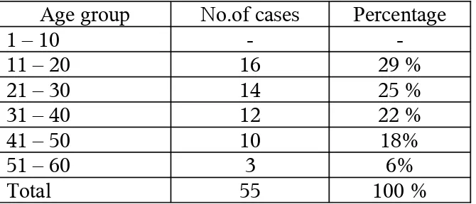
[image:34.612.141.476.263.407.2]Age Distribution :
Table : 1
Age group No.of cases Percentage
1 – 10 -
-11 – 20 16 29 %
21 – 30 14 25 %
31 – 40 12 22 %
41 – 50 10 18%
51 – 60 3 6%
Total 55 100 %
Sex Distribution :
Table - 2
Sex No.of cases Percentage
Male 32 58 %
Female 23 42 %
Total 55 100 %
Clinical Data
Table – 3
Clinical Data No.of cases Percentage
Pain Abdomen 55 100%
Diabetes 18 32%
Steatorrhea 3 6%
Weight loss 14 26%
Alcoholic 15 27 %
Smoker 17 30 %
IGTT 18 32%
IGTT – Impaired Glucose Tolerance Test
[image:35.612.169.450.563.672.2]Surgical Outcome :
Table - 4
Pain relief No.of cases Percentage
Yes 35 63.6 %
No 20 36.4 %
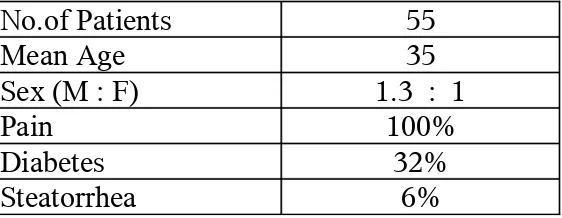
Clinical features- Chronic pancreatitis
Table - 5
No.of Patients 55
Mean Age 35
Sex (M : F) 1.3 : 1
Pain 100%
Diabetes 32%
DISCUSSION
Age Distribution :
The youngest patient in this study was 12 years old female and oldest patient
was 59 years old male. Most of the patients presented in the 2nd decade of life in
this study group ie. 11 to 20 years (29%).
Sex Distribution :
Males are affected more than female patient in the ratio of 1.3: 1 in this
study group.
Etiological distribution :
In this study (27%) of the patients are alcoholic. The etiological factor in
this group of patients are undetermined that may be attributed to nutritional,
idiopathic, hereditary etc.
Chronic alcoholism is one of the important etiological factor in chronic
pancreatitis. Alcohol abuse also affects the clinical feature, course and prognosis
of disease. In upto 70% of adult patient chronic pancreatitis appears to be caused
by alcoholism. This form is more common in men than women between age of 30
to 40.
Heriditary pancreatitis usually begins in childhood but may not be diagnosed
pancreatitis in more than one generation.
In our series 30% of the patients were smoker. Smoking is a recognized risk
factor associated with chronic pancreatitis. It also accelerates the progression of
the disease. It is now recognized as an independent risk factor for chronic
pancreatitis.
Idiopathic chronic pancreatitis was the most common from followed by
alcoholic pancreatitis in India. This was published by Amritha Institute of
Medical Science, Cochin, India. (chronic pancreatitis : prospective
nationwide study of 1086 subjects from India on Sep 2008 in pub-med.)
Clinical Presentation :
In our study common clinical presentation was recurrent episodes of pain in
epigastrium radiating to back, other features include diabetes, weight loss,
steatorrhea, anorexia etc.
For most patient with chronic pancreatitis abdominal pain is the presenting
symptom. Either the patient’s age or the etiology of the disease has some
influence in it. Most patients experience intermittent attack of pain at
unpredictable intervals, while minority of patients experience chronic pain. The
natural history of pain in chronic pancreatitis is highly variable.
fear of eating (post prandial exacerbation of pain) or due to pancreatic exocrine
insufficiency and steatorrhea).
A small percentage of patients (20%) have painless chronic pancreatitis, and
present with signs and symptoms of exocrine and endocrine insufficiency.
Diagnosis of chronic pancreatitis :
In this study diagnosis of chronic pancreatitis is based on the thorough
history, physical examination laboratory data or imaging abnormalities. Imaging
methods done in this study were plain abdominal radiographs which revealed the
presence of focal or diffuse pancreatic calcifications in 25 to 30% of the cases,
transabdominal ultrasound which showed pancreatic duct dilatation irregularly of
main pancreatic duct, loss or reduction of pancreatic parenchymal echogenicity,
calculi and calcifications. CT scan abdomen findings showed pancreatic duct
dilation, calcification and cystic lesions.
Imaging, modalities like ERCP and EUS are more sensitive and specific in
diagnosis of chronic pancreatitis. They are expensive and the cost factor and non
availability remains a constrain.
Diagnosis of chronic pancreatitis is based on a thorough history and physical
examination, laboratory data and imaging studies. Today pancreatic function tests
The two main reasons for this minor role are that
i) Non invasive test of exocrine pancreatic function show high sensitivity
only in advanced stage of chronic pancreatitis.
ii) Clinical manifestations of an exocrine pancreatic insufficiency occurs late
in the course of disease after approx 90% of exocrine parenchyma is
destroyed.
Exocrine pancreatic function test :
Invasive test :
1. Secretin cerulin test
2. Lundh test
Non invasive test :
1. Bentiranide test
2. PLT
3. FE – 1 (Stool test)
Endocrine pancreatic function test :
1. Fasting blood sugar
Imaging methods :
Non invasive imaging methods are method of choice for diagnosis of
chronic pancreatitis in clinical situations. Currently ERCP is still the “Gold
standard” among the all imaging methods. But in the future it may be replaced by
further significant refinement of magnetic resonance imaging cholangiography.
Plain Abdominal Radiography :
Shows focal or diffuse pancreatic calcifications in 30 to 40% of cases makes
the diagnosis of advanced chronic pancreatitis.
Transabdominal USG is an essential tool to visualize the entire pancreas. It
is inexpensive, simple, noninvasive, widely distributed, well tolerated and often
first imaging method in patient with abdominal complaint. In routine clinical
situation, USG is the easiest method to detect the complication of chronic
pancreatitis and to follow patients with chronic pancreatitis. Use of ultrasound for
diagnosing chronic pancreatitis is limited to advanced stage.
CT scan is as specific as ultrasound but more sensitive. CT scan cannot
detect early parenchymal changes and effects on small pancreatic ducts, but
advanced stages and complications of the disease can be evaluated with high
reliability. CT is most sensitive to detect calculi. Chronic pancreatitis is excellent
method to detect advanced stage but not for early stage of chronic pancreatitis.
based in pancreatic ductal changes has been developed for diagnosis of chronic
pancreatitis which was published in 1984 as the Cambridge criteria. Changes of
early chronic pancreatitis may not be seen on ERCP. ERCP is invasive method
(post ERCP pancreatitis of 3 to 7%), expensive, specialised equipment and trained
personal are necessary to perform the produce and to interpret the pancreatograms.
ERCP may be useful in distinguishing chronic pancreatitis from pancreatic cancer.
The advantage of ERCP are standardization and evaluation method in multi center
trials and possibility of intervention. The advantages are complications, costs and
invasiveness.
Magnetic Resonance Imaging pancreatography an imaging method is created
that enables clinicians to visualize ductal of chronic pancreatitis. The advantage of
this modality is noninvasiveness. The major disadvantage is that changes of side
branches are not visualized with same accuracy as in ERCP and not sensitive to
detect early stages of chronic pancreatitis.
Endoscopic ultrasound visualize the pancreatic duct and the parenchyma and
has the ability to detect chronic pancreatitis in patients with early stages of the
disease and with advanced chronic pancreatitis. Major advantage of EUS when
compared to other imaging modalities is that its ability to detect early stages of
chronic pancreatitis without any complications. The disadvantage of this method
MANAGEMENT :
In this study patients with refractory intractable abdominal pain are selected
and taken up for surgical drainage procedure. The most common surgical drainage
procedure done was modified peustows LPJ.
The over all surgical outcome in the form of pain relief in the study group
was approximately 64%. In the study group out of 55, 20 (36%) patients in the
follow up period came with recurrence of pain.
The treatment of chronic pancreatitis is complex and often an
interdisciplinary approach is indicated with the possibility of conservative
endoscopic and Surgical therapy.
Conservative treatment :
A major component of the conservative treatment is the management of
complications. This complex interaction requires individualization in nearly every
case. Hence the importance of the team approach.
Pancreatic exocrine enzyme supplemention :
When weight loss or steatorrhea (15g/day) or both develop supplementation
is indicated. The main goal is to ensure the optimal amounts of lipase reach the
duodenum together with the delivered food. With the currently available
steatorrhea can be reduced but not totally corrected. Side effects are rare except
soreness of mouth, perianal irritation, abdominal pain, diarrhea, constipation,
allergic reaction and fibrosing colonopathy in cystic firbrosis patiet. Dose of lipase
: 2500 u lipase / kg body weight per meal.
Conservative treatment of pain :
Pain significantly reduces patient’s quality of life and so main goals of
conservative treatment is to manage it, Medical treatment is generally the first line
therapy in patients with painful chronic pancreatitis.
Alcohol abstinence and diet advice are recommended but only 50% of
patients achieve pain relief.
Analgesies such as non narcotics and NSAID agents are recommended as
first step. An antidepressant may have an effect on pain and increase the effect of
opiates.
Interventional endoscopy and lithotripsy seems to be beneficial in cases with
man pancreatic duct stenosis and obstructing calculi. Further studies are needed to
evaluate the effect of pancreatic duct interventions on pancreatic pain.
About 80% of patient with chronic pancreatitis can be manage by direct
recommendations and pancreatic enzyme supplements. 10 to 15% of patients need
oral supplements, 5% need enteral tube feeding and approximately 1% need total
the main goals of nutritional therapy in chronic pancreatitis. Treatment of exocrine
insufficiency starts with dietary recommendations and pancreatic enzyme
supplementation.
Endoscopic Treatment :
1. Decompression of pancreatic cysts by transgastric, transduodenal, and
transpupillary approaches are preferred procedures in case of
interventional treatment for pancreatic pseudocyst. In case of poor
anatomic conditions or if there is contra indications for endoscopy patient
should be referred to a surgeon.
2. Common bile duct stenting is a another indication of endoscopic
intervention. A team approach between endoscopist and surgeon should
guide the treatment of CBD stenosis in patient with chronic pancreatitis.
The descision depends on patient age, comorbidities and cause of the
stricture. Major limitations of endoscopic intervention are stent clogging,
migration and cholangitis.
SURGERY :
The surgical treatment of chronic pancreatitis is based on two main concepts.
Preservations of tissue via drainage operation is the goal for protection against
of a non dilated pancreatic duct, if the pancreatic head is enlarged or if a
pancreatic carcinoma is suspected in addition to chronic pancreatitis.
Drainage procedure :
Pancreatic duct spinceterotomy was one of the first surgical procedures
proposed for patient with chronic pancreatitis and (stenosis) at the papilla of vater.
This procedure was recognized as a dangerous approach and lower success rates
for amelioration of pain.
More successful in patient with chronic pancreatitis and with dilated
pancreatic duct is the original Puestow procedure or its modification by Partington
and Rochelle. The procedure include resection of the tail of pancreas followed by
a longitudinal incision along the body of the pancreas and an anastomosis with a
Roux en Y lop of jejunum. The modification of Partington and Rochelle is the
elimination of the resection of the pancreatic tail. Patient with a dominant mass in
the head of the pancreas and a dilated pancreatic duct do not profit from a drainage
procedure only. In addition to the drainage approach Beger and Izbicki proposed
excavation of the head of the pancreas or the V shaped exacavation of the body
along the main pancreatic duct followed by a pancreatico jejunostomy.
Whipple’s procedure, Pylorus preserving pancreatico duodenectomy, Beger
procedure, Berne modification of the Beger procedure (DPPHR) are the various
resection procedured with merits and demerits.
Beger’s procedure (DPPHR) preserved the duodenum when compared to the
whipples procedure, which was the standard procedure in patient with chronic
pancreatitis for a long time. Patient who underwent Beger Procedure had greater
weight gain, better glucose tolerance and high insulin secretion capacity.
Improved pain status, lower frequency of acute episodes of chronic pancreatitis,
rate need for further hospitilization, low early and late mortality rates and
restoration of quality of life, DPPHR seems to be able to delay the natural course
of the chronic pancreatitis.
Frey procedure which involve local pancreatic head excision. combined with
longitudinal pancreatico jejunostomy can be considered as a standard procedure in
chronic pancreatitis and has under gone evaluation in multiple trails, confirming
its effectiveness as a surgical procedure for chronic pancreatitis.
Pancreatic left resection and central pancreatectomy (segmentectomy) in
chronic pancreatitis are procedure which further studies to prove the effectiveness
in chronic pancreatitis.
Laproscopic Surgery :
with distal chronic pancreatitis and patients with pancreatic pseudocyst. Analysis
of literature suggests that laparoscopic resection of the left part of pancreas, when
indicated in selected groups, produces excellent results, pain relief was achieved in
72% of patients. Laproscopic intraluminal cystogastrostomy and laproscopic
anterior cystogastrostomy are safe and effective methods to treat symptomatic
large pseudocyst (> 6cm in diameter)
Definite evidence from large RCT or meta analysis is still missing regarding
which surgical regimen is superior in patient with chronic pancreatitis. Many still
unresolved questions in the surgical treatment of chronic pancreatitis have to be
CONCLUSION
In this prospective study which was conducted in 55 patients with chronic
pancreatitis at GRH, Madurai between 2006 to 2008, the following conclusions
were derived out.
1. Most of the patients presented in their 2nd decade of life and mean age is
35.
2. Males are affected more than females in the ratio of 1.3 : 1
3. In this study 27% of the patients are alcoholic the etiological factor in this
group of patients are undetermined, that may be attributed to Nutritional,
Idiopathic, hereditary etc.
4. Recurrent episodes of pain in epigastrium radiating to back is the most
common presentation in this study group. Other features include diabetes,
weight loss, anorexia steatorrhea etc.
5. Modified Puestow’s is the most commonly performed surgical drainage
procedure and has good results in pain relief post operatively to 64% in
this study group.
PROFORMA
Name : Age / Sex :
Place : Ward : IP No.
Clinical Data : 1. Abdominal pain
2. Diabetes
3. Steatorrhea
4. Loss of weight
5. Jaundice
6. Physical findings
Epigastric tenderness Epigastric Mass Hepatomegaly Ascites
Investigations :
1. Blood – Sugar, Urea, Creatinine
2. Serum Amylase, lipase
3. Oral glucose tolerance test
4. Haemogram
6. pancreatic function test
Imaging
1. Plain X ray – abdomen
2. Ultra sound – abdomen
3. CT Scan – abdomen
4. ERCP
5. MRCP
Treatment
BIBLIOGRAPHY
1. Lee John Skandalakis, Joseph S.Rowe Jr, Stephen N. Gray, and John E.
Skandalakis Surgical Embryology, Anatomy of the pancreas, 2004
edition.
2. Lawrence H. Bannister : Alimentary system, Gray’s Anatomy, 39 Edition
Livingston 1995.
3. Russel T. noodbuma: Historical Development, Epidemiology of pancreas.
Surgery of the pancreas by John R. Brooks, MD, WB Saunders 1983.
4. Surgery of the Liver, Biliary tract and pancreas. Leslie H. Blumgart 4th
edition volume 1 page (713-38).
5. WR Muncy and Colin Mackey : Pancreas. Text book of Surgical
physiology, 4th edition Churchill Livingston 1988.
6. Michael B.L. Stear MD : The pancreas, Sabiston Text book of Surgery
18th edition, WB Saunders 2008 (page 1589-1610).
7. Lankisch PG : Chronic pancreatitis. Bockins Gastroenterology 5th edition
volume 4, Philadelphia WB Saunders 1995.
8. Stear ML : Classification and pathogenesis of pancreatitis, Surgical
Clinical North America 69 : June 1989.
Surgical disease of the pancreas, Lea and Feblger, Philadelphia – 1987.
10. Johnson CD : Pathogenesis of pancreatitis, Recent advances in Surgery
1995.
11. Shakelford’s Surgery of the Alimentary tract 5th edition WB Saunders
2000 page 628-650.
12. Malfortheiner P, Dominguez JE and Buchler M : Diagnosis and staging
of chronic pancreatitis, Standard in pancreatic surgery, springer Verlag
Berlin Heidelberg 1993.
13. Tittobella A et al : Endoscopic treatment facing the pancreatic Dilemma
springer – Verlag 1 994.
14. Yeung Ey : Image guided biopsy practical Interventional Radiology of
Hepatobiliary system and Gastraintestinal tract, Edward Arnold 1994.
15. Russell RCG : Indications for Surgical treatment in chronic pancreatitis.
Sterrhans in pancreatic surgery, springer – Verlag Berlin Heidelberg –
1993.
16. Prinz RA : Pancreatic duct drainage in chronic pancreatitis, standards in
pancreatic surgery springer – verlag Berlin Heidelberg – 1993.
17. Geerrghese PJ, Pillai VK, Joseph MP, Pitchumani CS. The diagnosis of
pancreatogenesis and Diabetes Mellitus J Assoc. phys India 1962.
pancreatitis. Ind J Surg 1981.
19. Tranverse et al : Surgical treatment of chronic pancreatitis, Ann Surg
1988 Sep.
20. Sharad Shah : chronic pancreatitis follow up operated cases. Ind J Gastro
1986.
21. Rajan N. Surgery for chronic calcific pancreatitis. In : Tandon BN (Ed)
chronic calcific pancreatitis in India : Proceedings of Round table
discussion on chronic calcific pancreatitis ; 1994 Trivandrum : Roussel
scientific Institute.
22. Pande, GK. Discussion on Surgical management of chronic calcific
pancreatitis. In : Tandem BN (ed) Chronic calcific pancreatitis in India :
proceeding of Round India.
23. Chronic pancreatitis : Author Kamil obideen MD, Division of Digestive
disease, Atlanta Veterans Medical centre. Article update June 16, 2008.
24. Wittel UA, Hopt UT, Batra SK, Dept of General and Visceral Surgery,
Fresburg Germany, Cigarette Smoking induces pancreatic damage
experimental data Langerbecks Arch Surg Jul; 2008.
25. Maisoneuve P, Frulloni L, Mullhaupt B. Epidemiological unit European
Institute of Oncology Milan Italy. Impact of Smoking on patient with