0022-538X/94/$04.00+0
Copyright © 1994,AmericanSocietyforMicrobiology
Modulation of Cellular Macromolecular
Synthesis by
Coronavirus: Implication for Pathogenesis
SHIGERUKYUWA,1t MICHAELCOHEN,1 GARYNELSON,1 STANLEY M.TAHARA,12
ANDSTEPHEN A. STOHLMANl 2*
Departmentsof
Microbiology'
andNeurology,2University ofSouthem Califomia Schoolof
Medicine,
LosAngeles, Califomia
90033Received 10 March 1994/Accepted 18 July 1994
Infectionwith the murine coronavirus strain JHMdecreases cellsurface expression of major histocompat-ibilitycomplex class I antigens. Northern blots showed thatJHMvirusinfection rapidlyreducedthe level of actinmRNA, whereas the levels of majorhistocompatibilitycomplexclass I andtubulin mRNAswerereduced
onlyslightly.Bycontrast,themRNA levels of interleukin 1ij,colony-stimulating factor 1 receptor,andtumor
necrosis factor alpha increased following infection.
JHM virus (JHMV),aneurotropicstrain of murine
corona-virusconsists of three glycoproteins designated the spike (S),
membrane (M), and hemagglutinin-esterase (HE) proteins and a nucleocapsid (N) protein (16). Although wild-type
JHMV induces fatal encephalomyelitis andprimary
demyeli-nation in mice, asmallpercentageofsurvivors show paralytic diseasewith chronicdemyelination (15). Thissystemprovides
a valuable animal model of demyelinating diseases in the
central nervous system (CNS) such as multiple sclerosis. In
addition, isolation of monoclonal antibody (MAb) neutraliza-tion-resistant viral variants, which induce demyelinating
dis-easewithoutfatalencephalitis, have extended the usefulnessof thismodel (5, 8, 40).A number of laboratories,including our
own, have beenpursuing the immunopathology of this model
(15). These studiessuggest that virus clearance from the CNS
ismediatedby CD8+ cytotoxicTlymphocytes (CTL) (31, 32,
35, 41, 42). However, CD4+ Tcellsarealso required, perhaps ashelperTcells fortheinduction ofvirus-specific CTLor as
effectorcellsthemselves(15, 35).TheCD8+ T-cellpopulation might be involvednotonlyinthe clearance of virus but also in
thedevelopment ofdemyelination following JHMV infection (9, 39, 42).CD8+CTLrecognize viral antigen in thecontextof major histocompatibility complex (MHC) class I molecules. Accumulatingevidencesuggeststhatoctapeptidesor
nonapep-tideswithallele-specificmotifs bind inthegrooveofthe MHC
class I molecules within the endoplasmic reticulum (26, 38).
These complexes aretransported to the cell surface for
pre-sentationtoCD8+ CTL withappropriateT-cellreceptors(37).
Since the CD8+ CTL-mediated immuneresponse is essential in JHMV infection, the expression of MHC class I antigens during JHMV infection is an important consideration in pathogenesis.
Murine coronavirus infection inhibits cellular macromolec-ularsynthesisin L-2cells(13),DBTcells(36),andJ774.1 cells
(datanot shown). Bycontrast, theexpressionofMHC classI antigen on astrocytes, oligodendroglia (17, 34), and brain endothelial cells(14)increases after murine coronavirus infec-tion. Therefore, the effect of JHMV infection on cell surface
*Correspondingauthor.Mailingaddress: USC School ofMedicine,
2025 Zonal Ave., MCH 142,Los Angeles, CA90033. Phone: (213) 342-1063.Fax:(213)225-2369.
tPresent address: Department ofAnimal Pathology, Institute of MedicalScience, UniversityofTokyo, Shirokanedai, Minato-ku, To-kyo 108, Japan. i
expressionof MHCclassI moleculesonJ774.1 cells, atarget
cell lineusedforanalysis ofJHMV-specific CTL (31, 32, 43),
was examined by radioimmunoassay. J774.1 cells grown in
96-wellplateswereinfectedwith the DL variant ofJHMVata
multiplicity of infection of4 and incubated at 37°C for 10 h.
Cells were washed with chilled phosphate-buffered saline
(PBS)andincubatedwith50 pLlofMAb (1 ,ug/ml)perwellat
4°Cfor 4 h.Cellswerethenwashed five times withchilledPBS
and incubatedwith 50 [lI of 125I-labeled protein A (approxi-mately 105cpm) at4°C overnight. Unbound radioactivity was
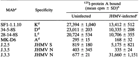
removed with five washes of chilled PBS, and the bound fractionwasquantitatedwithagammacounter.Table1 shows
decreasedexpressionof MHC class I andincreasedexpression
of the JHMV S glycoprotein 10 h postinfection (p.i.). No expression ofI-Adwasdetected.Invivotreatmentwith anti-N
MAbprotectsmicefromlethalcoronavirus infection, perhaps
by antibody-dependent cellular cytotoxicity response (18, 23,
24). Consistent with the binding of anti-N antibody to the
surface ofinfected DBTcells (24), a determinant defined by
MAb J.3.3 (7) was detected on JHMV-infected J774.1 cells,
whilebinding of another anti-N MAb (J.3.5)wasnotdetected (Table 1). We haverecently mappedthe bindingdomains for
a panel of anti-N MAbs, includingJ.3.3 and J.3.5 (30). J.3.3
binds to the acidiccarboxy terminus of the N protein, while J.3.5 bindstoa morebasic internal domain(aminoacids 249to
277) (30).Thedetailedmechanism of J.3.3bindingisnotclear; however,therapid cleavageof thecarboxy-terminal portionof the N protein during infection (unpublished data) and the
expression of this epitope on the surface of infected cells
provideapossible explanationfortheprotectionofmice from coronavirus infection mediated byanti-N MAb (18, 23, 24).
Toexamine the downregulationof MHC classI duringthe 6- to 10-h incubation used for CTL assays (31, 32, 43), the kinetics ofdownregulationof MHC classIwascomparedwith the increasedexpressionoftheJHMV S glycoprotein on the cell surface. Therewas nodetectableloss of MHCexpression
for thefirst6hp.i. (Fig. 1). However, expression of all three
H-2d MHC class I molecules was decreased by 8 h p.i. In
contrastto decreased MHCclass I expression, theexpression
of the JHMV S protein onthe cell surface of infected J774.1
cellswasclearlydetectable at8 hp.i.and increased with time
following JHMV infection. These data contrast with results
showingup regulationof MHC class I expressionon primary
astrocytes,oligodendroglia,and brain endothelialcells (14, 17, 34). Differences among cell types might account for this 6815
on November 9, 2019 by guest
http://jvi.asm.org/
TABLE 1. Cell surfaceexpression of MHC class I, class II and viral proteins onJHMV-infected J774.1 cells
'25I-proteinAbound
MAba Specificity (mean cpm±
SD)'
Uninfected JHMV-infectedc
SF-1.1.10 Kd 27,394±1,040 13,412±512
34-5-8S Dd 23,011±203 10,335- 208
28-14-8S Ld 20,724±534 10,706+355
MK-D6 Ad 295± 15 168±32
J.2.5 JHMVS 819± 180 5,175±821
J.3.5 JHMV N 403±345 335 ±24
J.3.3 JHMV N 677±21 31,660- 1,151
a Antibodieswerepurifiedfrom serum-free culture supernatants byproteinA columnchromatography.Thefollowinghybridomaswereobtained from
Amer-ican Type Culture Collection: SF1-1.1.10 (specific for Kd; IgG2a), 34-5-8S (specific fora2domainofDd; IgG2a), 28-14-8S (specific for a3domainofLd; IgG2a),andAF6-88.5.3(specific forKb; IgG2a).MAb J.2.5(specific forJHMV Sprotein;IgG2a), J.3.3(specific forJHMV Nprotein;IgG2a), and J.3.5 (specific
for JHMV Nprotein; IgG2b)wereproduced in ourlaboratory (7).
bEach value is the mean ±standard deviation ofthevaluesfromthreewells. 10 hp.i.
discrepancy, suggesting thatregulation of MHC classI expres-sion may be celltypedependent.Inaddition, different strains of MHV havedifferent abilitiestodownregulate MHC class I
(4).Differentialregulationof MHC class Iby MHV strains and amongdifferent cell types maypartially explain the absence of detectable CTL responses in somehaplotypes (21).
Control of viral and cellular geneexpression, either at the
~4=
A
-..
l
X 40 - 0 1
I
:0
0
2I46J
0 1ILHours aftr infecion
dr 60
-CC
-40
I
I
.c 20
-i
O-0 2 4 6 8 10 12
Hours aftsr knodon
transcriptional or posttranscriptional levels, is an important consequence of viral infection. Most viruses modify host cell macromolecular machineryfor viralreplication, andshutoff is a commonpropertyoflytic infections, although the underlying mechanisms vary amongviruses(27).RNA wasprepared from J774.1 cells atvarious times p.i., and MHC class I and actin mRNAlevels werecompared with the stability of MHC classI
mRNAfollowing JHMV infection. J774.1 cellswere infected with JHMV at a multiplicity of infection of2for1hat37°C. Atvarious timesp.i., the cells werelysed by the addition of 2.5 ml ofguanidine thiocyanate homogenization buffer and the DNA was sheared. RNAwas isolated by centrifugation over
2.5 mlof 5.7 MCsCl for 18 h at 100,000 xg,resuspended in sterile distilled water, and quantitated by UV absorbance. RNA samples were stored in 70% ethanol at -70°C until
analysis. For Northern analysis, RNA samples (30 p.g) were
suspended insamplebufferconsistingof50% formamide, 2.2
M formaldehyde, lx MOPS buffer (0.02 M
morpholinopro-panesulfonic acid, 5 mM sodium acetate, 1 mM EDTA; pH
7.0),and0.005%bromophenol blue andseparated by electro-phoresis in 1.5% agarose gels containing MOPS buffer and
formaldehyde (0.66 M) at 15 V for 18 to 24 h. RNA was
transferredtoHybond-N+ membranes(Amersham)with 1ox SSC(1xSSC is 150mMNaCl and 15mMsodiumcitrate)and fixed with 0.05 MNaOH for 5 min, and then the membranes werewashed for1 minwith 2x SSC.
Northern blots were analyzed with the following DNA
probes.A0.4-kbcDNAencodingaconserved sequence of the murine MHC classIgenewasexcised frompH-211a(29) with
011 60
B
'W-40
I
c 20
c
J0
0 2 4 6 8 10 12
Hours afmr infecion
.r- 15
D°
110
4 5
i
0 2 4 6 8 10 12
Hours after
infcUon
FIG. 1. Expression of MHC class I molecules and JHMV S protein on J774.1 cells during JHMV infection. J774.1 cells were infected with JHMV and incubated at37°C for the times indicated. Cells were incubated with MAbs specific forKd(A),Dd (B),Ld(C) and JHMV S protein (D) and further incubated with '25I-labeled protein A as described in footnote a of Table 1. Results represent mean counts per minute of
125I-labeled
protein A bound + standard deviation of three values.on November 9, 2019 by guest
http://jvi.asm.org/
[image:2.612.57.299.100.204.2]A 02 468 10 12
28S
-B 02 4 68 10 12 C 0 2 4 6 8 10 12
28S
-18S- 18S
28S-
l1s-D
°;
3Ab _
.... .
0.1
0.01
2 4 6 8 10
Hours after infection
FIG. 2. MHCclassI,actin and viralmRNAs in J774.1 cells following JHMV infection. J774.1 cells were infected with JHMV and incubated at37°C, and RNA was extracted at the times (in hours) indicated. MHC class I (A), actin (B), and JHMV (C) RNA levels were analyzed by Northernblot hybridization. The positions of 28S and18SrRNA areindicated at the left, and the numbers of JHMV-specific mRNAs are indicated atthe right(C).Autoradiographs were scanned by densitometry, and relative levels of actin(0)and MHC class I(O) RNA are presented (D).
restriction enzymesHhaIandSacl. A 0.85-kb cDNA encoding
a conserved sequence of the murine MHC class II gene was excised by digestion with EcoRI from pABk (6). A 1.4-kb cDNA encoding the murine interleukin 1B (IL-1,B) sequence was excised by digestion of pMuIL-l,B with EcoRI, kindly supplied byP. Gray, Genentech (11). A 0.6-kb insert encoding the IL-6 sequence was excised by digestion of pIL-6, with HindlIl and EcoRI kindly supplied by K. Uyemura, University of CaliforniaatLosAngeles(44). A 1.1-kb insert encoding the
tumornecrosis factor alpha(TNF-a) sequence was excised by digestion of pGEM-3-Cach, withEcoRI and PstI, kindly sup-plied by L. Schook, University of Illinois (22). Colony-stimu-lating factor 1 (CSF-1) receptor mRNA was quantitated by using the 1.0-kb insert encoding the
v-fins
oncogenederived by digestion of pSM7C (ATCC 41016) with XhoI. A 1.4-kb cDNA encoding the human y actin gene was excised from plasmid ActBluby digestion with PstI(2). A 0.5-kb insert encoding the murine tubulin gene was excised by digestion of plasmid Malwith EcoRI and Hindlll kindly supplied by N. Cowan, New
YorkUniversity (19).AcDNAencoding the JHMV N gene
was excised bydigestion withHindIll andEcoRIfrom pT7-4
(2), kindly supplied by M. Lai. The cDNA fragments were isolated by electrophoresis in
low-melting-point
agarose(FMC, Rockland, Maine), labeled with [a-3 P]dCTP (300
Ci/mmol) using a random-primed DNA labeling kit
(Boehr-inger Mannheim, Indianapolis, Ind.), followed by desalting with aSephadex G-50Quick Spin column (Boehringer
Mann-heim). Membranes were prehybridized for 15 to 45 min in
Hybrisol 1 (Oncor, Gaithersburg, Md.) at 42°C. Denatured
probe was added (approximately 2 x 10⁶ cpm/ml) and incu-bated for 18 to 72 h at 42°C. Membranes werewashed four times in 2x SSC containing 0.1% sodium dodecyl sulfate at roomtemperature and thenwashed twice in 2x SSCat65°C.
Membranes were air dried and exposed to XAR-5 film
(Kodak).
MHC classImRNAlevelsgraduallydecreasedafter JHMV infection (Fig. 2A). In contrast, actin mRNA levels
rapidly
decreased after infection (Fig. 2B), as
previously
observedfollowingMHV-A59infection of L2 cells
(13).
Thesechanges
inhostcellmRNAlevelsoccurred when viral mRNA
synthesis
increased(Fig. 2C). Comparisonof therelative mRNAlevels of actin andMHC classIfollowinginfection
(Fig. 2D) clearly
show that theamountof actin mRNA decreasesmore
rapidly
than the levels of MHC class I mRNA
following
JHMV infection.Since degradation of cellular mRNA is involved in the
shutoffphenomenoninother virus infections(27), the effect of JHMVinfection onmRNAlevels in the absence of transcrip-tion was examined by using actinomycin D treatment. The
mRNAlevels were examined by preparing RNA from JHMV-infected cells, uninfected actinomycin D-treated cells, and JHMV-infected cells treated with actinomycin D. The levels of MHC classI and actin mRNA in JHMV-infected cells were clearly reduced more rapidly than in actinomycin D-treated uninfected cells (Fig. 3). The MHC class I and actinmRNA
levels in JHMV-infected actinomycin D-treated cells at 4, 6, and 8 hp.i.weresimilartothose in untreated JHMV-infected cells. These results indicate that JHMV infection results in degradation of these cellular mRNAs, although the effect varies among mRNA species (see Fig. 4). It is not clear whether this degradation is due to a direct effect of a viral componentor avirus-induced effect mediated viainterruption of a host component regulating mRNA levels. Although the effect ofactinomycin D onthe levels of JHMV mRNA was
examined, therewas noobviouschangesin either the levelsor
kinetics(data not shown), consistent with the growth of virus in enucleatedand/or actinomycin D-treated cells(16).
The decrease inMHC classImRNArelativetoactinmRNA suggestedthatnotall mRNAs weredegraded with the same
kineticsfollowing JHMV infection. We tookadvantage of the
monocyte-macrophage nature of J774.1 cells to examine the
JHMV
JHMV
AMD
0 2 4 6 8 10 2 4 6 8 10
AMD
4 6 8
MHC
-,"
f:, 'p:class I
[image:3.612.97.534.82.214.2]actin
*_
FIG. 3. Stabilityof MHC class I andactinRNAinuninfectedand JHMV-infectedJ774.1 cells.J774.1 cells eitheruntreated ortreated with2 ,ugofactinomycinD(AMD)per mlwereinfected with JHMV andincubatedat37°Cforthe times(inhours)indicated. MHC classI and actin mRNAlevelswereanalyzedbyNorthernblothybridization.
fA 40 0-It
lillif.-IFY-Of
...on November 9, 2019 by guest
http://jvi.asm.org/
[image:3.612.342.542.549.674.2]az
-9
z c:
E
0
101
0.1
-v-v1r--r
0 2 4 6 8 10 12
Hours After Infection
FIG. 4. Relative host cell mRNA levels in J774.1 cells following JHMV infection. J774.1 cellswereinfected with JHMV and incubated
at 37°Cfor the times indicated. RNAwasisolated and analyzed by
Northern blothybridization.RNAlevelsof tubulin (U),CSF-1 recep-tor(A),TNF-a(*),IL-13 (v), and 28S rRNA(0)relativetothose
found in uninfected cellsarepresented.
stability of a number of other mRNAs, including factors involved in immuneregulation. Figure4shows the levelof28S rRNA was relatively unchanged for the first 10 h p.i. and decreasedby50% atthe last timepointexamined(12hp.i.).
Tubulin mRNA was degraded with kinetics similar to actin mRNA(Fig. 2) buttoalesserextent. No mRNAsspecificfor MHC class II orIL-6 were detected in either uninfected or
infected J774.1 cells(data not shown).The inabilityto detect MHC classIImRNAisconsistent with theradioimmunoassay
data(Table 1). Bycontrast,the levels ofmRNAsencodingthe CSF-1receptor,TNF-a,andIL-13 increasedrapidly following
JHMV infection andweresensitivetoactinomycinD(datanot
shown). Thisrapid transcriptional activation appears tobe a
direct result of virus replication, since infectionwith an iden-tical UV-inactivatedpoolof virusdid notactivatetranscription (data not shown). Interestingly, the level of CSF-1 receptor mRNAincreasedearly followinginfection butappearedtobe
susceptibletothesamedegradation mechanismaffectingactin
andtubulin mRNAsby 4 hp.i. Bycontrast,both TNF-a and IL-13 mRNA appeared to be resistant to JHMV-induced degradation. A striking feature is that these two cytokine
mRNAs have normal half-lives of 30 min or less. This
insta-bility is due to the presence of AUUUA motifs in their
3'-untranslatedregions which promote mRNAinstabilityand turnover.A32-kDaprotein (AUBF)bindstothisregionand is believed to stabilize the mRNA from degradation by protec-tion of this sequence motif. AUBF activation is believed to
require a posttranslational mechanism and not de novo
syn-thesis(20). Recentlyitwasreportedthat thedestabilization of
AUUUA-containing mRNAs is a cotranslational event (1); thus, JHM-induced inhibition of host protein synthesis may
stabilize AUUUA-containing mRNAsby preventing associa-tion withribosomes andsubsequent translation. Alternatively
an increase in AUBF activity or a decline in the
AUUUA-specific nuclease activity during JHMV infection could be
responsible for the increased steady-state levels of these mRNAs. Inadditionto RNAdegradation, JHMV alsousesa mechanism for preferential translation ofviral proteins (36),
i.e., mRNAs containing JHMV leader RNA sequences are
more efficiently translated thanmRNAwhich donot contain
the sequence.Takentogether,coronavirus-induced shutoff of
host cell synthesis appears to involve a number of different
mechanisms includingdifferential effects on host mRNA sta-bilityandpreferentialtranslation of viral mRNA.
Althoughthisstudywascarriedoutinvitro,it istemptingto
suggest that JHMVinfection also induces similar modifications
of gene expression in vivo. MHV infection increases MHC
class I expression (10, 25) and cytokine mRNA in vivo (25;
unpublished data). Furthermore, a variety of cell types are
susceptible to infection, including infiltrating monocytes as well as CNS resident cells. These cells may be
differentially
affectedbyJHMVinfection; therefore,theeventsthatoccurin
vivo may represent a combination of both direct effects of
JHMV infection andcytokine-mediated effectson uninfected
cells. From thispointofview,it is veryinterestingthat JHMV
infection increasedTNF-aandIL-13 mRNA levelsin infected
cells. Both ofthese cytokines have been implicated in CNS
disease (3) and TNF-ot has been implicated in a variety of demyelinatingdiseases(3, 12, 28, 33). However,since corona-virus-infected cells preferentially translate viral mRNA (36),
increased mRNA levels may not correlate with increased cytokine secretion; therefore, cytokine synthesis following
JHMV infection must be measured to
clarify
their role indevelopmentofJHMV-induced
demyelination.
This workwassupported byPublic Health Service grantsNS18146,
NS07149,andNS30880.
The technical assistanceofEmmanuelDimacalli,thecriticalreading andgraphicassistanceof ConniBergmann,andthe editorialassistance of SoniaQ. Garciaaregratefullyacknowledged.
REFERENCES
1. Aharon,T.,and R.J.Schneider.1993.Selectivedestabilization of short-lived mRNAs with the granulocyte-macrophage colony-stimulatingfactor AU-rich3' noncoding regionismediatedbya
cotranslational mechanism. Mol. Cell.Biol. 13:1971-1980. 2. Baric,R.S.,G. W.Nelson, J. 0.Fleming,R.J. Deans, J.G.Keck,
N.Casteel, and S. A. Stohlman. 1988. Interactionsbetween the coronavirusnucleocapsidproteinand viral RNAs:implicationsfor viraltranscription.J.Virol.62:4280-4287.
3. Benvenisti,E. N.1992.Inflammatory cytokineswithinthecentral
nervoussystem: sources,functionandmechanism of action. Am. J. Physiol.263 (Cell Physiol.32):C1-C16.
4. Bergmann, C.,M.McMillan,and S. Stohlman.1993. Character-ization of theLd-restrictedcytotoxic T-lymphocyte epitopeinthe
mousehepatitis virus nucleocapsidprotein.J. Virol. 67:7041-7049. 5. Dalziel,R.G.,P. W.Lampert,P.J. Talbot,and M.J.Buchmeier. 1986. Site-specific alteration of murine hepatitis virus type 4 peplomerglycoprotein E2 results in reduced neurovirulence. J. Virol.59:463-471.
6. Estess, P., A. B. Begovich, M. Koo, P. P. Jones, and H. 0. McDevitt.1986.Sequence analysisandstructure-function correla-tionof murineq,k,u,sand fhaplotypeI-AbetacDNAclones.Proc. Natl. Acad. Sci. USA83:3594-3598.
7. Fleming, J.O., S.A.Stohiman,R. C.Harmon,M. M.C.Lai, J.A.
Frelinger, and L. P. Weiner. 1983. Antigenic relationship of murine coronaviruses: analysis using monoclonal antibodies to
JHM(MHV-4)virus.Virology 131:296-307.
8. Fleming, J. O., M. D. Trousdale, F. A. K. El-Zaatari, S. A.
Stohlman, and L. P. Weiner. 1986. Pathogenicity of antigenic variants of murine coronavirus JHM selected with monoclonal antibodies. J.Virol.58:869-875.
9. Fleming, J. O., F.-I.Wang,M. D.Trousdale, D. R.Hinton, and S. A.Stohlman. 1993. Interactionofimmuneand centralnervous
system: contribution of anti-viral Thy-I+ cells to demyelination inducedby coronavirusJHM.Reg.Immunol. 5:37-43.
10. Gombold, J. L.,andS. R. Weiss. 1992. Mousehepatitisvirus A59 increasessteady-statelevels of MHC mRNAs inprimary glialcell cultures and in the murine central nervous system. Microb.
Pathog. 13:493-505. -0
0 8-10.0
on November 9, 2019 by guest
http://jvi.asm.org/
[image:4.612.89.258.79.240.2]11. Gray, P. W., D.Glaister, E.Chen,D.V. Goeddel, and D. Pennica.
1986. Twointerleukin 1 genes in the mouse: cloning and expres-sion of the cDNA for murine interleukin 1f. J. Immunol. 137: 3644-3648.
12. Hauser, S. L., T. H. Doolittle, R. Lincoln, R. H. Brown, and C. A. Dinarello. 1990. Cytokine accumulation in CSF of multiple scle-rosis patients: frequent detection of interleukin-1 and tumor necrosis factor but notinterleukin-6. Neurology 40:1735-1739. 13. Hilton, A., L.Mizzen, G. Macintyre, S. Cheley, and R. Anderson.
1986.Translational control in murine hepatitis virus infection. J. Gen. Virol. 67:923-932.
14. Joseph, J., R. L. Knobler, F. D. Lubin, and M. N. Hart. 1989. Differential modulation of MHC class I antigen expression on mouse brain endothelial cells by MHV-4 infection. J. Neuroim-munol. 22:241-253.
15. Kyuwa, S., and S. A. Stohlman. 1990. Pathogenesis ofa neuro-tropic murine coronavirus, strain JHM in the central nervous system of mice.Semin. Virol. 1:273-280.
16. Lai, M. M. C. 1990. Coronavirus: organization, replication and expression of genome. Annu. Rev. Microbiol. 44:303-333. 17. Lavi, E., A. Suzumura, E. M. Murray, D. H.Silberberg, and S. R.
Weiss. 1989. Induction of MHC class I antigens on glial cells is dependent onpersistent mouse hepatitis virus infection. J. Neu-roimmunol. 22:107-111.
18. Lecomte, J., V. Cainelli-Gebara, G. Mercier, S. Mansour, P. J. Talbot, G. Lussier, and D. 0th. 1987. Protection from mouse hepatitis virus type 3-induced acute disease by an anti-nucleopro-tein monoclonal antibody. Arch. Virol. 97:123-130.
19. Lewis, S. A.,M.G. Lee, and N. J.Cowan. 1985. Five mouse tubulin isotypes and their regulated expression during development. J. Cell Biol. 101:852-861.
20. Malter, J. S.,andY. Hong. 1991.Aredoxswitch and phosphory-lation are involved in the post-translational up-regulation of the adenosine-uridine binding factor byphorbolester and ionophore. J. Biol. Chem. 266:3167-3171.
21. Mobley,J., G. Evans, M.0. Dailey,andS.Perlman. 1992. Immune response to a murine coronavirus: identification of a homing receptor-negative CD4+ T cell subset that responds to viral glycoproteins. Virology 187:443-452.
22. Myers, M.J., J.F. Pullen,N.Ghildyal, E. Eustis-Turf, and L. B. Schook. 1989. Regulation of IL-I and TNF-ot expression during thedifferentiation of bonemarrowderivedmacrophage.J. Immu-nol. 142:153-160.
23. Nakanaga, K., K. Yamanouchi, andK.Fujiwara. 1986. Protective effect of monoclonal antibodies on lethal mouse hepatitis virus infection in mice. J.Virol. 59:168-171.
24. Nakanaga, K.,K.Yamanouchi,andK.Fujiwara. 1987.Protective effect of theF(ab')2fragments of monoclonal antibodiesto mouse
hepatitis virus. Adv. Exp. Med. Biol. 218:365-371.
25. Pearce,B.D.,M.V.Hobbs,and M.J. Buckmeier.1994.Cytokine induction during T-cell-mediated clearance of mouse hepatitis virus fromneuronsinvivo. J.Virol. 68:5483-5495.
26. Rotzschke, O.,K.Falk,K.Deres,H.Schild,M.Norda, J. Metzger,
G.Jung, and H.-G. Rammensee. 1990. Isolation and analysisof naturally processed viral peptides as recognized by cytotoxic T
cells. Nature(London)348:252-254.
27. Schneider, R. I., andT.Shenk. 1987.Impactof virus infectionon hostcellproteinsynthesis. Annu.Rev. Biochem.56:317-332.
28. Selmaj, K., C. S. Raine, and A. H. Cross. 1991. Anti-tumor necrosis factor therapy abrogates autoimmune demyelination. Ann. Neurol.30:694-700.
29. Steinmetz, M., J. G. Frelinger, D. Fisher, T. Hunkapiller, D. Pereira, S. M.Weissman, H. Uehara, S. Nathanson, and L. Hood. 1981. Three cDNA clones encoding mouse transplantation anti-gens: homologytoimmunoglobulin genes. Cell 24:125-134. 30. Stohlman, S. A., C. B. Bergmann, D. Cua, H. Wege, and R. van der
Veen. 1994. Location of antibody epitopes within the mouse hepatitis virus nucleocapsid protein. Virology 202:146-153. 31. Stohlman, S. A., S. Kyuwa, M. Cohen, C. Bergmann, J.M.Polo, J.
Yeh, R. Anthony, and J. G. Keck. 1992. Mouse hepatitis virus nucleocapsid protein-specific cytotoxic Tlymphocytes are Ld re-strictedandspecificfor the carboxy terminus. Virology 189:217-224.
32. Stohlman, S. A., S. Kyuwa, J.M.Polo, D. Brady,M. M. C.Lai,and
C. C. Bergmann. 1993.Characterization of mousehepatitis virus-specific cytotoxicTcellsderived from the central nervous system of mice infected with the JHM strain. J. Virol.67:7050-7059. 33. Stoll, G., S. Jung,S.Jander,P. van derMeide,and H. P.Hartung.
1993. Tumor necrosis factor-ot inimmune-mediateddemyelination andWalleriandegeneration.J. Neuroimmunol. 45:175-182. 34. Suzumura, A., E.Lavi, S.R. Weiss,and D. H. Silberberg. 1986.
Coronavirus infection induces H-2 antigen expression on oligo-dendrocytes and astrocytes. Science 232:991-993.
35. Sussman, M. A., J. 0. Fleming, H. Allen, and S. A. Stohlman. 1989. T-cell-mediated clearance of mouse hepatitis virus strain JHMfrom the central nervous system. J. Virol. 63:3051-3056. 36. Tahara, S. M., T.A. Dietlin, C. C. Bergmann, G.W.Nelson, S.
Kyuwa, R.P. Anthony, and S. A. Stohlman. 1994. Coronavirus translational regulation:leader affectsmRNAefficiency. Virology 202:621-630.
37. Townsend, A.,and H. Bodmer. 1989.Antigen recognitionby class I-restrictedTlymphocytes.Annu. Rev.Immunol. 7:601-624. 38. Van Bleek, G. M., and S. G. Nathenson. 1990. Isolation of an
endogenously processed immunodominant viralpeptidefromthe class IH-2kdmolecule. Nature(London)348:213-216.
39. Wang,F., S.A.Stohlman,andJ.0. Fleming. 1990.Demyelination induced by murinehepatitis virus JHM strain (MHV-4) is immu-nologically mediated. J. Neuroimmunol. 30:31-41.
40. Wege, H., J. Winter, and R. Meyermann. 1988. The peplomer protein E2 of coronavirus JHM as a determinant of neuroviru-lence:definition of criticalepitopesvariantanalysis.J.Gen. Virol. 69:87-98.
41. Williamson, J. S.P.,andS.A.Stohlman. 1990. Effective clearance ofmousehepatitisvirus from the centralnervoussystemrequires bothCD4+ andCD8' Tcells.J. Virol. 64:4589-4592.
42. Yamaguchi, K., N.Goto, S. Kyuwa, M. Hayami, and Y. Toyoda.
1991.Protection of mice fromalethalcoronavirus infection in the centralnervoussystembyadoptivetransfer ofvirus-specificTcell clones.J. Neuroimmunol. 32:1-9.
43. Yamaguchi, K., K. Kyuwa,K. Nakanaga,and M. Hayami. 1988. Establishment ofcytotoxicT-cell clonesspecificforcells infected withmouse hepatitisvirus. J. Virol. 62:2505-2507.
44. Yamamura,M.,X.-H.Wang,J.D.Ohmen,K.Uyemura,T. H.Rea,
B. R. Bloom, and R. L. Modlin. 1992. Cytokine patterns of immunologicallymediated tissuedamage.J. Immunol. 149:1470-1475.