INTERVENTIONAL STUDY
A Dissertation submitted in
partial fulfilment of the requirements
for the degree of
MASTER OF DENTAL SURGERY
BRANCH – II
PERIODONTOLOGY
THE TAMIL NADU DR. M.G.R. MEDICAL UNIVERSITY
Chennai – 600 032
This is to certify that Dr. C. KANNAN, Post Graduate student (2010-2013) in the Department of Periodontics, Tamil Nadu Government Dental College and Hospital, Chennai - 600 003, has done
this dissertation titled "EVALUATION OF SERUM ADIPONECTIN
LEVELS IN TYPE II DIABETES MELLITUS WITH CHRONIC PERIODONTITIS – AN INTERVENTIONAL STUDY" under our direct guidance and supervision in partial fulfillment of the regulations laid
down by The Tamil Nadu Dr. M.G.R. Medical University, Chennai -
600 032 for M.D.S., (Branch-II) Periodontics degree examination.
Dr. S. Kalaivani
Professor and Guide
Dr. K. Malathi
Professor & H.O.D.
Department of Periodontics
Tamil Nadu Government Dental College and Hospital
Chennai - 600 003
Dr. K.S.
G.A. NASSER
PRINCIPAL
Tamil Nadu Government Dental College and Hospital
ACKNOWLEDGEMENTS
I express my earnest gratitude to my Guide Dr. S. KALAIVANI
M.D.S., Professor and Guide, Department of Periodontics, Tamil Nadu
Government Dental College and Hospital, Chennai – 600 003 without her
esteemed guidance and support this study would have not been possible.
I am extremely grateful to Dr. K. MALATHI M.D.S., Professor &
H.O.D., Dr. MAHEASWARI RAJENDRAN M.D.S., Professor, Tamil Nadu
Government Dental College and Hospital, Chennai – 600 003 for their
suggestions, source of inspiration and for the betterment of this dissertation.
I sincerely thank to Dr. K.S.G.A. NASSER, M.D.S., Principal, Tamil
Nadu Government Dental College and Hospital, Chennai – 600 003 for his
kind permission and encouragement.
I am extremely grateful to Dr. A. MUTHUKUMARASWAMY
M.D.S., Dr. P. KAVITHA M.D.S., Dr. M. JEEVA REKHA M.D.S.,
Assistant Professors, Tamil Nadu Government Dental College and Hospital,
Chennai – 600 003, for helping me with my dissertation and during my study
period.
I extent my sincere thanks to Dr. PRAGNA B. DOLIA M.D., Director,
Department of Biochemistry, Government General Hospital, Chennai-600 003,
for granting me permission to conduct this study and her supportive guidance
and avail the lab facilities throughout this study.
I specially thank to my colleague Dr. K. GOKUL M.D.S., for helping
A special mention of thanks to all my patients for their very kind
cooperation throughout my study.
I wish to express special thanks to my colleagues who have stood by me
and have been a constant source of encouragement for me during this period.
I dedicate this work to my wife Dr. S.R.N. RAMYA KANNAN
M.B.B.S., and my PARENTS for their love, support and prayers to overcome
hardships and pursue my goals.
TITLE OF DISSERTATION “Evaluation of serum adiponectin
levels in type II diabetes mellitus with
chronic periodontitis – An
interventional study”
PLACE OF STUDY Tamil Nadu Government Dental
College & Hospital, Chennai-600003
DURATION OF THE COURSE 3 Years
NAME OF THE GUIDE DR. S. KALAIVANI
HEAD OF THE DEPARTMENT DR. K. MALATHI
I hereby declare that no part of the dissertation will be utilized for gaining
financial assistance/any promotion without obtaining prior permission of the
Principal, Tamil Nadu Government Dental College & Hospital,
Chennai-600003. In addition, I declare that no part of this work will be published either
in print or in electronic media without the guide who has been actively
involved in dissertation. The author has the right to reserve for publish of work
solely with the prior permission of the Principal, Tamil Nadu Government
Dental College & Hospital, Chennai-600003.
This agreement herein after the “Agreement” is entered into on this day --- between the Tamil Nadu Government Dental College and Hospital represented by its Principal having address at Tamil Nadu Government Dental College and Hospital, Chennai – 600 003, (hereafter referred to as, „the college‟)
And
Mrs. Dr. S. Kalaivani aged 56 years working as Professor in Department of Periodontics at the college, having residence address 13th Mother Thersa Street, Srinagar colony, Thirumullaivoyal, Chennai – 62 (herein after referred to as the „Principal Investigator‟)
And
Dr. C. Kannan aged 31 years currently studying as Post Graduate student in Department of Periodontics, Tamil Nadu Government Dental College and Hospital, Chennai – 600 003, (hereafter referred to as „the PG student and co-investigator‟)
Whereas the PG student as part of her curriculum undertakes to research on “EVALUATION OF SERUM ADIPONECTIN LEVELS IN TYPE II
DIABETES MELLITUS WITH CHRONIC PERIODONTITIS – AN INTERVENTIONAL STUDY” for which purpose the Principal Investigator and the college shall provide the requisite infrastructure based on availability and also provide facility to the PG student as to the extent possible as a Co – investigator
Whereas the parties, by this agreement have mutually agreed to the various issues including in particular the copyright and confidentiality issues that arise in this regard.
Now this agreement witnessed as follows
1. The parties agree that all the Research material and ownership therein shall become the vested right of the college, including in particular all the copyright in the literature including the study, research and all other related papers.
2. To the extent that the college has the legal right to do go, shall grant to licence or assign the copyright so vested with it for medical and/or commercial usage of interested persons/ entities subject to a reasonable terms/ conditions including royalty as deemed by the college.
3. The royalty so received by the college shall be shared equally by all the three parties.
to anyone in any manner whatsoever and for any purpose without the express written consent of the college.
6. All expenses pertaining to the research shall be decided upon by the Principal investigator/ Co-investigator or borne sole by the PG student. (Co-investigator)
7. The college shall provide all infrastructure and access facilities within and in other institutes to the extent possible. This includes patient interactions, introductory letters, recommendation letters and such other acts requires in this regard.
8. The Principal Investigator shall suitably guide the Student Right from selection of the Research Topic and Area till its completion. However the selection and conduct of research, topic and area of research by the student researcher under guidance from the Principal Investigator shall be subject to the prior approval, recommendations and comments of the Ethical Committee of the College constituted for the purpose.
9. It is agreed that as regards other aspects not covered under this agreement, but which pertain to the research undertaken by the PG student, under the guidance from the Principal Investigator, the decision of the college may be binding and final.
10.If any dispute arises as to the matters related or connected to this agreement herein, it shall be referred to arbitration in accordance with the provisions of the Arbitration and Conciliation Act, 1996.
In witness whereof the parties hereinabove mentioned have on this day month and year herein above mentioned set their hands to this agreement in the presence of the following two witnesses.
College represented by its Principal PG Student
Witness Student Guide
1.
SL.NO. TITLE PAGE NO.
1 INTRODUCTION 1
2 AIM AND OBJECTIVES 3
3 REVIEW OF LITERATURE 4
4 MATERIALS AND METHODS 19
5 STATISTICAL ANALYSIS 45
6 RESULTS 48
7 DISCUSSION 67
8 SUMMARY AND CONCLUSION 72
9 BIBLIOGRAPHY 74
AAP American Academy of Periodontology
BOP Bleeding on probing
CAL Clinical Attachment Level
CBC Complete Blood Count
CEJ Cemento Enamel Junction
CRP C-Reactive Protein
DM Diabetes Mellitus
ELISA Enzyme Linked Immuno Sorbent Assay
FBS Fasting Blood Sugar
IL-6 Interleukin-6
IR Insulin Resistance
kDa Kilo Dalton
LPS Lipopolysaccharide
NF-κB Nuclear Factor kappa B
NSPT Non Surgical Periodontal Therapy
OPG Orthopantomogram
PI Plaque Index
PPD Pocket Probing Depth
T2DM Type-2 Diabetes Mellitus
SL.NO. TITLE PAGE
1 Control group 33
2 Study group-I: Type-2 diabetes with chronic periodontitis 33
3 Orthopantomogram for study group 34
4 Armamentarium for periodontal examination 34
5 Measurement of probing depth with Williams periodontal probe 35
6 Armamentarium for phase I therapy 35
7 After non surgical periodontal therapy - Study group II 36
8 Collection of blood sample 36
9 Collected blood sample 37
10 Centrifuge machine 37
11 Centrifuged serum 38
12 Serum in Eppendorf microfuge tube 38
13 Deep freezer and samples stored at -20˚C 39
14 ELISA kit 39
15 ELISA kit contents 40
16 96 well microplate of ELISA kit 40
17 Micropipette 41
18 Collected serum samples for assay 41
19 ELISA reader and washer 42
20 96 well microplate on autowasher 42
21 96 well microplate with conjugate solution 43
22 96 well microplate after adding substrate solution 43
23 96 well microplate on ELISA reader 44
SL.NO. TITLE PAGE
1 Master chart - Control group 51
2 Master chart – Study group-I 52
3 Master chart – Study group-II 53
4 Comparison of age between study and control group 55
5 Comparison of gender between study and control group 55
6 Comparison of clinical parameters between study group I and II 56
7 Comparison of mean adiponectin level between study group I and II 56
8 Comparison of mean CAL between study group I and II 57
9 Comparison of mean PPD between study group I and II 58
10 Comparison of mean FBS level between study group I and II 58
11 Correlation between clinical parameters, fasting blood sugar level and adiponectin levels in study group I and II 59
SL.NO. TITLE PAGE
1 The relationship among periodontitis and adipokines 10
2 Dilution of adiponectin standard 30
3 Adiponectin standard curve 31
4 Comparison of age between study and control group 60
5 Comparison of gender between study and control group 60
6 Comparison of mean Plaque Index between study group I and II 61
7 Comparison of mean % of sites with BOP between study gr. I & II 61
8 Comparison of mean Adiponectin level between study group I & II 62
9 Comparison of mean CAL between study group I and II 62
10 Comparison of mean PPD between study group I and II 63
11 Comparison of mean FBS level between study group I and II 63
12 Correlation of PPD and Adiponectin level in study group I 64
13 Correlation of PPD and Adiponectin level in study group II 64
14 Correlation of CAL and Adiponectin level in study group I 65
15 Correlation of CAL and Adiponectin level in study group II 65
16 Correlation of FBS and Adiponectin level in study group I 66
17 Correlation of FBS and Adiponectin level in study group II 66
BACKGROUND
Chronic periodontitis is primarily an infection wherein pro-inflammatory cytokines are instrumental in host mediated tissue destructive immune response. Among the cytokines, Tumor necrosis factor-α (TNF-α) has been considered to cause insulin resistance. In type-2 diabetes mellitus, insulin resistance is the main etiological factor which is linked to decreased serum adiponectin level, which has been found recently. Many studies have hypothesized that non-surgical periodontal therapy improves glycemic control and adiponectin level in type-2 diabetes mellitus with chronic periodontitis.
AIM
To evaluate the effect of non-surgical periodontal therapy on serum adiponectin level in Type-II diabetic patients with moderate to severe chronic periodontitis.
METHODS
50 systemically healthy age & gender matched controls with healthy periodontium and 50 type-2 diabetes mellitus patients with moderate to severe chronic periodontitis were enrolled. Venous blood samples were collected at baseline from 100 subjects (Control group and Study group-I) and 3 months after the non-surgical therapy from diabetic patients with moderate to severe chronic periodontitis (Study group-II). Serum level of adiponectin was analysed by Enzyme linked immunosorbent assay (ELISA).
RESULTS
The study group-II showed significant (p=0.000) improvement in clinical periodontal parameters 3 months after the non-surgical therapy. After 3 months, Fasting blood sugar level was significantly (p=0.000) decreased and serum adiponectin level was significantly (p=0.000) increased (8.39µg/mL) in study group-II compared to those in the study group-I (7.52µg/mL).
CONCLUSION
In the present study, type-2 diabetes mellitus patients with moderate to severe chronic periodontitis exhibited improvement compared to baseline value. Non-surgical periodontal therapy improves fasting blood sugar level with elevation of serum adiponectin level in type-2 diabetic patients with moderate to severe chronic periodontitis.
1 Diabetes mellitus is a metabolic disorder characterized by altered glucose
tolerance and impaired lipid and carbohydrate metabolism. More than 300 million
people worldwide will have diabetes by 2025 and at least 366 million people will
have diabetes by 203043. Diabetes mellitus is one among the systemic conditions that
can aggravate the progression of chronic periodontitis. Chronic periodontal disease is
considered to be the sixth complication of diabetes mellitus27.
Chronic inflammatory periodontal disease represents a primarily anaerobic
gram-negative oral infection that leads to gingival inflammation, destruction of
periodontal tissue, loss of alveolar bone and eventually loss of teeth in severe cases.
The endotoxin of these micro-organisms induce pro-inflammatory cytokines such as
Tumor necrosis factor-α (TNF-α), Interleukin-6 (IL-6) and are instrumental in
generating a host-mediated tissue destructive immune response.
Studies3, 7, 40 have established that adipose tissue is no longer a fatty tissue and
it does produce many cytokines, grouply named as ‘adipokines’. Among adipokines,
adiponectin is a protein hormone or cytokine of 30-KDa complement-related protein
(Acrp 30), exclusively produced by adipose tissue7. Adiponectin is a
insulin-sensitizing hormone with anti-diabetic, anti-inflammatory and anti-atherosclerotic
properties46.
The etiology of type-2 diabetes mellitus is insulin resistance in spite of hyper
insulinemia, there is increased hyperglycemia. Increased release and activity of serum
C-reactive protein (CRP) and Tumor necrosis factor-α (TNF-α) are mainly considered
to be responsible for the development and progression of insulin resistance in type-2
2 resistance21 and plays an important negative regulatory role in some physiological and
pathological processes53. Few studies8,22,36,50 have been done regarding initial
periodontal treatment for type-2 diabetes patients with moderate to severe chronic
periodontitis to evaluate the impact on pro-inflammatory cytokines, glycemic control
and serum adiponectin level.
Hence, the present study was undertaken to evaluate the effects of
non-surgical periodontal therapy on serum adiponectin levels in type-II diabetes mellitus
3 AIM AND OBJECTIVES
The aim of the study was to evaluate the effect of non-surgical periodontal
therapy on serum adiponectin level in Type-II diabetic patients with moderate to
severe chronic periodontitis.
For this purpose the following objectives were undertaken:
1. To estimate the level of adiponectin in type-II diabetic patients with moderate to
severe chronic periodontitis.
2. To investigate the effect of non-surgical therapy on serum adiponectin level as a
marker of insulin resistance.
3. To correlate and compare the serum adiponectin level with blood sugar level and
periodontal status of type-II diabetes mellitus patients with moderate to severe
4 ADIPONECTIN
HISTORICAL BACKGROUND AND GENERAL PROPERTIES
In 1992, Grimshaw et al14, stated that mice fed with omega-3 fatty acids
eicosapentaenoic acid (EPA) and docosahexaenoic acid (DHA) have shown increased
plasma adiponectin levels. Furthermore, Berberine is an herbal folk medicine, which
also has been shown to increase adiponectin expression which partly explains its
beneficial effects on metabolic disturbances.
Lodish et al. 199526 identified a secretory protein from murine 3T3-L1
adipocytes and named it adipocyte complement-related protein of 30 kDa (Acrp30). It
is a structural homolog to complement factor C1q and to a hibernation-specific
protein isolated from the plasma of Siberian chipmunks. It forms large
homo-oligomers that undergo a series of post-translational modifications.
Scherer PE et al. 199540 observed that the adiponectin is a 147 amino acid
protein and consisits of four distinct regions. The first is a short signal sequence that
targets the hormone for secretion outside the cell; next is a short region that varies
between species; the third is a 65-amino acid region with similarity to collagenous
proteins; the last is a globular domain. Overall this gene shows similarity to the
complement 1q factors (C1q). It possesses a short N-terminal variable region followed
by several collagen repeats (Gly-X-Y) and finally a large C-terminal globular domain.
Maeda K et al. 199630 assessed the human adiponectin gene which was
cloned through systematic sequencing of an adipose-tissue library. The apM1 gene
5 repeat (66 amino acids) followed by a cluster of aromatic residues near the C terminus
having high local resemblance to collagens X and VIII and complement factor C1q.
Based on SDS-PAGE1 and crystallographic studies, structure of adiponectin
appears to form a variety of higher order structures. Also Shapiro L et al. 199841
demonstrated that the adiponectin monomers assemble into homotrimers with the
three globular domains adjacent to one another and the three collagen-like regions
forming a collagen triple helix. These trimers then assemble into hexamers and other
high molecular weight (HMW) complexes.
In 1999, a group at Osaka University3 isolated the human adipose-specific
transcript, the apM1 gene product, which was found to be a soluble matrix protein,
and named it adiponectin. It was identified as a distinct protein among the adipokines
because the plasma concentration of adiponectin decreases upon accumulation of
visceral fat.
In 2003, a group from Japan51 isolated complementary DNAs encoding the
adiponectin receptors 1 and 2(AdipoR1 and AdipoR2) by expression cloning. These
two adiponectin receptors have seven-transmembrane domains, but they are distinct
from the topology of G-protein-coupled receptors. The AdipoR1 gene encodes for a
375-amino-acid protein with an estimated molecular mass of 42.4 kDa, whereas
AdipoR2 encodes for a 311-amino-acid protein of 35.4 kDa.
Waki et al. 200347 described that the various adiponectin species affect liver
hepatocytes and skeletal muscle myocytes differently. Only the high molecular weight
(HMW) multimer and the hexamer forms of adiponectin act on hepatocytes via
AMP-activated protein kinase (AMPK) to inhibit glucose production and reduce
intracellular triglycerides (TGs) and insulin resistance. By contrast, the Cys39ser
6 species, act on myocytes via AMPK to stimulate glucose uptake and reduce
intracellular TGs and insulin resistance.
Wang J et al. 200448 revealed that the T-cadherin is an adiponectin-binding
protein through subsequent DNA analysis. T-cadherin is a unique cadherin molecule
that lacks the transmembrane and cytoplasmic domains and is bound to the surface
membrane through a glycosylphosphatidylinositol (GPI) anchor and it can bind to the
hexameric and HMW forms of adiponectin but not to monomer globular and trimeric
forms. T-cadherin is ubiquitously expressed, with the highest expression found in the
heart and the aortic, carotid, iliac, and kidney arteries and it is critical for the
association of adiponectin protection against cardiac stress in mice.
Nedvídkova J et al. 200534 showed that the gene was localized to chromosome
3q27, a region highlighted as affecting genetic susceptibility to type-2 diabetes and
obesity. The gene was investigated for variants that predispose to type–2 diabetes.
Several single nucleotide polymorphisms in the coding region and surrounding
sequence were identified from several different populations, with varying prevalence,
degrees of association and strength of effect on type-2 diabetes.
Choi BH et al. 20097 revealed that the adiponectin (also known as GBP28,
apM1, Acrp30, or AdipoQ) is a 244-residue protein that is produced largely by white
adipose tissue (WAT). Adiponectin has structural homology with collagens VIII and
X and complement factor C1q, and circulates in the blood in relatively large amounts
in different molecular forms. Furthermore, adiponectin circulates in the bloodstream
in trimeric, hexameric, and high-molecular-mass species, while different forms of
adiponectin have been found to play distinct roles in the balance of energy
7 ASSOCIATION BETWEEN ADIPONECTIN AND TYPE-2 DIABETES
MELLITUS, PERIODONTITIS
Uysal et al. 199746 observed that the adiponectin mainly affects insulin
receptors which are a part of insulin sensitivity cascade rather than HbA1c level and
plays an important negative regulatory role in some physiological and pathological
processes including multiple protective roles such as anti-diabetic, anti-atherosclerotic
and anti-inflammatory factors. It regulates glucose and lipid metabolism, improves
insulin sensitivity, reduces hepatic glucose production and also they stated that the
hypoadiponectinemia is correlated with increased insulin resistance during the
development Type-2 Diabetes mellitus.
Hotta et al. 200016 reported that adiponectin is a novel, adipose-specific
protein abundantly present in the circulation, and it has anti-atherogenic properties.
Plasma levels of adiponectin in the diabetic subjects without CAD were lower than
those in nondiabetic subjects. The plasma adiponectin concentrations of diabetic
patients with CAD were lower than those of diabetic patients without CAD.
Significant, univariate, inverse correlations were observed between adiponectin levels
and fasting plasma insulin (r=-0.18, P<0.01) and glucose (r=-0.26, P<0.001) levels.
These results suggested that the decreased plasma adiponectin concentrations in
type-2 diabetes patients compared with non-diabetic.
Imagawa A et al. 200219 stated that the serum adiponectin levels are elevated
in type I diabetic patients (ie: patients with reduced levels of circulating insulin) as
well as in patients with genetically defective insulin receptors when compared with
healthy controls. Furthermore, hyperinsulinaemia significantly lowers plasma
8 adiponectin is selectively down-regulated in hyperinsulinaemia and type II diabetes. It
is possible that insulin may activate some signaling pathways that indirectly suppress
adiponectin biosynthesis and secretion.
Yamauchi T et al. 200252 confirmed that adiponectin (Ad) is a hormone
secreted by adipocytes that regulates energy homeostasis and glucose and lipid
metabolism. Further, they showed that phosphorylation and activation of the
5'-AMP-activated protein kinase (AMPK) are stimulated with globular and full-length Ad in
skeletal muscle and only with full-length Ad in the liver. In parallel with its activation
of AMPK, Ad stimulates phosphorylation of acetyl coenzyme A carboxylase (ACC),
glucose uptake and reduction of molecules involved in gluconeogenesis in the liver.
Blocking AMPK activation by dominant-negative mutant inhibits each of these
effects, indicating that stimulation of glucose utilization and fatty-acid oxidation by
Ad occurs through activation of AMPK.
He W et al. 200315identified Peroxisome Proliferator- Activated Receptors- γ
(PPARγ) is a member of the PPAR subfamilies of transcription factors, which is
expressed mainly in adipose tissue and which is considered to be a positive regulator
of adiponectin gene expression. Targeted deletion of PPARγ in adipose tissue of mice
results in marked adipocyte hypocellularity and hypertrophy, elevated levels of
plasma free fatty acids and triglyceride, and decreased levels of adiponectin.
Furthermore, PPARγ increases adiponectin levels and secretion by stimulating the
expression of proteins involved in adiponectin assembly.
Kadowaki et al. 200621 reported that adiponectin, the major adipocyte
secretory protein, has been thought to be associated with insulin resistance (IR) and
hypoadiponectinemia, caused by interactions of genetic factors such as SNPs in the
9 important causal role in insulin resistance, type 2 diabetes, and the metabolic
syndrome, which are linked to obesity. The adiponectin receptors, AdipoR1 and
AdipoR2, which mediate the antidiabetic metabolic actions of adiponectin, have been
cloned and are downregulated in obesity-linked insulin resistance. Upregulation of
adiponectin is a partial cause of the insulin-sensitizing and antidiabetic actions of
thiazolidinediones.
Lu HL et al. 200629 showed that plasma adiponectin was associated with the
disorder of metabolism of glucose and lipid in diabetes. The levels of plasma
adiponectin was correlated negatively with BMI, blood glucose, insulin resistance
index and triglyceride (respectively, r=-0.55, P<0.01; r=-0.51, P<0.05; r=-0.52,
P<0.05; r=-0.39, P<0.05), while it was positively correlated with insulin sensitive
index (r=0.45, P<0.05). They concluded that the relationship between adiponectin
hormone and insulin sensitivity suggests that it may take part in the development of
insulin resistance of type 2 diabetes and levels appear to correlate negatively with
insulin sensitivity.
Yamaguchi N et al. 200753 explored the role of adiponectin in the etiology of
periodontitis using the D clone of RAW264, a clone that exhibits highly efficient
osteoclast formation, to determine whether adiponectin acts as a regulatory molecule
in osteoclast formation stimulated by lipopolysaccharide of periodontopathic bacteria.
Further, they observed that adiponectin acted as a potent inhibitor of osteoclast
formation stimulated by Toll-like receptor-4 (TLR4) ligand and receptor activator of
NF-κB ligand (RANKL). These results strongly suggested that adiponectin may
function as a negative regulator of lipopolysaccharide/RANKL-mediated osteoclast
10 Kopp A et al. 201024 investigated that the mechanisms of Toll-like receptor
(TLR)-induced prodiabetic and proinflammatory activation of adipocytes. They
concluded that macrophage activating lipopeptide-2 (MALP-2) is able to induce IL-6
and monocyte chemoattractant protein-1 (MCP-1) release in adipocytes isolated from
inflamed adipose tissue, whereas these adipocytes lost their ability to respond to LPS.
The present results shows a role of the adipose tissue in innate immunity and
TLR-ligand induced proinflammatory and prodiabetic activation of adipocytes might
couple visceral adipose tissue dysfunction with insulin resistance and type 2 diabetes
mellitus.
Reiko Furugen et al. 201039 stated that pro-inflammatory cytokines such as
TNF-α and IL-6 are induced after stimulation with lipopolysaccharide (LPS) of
gram-negative periodontopathic bacteria. These cytokines increased by the progression to
severe forms of periodontitis may affect glucose metabolism. Hence, the elevation of
these cytokines attributable to periodontitis could increase the risk for insulin
resistance and suppress the adiponectin production leads to reduce insulin sensitivity
and worsen glycemic control.
11 Zhang X et al. 201154 stated that on a high-fat diet (HFD), the ap2-dn-JNK
mice displayed a marked suppression of both JNK1 and JNK2 activation in their
adipose tissue, accompanied by a marked reduction in weight gain, fat mass, and size
of the adipocytes. The transgenic mice were resistant to the deleterious impact of an
HFD on systemic insulin sensitivity, glucose tolerance. These changes were
accompanied by reduced macrophage infiltration in adipose tissue, decreased
production of proinflammatory adipokines and increased expression of adiponectin.
These results concluded that the selective suppression of JNK activation in adipose
tissue leads to decreased systemic inflammation and increased serum adiponectin
level.
Ioanna Xynogala et al. 201220 showed that the serum levels of adiponectin
were increased in insulin treated diabetic rats in the presence of periodontitis, while
serum IL-6 levels did not change. Furthermore, adiponectin levels were statistically
significantly higher at the end of the experiment compared with levels on day 16 in
the periodontitis group (p<0.01), but did not change in insulin treated diabetic rats
without periodontitis.
Dominik Kraus et al. 201210 revealed that adiponectin modulates critical
effects of lipopolysaccharide (LPS) from P.gingivalis on oral epithelial cells (OECs).
Gingival tissue sections showed a strong synthesis of adiponectin and its receptors in
the epithelial layer. Adiponectin abrogated significantly the stimulatory effect of LPS.
Similarly, it inhibited significantly the LPS-induced decrease in cell viability and
increase in cell proliferation and differentiation. Also, adiponectin led to a
time-dependent induction of the anti-inflammatory mediators IL-10 and hemeoxygenase-1
12 ASSOCIATION BETWEEN PERIODONTITIS AND TYPE-2 DIABETES
MELLITUS
Pociot F et al. 199337 stated that the circulating monocytes from diabetic
patients exhibit an exaggerated inflammatory response to gram-negative bacterial
lipopolysaccharides, releasing large amounts of inflammatory mediators and
pro-inflammatory cytokines such as IL-1β, IL-6 and TNF-α. This hyper responsive
monocytic phenotype is not associated with hyperglycemia and potential
predisposition to tissue breakdown.
Weidman E et al. 199649 reported that individuals with sustained
hyperglycemia, proteins become irreversibly glycated to form advanced glycation end
products (AGEs). These stable carbohydrate-containing proteins have multiple effects
on cell-to-cell and cell-to-matrix interactions and are commonly thought to be a major
link between the various diabetic complications. The formation of AGEs also occurs
in the periodontium and higher levels of periodontal AGE accumulation and
periodontal destruction are found in those with diabetes than in non-diabetic subjects.
Loesche WJ et al. 199828 stated severe chronic periodontitis represents a
sub-clinical septicemic state. It can produce some inflammatory cytokines (e.g. CRP,
TNF-α and IL-6) in the local tissue, as well as elevating their circulating levels. CRP
is an important mediator of inflammation, mainly synthesized in the liver. TNF-α is
another important inflammatory cytokine, closely linked to insulin resistance, which
plays a role in the regulation of CRP expression and both the levels are increased in
Type – 2 Diabetes patients with periodontitis.
Qi C et al. 200038 reported that the elevated levels of TNF-α alter intracellular
13 the synthesis of insulin responsive glucose transporter, creating an insulin resistance
syndrome similar to insulin resistance that characterizes type-2 diabetes.
Hujoel PP et al. 200118 described that the dentogingival surface area (DGES)
comprises both the sulcular and junctional epithelium, present in health, as well as
any intervening pocket epithelium present in periodontitis. Individuals without
periodontitis had a typical DGES of 5 cm2. Among individuals with periodontitis, the
mean DGES in the three samples ranged from 8 cm2 (ranging from 1 to 29 cm2) to 20
cm2 (ranging from 2 to 44 cm2). It was concluded that the mean DGES among
individuals with periodontitis or the cumulative surface area of ulcerated pocket
epithelium has been estimated to ranges from 8 to 20 cm2, which is approximately the
size of the palm of an adult hand.
Arner P et al. 20054 confirmed that the periodontal treatment not only reduces
clinically evident inflammation, but also improves the glycemic control and reduces
insulin resistance (IR) and improves β-cell function in type-2 diabetes mellitus
(T2DM) patients. These findings indicate that inflammation is involved in the
pathogenesis of IR and T2DM, which is regarded as key processes in the mechanism
of T2DM. Furthermore, IR plays a central role in the development of T2DM.
Nishimura et al. 200535 found that chronic periodontal inflammation can lead
to increased serum levels of TNF-α, thus inducing the phosphorylation of serine
residues in the insulin receptor substrate-1, prompting the target cells to produce
insulin resistance (IR). TNF-α and other inflammatory mediators may activate the
intracellular pathways, such as the I-kappa-B (IκB), I-kappa-B kinase-β (IκKβ),
nuclear factor-kappa B (NF-κβ) and the protein c-Jun N-terminal Kinase (JNK) axes,
self-14 perpetuating through a positive feedback loop created by the pro-inflammatory
cytokines and lead to IR and diabetes.
Engebretson et al. 200711 suggested that the elevated circulating tumour
necrosis factor-alpha (TNF-α) may contribute to insulin resistance in patients with
type 2 diabetes. The source of plasma TNF-α has been thought to be adipocytes
associated with obesity, but inflammation and infection result in TNF-α production as
well. Furthermore, chronic periodontitis is associated with plasma TNF-α levels in
subjects with type-2 diabetes supports the hypothesis that periodontal infection and
inflammation may contribute to insulin resistance. TNF-α showed a significant
positive correlation with attachment loss (r=0.40, p=0.009), plasma endotoxin
(r=0.33, p=0.03). A dose-response relationship was observed between periodontitis
severity and TNF-α (p=0.012)
Mealey BL et al. 200832 evaluated that chronic systemic inflammatory state
induced by periodontal disease which may contribute to insulin resistance through a
“feed-forward” mechanism that worsens glycemic control. There was a dose-response
relationship between the severity of PD and serum TNF-α level, which suggested that
periodontal disease may play a major role in elevating levels of this cytokine, which is
closely linked to insulin resistance and thereby aggravate metabolic control.
Lei Chen et al. 201025 stated chronic periodontitis was associated with
glycemic metabolic and serum hs-CRP levels in patients with type-2 diabetes. The
results revealed that an increased mean PD had significantly higher levels of HbA1c
and hs-CRP (P<0.05) and no significant difference was found among different groups
in the levels of serum TNF-α , fasting glucose and lipid profiles but, after controlling
for age, gender, body mass index, duration of diabetes mellitus, smoking, regular
15 mean PD and HbA1c (r=0.2272; P=0.009) and between mean PD and hs-CRP
(r=0.2336; P=0.007).
EFFECT OF PERIODONTAL TREATMENT ON SERUM ADIPONECTIN
AND BLOOD GLUCOSE LEVEL
Miller LS et al. 199233 demonstrated that combination of scaling and root
planing with systemic doxycycline therapy is associated with an improvement in
periodontal status that is accompanied by significant improvement in glycemic
control, as measured by the glycated hemoglobin assay (HbA1c) as well as fasting
plasma glucose level (FPG).
Stewart JE et al. 200144 reported that significant improvement in glycemic
control after treatment. After periodontal therapy, the weighted average decrease in
absolute HbA1c values was 0.4%, but this was not found to be statistically significant.
The addition of adjunctive systemic antibiotics to the mechanical therapy regimen
resulted in an average absolute reduction of 0.7%. Again, this reduction did not
achieve a level of statistical significance.
Kiran et al. 200523 showed that non-surgical periodontal treatment is
associated with improved glycemic control in type-2 diabetes patients. After SRP,
poorly controlled diabetes patients were reported to exhibit significant reductions in
glycated haemoglobin (HbA1c) and fasting plasma glucose levels at the 3-month time
point compared to levels at baseline and 1-month and also exhibit significant
reductions in PD, PI, BOP and CAL. However, the baseline and 1-month HbA1c
levels were indifferent for these clinical parameters.
Furugen R et al. 200812 showed that elderly Japanese people with
16 significantly associated with higher serum resistin levels and total leukocyte counts.
No significant differences were observed in adiponectin, IL-6 and TNF-α levels
between subjects with and without periodontitis, but serum adiponectin tended to
decrease in patients with periodontitis.
Debora C et al. 20099 demonstrated that the periodontal therapy improved
glycemic control in patients with type-2 diabetes. However, the reduction in HbA1c
values for the group who received full mouth scaling and root planning, was
statistically significant, but not for the group who received full mouth scaling and root
planning and antibiotics. The changes in fasting glucose levels were not significant
for either group. However, the reduction in HbA1c values reached statistical
significance only in the group received scaling and root planing alone.
Matsumoto et al. 200931 revealed that anti-microbial periodontal treatment
(APT) and periodontal maintenance (PM) not only improve periodontal disease but
also increase serum adiponectin in T2DM patients. Test group received scaling with
ultrasonic devices at baseline and APT biweekly for 2 months while control group
received scaling at baseline and mechanical tooth cleaning (MPT) at the same
interval. At 6 months, all patients received mechanical tooth cleaning as PM.
Adiponectin concentrations in test group had significantly increased by 31.4% after
APT (p=0.024) and by 30.4% after PM (p=0.002) compared with baseline. The
percentage of ≥4 mm probing depths (PD) had shown 8.3% and 9.3% reduction after
APT and PM (p=0.046, 0.02) in test group while 5.0% reduction after MPT in control
group.
Correa FOB et al. 20108 reported that clinically successful non-surgical
17 some circulating cytokines. Further, the results showed that all clinical parameters
(plaque index, gingival bleeding index, bleeding on probing, probing depth and
clinical attachment level) were significantly improved 3-months after the periodontal
therapy. A univariate comparison suggested a tendency towards a decrease of the
measured biomarkers, most pronounced for TNF-α and FIB, after therapy.
Periodontal treatment also reduced fasting plasma glucose and hs-CRP levels, albeit
not significantly.
Kardesler L et al. 201022 demonstrated that the patients with type 2 diabetes
and chronic periodontitis exhibited similar clinical periodontal improvements as their
systemically healthy counterparts. Initial periodontal treatment appeared to decrease
fasting plasma glucose (FPG) and HbA1c level, thereby improve glycemic control in
poorly controlled patients with diabetes. Decreases in levels of IL-6, TNF-α, CRP and
leptin and an increase in adiponectin levels after periodontal therapy may be a
function of glycemic control in patients with type-2 diabetes.
Pariksha Bharti et al. 201136 stated that periodontal treatment improves
periodontal status and glycemic control with elevation of serum adiponectin in type-2
diabetes patients. The results suggested that HbA1c is reduced by amelioration of
insulin resistance due to elevated serum adiponectin after periodontal treatment and
improvements of PPD and BOP also were observed. Generalized linear model
revealed that changes of serum adiponectin and TNF-α and change of BOP correlated
significantly with the reduction of HbA1c at 6 months after periodontal treatment.
Wei-Lian Sun et al. 201150 showed that periodontal intervention can improve
glycemic control, lipid profile and IR, reduce serum inflammatory cytokine levels and
increase serum adiponectin levels in moderately to poorly controlled T2DM patients.
18 bleeding index, and plaque index were improved significantly in T2DM-Treated
group after 3 months compared to T2DM-Non treated group (all p<0.01). After 3
months, the serum levels of hs-CRP, TNF-α, IL-6, fasting plasma glucose (FPG),
glycosylated hemoglobin (HbA1c), fasting insulin (FINS) and homeostasis model of
assessment - insulin resistance (HOMA-IR) significantly decreased and adiponectin
was significantly increased in Treated group compared to those in the
19
STUDY DESIGN AND SUBJECT SELECTION:
The study was approved by the Institutional Ethical Committee. About 100
subjects in the age group of 35-60 years who attended the outpatient section of
Department of Periodontics, Tamil Nadu Government Dental College, Chennai
participated in the study. The patients were divided into 3 Groups, Control group - 50
subjects with healthy periodontium and Study group - 50 type-2 diabetes mellitus
subjects with moderate to severe chronic periodontitis, the same study group before
treatment considered as Study group-I and after treatment considered as Study
group-II.
INCLUSION CRITERIA:
• Patients willing for voluntary participation and signing the informed consent.
• Age : 35-60 years
• Gender : Both males and females
• Healthy subjects: (CONTROL GROUP)
a. Probing depth <3mm
b. Less than 20% of sites with gingival bleeding
c. Fasting blood sugar level 80-110 mg/dl
d. Good oral hygiene status with plaque index score less than 1
e. Absence of Clinical Attachment Loss as determined by CAL (Clinical
20
• Type-II Diabetes mellitus with moderate to severe generalized chronic
periodontitis: (STUDY GOUP-I)
a. To obtain physician opinion prior to non surgical periodontal therapy.
b. No change in courses of treatment
i.e. there was no change in oral anti-diabetic drug 3 months before and during
the study.
c. Patients having at least 20 teeth
d. ≥ 2 teeth per quadrant, excluding third molars with Probing Pocket Depth
≥5mm.
e. Presence of Clinical Attachment Loss as determined by CAL measurements
≥3mm2.
f. Patients having greater than 126mg/dl of fasting blood glucose level
g. Radiographic evidence of alveolar bone loss
EXCLUSION CRITERIA:
• History of Smoking or use of tobacco in any forms
• History of any systemic diseases except Diabetes Mellitus (Hypertension,
Cardiovascular diseases and Kidney, Liver or Lung diseases)
• Patients with a history of periodontal treatment in the past 6 months
21
• Pregnancy and lactation
• Alcohol consumption
STUDY PROTOCOL:
1. Institutional Ethical Committee approval.
2. Medical history and Informed consent.
3. Periodontal examination using clinical parameters namely, Gingival Bleeding
Index, Plaque Index, Probing Pocket Depth and Clinical Attachment Level.
4. Orthopantomogram (OPG) for radiographic evaluation of generalized chronic
periodontitis.
5. Complete blood count (CBC)
6. Estimation of Fasting blood glucose (FBS) level by Glucometer.
7. Collection of blood samples.
8. Estimation of Adiponectin level in serum by ELISA method.
Following selection of subjects, written informed consent was
obtained after explaining the study procedure. Examination was preceded by a
thorough medical and dental history of the subjects. Intra-oral examination
was done using Mouth mirror and William’s periodontal probe. Periodontal
evaluation was done by measuring Gingival Bleeding Index (GBI), Plaque
Index (PI), Probing Pocket Depth (PPD), Clinical Attachment Level (CAL)
and Orthopantomogram (OPG).
22
ARMAMENTARIUM
CLINICAL EXAMINATION AND SAMPLE COLLECTION:
Mouth mirror
William’s Periodontal Probe
Tweezer
Head cap
Surgical gloves
Face mask
Spirit
Sterile cotton
Torniquette
5ml disposable syringe with 23 gauge disposable needle
Vaccutainer tubes
Centrifuge machine
Micropipette
Eppendorf tubes
Thermos box with gel pack
23
FOR NON-SURGICAL PERIODONTAL THERAPY:
Mouth mirror
Explorer
Tweezer
Scalers and Curettes
Kidney Tray
Cotton Rolls
Disposable Gloves
Disposable Facemask
Disposable Headcap
Disposable syringe
Local Anaesthetic solution
SAMPLE STORAGE:
-20 ˚C Freezer
ELISA PROCEDURE:
Test tubes
Eppendorf tubes
Plastic rack
24
Micropipette
ELISA washer
ELISA reader
PERIODONTAL EXAMINATION
CLINICAL PARAMETERS:
GINGIVAL BLEEDING INDEX1:
Starting distobuccally, the probe was inserted slightly into the sulcus and run
to the buccal and mesial surfaces of every tooth at an angle of about 45’. This was
repeated for all teeth present. Probing was similarly carried out at palatal/lingual sites.
Any gingival units that exhibited bleeding were recorded. The total number of
bleeding sites per tooth was recorded for every tooth except the third molars.
Criteria for scoring:
Positive score (+) - Presence of bleeding within 10 seconds
Negative score (-) - Absence of bleeding
Total number of positive score
% of bleeding sites = --- x 100
25
PLAQUE INDEX42 :
All teeth were examined at 4 surfaces (disto-facial, facial, mesio-facial,
lingual/palatal) and were scored as follows
Criteria for scoring:
Score 0 No plaque
Score 1 Plaque not visible to the naked eye, detected by explorer
Score 2 Thin to moderate accumulation of soft deposits within the gingival pocket or
the tooth and gingival margin, visible to the naked eye
Score 3 Abundance of soft matter within gingival pocket or on tooth surface and
gingival margins, inter-dental area stuffed with soft debris
Calculation:
Plaque index for the tooth = Total score from 4 areas / 4
Plaque index for the individual = Total plaque indices for all teeth /
No. of teeth examined.
Interpretation:
Score 0 - Excellent oral hygiene
0.1 to 0.9 - Good oral hygiene
1.0 to 1.9 - Fair oral hygiene
26
PROBING POCKET DEPTH (PPD)13, 6 (mm):
Probing Pocket Depth was measured from the gingival margin to the base of the
pocket using William’s Periodontal Probe. The probe was walked within the gingival
sulcus along the circumference of the tooth. Six measurements were made per tooth
- Mesiobuccal, Midbuccal,
Distobuccal, Mesiolingual,
Midlingual, Distolingual.
CLINICAL ATTACHMENT LEVEL (CAL) 6 (mm):
• Clinical Attachment Level was measurement from the Cemento – enamel
junction (CEJ) to the base of the pocket in millimeters using William’s Periodontal
Probe. Three measurements were made on the buccal aspect and three on the lingual
aspect of each tooth – total of six sites per tooth such as Mesiobuccal, Midbuccal,
Distobuccal, Mesiolingual, Midlingual, and Distolingual sites.
• When the gingival margin was located on the anatomic crown, the level of the
attachment was determined by subtracting from the probing pocket depth, the distance
from the gingival margin to the Cemento-Enamel Junction (CEJ). If both were the
same, the loss of attachment was calculated to be Zero.
• When the gingival margin coincided with the CEJ, the loss of attachment was
calculated as equaling the probing pocket depth.
• When the gingival margin was located apical to the CEJ, the loss of
attachment was greater than the probing pocket depth and therefore the distance
27
FASTING BLOOD SUGAR ESTIMATION:
After cleaning of the middle finger of the left hand for each patient using spirit
and lancet, a drop of blood was obtained and fasting blood glucose level was
estimated using One-touch Horizon glucometer and recorded.
BLOOD SAMPLE COLLECTION FOR CONTROL AND STUDY GROUP-1
(PRE-TREATMENT) SUBJECTS:
After skin preparation, 5ml of fasting venous blood sample was obtained from
each patient by using disposable hypodermic syringe with 23 gauge needle by
venipuncture without stasis from antecubital fossa between 8.00 to 10.00 AM and
transferred to a plain vaccutainer tube. The blood sample was allowed to clot for 30
minutes in the vaccutainer tube and then centrifuged for 15 minutes at 3000 rpm to
separate the serum. Then 500µL of serum sample was divided in aliquots and stored
at -20˚C until analysis.
BLOOD SAMPLE COLLECTION FOR STUDY GROUP - 11
(POST-TREATMENT) SUBJECTS:
Non-surgical periodontal therapy (Scaling and Root planing) was performed
and the patient was instructed to maintain oral hygiene. After 3 months, periodontal
status were re-evaluated and blood samples obtained same as previously mentioned
and then centrifuged for 15 minutes at 3000 rpm to separate the serum. Then 500µL
of serum sample was divided in aliquots and stored at -20˚C until analysis.
PROCEDURE FOR ADIPONECTIN ANALYSIS:
28
The circulating adiponectin, is expressed and secreted exclusively by adipose
tissue and is measurable in serum by ELISA.
In this study Orgenium Human Adiponectin, R&D Laborateries ELISA Kit,
FINLAND was used.
Contents of ELISA kit:
• Adiponectin Microplate (Item A) : 1 plate
96 well polystyrene microplate coated with anti-human Adiponectin.
• Wash Buffer Concentrate (20x) (Item B) :
25ml of 20x concentrated solution.
• Adiponectin Standards (Item C) : 2 vials
80 ng of recombinant adiponectin in a buffered protein base added with
preservatives.
• Assay Diluent A (Item D) : 2 bottles
30ml of diluents buffer, 0.09% sodium azide as preservative.
• Assay Diluent C (Item L) : 2 bottles
15ml of diluents buffer
• Assay Diluent B (Item E) :
15ml of 5x concentrated buffer
• Detection Antibody Adiponectin (Item F) : 2 vials
Biotinylated anti-human Adiponectin (each vial is enough to assay half
microplate).
• HRP-Streptavidin Concentrate (Item G) :
8 µl 4000x concentrated HRP-conjugated streptavidin.
29
12ml of 3,3’ ,5,5’- tetramethylbenzidine (TMB) in buffer solution.
• Stop solution (Item I) : 1 vial
8 mL of 2 M sulfuric acid.
REAGENT PREPARATION:
1. All reagents and samples allowed to attain room temperature (18- 25˚C).
2. Sample dilution :
A 200 – fold dilution of serum sample was done - 2µl sample + 398 µl Assay
Diluent A (Item D).
3. Assay Diluent B should be diluted 5 – fold with deionized or distilled water.
4. Preparation of standard :
The vial of Item C was briefly spined and then added 800 µl Assay Diluent A
or Assay Diluent C into Item C vial, a 50 µg/ml standard prepared. The powder
thoroughly mixed and dissolved. Added 180 µl Adiponectin standard (50 µg/ml)
from the vial of Item C, into a tube with 320 µl Assay Diluent A or Assay Diluent
C, a 80 µg/ml stock standard solution prepared. The solution was Pipetted 500 µl
Assay Diluent A or Assay Diluent C into each tube. The stock standard solution
used to produce a dilution series. Each tube mixed thoroughly before the next
transfer. Assay Diluent A or Assay Diluent C served as the Zero standard
(0 µg/ml).
5. The Detection Antibody vial (Item F) was briefly spined before use. Then added
100µl of 1 x Assay Diluent B into the vial, a detection antibody concentrate
prepared. Pipetted up and down and gently mixed. The detection antibody
concentrate was diluted 80 – fold with 1 x Assay Diluent B and used in step 4 of
30
6. The HRP-Streptavidin concentrate vial (Item G) was briefly spined and pipetted
up and down. HRP-Streptavidin concentrate was diluted 4,000 – fold with 1x
Assay Diluent B.
Figure 2: Dilution of adiponectin standard
80 8 4 2 1 0.5 0.25 0.125
µl/mL µl/mL µl/mL µl/mL µl/mL µl/mL µl/mL µl/mL
ASSAY PROCEDURE:
1. All reagents and samples were allowed to attain the room temperature before use.
2. 100 µl of each standard and sample were added into appropriate wells and
incubated for 2.5 hours at room temperature.
3. The solution was discarded and washed 4 times with 1 x Wash Solution. Washed
by filling each well with Wash Buffer (300 µl) using a multi-channel Pipette. After
the last wash, any remaining Wash Buffer was removed by aspirating or decanting.
The plate was inverted and blotted against clean paper towels.
4. Then 100 µl of 1 x prepared biotinylated antibody were added to each well.
Incubated for 1 hour at room temperature with gentle shaking.
31
6. 100 µl of prepared Streptavidin solution were added to each well. Incubated for 45
minutes at room temperature with gentle shaking.
7. The solution was discarded. Repeated the wash as in step 3.
8. Then 100 µl of TMB One-step Color Substrate Reagent (Item H) were added to
each well. Incubated for 30 minutes at room temperature.
9. 50 µl of Stop Solution (Item I) were added to each well. The color in the wells
changed from blue to yellow.
10. The optical density of each well was determined immediately using a micro plate
reader set to 450 nm.
Calculation of results:
The optical density of each sample was plotted against its concentration and a
curve was drawn through the points. Because the samples were diluted, the
concentrations was read from the standard curve and multiplied by the dilution factor.
Figure 3: Adiponectin standard curve
32
ASSAY PROCEDURE SUMMARY:
Reagents, samples and standards were prepared as instructed
↓
100 µl sample was added to each well.
Incubated for 2.5 hours at room temperature
↓
Aspirated and washed four times
↓
100 µl prepared biotin antibody to each well.
Incubated for 1 hour at room temperature.
↓
Aspirated and washed four times
↓
100 µl prepared Streptavidin solution.
Incubated 45 minutes at room temperature.
↓
100 µl TMB One-step Substrate Reagent to each well,
Incubated 30 minutes at room temperature.
↓
50 µl Stop Solution to each well
↓
33
Photograph 1 : Healthy periodontium - Control group
34
Photograph 3 : Measurement of probing depth with
William’s periodontal probe
35
Photograph 5: After Non-surgical periodontal therapy – Study group-II
36
Photograph 7: Collection of blood sample
37
Photograph 9: Centrifuge machine
38
Photograph 11: Serum in Eppendorf microfuge tube
39
Photograph 13 - C
40
Photograph 15: ELISA kit contents
41
Photograph 17: Micropipette
42
Photograph 19: ELISA reader and washer
43
Photograph 21: 96 well microplate with conjugate solution
44
Photograph 23: 96 well microplate on ELISA reader
45 The statistical analysis was done using the computer software program SPSS
(Statistical Package for Social Sciences) for Windows version 17.
Mean and Standard Deviation were estimated for different variables in each
group.
Mean values were compared between levels before & after treatment using
Student’s Paired t-test.
Pearson’s chi-square test was done to compare the gender distribution
between the two groups.
Kendall’s tau-b rank correlation coefficient test was used to analyze the
correlation between the clinical parameters, fasting blood sugar level and adiponectin
level.
In the present study, P-value <0.05 was considered as the level of significance.
STATISTICAL FORMULAE USED FOR DATA ANALYSIS
Student’s Paired t-test:
The Paired t-test was used to compare the statistical significance of a possible
difference between the means of same group on some dependent variable and the two
groups were dependent of one another.
46
The top of the formula is the sum of the differences (i.e. the sum of d). The bottom of
the formula reads as:
The square root of the following: n times the sum of the differences squared minus the
sum of the squared differences, all over n-1.
The sum of the squared differences: ∑d2 means take each difference in turn, square it,
and add up all those squared numbers.
The sum of the differences squared: (∑d)2 means add up all the differences and
square the result.
The P value or calculated probability was the estimated probability of
rejecting the null hypothesis (H0) of a study question when that hypothesis was true.
The smaller the p-value, the more significant the result was said to be. All P-values
are two tailed, and confidence intervals were calculated at the 95% level. Differences
between the two populations were considered significant when p < 0.05.
Pearson’s Chi-square Test:
The formula used was
47 E = Expected frequency in a cell
Kendall’s tau-b Test:
Kendall’s tau-b was used to measure the strength of relationship between the
two variables and the coefficient was used to test for statistical dependence. Values of
tau-b ranges from −1 (100% negative association or perfect inversion) to +1 (100%
positive association or perfect agreement). A value of zero indicates the absence of
association.
The Kendall tau-b coefficient is defined as:
48 Hundred subjects were incl uded i n t h is pres ent stud y. The
subj ects were categorized int o t hree groups as foll ows:
50 subjects with healthy periodontium as the Control group
50 subjects type-2 diabetes mellitus with moderate to severe generalized chronic
periodontitis as the Study group and it is considered as the following:
Baseline level (Pre-treatment) – Study Group-I
After initial treatment (Post-treatment) – Study Group-II
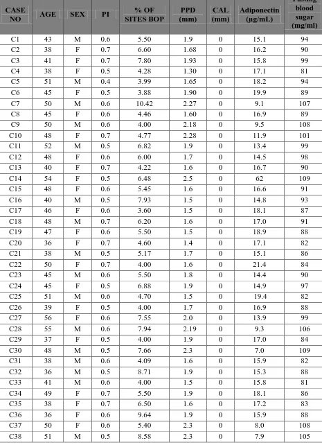
Table 1, 2 and 3 s hows the m aster chart of the cont rol group, stud y
group - I and II with the clini cal param et ers, fast ing blood sugar level
and s erum adi ponecti n l evel .
Table 4 and Figure 4 shows the comparison of age between the study and control
group. The mean age in the study group was 46 years and 41 years in the control
group respectively. There was no s tat isti cal si gni fi cant difference in t he
distribut ion of s ex in both the groups.
Table 5 and Figure 5 shows the comparison of gender distribution between the study
and control group. The males constituted 46% while females constituted 54% in the
study group. Both males and females constituted about 44% and 56% respectively in
the control group. There was no st atisti cal s i gnificant di fference in
49 Table 6 and Figure 6 shows the comparison of clinical parameter mean Plaque Index
between the study group I and II. The mean Plaque Index score in the study group-I
was 2.494±0.147 and 1.187 ±0.079 in the study group-II, which was statistically
highly significant (p=0.000).
Table 6 and Figure 7 shows the comparison of clinical parameter mean % of sites
with Bleeding on Probing between the study group I and II. The mean % of sites with
Bleeding on Probing was 90.407±5.385 in the study group-I and 8.563±3.289 in the
study group-II, which was statistically highly significant (p=0.000).
Table 7 and Figure 8 shows the comparison of mean adiponectin levels between the
study group I and II. The mean adiponectin levels in serum were 7.52±2.96 µg/mL i