0022-538X/95/$04.0010
Copyright 1995, American Society for Microbiology
Lactate Dehydrogenase-Elevating Virus Replication Persists in Liver,
Spleen, Lymph Node, and Testis Tissues and Results in
Accumulation of Viral RNA in Germinal Centers,
Concomitant with Polyclonal Activation of B Cells
GRANT W. ANDERSON, RAYMOND R. R. ROWLAND,† GENE A. PALMER,
CHEN EVEN,ANDPETER G. W. PLAGEMANN*
Department of Microbiology, University of Minnesota, Minneapolis, Minnesota 55455
Received 9 January 1995/Accepted 8 May 1995
Lactate dehydrogenase-elevating virus (LDV) replicates primarily and most likely solely in a subpopulation of macrophages in extraneuronal tissues. Infection of mice, regardless of age, with LDV leads to the rapid cytocidal replication of the virus in these cells, resulting in the release of large amounts of LDV into the circulation. The infection then progresses into life-long, asymptomatic, low-level viremic persistence, which is maintained by LDV replication in newly generated LDV-permissive cells which escapes all antiviral immune responses. In situ hybridization studies of tissue sections of adult FVB mice revealed that by 1 day postinfection (p.i.), LDV-infected cells were present in practically all tissues but were present in the highest numbers in the lymph nodes, spleen, and skin. In the central nervous system, LDV-infected cells were restricted to the leptomeninges. Most of the infected cells had disappeared at 3 days p.i., consistent with the cytocidal nature of the LDV infection, except for small numbers in lymph node, spleen, liver, and testis tissues. These tissues harbored infected cells until at least 90 days p.i. The results suggest that the generation of LDV-permissive cells during the persistent phase is restricted to these tissues. The continued presence of LDV-infected cells in testis tissue suggests the possibility of LDV release in semen and sexual transmission. Most striking was the accumulation of large amounts of LDV RNA in newly generated germinal centers of lymph nodes and the spleen. The LDV RNA was not associated with infected cells but was probably associated with virions or debris of infected, lysed cells. The appearance of LDV RNA in germinal centers in these mice coincided in time with the polyclonal activation of B cells, which leads to the accumulation of polyclonal immunoglobulin G2a and low-molecular-weight immune complexes in the circulation.
Lactate dehydrogenase-elevating virus (LDV) of mice, equine arteritis virus, simian hemorrhagic fever virus, and por-cine reproductive and respiratory syndrome virus constitute a new group of enveloped positive-stranded RNA viruses (for reviews, see references 24, 26, 27, 30, and 31). The genome
organization and replication of these viruses via a 39
-cotermi-nal nested set of subgenomic mRNAs resemble those of coro-naviruses. However, the genomes of these viruses (13 to 15 kb) are much smaller than those of coronaviruses, and so are the sizes of their virions (50 to 60 nm in diameter). The virions possess four structural proteins: a 12- to 13-kDa nucleocapsid protein (N/VP-1), an 18- to 19-kDa nonglycosylated envelope
protein (M/VP-2), a major envelope glycoprotein (VP-3/GL),
and probably a minor envelope glycoprotein (GS [12]).
VP-3/GLis heterogeneous in size (25 to 42 kDa) because of varying
degrees of glycosylation, and it seems to function as a virus attachment protein, since all neutralizing antibodies are di-rected to this protein (14, 27).
Viruses of this group have other properties in common. One is that macrophages are the primary or only host cells support-ing the replication of these viruses in the host animal (24, 26, 30). Another is that they can cause persistent infections in their
hosts. LDV replicates in a nonessential subpopulation of mac-rophages, and the infection is cytocidal. Upon an initial infec-tion of a mouse, LDV productively infects and destroys all permissive macrophages during the first 2 days postinfection (p.i.), resulting in the accumulation of large amounts of
infec-tious virus in the blood: up to 1010 50% infectious doses
(ID50)/ml at 1 day p.i. (24, 26). Thereafter, a life-long,
asymp-tomatic infection which is associated with a continuous viremia
of 105 to 106 ID
50/ml develops. The persistent infection is
maintained by the productive infection of new permissive mac-rophages that become continuously regenerated in the mouse from apparently nonpermissive precursor cells and escape of LDV replication from all host defense mechanisms (24, 27). Although no clinical symptoms are associated with LDV infec-tions, they bring about a permanent polyclonal activation of B cells that result in elevated plasma immunoglobulin G2a (IgG2a) or IgG2b concentrations (10, 11, 21, 26, 27) and pro-duction of autoantibodies (6, 26, 35) and immune complexes (5, 27, 29).
The present study was designed to inquire into the nature and location of the cells supporting the persistent infection and the mechanism of the associated polyclonal activation of B cells. Earlier immunohistocytochemical staining of tissue sec-tions had shown that at 1 day p.i., LDV antigen-positive cells were present in all tissues examined but that at 2 to 3 days p.i., all infected cells had disappeared (18, 28), consistent with the cytocidal nature of the LDV infection (18, 24, 26). However, the regional distribution and density of infected cells in the various tissues could not be deduced from the cryostat sections
* Corresponding author. Mailing address: Department of Microbi-ology, University of Minnesota, Box 196 UMHC, 420 Delaware St. S.E., Minneapolis, MN 55455. Phone: (612) 624-3187. Fax: (612) 626-0623.
† Present address: Department of Biology and Microbiology, South Dakota State University, Brookings, SD 57007-2142.
5177
on November 9, 2019 by guest
http://jvi.asm.org/
analyzed and the method was not sensitive enough to detect the low number of LDV-infected cells in persistently infected mice. We have estimated that the productive infection of 100 to 1,000 new permissive macrophages in a mouse per day is sufficient to account for the level of viremia in persistently infected mice (26). Therefore, we approached these questions using a more sensitive method: in situ hybridization.
FVB mice, 4 to 8 weeks of age, were supplied by the Uni-versity of Minnesota Division of Research Animal Resources
and were infected intraperitoneally with 106 ID
50 of LDV
strain P as required (4). Plasma was obtained from mice by retro-orbital bleeding, and LDV concentrations were deter-mined by an endpoint dilution (25). Tissues were removed from phosphate-buffered saline-perfused mice, fixed in
forma-lin, embedded in paraffin, and sectioned (8-mm-thick sections
for the brain and spinal cord and 6-mm-thick sections for all
other tissues) as described previously (1). The sections were
hybridized with 35S-labeled LDV-specific cDNA 4-55 as
de-scribed by Blum et al. (3) and Anderson et al. (1). cDNA 4-55
(437 bp) represents the 39end of the LDV genome (8) and was
radiolabeled by random priming with a Random Primed DNA Labeling Kit from Boehringer Mannheim (Indianapolis, Ind.)
and [a-35S]dCTP according to the procedure recommended by
the manufacturer of the kit. After hybridization, the slides were autoradiographed, stained with Mayer’s hematoxylin and eosin Y, and examined with a Leitz microscope (1). The cDNA probe hybridizes with both LDV genomic RNA and all sub-genomic mRNAs (8), but since the subsub-genomic mRNAs are present in infected macrophages far in excess of the amount of genomic RNA, the probe specifically detects LDV-infected cells in tissue sections (1, 2).
In a recent study, we demonstrated that at 1 day p.i. many LDV-infected cells are localized in macrophage-rich areas of many tissues of AKXD-16 mice, including the peritoneal cav-ity, bone marrow, skeletal muscle, thymus, liver, adipose tissue, spleen, and lymph nodes (2). In the present study, we have extended these studies to FVB mice, to additional tissues, and to persistently infected mice. At 1 day p.i., a small number of LDV-infected cells were found in kidney (Fig. 1A) and pan-creas (Fig. 1C) tissues and numerous infected cells were found in the leptomeninges of the brain (Fig. 1E) and of the spinal cord (data not shown), in the lung (Fig. 1G), and in the retic-ular layer of the skin (Fig. 1H). LDV-infected cells were also present in skeletal muscle and adipose tissue (data not shown). The results are in agreement with those observed for AKXD-16 mice (2). On the other hand, at 3, 7, and 90 days p.i. no infected cells could be detected in most of the tissues cited above. This is illustrated for kidney tissue from a 7-day-in-fected mouse (Fig. 1B), for leptomeninges from a 3-day-in-fected mouse (Fig. 1F), and for pancreas tissue from a 90-day-infected mouse (Fig. 1D). The results suggest that during the persistent phase little, if any, LDV replication occurs in most tissues that initially harbor LDV-permissive macrophages which become infected and destroyed during the first 1 to 2 days p.i. The LDV titer in plasma of 1-day-LDV-infected FVB
mice was 109.0to 109.5ID
50/ml, and at 3, 7, and 30 days p.i. this
had decreased to 108.0, 107.5, and 106.5ID
50/ml, respectively.
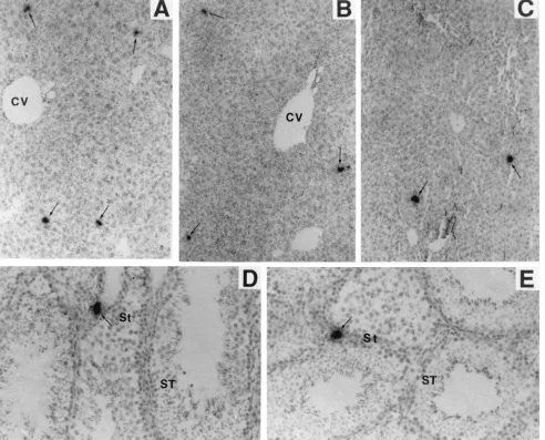
The only tissues in which we consistently have found cells productively infected with LDV were liver, spleen, lymph node, and testis tissues. Fig. 2A–C illustrates typical sections of livers from mice infected with LDV for 1, 7, and 90 days. Compared with numbers in the leptomeninges, lungs, skin (Fig. 1), lymph nodes, and spleen (see below), relatively few infected cells were present in the liver at 1 day p.i. However, the density and distribution of infected cells remained about the same, at least until 90 days p.i. (Fig. 2C). Since the grain density over the cells was comparable to that for positive cells in all tissues at 1 day p.i. and most of the LDV RNA in infected cells seems to represent the subgenomic mRNAs, it seems likely that the LDV RNA-containing cells in the liver were productively infected. This conclusion implies that some new LDV-permissive cells continuously arise or localize in the liver. The same conclusion applies to testis tissue (Fig. 2D and E). A small number of LDV-infected cells were consistently found in testis tissue in proximity to seminiferous tubules, where sperm are produced. This implies that infectious LDV might be con-tinuously released in semen (see below).
Massive LDV replication occurred in lymph nodes during the first day p.i. (Fig. 3). This was also indicated by previous electron microscope studies which suggested the presence of large numbers of virus-like particles in lymph nodes from 1-day-LDV-infected mice (30, 32). At this time, LDV-infected cells were found primarily in the marginal zone (Fig. 3A and C) but also interspersed through the paracortex (Fig. 3C) and throughout the capsule (Fig. 3A and G). Dark-field images greatly accentuated the recognition of silver grains over in-fected cells (Fig. 3B, D, and H). The radioactivity signal was completely abolished by treatment of sequential lymph node
sections with RNases A and T1 (Fig. 3F), and none was
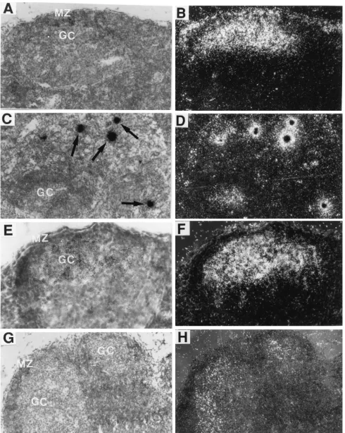
de-tected in lymph nodes from uninfected mice (Fig. 3E). By 3 days p.i., most of the LDV-infected cells in the marginal zone of the lymph nodes had disappeared (Fig. 4A and B). Instead, LDV RNA began to accumulate in germinal centers (Fig. 4A and B). In agreement with what was observed in earlier studies (30), these germinal centers were generated after LDV infection, since few germinal centers were detected in the lymph nodes of companion uninfected mice (data not shown). At times subsequent to 3 days p.i., no infected cells were detectable in the marginal zone (Fig. 4E and F) but a few LDV-infected cells continued to be present in the paracortex next to the germinal centers (Fig. 4C and D). However, the largest amounts of LDV RNA were located within germinal centers (Fig. 4E and F). The LDV RNA in the germinal cen-ters was clearly not present in LDV-infected cells such as those seen in the paracortex (Fig. 4C and D). Instead, it was diffusely distributed (Fig. 4E and F) and thus probably associated with LDV virions or debris from infected cells that became trapped in the germinal centers. Practically every germinal center con-tained a large amount of LDV RNA. Most of the diffusely distributed LDV RNA in the germinal centers was removed by
FIG. 1. Distribution of LDV-infected cells in various tissues from 4-week-old FVB mice infected with LDV for 1, 3, 7, and 90 days. Sections of formalin-fixed, paraffin-embedded tissues were hybridized with a35
S-labeled, LDV-specific probe (cDNA 4-55), washed, coated with photographic emulsion, exposed for 1 to 3 days, and thereafter stained with hematoxylin and eosin. Foci of autoradiographic grains that were detected in sequential sections were scored as LDV-infected cells (arrows). Sections from tissues of at least three LDV-infected mice and one uninfected mouse were analyzed on the same slide. Duplicate slides were treated with RNases A and T1before hybridization. No foci of autoradiographic grains were detected in the RNase-treated sections or sections from uninfected mice (data not shown). Stated
magnifications pertain to the microscope. Photographic enlargements are the same for all panels. (A and B) Kidney tissues from 1- and 7-day-infected mice, respectively. CT, collecting tubule. Magnification,3100. (C and D) Pancreas tissues from 1- and 90-day-infected mice, respectively. IL, islet of Langerhans. Magnification,3200. (E and F) Brain tissues from 1- and 3-day-infected mice, respectively. Le, leptomeninges. Magnification,3200. (G) Lung tissue from a 1-day-infected mouse. TB, terminal bronchioles. Magnification,3100. (H) Skin from a 1-day-infected mouse. RL, reticular layer. Magnification,3100.
5178 NOTES J. VIROL.
on November 9, 2019 by guest
http://jvi.asm.org/
on November 9, 2019 by guest
http://jvi.asm.org/
RNase A and T1treatment, but small amounts remained (Fig.
4G and H), perhaps being protected in virions.
The situation in the spleen was very similar to that in lymph nodes (Fig. 5). At 1 day p.i., large numbers of LDV-infected cells were present in the marginal zone between red and white pulp (Fig. 5A–D). By 3 days p.i., most of these infected cells had disappeared while LDV RNA accumulated in the newly formed germinal centers, generally at the edges of the germi-nal centers (Fig. 5E and F). The accumulation of LDV virions in the germinal centers persisted at least until 90 days p.i. (Fig. 5G and H). As in the lymph nodes, most of the LDV RNA in germinal centers was removed by treatment of the sections
with RNases A and T1(Fig. 5I and J).
Our results have led to several novel conclusions concerning the persistent infection of mice with LDV. First, the persistent infection is maintained by LDV replication in only a few tis-sues. Second, and related, is the finding that LDV replication persists in testis tissue, which might play an important role in its transmission in nature. Third, during the persistent phase
large amounts of viral material accumulate in the new germinal centers of the spleen and lymph nodes, which might explain some of the effects of LDV infection on the host immune system.
[image:4.612.64.556.74.471.2]The level of LDV viremia in persistently infected mice re-flects an equilibrium between LDV production resulting from infection of newly generated LDV-permissive cells and LDV clearance (26, 27). Clearance is clearly not immune mediated since the time course and level of LDV viremia are the same whether or not the mice mount anti-LDV immune responses. They are the same in nude mice or neonatally infected, toler-ized mice that fail to mount anti-LDV immune responses as they are in immunocompetent littermates of these mice (23, 29). Most likely clearance reflects physical inactivation of the virions in the circulation (27). LDV replication during the persistent phase seems limited only by the availability in the host of LDV-permissive cells, which become regenerated only slowly. Our present results show that the cells supporting LDV infection during the persistent phase are restricted to liver,
FIG. 2. Persistent LDV infection of liver (A–C) and testis (D and E) cells of FVB mice. Tissue sections were hybridized with an LDV-specific probe as described in the legend to Fig. 1. Stated magnifications pertain to the microscope. Photographic enlargements are the same for all panels. (A–C) Liver tissues from 1-, 7-, and 90-day-infected mice, respectively. CV, central vein. Magnification,3100. (D and E) Testis tissues from 1- and 90-day-infected mice, respectively. St, stroma; ST, seminiferous tubules. Magnification,3200.
5180 NOTES J. VIROL.
on November 9, 2019 by guest
http://jvi.asm.org/
FIG. 3. LDV-infected cells in lymph nodes of 1-day-infected FVB mice. Lymph node sections were hybridized with an LDV-specific probe as described in the legend to Fig. 1. Stated magnifications pertain to the microscope. Photographic enlargements are the same for all panels. (A and B) MZ, marginal zone; MC, medullary cord; Ca, capsule; PC, paracortex; C, cortex. Magnification,350. (C and D) Magnification,3200. (E) Section of lymph node of uninfected mouse; dark-field image. Magnification,3100. (F) RNase-treated dark-field image. Magnification,3100. (G and H) Capsule. Magnification,3200. Panels B, D, and H are dark-field images of the microscope fields shown in panels A, C, and G, respectively.
on November 9, 2019 by guest
http://jvi.asm.org/
FIG. 4. Lymph nodes from persistently infected FVB mice. The sections were hybridized with an LDV-specific probe as described in the legend to Fig. 1. Panels A, C, E, and G are bright-field images and frames B, D, F, and H are dark-field images of the same microscopic fields. Stated magnifications pertain to the microscope. Photographic enlargements are the same for all panels. (A and B) Three-day-infected mice. MZ, marginal zone; GC, germinal center. Magnification,3200. (C and D) Seven-day-infected mouse. Arrows indicate infected cells. Magnification,3200. (E and F) Ninety-day-infected mouse. Magnification,3400. (G and H) Ninety-day-infected mouse; RNase A- and T1-treated section. Magnification,3200.
5182 NOTES J. VIROL.
on November 9, 2019 by guest
http://jvi.asm.org/
FIG. 5. LDV-infected cells in spleens of FVB mice infected with LDV for 1 (A–D), 3 (E and F), and 90 (G–J) days. The sections were hybridized with an LDV-specific probe. Panels A, C, E, G, and I are bright-field images and frames B, D, F, H, and J are dark-field images of the same microscopic fields. WP and RP, white and red pulp, respectively; MZ, marginal zone; GC, germinal center. I and J represent RNase A- and T1-treated sections. Magnification,3100. Magnification
pertains to the microscope. Photographic enlargements are the same for all panels.
5183
on November 9, 2019 by guest
testis, spleen, and lymph node tissues (Fig. 2, 4, and 5) and probably thymus tissue (29). The origin and nature of the new LDV-permissive cells are unproven. However, the cells are probably macrophages, since they are found in the same areas that harbor LDV-infected cells during the acute phase of in-fection at 1 day p.i. (Fig. 1 and 3). These cells probably become LDV permissive as the result of a step in differentiation that results in the expression of a surface component acting as an LDV receptor (24, 26).
Of special interest is the persistent presence of LDV-in-fected cells in areas of sperm formation in the testis (Fig. 2D and E), which should result in the continuous release of infec-tious virions in semen. It follows that LDV may be sexually transmitted. The presence of LDV in semen has not been directly demonstrated, because of technical difficulties in ob-taining semen from mice (19), but the presence of both equine arteritis virus and porcine reproductive and respiratory syn-drome virus in semen and their sexual transmission have been proven (22, 24, 33). Thus, transmission via semen might be a common route of transmission of this group of viruses. In fact, infections of horses by EAV worldwide seem to be primarily generated via semen from persistently infected stallions (33). Sexual transmission of LDV might be responsible for the en-demic infections observed to occur in wild house mouse pop-ulations, since LDV is transmitted only poorly, if at all, via oral or intranasal routes, even though the virus is secreted by in-fected mice in feces, urine, and saliva (7, 24, 27).
The accumulation of LDV RNA in the germinal centers of the spleen and lymph nodes (Fig. 4 and 5) that are generated shortly after LDV infection correlates in time with anti-LDV antibody formation, as well as with the initiation of the poly-clonal activation of B cells (24, 27). Germinal-center hyperpla-sia becomes apparent within 2 to 3 days p.i. and is associated with splenomegaly and lymph node enlargement (30). The LDV RNA is probably associated with virions released from the large number of macrophages infected initially or with debris of infected and lysed cells. LDV virions are probably trapped by the follicular dendritic cells of the germinal centers via interaction with complement receptors or as immune com-plexes. During the persistent phase of infection, practically all LDV virions are associated with antibodies in infectious anti-body-virus complexes (5, 24, 27, 29, 31). It stands to reason that the production of very large amounts of LDV antigen in the marginal zones of the spleen and lymph nodes, the formation of germinal centers, the accumulation of LDV virions in these germinal centers, the initiation of anti-LDV immune re-sponses, and the polyclonal activation of B cells are causally related processes. The polyclonal activation of B cells might be induced by the large amounts of LDV antigens accumulating in the lymphoid tissues or by an LDV protein (17). The differ-entiation of the polyclonally activated B cells to IgG2a-produc-ing plasma cells in the germinal centers is then probably
in-duced by gamma interferon (IFN-g) produced by activated
Th1 cells, cytotoxic T lymphocytes, or NK cells (9, 27). Indeed,
we have found that the ability of spleen cells to produce IFN-g
in vitro in response to treatment with concanavalin A increases progressively beginning 3 to 4 days after infection with LDV
and that concomitantly increasing amounts of IFN-g mRNA
are detectable in the spleen (27, 28a). In the LDV-infected FVB mice, levels of IgG2a and low-molecular-weight immune complexes in the plasma began to increase about 4 days p.i. and reached maximum levels about 10 to 20 days p.i. (data not shown). The plasma IgG2a concentration increased from about 0.4 mg/ml to a maximum of 1.8 mg/ml.
The events associated with a persistent LDV infection of mice strongly resemble those which are observed to occur in
humans during the asymptomatic persistent phase of infection with human immunodeficiency virus (13, 15, 16, 20, 34) and which most likely occur in other asymptomatic persistent virus infections with associated viremia. These events include (i) continuous cycles of viral replication in a renewable population of cells, (ii) escape from antiviral humoral and cellular immune responses, (iii) accumulation of virus in germinal centers con-comitant with a polyclonal activation of B cells, and (iv) a potential sexual transmission of the infection as well as a trans-placental infection of the fetus from an infected mother.
G.W.A. and R.R.R.R. contributed about equally to this work. We thank Colleen O’Neill for competent secretarial help. R.R.R.R., G.W.A., and G.A.P. were supported by training grant CA 09138 from the National Institutes of Health, and R.R.R.R. was also supported by a postdoctoral fellowship from the American Cancer Society.
REFERENCES
1. Anderson, G. W., G. A. Palmer, R. R. R. Rowland, C. Even, and P. G. W. Plagemann.1995. Infection of central nervous system cells by ecotropic murine leukemia virus in C58 and AKR mice and in in utero-infected CE/J mice predisposes mice to paralytic infection by lactate dehydrogenase-ele-vating virus. J. Virol. 69:308–319.
2. Anderson, G. W., G. A. Palmer, R. R. R. Rowland, C. Even, and P. G. W. Plagemann.1995. Lactate dehydrogenase-elevating virus entry into the cen-tral nervous system and replication in anterior horn neurons. J. Gen. Virol. 76:581–592.
3. Blum, H. E., A. T. Haase, and G. N. Vyas. 1984. Molecular pathogenesis of hepatitis B infection: simultaneous detection of viral DNA and antigen in paraffin-embedded liver sections. Lancet ii:771–775.
4. Brinton-Darnell, M., and P. G. W. Plagemann. 1975. Structure and chemi-cal-physical characteristics of lactate dehydrogenase-elevating virus and its RNA. J. Virol. 16:420–433.
5. Cafruny, W. A., S. P. K. Chan, J. T. Harty, S. Yousefi, K. Kowalchyk, D. McDonald, B. Foreman, G. Budweg, and P. G. W. Plagemann.1986. Anti-body response of mice to lactate dehydrogenase-elevating virus during in-fection and immunization with inactivated virus. Virus Res. 5:357–375. 6. Cafruny, W. A., and D. E. Hovinen. 1988. Infection of mice with lactate
dehydrogenase-elevating virus leads to stimulation of autoantibodies. J. Gen. Virol. 69:723–729.
7. Cafruny, W. A., and D. E. Hovinen. 1988. The relationship between route of infection and minimum infectious dose: studies with lactate dehydrogenase-elevating virus. J. Virol. Methods 20:265–270.
8. Chen, Z., L. Kuo, R. R. R. Rowland, C. Even, K. S. Faaberg, and P. G. W. Plagemann.1993. Sequence of 39end of genome and of 59end of ORF 1a of lactate dehydrogenase-elevating virus (LDV) and common junction mo-tifs between 59-leader and bodies of seven subgenomic mRNAs. J. Gen. Virol. 74:643–660.
9. Clark, E. A., and J. A. Ledbetter. 1994. How B and T cells talk to each other. Nature (London) 367:425–428.
10. Coutelier, J.-P., P. G. Coulie, P. Wauters, H. Heremans, and J. T. M. van der Logt.1990. In vivo polyclonal B-lymphocyte activation elicited by murine viruses. J. Virol. 64:5383–5388.
11. Coutelier, J.-P., and J. Van Snick. 1985. Isotypically restricted activation of B lymphocytes by lactic dehydrogenase virus. Eur. J. Immunol. 15:250–255. 12. deVries, A. A. F., E. D. Chirnside, M. C. Horzinek, and P. J. M. Rottier. 1992.
Structural proteins of equine arteritis virus. J. Virol. 66:6294–6303. 13. Embretson, J., M. Zupancic, J. L. Ribas, A. Burke, P. Racz, K. Tenner-Racz,
and A. T. Haase.1993. Massive covert infection of helper T lymphocytes and macrophages by HIV during the incubation period of AIDS. Nature (Lon-don) 362:359–362.
14. Faaberg, K. S., and P. G. W. Plagemann. The envelope proteins of lactate dehydrogenase-elevating virus and their topography in membranes. Virol-ogy, in press.
15. Fauci, A. S. 1993. Multifactorial nature of human immunodeficiency virus disease: implications for therapy. Science 262:1011–1016.
16. Ho, D. D., A. U. Neumann, A. S. Perelson, W. Chen, J. M. Leonard, and M. Markowitz.1995. Rapid turnover of plasma virions and CD4 lymphocytes in HIV-1 infection. Nature (London) 373:123–126.
17. Hodgkin, P. D., and A. Basten. 1995. B cell activation, tolerance and antigen-presenting function. Curr. Opin. Immunol. 7:121–129.
18. Inada, T., and C. A. Mims. 1985. Pattern of infection and selective loss of Ia-positive cells in suckling and adult mice inoculated with lactic dehydro-genase virus. Arch. Virol. 86:151–165.
19. King, W. W., L. G. St. Amant, and W. R. Lee. 1994. A technique for serial spermatozoa collection in mice. Lab. Anim. Sci. 44:295–296.
20. Koup, R. A., and D. D. Ho. 1994. Shutting down HIV. Nature (London) 370:416.
5184 NOTES J. VIROL.
on November 9, 2019 by guest
http://jvi.asm.org/
21. Li, X., B. Hu, J. T. Harty, C. Even, and P. G. W. Plagemann. 1990. Polyclonal B cell activation of IgG2a and IgG2b production by infection of mice with lactate dehydrogenase-elevating virus is mediated by CD41lymphocytes. Viral Immunol. 3:273–288.
22. Meredith, M. J. 1993. Porcine reproductive and respiratory syndrome, 7th ed. University of Cambridge Press, Cambridge.
23. Onyekaba, C., J. T. Harty, C. Even, B. Hu, and P. G. W. Plagemann. 1989. Persistent infection of mice by lactate dehydrogenase-elevating virus. Effects of immunosuppression on virus replication and antiviral immune responses. Virus Res. 14:297–316.
24. Plagemann, P. G. W. Lactate dehydrogenase-elevating virus and related viruses. In B. N. Fields, D. M. Knipe, and D. M. Howley (ed.), Virology, in press. Raven Press, New York.
25. Plagemann, P. G. W., K. G. Gregory, H. E. Swim, and K. K. W. Chan. 1963. Plasma lactate dehydrogenase-elevating agent of mice: distribution in tissues and effect on lactate dehydrogenase isozyme patterns. Can. J. Microbiol. 9:75–86.
26. Plagemann, P. G. W., and V. Moennig. 1992. Lactate dehydrogenase-elevat-ing virus, equine arteritis virus and simian hemorrhagic fever virus: a new group of positive strand RNA viruses. Adv. Virus Res. 41:99–190. 27. Plagemann, P. G. W., R. R. R. Rowland, C. Even, and K. S. Faaberg. Lactate
dehydrogenase-elevating virus—an ideal persistent virus. In Seminars in immunopathology, in press. Springer Verlag, New York.
28. Porter, D. D., H. G. Porter, and B. B. Deerhake. 1969. Immunofluorescence assay for antigen and antibody in lactic dehydrogenase virus infection of
mice. J. Immunol. 102:431–436.
28a.Rowland, R. R. R., et al. Unpublished data.
29. Rowland, R. R. R., C. Even, G. W. Anderson, Z. Chen, B. Hu, and P. G. W. Plagemann.1994. Neonatal infection of mice with lactate dehydrogenase-elevating virus results in suppression of humoral antiviral immune response but does not alter the course of viraemia or the polyclonal activation of B cells and immune complex formation. J. Gen. Virol. 75:1071–1081. 30. Rowson, K. E. K., and B. W. J. Mahy. 1975. Lactic dehydrogenase virus.
Virology monograph, vol. 13. Springer-Verlag, New York.
31. Rowson, K. E. K., and B. W. J. Mahy. 1985. Lactate dehydrogenase-elevating virus. J. Gen. Virol. 66:2297–2312.
32. Snodgrass, M. J., and M. G. Hanna. 1970. Histoproliferative effect of Raus-cher leukemia virus in lymphatic tissue. III. Alteration in the thymic depen-dent area induced by the passenger virus lactic dehydrogenase virus. J. Natl. Cancer Inst. 45:741–759.
33. Timoney, P. J., and W. H. McCollum. 1993. Equine viral arteritis. Vet. Clin. North Am. Equine Pract. 9:295–309.
34. Wei, X., S. K. Ghosh, M. E. Taylor, V. A. Johnson, E. A. Emini, P. Deutsch, J. D. Lifson, S. Bonhoeffer, M. A. Nowak, B. H. Hahn, M. S. Saag, and G. M. Shaw.1995. Viral dynamics in human immunodeficiency virus type 1 infec-tion. Nature (London) 373:117–122.
35. Weiland, E., F. Weiland, and A. Grossmann. 1987. Lactate dehydrogenase-elevating virus induces anti-Golgi apparatus antibodies. J. Gen. Virol. 68: 1983–1991.