0022-538X/97/$04.0010
Copyright © 1997, American Society for Microbiology
Localization of the Labile Disulfide Bond between SU and TM
of the Murine Leukemia Virus Envelope Protein Complex to a
Highly Conserved CWLC Motif in SU That Resembles the
Active-Site Sequence of Thiol-Disulfide Exchange Enzymes
ABRAHAM PINTER,1,2* REBECCA KOPELMAN,1ZHIYONG LI,2† SAMUEL C. KAYMAN,1
ANDDAVID AVRAM SANDERS3
Laboratory of Retroviral Biology, Public Health Research Institute,1and Department of Microbiology,
New York University School of Medicine,2New York, New York 10016, and Department of
Bilogical Sciences, Purdue University, West Lafayette, Indiana 479073 Received 27 November 1996/Accepted 16 July 1997
Previous studies have indicated that the surface (SU) and transmembrane (TM) subunits of the envelope protein (Env) of murine leukemia viruses (MuLVs) are joined by a labile disulfide bond that can be stabilized by treatment of virions with thiol-specific reagents. In the present study this observation was extended to the Envs of additional classes of MuLV, and the cysteines of SU involved in this linkage were mapped by proteolytic fragmentation analyses to the CWLC sequence present at the beginning of the C-terminal domain of SU. This sequence is highly conserved across a broad range of distantly related retroviruses and resembles the CXXC motif present at the active site of thiol-disulfide exchange enzymes. A model is proposed in which rearrange-ments of the SU-TM intersubunit disulfide linkage, mediated by the CWLC sequence, play roles in the assembly and function of the Env complex.
Enveloped mammalian viruses generally enter cells via the fusion of viral and cellular membranes. Many viral proteins that mediate fusion are synthesized as polyprotein precursors that are processed by proteolytic cleavage into two subunits, only one of which is an integral membrane protein. These subunits are frequently linked by disulfide bonds. Examples of disulfide-linked viral envelope protein (Env) complexes in-clude the hemagglutinins of orthomyxoviruses (42) and rubella virus (43), the fusion proteins of paramyxoviruses (15, 36), parainfluenza virus (39) and pneumoviruses (respiratory syn-cytial virus) (12), and the gB proteins of cytomegalovirus (11) and other herpesviruses (26, 41). A potential advantage of this strategy is that it allows the activation of a cryptic fusogenic domain by proteolytic cleavage at a relatively late stage in the assembly process and provides for a covalent linkage between the cleaved subunits that may stabilize the surface protein complex.
Retroviral Envs are expressed as glycosylated polyproteins that are processed intracellularly by proteolytic cleavage into two subunits, a surface protein (SU), which bears the receptor-binding activity of the virus, and a transmembrane protein (TM), which is believed to mediate membrane fusion. A disul-fide linkage between SU and TM has been demonstrated for Rous sarcoma virus (19), several strains of ecotropic and dual-tropic murine leukemia viruses (MuLV) (20, 27, 28, 30, 31, 45), and feline leukemia virus (FeLV) (29). The intersubunit cys-tine bridge of MuLV and FeLV Envs differs from that of other viral Envs in that it is labile. Whereas all of Rous sarcoma virus Env is recovered as the disulfide-linked complex after the sol-ubilization of viral particles, only a fraction of the MuLV and
FeLV Envs was found as disulfide-linked heterodimers after the solubilization or denaturation of virions. This fraction var-ied among different strains of ecotropic MuLV and increased when virions were pretreated with thiol-reactive reagents, such as dithiobis(m-nitropyridine) or N-ethyl maleimide (NEM) (33). This suggested that the disulfide bond involved in the intersubunit linkage was stabilized by the blockage of one or more free thiols present in intact virions.
In this study we show that a labile SU-TM disulfide linkage is present in the Env complexes of previously unexamined classes of MuLV. In addition, the cysteine residue(s) of eco-tropic MuLV SU involved in the intersubunit linkage are lo-calized to the Cys-Trp-Leu-Cys (CWLC) motif in SU. This motif is broadly conserved in retroviral SUs and resembles the sequence found at the active site of a family of proteins in-volved in thiol-disulfide exchange reactions.
A labile SU-TM disulfide bond is present in ecotropic,
xe-notropic, and amphotropic strains of MuLV.The presence of
a labile disulfide bond between SU (gp70) and TM (p15E) was previously demonstrated for ecotropic and dual-tropic strains of MuLV. Analyses were performed to determine whether a similar linkage occurred between the Env subunits of xeno-tropic and amphoxeno-tropic MuLVs. Radiolabeled preparations of xenotropic (NZB-345), amphotropic (4070), and control eco-tropic (Friend and Moloney) MuLVs were pelleted and puri-fied by gel chromatography. Nonidet P-40 (NP-40) lysates of purified virions, with and without treatment of virions with 8.0 mM NEM prior to lysis, were examined by immunoprecipita-tion with polyclonal anti-gp70 antiserum followed by sodium dodecyl sulfate-polyacrylamide gel electrophoresis (SDS-PAGE) under both reducing and nonreducing conditions (Fig. 1).
Similar results were obtained for all four viruses. Without NEM pretreatment, almost all gp70 was recovered in uncom-plexed form under both reducing and nonreducing conditions. For virions treated with NEM, a large fraction of the material * Corresponding author. Mailing address: Public Health Research
Institute, 455 First Ave., New York, NY 10016. Phone: (212) 578-0879. Fax: (212) 576-8420. E-mail: pinter@phri.nyu.edu.
† Present address: Dept. of Medicine, L. I. Medical Center, New Hyde Park, NY 11042.
8073
on November 9, 2019 by guest
http://jvi.asm.org/
immunoprecipitated by anti-gp70 serum had an apparent mo-lecular mass of 90 kDa under nonreducing conditions, consis-tent with the expected size of the disulfide-linked gp70-p15E complex; reduction with dithiothreitol (DTT) converted the 90-kDa material to free gp70 and p15E. Parallel immunopre-cipitations showed that the 90-kDa forms, but not the 70-kDa forms, were recognized by an anti-p15E antibody (not shown), confirming that the lower mobility band represented disulfide-linked complexes of gp70 and p15E. Together with previously published data on dual-tropic viruses, the results of these ex-periments showed that following treatment of viral particles with the thiol-blocking agent, NEM, the predominant form of Env complex was a stable disulfide-linked heterodimer in all four classes of MuLV.
Localization of gp70 cysteines involved in disulfide linkage
to p15E.Proteolytic fragmentation experiments were used to
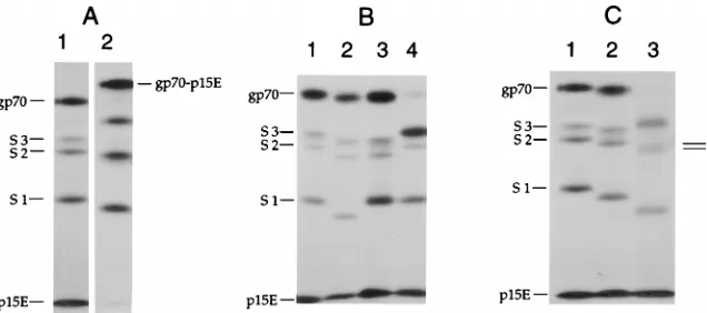
map the cysteines in gp70 involved in the disulfide linkage to p15E. Purified [35S]methionine-labeled Friend ecotropic MuLV particles were treated with NEM and solubilized with
NP-40, and fragments of gp70 were generated by proteolysis with subtilisin in the presence of excess goat anti-gp70 serum. Antibody-complexed fragments were then immunoprecipi-tated and analyzed by SDS-PAGE. The addition of anti-gp70 antibodies during the protease digestion step limited the ac-cessibility of potential proteolysis sites, resulting in the gener-ation of a reproducible pattern of discrete fragments. Along with uncleaved gp70 and p15E, three large gp70 fragments were detected, with apparent molecular masses of 53 kDa (S3), 50 kDa (S2), and 37 kDa (S1) (Fig. 2A). These represented N-terminal gp70 fragments, since Friend gp70 contains a single methionine at position 47 (18). The mobilities of the two smaller components, S2 and S1, increased slightly when ana-lyzed under nonreducing conditions, consistent with a globular structure maintained by internal disulfide bonds. In contrast, the mobilities of both gp70 and the S3 fragment decreased when the reducing agent was omitted, consistent with an ap-parent increase in molecular mass of approximately 20 kDa. The larger band had the expected mobility of the gp70-p15E disulfide-linked complex, and the p15E band was greatly di-minished in the nonreduced sample. These data indicated that intact gp70 and the S3 fragment were disulfide-linked to p15E, while the S2 and S1 fragments were not.
To delineate the region of gp70 present in S3, but not in S2, that contained the gp70 cysteine involved in the cystine bridge to p15E, subtilisin digests of mutant gp70s lacking specific N-linked glycans (17) were analyzed (Fig. 2B). Each of the mutant gp70s was;2 kDa smaller than wild-type gp70, con-sistent with the loss of one N-linked glycan, and they all yielded the same number of fragments after subtilisin treatment. For the gs12(N12D) mutant protein lacking the glycan normally present at Asn12 (Fig. 2B, lane 2), all three fragments had increased mobilities relative to those of the corresponding wild-type fragments (Fig. 2B, lane 1), indicating that all of the fragments contained position 12 and confirming that they were derived from the amino terminus of gp70. For the gs32 (N266D) mutant (Fig. 2B, lane 3) the S1 fragment had the same mobility as the wild-type S1 fragment, indicating that this fragment terminated before gs3, while the S2 and S3 fragments comigrated with the corresponding gs12fragments and were approximately 2 kDa smaller than the corresponding wild-type
FIG. 1. Demonstration of a liabile disulfide linkage between SU and TM proteins of various MuLV subtypes. [35S]cysteine-labeled virions of ecotropic Friend (lanes 1, 5, and 9), Moloney (lanes 2, 6, and 10), xenotropic NZB-345 (lanes 3, 7, and 11), and amphotropic 4070A (lanes 4, 8, and 12) strains of MuLV were purified, solubilized with NP-40, and immunoprecipitated with polyclonal anti-gp70 serum, as previously described (33). The first set of samples (lanes 1 to 4) was analyzed without prior treatment with NEM, while the last two sets (lanes 5 to 12) were treated with 8.0 mM NEM before solubilization. Immunoprecipi-tates were dissolved in SDS buffer and analyzed by SDS-PAGE without reduc-tion (lanes 1 to 8) or after reducreduc-tion with DTT (lanes 9 to 12). In all samples treated with NEM and analyzed under nonreducing conditions, the majority of the gp70 was present as a 90-kDa band corresponding to the gp70-p15E disulfide complex.
FIG. 2. (A) Analysis of proteolytic fragments of Friend MuLV (F-MuLV) gp70 covalently associated with p15E. Purified [35S]methionine-labeled virions were treated with 8 mM NEM, lysed with NP-40, and digested with subtilisin (50mg/ml) in the presence of a 1:10 dilution of polyclonal goat anti-gp70 serum (lot 79S-771, Microbiological Associates). The antibody-associated gp70 digestion products were immunoprecipitated with fixedStaphylococcus aureuscells (Pansorbin, La Jolla, Calif.) and analyzed by SDS-PAGE with (lane 1) and without (lane 2) reduction with DTT. (B) Analysis of subtilisin fragments of glycosylation site mutants of F-MuLV gp70. Subtilisin digestions were performed on gp70 from F-MuLV (lane 1) and F-MuLV mutants in which the glycosylation site at position 12 ([gs1] lane 2), 266 ([gs3] lane 3), or 334 ([gs5] lane 4) was removed by asparagine-to-aspartate mutation (17). gp70 fragments were immunoprecipitated with fixedS. aureuscells and analyzed by SDS-PAGE after reduction with DTT. (C) Effect of deglycosylation on mobilities of subtilisin fragments of F-MuLV gp70. Subtilisin fragments of wild-type Friend gp70 were prepared as in panel A and analyzed by SDS-PAGE under reducing conditions either directly (lane 1) or after digestion with endoglycosidase H (lane 2) orN-glycanase (lane 3), as previously described (30).
on November 9, 2019 by guest
http://jvi.asm.org/
[image:2.612.149.467.508.649.2]fragments. These data indicated that both the S2 and S3 frag-ments carried the gs3 glycan and thus were generated by cleav-age at a site C-terminal to position 266. All three gs52 (N334D) mutant fragments had mobilities similar to those of the corresponding wild-type fragments (Fig. 2B, lane 4), indi-cating that these fragments all terminated before position 334. Cleavage of gs52 gp70 to the S3 fragment was much more efficient than cleavage of the other gp70s, indicating that the gs5 glycan partially masked the cleavage site responsible for the generation of the S3 fragment.
The subtilisin fragments of wild-type gp70 were further char-acterized by digestion with endoglycosidases (Fig. 2C). After digestion with endo H, all three fragments shifted in mobility to an extent consistent with the loss of one N-linked glycan. This was due to the gs2 glycan at N168, which previous studies have shown is retained as an endo H-sensitive, high-mannose glycan (17), and indicates that the S1 fragment terminated between residues 168 and 266 of gp70. Upon removal of all N-linked glycans with N-glycanase, the S1, S2, and S3 frag-ments exhibited mobility shifts consistent with the loss of two, three, and four N-linked glycans, respectively. Thus, S3 carried the gs4 glycan present at position 302, while S2 did not. Fully deglycosylated S2 and S3 migrated as a closely spaced doublet, indicating that nearby cleavage sites on opposite sides of res-idue 302 were responsible for the formation of these two pro-teolytic products.
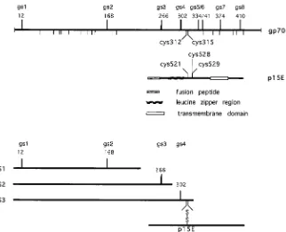
These results allowed the localization of the cysteines in gp70 involved in the disulfide linkage with p15E (Fig. 3). The S3 fragment was disulfide linked to p15E, while the S2 frag-ment was not. The glycosylation site mutant and endoglycosi-dase analyses showed that S2 carried the first three N-linked glycans and thus was formed by cleavage between gs3 and gs4, while the S3 fragment carried the first four N-linked glycans
and thus resulted from cleavage between gs4 and gs5. Since the only cysteines present between the gs3 and gs5 sites are the two closely spaced residues at positions 312 and 315, one of these must be the site of the disulfide linkage to p15E.
The SU cysteines at positions 312 and 315 in the Friend SU sequence form a CWLC motif, which is one of the most highly conserved features of retroviral SUs. As previously noted (17), the CWLC motif together with a nearby N-linked glycosylation site (gs4, at position 302 in Friend MuLV SU) that is essential for MuLVenvfunction (7, 17) is present in all MuLV SUs, in the closely related FeLV SUs, in the more divergent gibbon ape leukemia virus SU, and in the otherwise highly divergent RD114, avian reticuloendotheliosis virus (REV-A), simian ret-rovirus types 1 and 2, and Mason-Pfizer primate virus SUs. A related CXXC motif, with X representing hydrophobic amino acids, is also present at the same approximate location in bovine leukemia virus (BLV), human T-cell leukemia virus type 1 (HTLV-1), and HTLV-2 SUs; for BLV, this sequence is CAIC (35), for HTLV-1, it is CIVC (26), and for HTLV-2, it is CMVC (39). All of theseenvgenes also have a CX6CC motif in the ectodomain of TM. For a bacterially produced fragment of Moloney MuLV p15E, the first two cysteines have been shown to form a stable disulfide bond (6), leaving the third cysteine free to form the disulfide bond with gp70. The cocon-servation of these cysteine motifs across highly divergent ret-roviralenvgenes which have little other sequence similarity in SU (9) suggests a conserved function for these cysteines. Al-though a covalent interaction between TM and SU was not previously found for some of the more distant viruses (4), conditions required for the stabilization of labile disulfide bonds were not utilized in that study.
Similarity of the CWLC motif in SU to the active site
[image:3.612.146.468.67.328.2]se-quence of thiol-disulfide exchange enzymes.The CXXC motif
FIG. 3. Model showing structural features of Friend gp70 and p15E and localization of subtilisin cleavage sites. N-linked glycosylation sites are indicated as bars above the line for gp70, and cysteine residues are indicated as bars below the line for gp70 and above the line for p15E. Not shown are O-linked glycosylation sites that contribute to the sizes of the gp70 fragments (10, 32). The orientation of the fragments and approximate locations of subtilisin cleavage sites resulting in fragments S1, S2, and S3 are indicated. Molecular weights of fragments were determined from their SDS-PAGE mobilities with viral gp70 and p30 bands as molecular-weight markers.
on November 9, 2019 by guest
http://jvi.asm.org/
occurs relatively rarely in known protein sequences. A major class of proteins containing such a sequence consists of the thiol-disulfide exchange enzymes, such as DsbA, DsbB, thiore-doxin, and protein disulfide isomerase (PDI), in which these cysteines are the active site of the enzyme (reviewed in refer-ence 8). Some of these active-site sequrefer-ences have intervening hydrophobic residues very similar to those of the retroviral CWLC motif (e.g., CVLC for DsbB [3] and CWGC for a thioredoxin mutant that complements a PDI null mutation in
Saccharomyces cerevisiae [5]). A second class of protein with CXXC sequences is transition-metal binding proteins, in which these cysteines are present as free thiols and are thought to be involved in binding the metal ion (37). For other proteins containing a pair of cysteines separated by two residues, at least one of the cysteines is modified (by palmitylation in the case of H-ras [14]) or the cysteines are disulfide bonded to other cysteines in the protein (e.g., ovomucoid [44]) and sur-factant protein B [16]).
In the thiol-disulfide exchange enzymes, it is believed that the active-site cysteines cycle between a reduced form and an activated, oxidized state in which they form a reactive disulfide bond (2, 46). The activated enzyme undergoes disulfide ex-change with a free cysteine of a substrate protein to form an unstable mixed disulfide, which then reacts with a second free cysteine on the substrate to form a new disulfide bond and regenerate the reduced enzyme. Notably, biochemical evi-dence suggests that the CWLC cysteines in soluble Friend MuLV and Friend mink cell focus-forming virus SUs are di-sulfide linked to each other (23, 24), forming a structure re-sembling the activated state of the thiol-disulfide exchange enzymes. Taken together, these results suggest that the retro-viral CXXC sequence may also possess disulfide exchange ac-tivity, catalyzing disulfide isomerizations with one or more of the TM ectodomain cysteines and leading to the observed instability of the SU-TM disulfide bond.
Possible roles for the labile SU-TM disulfide bond in
retro-viral env assembly and function. Site-directed mutagenesis
studies have shown that each of the cysteines participating in SU-TM disulfide bonding is essential for the correct folding of the envelope precursor protein. Mutation of each of the three ectodomain cysteines of Moloney MuLV p15E, either singly or in combinations, blocked transport of gPrEnv into the Golgi apparatus and processing to matureenvproducts (40). Simi-larly, mutation of each of the CWLC cysteines of REV-A SU resulted in noninfectious virions (13), and mutation of the CWLC cysteines of Moloney MuLV to serine, either sepa-rately or together, also strongly blocked transport of gPrEnv into the Golgi apparatus (21). These results suggest that the ability to form the intersubunit disulfide bond is required for correct processing of MuLVenv.
A possibility also suggested by the data presented and re-viewed in this paper is that a CXXC-mediated isomerization of the SU-TM disulfide bond occurs during infection, perhaps triggered by binding of virion SU to its cell surface receptor, leading to conformational changes necessary for fusion. Roles for thiol-disulfide exchange reactions in the fusion of viral and cellular membranes have recently been proposed for other viral systems. For example, Sindbis virus-induced fusion of cells was enhanced by exogenous reducing agents and inhibited by thiol-alkylating agents (1); this was explained by a model in which the binding of virions to cells led to the reduction of critical disulfide bonds in Sindbis virus Envs, resulting in in-creased flexibility required for membrane fusion. Also, infec-tion of lymphoid cells by human immunodeficiency virus type 1 (HIV-1) was inhibited by a membrane-impermeant sulfhy-dryl blocking reagent and by inhibitors of cell surface PDI (35);
these results were interpreted as implying that PDI mediates a thiol-disulfide exchange within HIV-1 Envs, triggering the con-formational changes required for HIV-1 entry.
Evidence that a similar disulfide bond rearrangement may be required during infection by retroviruses with CXXC-con-tainingenvgenes is provided by data showing that treatment of MuLV virions with thiol-reactive reagents such as NEM and dithiobis(m-nitrobenzoic acid) resulted in loss of both infectiv-ity and fusion activinfectiv-ity (21). This functional isomerization may have a counterpart that occurs during solubilization and dena-turation of the MuLV Env complex, accounting for the ob-served lability of the intersubunit disulfide bond. This model provides for a covalent association between SU and TM that may be required for assembly and stability of the mature Env complex, along with an efficient disulfide bond isomerization that allows a structural reorganization in Env to occur when it is needed for fusion. Additional experimentation is required to validate this model.
This work was supported by PHS grant AI-23884 to A.P. and Amer-ican Cancer Society grant IRG-17-36 to D.A.S. S.C.K. was a scholar of the American Foundation for AIDS Research.
REFERENCES
1.Abell, B. A., and D. T. Brown.1993. Sindbis virus membrane fusion is mediated by reduction of glycoprotein disulfide bridges at the cell surface. J. Virol.67:5496–5501.
2.Bardwell, J. C. A., and J. Beckwith.1993. Minireview. The bonds that tie: catalyzed disulfide bond formation. Cell74:769–771.
3.Bardwell, J. C. A., J.-O. Lee, G. Jander, N. Martin, D. Belin, and J. Beckwith. 1993. A pathway for disulfide bond formationin vivo. Proc. Natl. Acad. Sci. USA90:1038–1042.
4.Brody, B. A., M. G. Kimball, and E. Hunter.1994. Mutations within the transmembrane glycoprotein of Mason-Pfizer monkey virus: loss of SU-TM association and effects on infectivity. Virology202:673–683.
5.Chivers, P. T., M. C. A. Laboissiere, and R. T. Raines.1996. The CXXC motif: imperatives for the formation of native disulfide bonds in the cell. EMBO J.15:2659–2667.
6.Fass, D., and P. S. Kim.1995. Dissection of a retrovirus envelope protein reveals structural similarity to influenza hemagglutinin. Curr. Biol.15:1377– 1383.
7.Felkner, R. H., and M. J. Roth.1992. Mutational analysis of the N-linked glycosylation sites of the SU envelope protein of Moloney murine leukemia virus. J. Virol.66:4258–4264.
8.Freedman, R. B., T. R. Hirst, and M. F. Tuite.1994. Protein disulphide isomerase: building bridges in protein folding. Trends Biochem. Sci.19:331– 336.
9.Gallaher, W. R., J. M. Ball, R. F. Garry, A. M. Martin-Amedee, and R. C. Montelaro.1995. A general model for the surface glycoproteins of HIV and other retroviruses. AIDS Res. Hum. Retroviruses11:191–202.
10. Geyer, R., J. Dabrowski, U. Dabrowski, D. Linder, M. Schluter, H.-H. Schott, and S. Stirm.1990. Oligosaccharides at individual glycosylation sites in glycoprotein 71 of Friend murine leukemia virus. Eur. J. Biochem.187:95– 110.
11. Gretch, D. R., R. C. Gehrz, and M. F. Stinski.1988. Characterization of a human cytomegalovirus glycoprotein complex (gcI). J. Gen. Virol.69:1205– 1215.
12. Gruber, C., and S. Levine.1983. Respiratory syncytial virus polypeptides. III. The envelope-associated proteins. J. Gen. Virol.64:825–832.
13. Gu, J., S. Parthasarathi, A. Varela-Echavarria, Y. Ron, and J. P. Dougherty. 1995. Mutations of conserved cysteine residues in the CWLC motif of the oncoretrovirus SU protein affect maturation and translocation. Virology 206:885–893.
14. Hancock, J. F., A. I. Magee, J. E. Childs, and C. J. Marshall.1989. All ras proteins are polyisoprenylated but only some are palmitoylated. Cell57: 1167–1177.
15. Hardwick, J. M., and R. H. Bussell.1978. Glycoproteins of measles virus under reducing and nonreducing conditions. J. Virol.25:687–692. 16. Johansson, J., T. Curstedt, and H. Jornvall.1991. Surfactant protein B:
disulfide bridges, structural properties, and kringle similarities. Biochemistry 30:6917–6921.
17. Kayman, S., R. Kopelman, D. Kinney, S. Projan, and A. Pinter.1991. Mutational analysis of N-linked glycosylation sites of the Friend murine leukemia virus envelope proteins. J. Virol.65:5323–5332.
18. Koch, W., G. Hunsmann, and R. Friedrich.1983. Nucleotide sequence of the envelope gene of Friend murine leukemia virus. J. Virol.45:1–9. 19. Leamnson, R. N., and M. S. Halpern.1976. Subunit structure of the
on November 9, 2019 by guest
http://jvi.asm.org/
protein complex of avian tumor virus. J. Virol.18:956–968.
20. Leamnson, R. N., M. H. M. Shander, and M. S. Halpern.1977. A structural protein complex in Moloney leukemia virus. Nature227:680–685. 21. Li, Z.1996. Characterization of a conserved N-linked glycosylation site/
cysteine motif involved in the processing and function of murine leukemia virus env protein. Ph.D. thesis. Sackler Institute of Graduate Biomedical School of Arts and Sciences, New York University, New York, N.Y. 22. Li, Z., A. Pinter, and S. C. Kayman.1997. The critical N-linked glycan of
murine leukemia virus envelope protein promotes both folding of the C-terminal domains of the precursor polyprotein and stability of the post-cleavage envelope complex. J. Virol.71:7012–7019.
23. Linder, M., D. Linder, J. Hahnen, H.-H. Schott, and S. Stirm.1992. Local-ization of the intrachain disulfide bonds of the envelope glycoprotein 71 from Friend murine leukemia virus. Eur. J. Biochem.203:65–73.
24. Linder, M., V. Wenzel, D. Linder, and S. Stirm.1994. Structural elements in glycoprotein 70 from polytropic Friend mink cell focus-inducing virus and glycoprotein 71 from ecotropic Friend murine leukemia virus, as defined by disulfide-bonding pattern and limited proteolysis. J. Virol.68:5133–5141. 25. Malik, M. T. A., J. Even, and A. Karpas.1988. Molecular cloning and
complete nucleotide sequence of an adult T cell leukaemia virus/human T cell leukaemia virus type I (ATLV/HTLV-I) isolate of Caribbean origin: relationship to other members of the ATLV/HTLV-I subgroup. J. Gen. Virol.69:1695–1710.
26. Meredith, D. M., J.-M. Stocks, G. R. Whittaker, I. W. Halliburton, B. W. Snowden, and R. A. Killington.1989. Identification of the gB homologues of equine herpesvirus types 1 and 4 as disulfide-linked heterodimers and their characterization using monoclonal antibodies. J. Gen. Virol.70:1161–1172. 27. Montelaro, R. C., S. J. Sullivan, and D. P. Bolognesi.1978. An analysis of type-C retrovirus polypeptides and their associations in the virion. Virology 84:19–31.
28. Pinter, A., and E. Fleissner.1977. The presence of disulfide-linked gp70-p15(E) complexes in AKR MuLV. Virology83:417–422.
29. Pinter, A., and E. Fleissner.1979. Characterization of oligomeric complexes of murine and feline leukemia virus envelope and core components formed upon crosslinking. J. Virol.30:157–165.
30. Pinter, A., and W. J. Honnen.1983. Comparision of structural domains of gp70s of ecotropic Akv and its dualtropic recombinant MCF-247. Virology 129:40–50.
31. Pinter, A., and W. J. Honnen. 1984. Characterization of structural and immunological properties of specific domains of Friend ecotropic and dual-tropic murine leukemia virus gp70s. J. Virol.49:452–458.
32. Pinter, A., and W. J. Honnen.1988. O-linked glycosylation of retroviral
envelope gene products. J. Virol.62:1016–1021.
33. Pinter, A., J. Lieman-Hurwitz, and E. Fleissner.1978. The nature of the association between the murine leukemia virus envelope proteins. Virology 91:345–351.
34. Rice, N. R., R. M. Stephens, D. Couez, J. Deschamps, R. Kettmann, A. Burny, and R. V. Gilden.1984. The nucleotide sequence of theenvgene and post-envregion of bovine leukemia virus. Virology138:82–93.
35. Ryser, H. J.-P., E. M. Levy, R. Mandel, and G. J. DiSciullo.1994. Inhibition of human immunodeficiency virus infection by agents that interfere with thiol-disulfide interchange upon virus-receptor interaction. Proc. Natl. Acad. Sci. USA91:4559–4563.
36. Scheid, A., and P. W. Choppin. 1977. Two disulfide-linked polypeptide chains constitute the active F protein of paramyxoviruses. Virology80:54–66. 37. Skarfstad, L. S. G.1983. Mercuric ion binding abilities of MerP variants containing only one cysteine. Biochem. Biophys. Res. Commun.196:583– 588.
38. Sodroski, J., R. Patarca, D. Perkins, D. Briggs, T.-H. Lee, M. Essex, J. Coligan, F. Wong-Staal, R. C. Gallo, and W. A. Haseltine.1984. Sequence of the envelope glycoprotein gene of type II human T lymphotropic virus. Science225:421–424.
39. Storey, D. G., K. Dimock, and C. Y. Kang.1984. Structural characteristics of virion proteins and genomic RNA of human parainfluenza virus 3. J. Virol. 52:761–766.
40. Thomas, A., and M. J. Roth.1995. Analysis of cysteine mutations on the transmembrane protein of Moloney murine leukemia virus. Virology211: 285–289.
41. Van Drunen Littel-van den Hurk, S., and L. A. Babiuk.1986. Synthesis and processing of bovine herpesvirus 1 glycoproteins. J. Virol.59:401–410. 42. Waterfield, M., G. Scrace, and J. Skehel.1981. Disulphide bonds of
haem-agglutinin of Asian influenza virus. Nature289:422–424.
43. Waxham, M. N., and J. S. Wolinsky.1983. Immunochemical identification of Rubella virus hemagglutinin. Virology126:194–203.
44. Weber, E., E. Papamokos, W. Bode, R. Huber, I. Kato, and J. M. Laskowski. 1981. Crystallization, crystal structure analysis and molecular model of the third domain of Japanese quail ovomucoid, a Kazal type inhibitor. J. Mol. Biol.149:109–123.
45. Witte, O. N., A. Tsukamoto-Adey, and I. L. Weissman.1977. Cellular mat-uration of oncornavirus glycoproteins: topological arrangement of precursor and product forms in cellular membranes. Virology76:539–553.
46. Zapun, A., J. C. Bardwell, and T. E. Creighton.1993. The reactive and destabilizing disulfide bond of DsbA, a protein required for protein disulfide
bond formation in vivo. Biochemistry18:5083–5092.