INCIDENCE OF BILE DUCT INJURIES IN
LAPAROSCOPIC Vs OPEN CHOLECYSTECTOMY
A REVIEW OF METHYLENE BLUE INJECTION TECHNIQUE TO PREVENT BILE DUCT INJURIES IN
LAPAROSCOPIC CHOLECYSTECTOMY
DISSERTATION SUBMITTED FOR THE DEGREE OF
M.S. GENERAL SURGERY
(
BRANCH – I )
MARCH - 2008
THE TAMILNADU
DR. M.G.R. MEDICAL UNIVERSITY
BONAFIDE CERTIFICATE
This is to certify that the dissertation entitled “
INCIDENCE OF BILE DUCT INJURIES IN LAPAROSCOPIC Vs
OPEN CHOLECYSTECTOMY - A REVIEW OF METHYLENE BLUE
INJECTION TECHNIQUE TO PREVENT BILE DUCT INJURIES IN
LAPAROSCOPIC CHOLECYSTECTOMY” is bonafide record work
done by Dr. P.J.GOKULAKRISHNAN under my direct
supervision and guidance, submitted to the Tamil Nadu Dr. M.G.R.
Medical University in partial fulfillment of University regulation for
M.S. General Surgery, Branch I.
Dr.S.M. Sivakumar. M.S., Dr.M. Gobinath. M.S.,
Professor, Professor & Head of the Department
Dept. of General Surgery, Department of Surgery,
Madurai Medical College, Madurai Medical College
DECLARATION
I Dr. P.J. GOKULAKRISHNAN solemnly declare that the dissertation titled “INCIDENCE OF BILE DUCT INJURIES IN
LAPAROSCOPIC Vs OPEN CHOLECYSTECTOMY - A REVIEW OF
METHYLENE BLUE INJECTION TECHNIQUE TO PREVENT BILE
DUCT INJURIES IN LAPAROSCOPIC CHOLECYSTECTOMY” has
been prepared by me. I also declare that this bonafide work or a part
of this work was not submitted by me or any other for any award,
degree, diploma to any other University board either in India or
abroad.
This is submitted to The Tamilnadu Dr. M. G. R. Medical
University, Chennai in partial fulfillment of the rules and regulation for
the award of M.S.(General Surgery) Branch – I to be held in March 2008.
Place : Madurai Dr. P.J. GOKULAKRISHNAN
ACKNOWLEDGEMENT
At the out set I wish to thank our Unit Chief Prof. Dr. S.M.
SIVAKUMAR. M.S., and my Asst. Professors Dr. S.
DHAMOTHARAN M.S., Dr. P. AMUTHA M.S., and Dr. S.
SHANTHI NIRMALA M.S., D.G.O., for their valuable guidance and
advices.
I wish to express my sincere gratitude to Prof. Dr. M.GOBINATH
M.S., Head of the Department of Surgery, GRH, Madurai and Dr.
M.KALYANASUNDARAM M.S., former Prof. & HOD of Surgery for
their expert supervision, due to which, I could complete this study
successfully.
I am greatly indebted to our ‘DEAN’ Prof. Dr. V.RAJI M.D.,
Govt. Rajaji Hospital, Madurai for her kind permission to allow me to
utilize the clinical material from the hospital.
I whole heartedly thank all the patients who willingly co-operated
and rendered themselves for the study without whom this study couldn’t
CONTENTS
S.No.
Title
Page No
1. INTRODUCTION 1
2. AIM OF THE STUDY 2
3. HISTORY 3
4. SURGICAL ANATOMY 5
5. PHYSIOLOGY 11
6. MATERIALS AND METHODS 15
7. RESULTS OF THE STUDY 17
8. REVIEW OF LITERATURE 25
9. DISCUSSION 29
A) BILE DUCT INJURIES
B) METHYLENE BLUE DYE INJECTION
C) TECHNIQUE OF SAFE LAP.CHOLE.
10. CONCLUSION 58
BIBLIOGRAPHY
PROFORMA
INTRODUCTION
Gall stones are a major cause of morbidity, to tackle this,
Medical fraternity has devised and refined various therapeutic
modalities, over these years. To this date surgical modality is the
mainstay of treatment and in last decade laparoscopic
cholecystectomy undoubtedly has become the gold standard and one
of the commonest operations performed today.
Laparoscopic cholecystectomy is an indispensable weapon in
armamentarium of today’s new age practicing surgeons, hence there
is growing need for safer procedures in this era of consumer rights
and minimal access surgery.
This study intends to throw some light on safe laparoscopic
AIM OF THE STUDY
A study of 71 cases of cholecystectomies (34 lap & 37 open)
for symptomatic cholelithiasis over a period of 2 years from July
2005 to July 2007 from a surgical unit.
¾ To find out the incidence of Bile duct injuries in laparoscopic cholecystectomy with methylene blue dye injection versus
routine open cholecystectomy.
¾ To highlight the use of Methylene blue dye injection to prevent bile duct injuries and to identify congenital anomalies of biliary
tract in laparoscopic cholecystectomies.
HISTORY
Cholecystectomy is the commonest operation of the biliary
tract and second most common operative procedure performed
today. Though the technique was developed a century ago by a
German Surgeon Carl Johan August Langenbuch, it received little
recognition till it became the gold standard for the definitive
management of symptomatic cholelithiasis.
Carl Langenbuch is credited with having pioneered the concept
and execution of the first gall bladder extirpation.
The first account of Gall stones was given in 1420 by
pathologist Antonio Benevieni in a woman who died with abdominal
pain.
Since then treatment of gall stones has undergone the process
of metamorphosis dating back from 1733 when Jean-Louis Petit
removed gall stones and drained gall bladder by creating external
fistula.
In 1859, JL.W.Thudichum proposed two stage elective
cholecystectomy. Marion Simms performed cholecystostomy on a
It was Carl Lagenbuch who realized the temporary relief
provided by above procedures which inspired him to develop the
technique of cholecystectomy through cadaver dissection, which he
implemented in a 43 yr old patient on July 15 1882.
Lagenbuch’s cholecystectomy with few initials denials became
the gold standard for years to come.
Technique was further refined by introduction of operative
cholangiography by Mirizzi 60 year ago.
With the advent of safer laparoscopic technique which itself
has evolved in past 70 years found its use in performing
cholecystectomy in last decade.
If Phillippe Mauret who performed the first successful
laparoscopic cholecystectomy in 1987. Since then the procedure has
enjoyed vast popularity and patient satisfaction and is still evolving.
In September 1992 a NIH consensus conference held in
Bethesda concluded that Laparoscopic cholecystectomy was
SURGICAL ANATOMY
GALL BLADDER :
This is a pear shaped sac about 10 cm in length with 30-60 ml
capacity. Main function being concentration of bile (hence its
tendency to form stones) and emptying in the gut during a meal.
Position :
It is situated on the inferior surface of segment V of the right
liver in a shallow fossa. It is covered with a layer of peritoneum that
contains many small veins that require coagulation during
cholecystectomy.
It is divided into fundus which has the poorest blood supply
especially when distended, the body and the neck or infundibulum
which leads to cystic duct.
Frequently infundibulum has an abnormal sacculation which is
referred to as HARTMANN’s pouch. This may become adherent to
the surrounding structures in porta hepatis esp. CBD, obscuring
Relations :
Superiorly it abuts the liver with fundus protruding beyond the
inferior margin of the liver. Surface marking of which lies at the
intersection of linea semilunaris and ninth costal cartilage. Neck or
infundibulum lies near the right of porta hepatis.
Inferior surface of Gall bladder is related to transverse colon,
first part of duodenum.
Nerve and Blood supply :
The Gall bladder is supplied by the cystic artery a branch of
right hepatic artery. Its an end artery, its occlusion leads to gangrene
of the Gall bladder. Venous drainage is by multiple small veins
draining into hepatic and portal venous system.
Nerves reach it along the artery from celiac plexus
(sympathetic), the vagus (parasympathetic) and right phrenic nerve
(sensory).
Lymphatic drainage :
Distally it communicates with those of Glisson’s capsule of the
Proximally it drains into the cystic lymph node of Lundh in
Calot’s triangle and nodes in the lateral aspect of lower end of the
bile duct.
Cystic duct :
Cystic duct has variable course to its termination into CBD,
measuring about 2 cm or more in length. Drainage most commonly
occurs posteriorly or anteriorly rather than into the right lateral
margin of common bile duct as was commonly believed earlier.
Cysto hepatic triangle or Calot’s triangle
It is a triangular fold of peritoneum containing cystic duct,
cystic artery and cystic node and variable amount of fat. This
triangle must be well defined before proceeding with dissection
during any cholecystectomy. Cystic lymph node usually located at
junction of cystic artery and right hepatic artery. Vast Majority of
aberrant / anomalous bile ducts arise from right ductal systems 80%
of which are located in Calot’s triangle. This triangle is obliterated
Common Bile Duct :
Common hepatic duct is formed by union of right and left
hepatic duct joined at variable distance by cystic duct to form
common bile duct. For surgical understanding both are considered
same due variable site of drainage of cystic duct.
Bile duct is divided into supraduodenal, retroduodenal,
intrapancreatic and intraduodenal. Measures about 11-12 cm and
average diameter of 7mm (4-10mm).
Supraduodenal portion is most important in surgical point of
view as it is here, where all injuries occur. It lies in the free edge of
hepato duodenal ligament to the right of hepatic artery and
anterolateral to the portal vein.
Retroduodenal segment curves to right before entering the head
of pancreas (Intra pancreatic segment) though 20% have partial or
complete extrapancreatic course.
Transduodenal segment which traverses the duodenal wall
obliquely, joins the pancreatic duct and opens into the duodenal
lumen at the summit of major duodenal papilla surrounded by
Anomalies of Gall Bladder :
• Agenesis of Gall Bladder - Rare can be diagnosed only
during surgery
• Phrygian cap. – Most common anomaly
• Floating gall bladder (with mesentery)
• Double or triple gall bladder
• Partial or totally intra hepatic gall bladder
• Accessory cholecystohepatic duct
• Medioposition (under segment IV)
• Sinistroposition (under segment III)
Anomalies of Ducts :
¾ Absent cystic duct
¾ Long cystic duct with or without low insertion
¾ Long cystic duct winding around common hepatic duct
¾ Cystic duct draining to right hepatic duct
Anomalies of cystic artery :
1. Origin of cystic artery to left of bile duct anterior to CBD
2. Low origin of cystic artery from common hepatic and or
gastro duodenal artery
3. Accessory cystic artery arising from the common hepatic
artery.
4. Looped right hepatic artery (Moynihan’s hump or caterpillar
turn) with cystic artery arising from the summit
5. The right hepatic artery runs close to the cystic duct and the
neck of the gall bladder before giving anterior or posterior
PHYSIOLOGY
Functions of Gall Bladder :
1. Absorption : Concentration of bile by removing 80-90% of water and simple solutes Na+, K+, Cl- and HCO3- by active transport
whereas water is extracted by
1. Associated active ion transport
2. Osmotic gradient
In diseased Gall bladder
a) Water absorption is decreased
b) Probably excretes more cholesterol into the lumen
c) Secretin decreases the absorption and thereby the
concentrating capacity of Gall bladder
2. Secretion : Gall bladder secretes – mucus, mucins, mucoproteins, mucopoly saccharides and glycoprotein. Increased
secretion occurs in a diseased Gall bladder well known as white bile
(Misnomer as its neither white nor bile) actually it is mucus secreted
by gall bladder where cystic duct is blocked by stone, constitutes the
Gall bladder kinetics :
Liver secretes bile continuously and is capable of maintaining
secretion against all pressures normally encountered. During periods
of fasting, bile enters gall bladder, to be stored and concentrated as
the pressure in the gall bladder is less than the resistance of the
sphincter at lower end of common duct. At the sight of food, there
may be some escape of bile into duodenum, but the main out pouring
of bile begins about half an hour after food intake.
Control of Gall Bladder emptying :
1. Parasympathetic system is responsible for maintenance of
gall bladder tone. After vagotomy there is gall bladder
stasis, causes increased risk for stone formation.
2. Cholecystokinin released from duodenal mucosa in response
to essential amino acids in food it is a potent stimulant of
gall bladder contraction and relaxation of sphincter
mechanism.
3. Secretin : i) Potentiates the action of cholecystokinin
ii) Increases bile secretion by liver
5. Drugs :
i) Morphine – Causes pronounced increase in sphincter
resistance alleviated by atropine
ii) Nitrites - relaxes the sphincter
iii) MgSO4 – increases Gall bladder tone and relaxes the
sphincter
Composition of Bile
Liver Bile GB Bile
Water 97.5% 92 %
Bile salts 1.1 gm/dl 6g/dl
Bilirubin 0.04 gm/dl 0.3gm/dl
Cholesterol 0.1 gm/dl 0.3-0.9 gm/dl
Fatty acids 0.12gm/dl 0.3-1.2gm/dl
Lecithin 0.04 gm/dl 0.3gm/dl
Na+ 145 in mEq/L 130 mEq/L
K+ 5 mEq/L 12mEq/L
Ca2+ 5mEq/L 23mEq/L
HCO3 28mEq/L 10mEq/L
Cl- 100 mEq/L 25 mEq/L
Functions of bile :
1. Enhances digestion and absorption of fat by reducing
surface tension and emulsifying the fat
3. Bile acids act by formation of micelles, maintain cholesterol
and bile pigments in solution and are useful in excretion.
4. Due to presence of HCO3- in it neutralizes the acid chyme
and provides optimum environment for the action of
MATERIALS AND METHODS
1. Patients subjected to this study are taken from surgical unit
of Government Rajaji Hospital, Madurai over a period of
two years.
2. Patients were operated (37 open and 34 laparoscopic
cholecystectomies) for symptomatic cholecystitis.
3. Epidemiological factors such as age, sex, body mass index,
previous surgery, duration and number of attacks were taken
into account.
4. Patients were routinely investigated with LFT, OGD, USG
abdomen and CT abdomen (if indicated)
5. All cases of laparoscopic cholecystectomies were subjected
to Methylene blue dye injection with informed consent,
where as open cases were excluded.
6. Material used for dye injection were
a) Medical grade sterile methylene blue dye
b) 20 ml disposable syringe
c) Normal saline
7. Cholecystectomies were performed by surgeons adequately
trained in open surgery and for laparoscopic approach
surgery with experience of at least 5 cases, were included.
8. The entity of difficult cholecystectomies was assigned to
cases with following criteria
a) Dense adhesions in the triangle of Calot’s
b) Chronic cholecystitis with fibrotic gall bladder
c) Previous surgery (upper abdominal)
d) Gangreneous gall bladder
e) Acutely inflamed gall bladder
f) Empyema gall bladder
g) Mirizzi syndrome
h) Congenital anomalies of biliary tract
9. Patients were followed up for time period of 3-20 months
and those suspected have bile duct injuries, were subjected
RESULTS OF STUDY
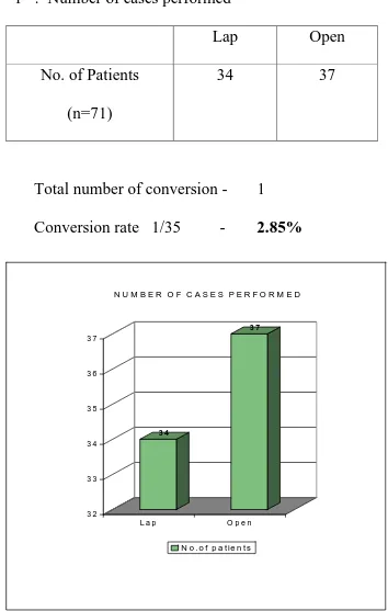
Table – 1 : Number of cases performed
Lap Open
No. of Patients
(n=71)
34 37
Total number of conversion - 1
Conversion rate 1/35 - 2.85%
3 4
3 7
3 2 3 3 3 4 3 5 3 6 3 7
L a p O p e n
N U M B E R O F C A S E S P E R F O R M E D
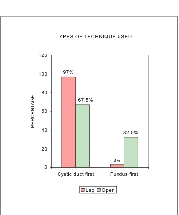
Table – 2 : Type of technique used
Procedure / Technique Lap (n=34) Open (n=37)
Cystic duct first 33 (97%) 25(67.5%)
Fundus first 1 (3%) 12 (32.5%)
TYPES OF TECHNIQUE USED
3% 97%
32.5% 67.5%
0 20 40 60 80 100 120
Cystic duct first Fundus first
PE
RC
E
N
T
A
GE
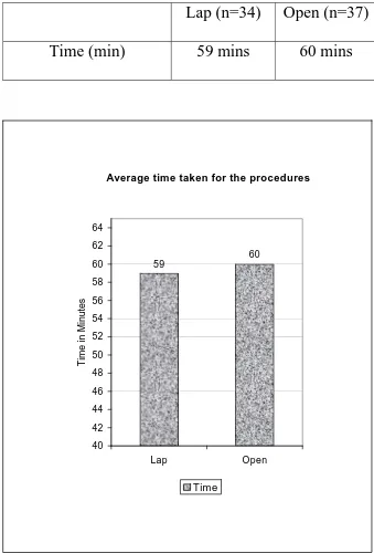
Table – 3 : Average time taken for the procedure (Including the time
taken for methylene blue injection in laparoscopic approach)
Lap (n=34) Open (n=37)
Time (min) 59 mins 60 mins
Average time taken for the procedures
59 60
40 42 44 46 48 50 52 54 56 58 60 62 64
Lap Open
Time in
Minutes
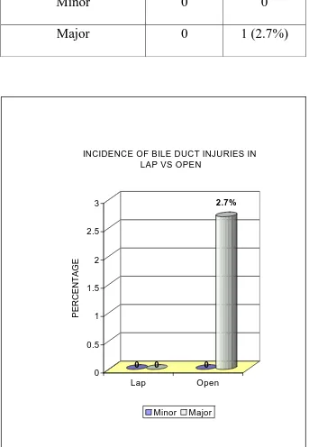
Table – 4 : Incidence of bile duct injuries in Lap (with methylene blue injection) Vs Open
Procedure
Bile duct inj.
Lap (n=34) Open (n=37)
Minor 0 0
Major 0 1 (2.7%)
0 0 0
2.7%
0 0.5 1 1.5 2 2.5 3
PERCENTAGE
Lap Open
INCIDENCE OF BILE DUCT INJURIES IN LAP VS OPEN
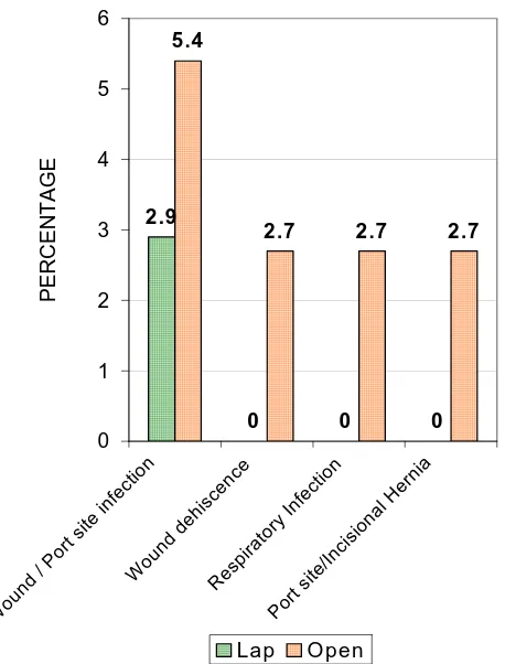
Table – 5 : Incidence of Other Complications
Lap (n=34) Open (n=37)
Wound / Port site infection 1 (2.9%) 2 (5.4%)
Wound dehiscence 0 1 (2.7%)
Respiratory Infection 0 1 (2.7%)
Port site / Incisional hernia 0 1 (2.7%)
Overall complication 2.9% 13.5%
INC IDEN CE O F O TH ER C O M PLIC ATIO NS
2.9
0 0 0
5.4
2.7 2.7 2.7
0 1 2 3 4 5 6
Wound / Por
t site infe
ctio n
Wound dehi scen
ce
Resp irato
ry In fecti on Port site /Inci sional Her nia PE R C E N T A G E
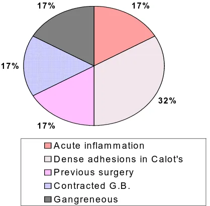
Table – 6 : Incidence of difficult cholecystectomies and its
distribution (Laparoscopic approach only)
Difficult cholecystectomies n = 6
Acute inflammation 1 (16.6%)
Dense adhesions in Calot’s triangle 2 (33.33%)
Previous surgery 1(16.6%)
Contracted GB 1 (16.6%)
Gangreneous GB 1 (16.6%)
Incidence : 6/35 : 17.5%
Conversion Rate: 1/6 : 16.66%
IN C ID E N C E O F D IF F IC U L T C H O L E C Y S T E C T O M IE S A N D IT S D IS T R IB U T IO N
17 %
3 2 %
17 % 1 7%
17 %
A cu te in fla m m a tio n
D e n s e a d h e s io n s in C a lo t's P re vio u s s u rg e ry
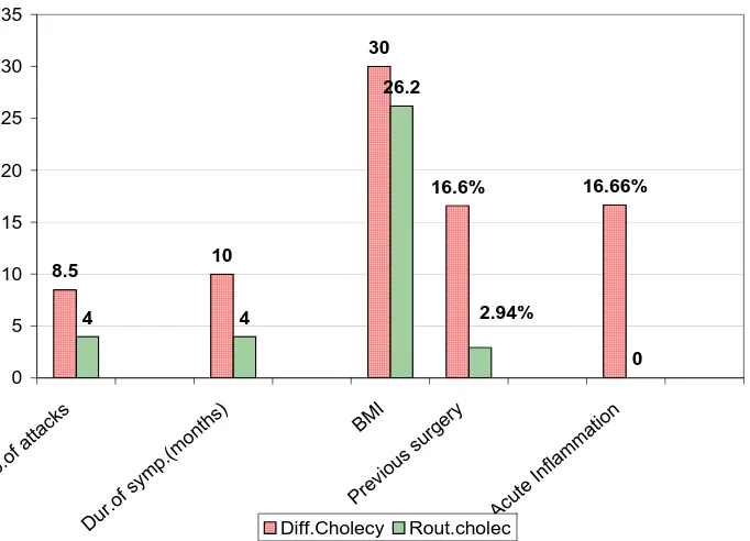
Table – 7 : Pre-operative predictors for difficult Laparoscopic cholecystectomies Predictive Factors No.of attacks Duration of symptoms (months) BMI Previous Surgery Acute inflammation Difficult cholecystectomy
8.5 10 months 30 16.66% 16.66%
Routine
cholecystectomy
4 4 months 26.2 2.94% Nil
PRE-OPERATIVE PREDICTORS FOR DIFFICULT LAPAROSCOPIC CHOLECYSTECTOMIES 8.5 10 30 4 4 26.2 0 16.66% 16.6% 2.94% 0 5 10 15 20 25 30 35 No. of a
ttack s
Dur .of s
ymp. (mon
ths) BM I Prev ious sur gery Acut e In
flam mat
ion
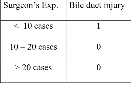
Table – 8 : Surgeon’s Experience as a factor in bile duct injury
Surgeon’s Exp. Bile duct injury
< 10 cases 1
10 – 20 cases 0
[image:29.612.90.532.458.611.2]> 20 cases 0
Table – 9 : Mode of management adopted for Bile duct injury
Management
Type of Inj.
Serial
LFT
Conservative
Management
MRCP ERCP
Management
Surgical
Management
(Bilary enteric
bypass)
Minor (n=0) - - - - -
REVIEW OF LITERATURE
Incidence of bile duct injury in our study was restricted only to
open cholecystectomy (2.7%) which was a major transection injury
involving common bile duct, way higher than reported in standard
literature of about 0.125% (1 in 800). Biliary continuity was restored
with Roux-en-Y hepatico jejunostomy electively, months after a
emergency laparotomy where in a large bilioma was drained.
Interestingly incidence of bile duct injury in laparoscopic
cholecystectomy was nil (0%) in comparison to Strassberg et al data
of 0.55% to 0.85%. This could be attributed to following reasons.
¾ Meticulous technique of Safe Laparoscopic cholecystectomy
¾ Use of Methylene blue injection to delineate biliary anatomy Bile Duct Injury (%)
LC OC
Our study 0 % 2.7%
Strassberg et al 0.55-0.85% 0.125%
Daziel et al 0.6% -
Fullarton et al 0.7% -
Brune et al 0.2% -
Overall conversion rate in our study was 2.85% in comparison to
various series which ranges from 1.2% - 17%
Conversion Rate %
Fullarton et al 17%
Liturin et al 4.3%
Brune et al 1.2%
Our study 2.85%
Conversion of laparoscopic approach to open was in a case
gangreneous gall bladder, distended, fragile and was difficult to
grasp and with laparoscopic instruments. Moreover methylene blue
could not be injected due to obvious reasons as the color of Gall
bladder wall bluish black.
Average operative time in our study of laparoscopic
cholecystectomy was significantly low (59 min) in spite of the time
consumed in methylene blue injection. This probably may be due to
color contrast offered by methylene blue colored ducts which
Operative time (mins)
LC OC
Our study 59 60
Barkun et al 86 73
Trondsen et al 100 50
Majeed et al 65 40
Overall incidence of other complications were significantly
higher in open cholecystectomy (13.5%) whereas it was minor
wound infection in one case in laparoscopic arm (2.9%).
Incidence ‘Difficult cholecystectomy based on intraoperative
pathology in our study was 17.5%, commonest presentation being of
dense adhesions in the triangle of Calot`s. Incidence reported by
Kuldhip Singh et al in large series from North India was 22.67 where
in commonest presentation was acute inflammation of Gall bladder.
There was no case of congenital anomalous biliary ducts in our
study.
As shown by our study, number of attacks of pain , duration of
abdominal surgery are found to valuable preoperative indicators to
anticipate difficult cholecystectomy.
Age and sex did not bear any significance in this regard.
Surgeon experience with both approaches especially laparoscopic
can be considered a risk factor for bile duct injury as shown in our
study and in literature (Southern surgeons club).
Most of the cases in laparoscopic arm were accomplished by
‘cystic duct first’ technique (97%) where as one third of cases in
open approach was done by ‘fundus first’ technique. This can be
attributed to magnified and clear vision of Calot’s triangle through a
DISCUSSION
BILE DUCT INJURIES :
Bile duct injuries are associated with significant morbidity,
prolonged hospitalization, increased financial burden, potential
litigation and occasional mortality. It is the third most common
litigated general surgical complications in western statistics, also it
has been reported that average two procedures (between 1 to 8) are
required for definitive repair of bile ducts. Bile duct injury if
fortunately identified and repaired peroperatively, carry less
morbidity and mortality.
In the era of laparoscopic cholecystectomy bile duct injuries
has gained tremendous amount of attention. Laparoscopic
cholecystectomy in its earlier days was ill famed due to the high
incidence of bile duct injury. With refinement of technique and
various factors incidence of bile duct injury has become surprisingly
With the huge number of Laparoscopic cholecystectomy
performed today even this fraction carries a substantial economic
impact.
Many advancements in this field are in vogue to decrease the
incidence of bile duct injuries to low minimum like, IOC, defining
technique for safe cholecystectomy, sophisticated new generation
laparoscopic instruments, per operative dye injection are few to
mention.
Types of Injury
• Bile leaks (Usually minor)
• Bile duct transections / stricturing type (Major)
Bile leaks :
Minor, Bile duct injuries occur in a frequency of 0.3%
worldwide. Common causes are :-
• Leak from cystic duct stump (may be due to slippage of clip
during suction and irrigation after removal of GB)
• Transected aberrant right hepatic duct
• Lateral injury to the main bile duct (<25% of circumference)
These injuries usually present within 1 week of laparoscopic
cholecystectomy with pain, fever and mild hyper bilirubinaemia ( up
to 2.5 mg /dl) from a bilioma or bile peritonitis. Symptoms may be
subtle initially. If drain is placed, bile may leak from it or through
one of the port sites. Diagnosis should be considered in patients
presenting with bloating or anorexia more than few days after
laparoscopic cholecystectomy.
Even though minor, it can present very late with bile duct
strictures (esp. lateral wall injuries months to years after the
procedure).
Bile duct Transections / Stricturing injury :
The incidence of these major injuries are 0.55% - 0.6 % world
wide ,commonest of these are
1. Clip placement in common bile duct or right heptatic duct
mistaken for cystic duct.
2. Excessive use of monopolar cautery to control bleeding and in
difficult dissections.
Recognized fairly late in post operative period and there are no
painless or with pain if cholangitis complicates the situation. Less
commonly, patient may present fairly late (months to years) with
cholangitis, cirrhosis and portal hypertension.
Classification of Bile duct injuries :
Commonest used is Corlette –Bismuth classification which
classifies major Bile duct transections and strictures of extra hepatic
biliary type.
Bismuth Classification
Type I - Low common hepatic stricture,
length of the stump > 2cm
Type II - Higher strictures
Length of the CHD stump < 2 cm
Type III - High hilar strictures – no serviceable CHD but the
confluence of right and left hepatic duct is intact
Type IV - Involvement of confluence with no communication
between right and left hepatic ducts
Fibrosis in between the two ducts may be thin
Type V - Combined common hepatic and aberrant right
hepatic duct injury separating both from distal
biliary tract.
Advantages of Bismuth classification :
1. Length of the remnant stump determines the type of repair
2. Indicates prognosis, morbidity and chance of recurrence
after the indicated repair.
Disadvantage :
• Does not indicate the length of the stricture as in present
era small length strictures can be dealt non-operatively.
• Does not include minor biliary tract injuries which
require management
• Does not mention the continuity across the injury.
More recently Strassberg classification, out lines a comprehensive
STRASSBERG CLASSIFICATION
Type A :
Bile leak from a minor duct that is still in continuity with
common bile duct. Usually from cystic duct stump or gall bladder
bed. Does not cause strictures or require tertiary referral.
Type B :
Occlusion of part of biliary tree usually it is aberrant right
hepatic duct mistaken for cystic duct. Often asymptomatic may
present later with pain and cholangitis.
Type C :
Bile leak from a duct not in communication with distal
common bile duct. Usually transection of right aberrant hepatic duct
with drainage of bile into peritoneal cavity presents early in post
operative period.
Type : D
Lateral injury to extrahepatic bile duct. The hepatic
parenchyma remains in communication with the distal end of biliary
Type E :
Circumferential injury of major extrahepatic ducts with
separation of liver parenchyma from the lower ducts and duodenum.
(Type E1- E5 is same as type 1-5 of Bismuth classification)
Modes of Injury during Laparoscopic Cholecystectomy Basic two error groups which lead to bile duct injuries are :
1. Misinterpretation of anatomy
2. Technical Error
Misinterpretation of anatomy: As a broad term is responsible for 70% of Bile duct injuries as
concluded in retrospective analysis. Confirming and reconfirming
the key anatomical structures before dividing or clipping is the
key to avoid bile duct trauma.
Technical errors :
Technique of ‘Safe cholecystectomy’ is crucial for any
uneventful cholecystectomies. ‘Classical injury’ which leads to
of lateral traction which brings BD in line with cystic duct, this
eventually leads to application of clips partially or totally on to
CBD which leads to total transection of CBD without continuity.
(Strassberg type E)
Hilar bleeding and its desperate control accounts for many high
injuries, this usually is due to cystic artery bleeding or due looped
right hepatic artery (Moynihan’s Hump).
Other causes of injuries are :
1. Occlusion of lumen of common bile duct by ligating cystic
duct flush at its origin
2. Excessive dissection
3. Excessive use of Monopolar diathermy (conduction and
transmission of current while using monopolar diathermy after
clipping of cystic artery.)
Risk factors for Bile duct injuries :
Experience of the surgeon : The learning curve
This is the most crucial factor with regards to iatrogenic injury
to bile duct during laparoscopic cholecystectomy.
Its not only the technical competence of the surgeons but also
the ability to adjust to two dimensional images on the monitor and
the lack of depth perception. This is the learning curve which every
beginner laparoscopic surgeon must go through and indeed the curve
isn’t of a similar shape in every one’s case.
Southern surgeons club reported initial high rate of bile duct
injury (2.2%) during first 13 laparoscopic cholecystectomies per
surgeon. This rate fell to 0.1% for subsequent operation.
Improper use of energy sources for dissection :
Any thermal source causes collateral damage and hence their
lies a potential risk of delayed injuries to surrounding structures,
incidence of such damage is higher in use of monopolar diathermy
when compared to bipolar diathermy , and damage is claimed to be
Patient factors :
Apart from disease pathology per operatively other factor
which predict difficult dissection are :
1. Obesity (as the excessive fat obscures the anatomy of
Calot’s triangle)
2. Number and duration of attacks
These directly relate to difficult dissection due formation of
dense adhesion with many attacks and its long duration
before patient is subjected to Laparoscopic
Cholecystectomy.
3. Age and male gender :
Though considered by few as predictor of difficult
cholecystectomies, it only increases the post operative morbity
and are not considered as risk factor for difficult cholecystectomy.
Anomalous and Morbid anatomy :
Grossly these conditions are grouped under the entity ‘Difficult
cholecystectomy’ as mentioned later. Broadly these conditions
Dangerous Anatomy :
These include adhesions / excessive fat in the porta hepatis
which obscure the view of vital structures.
Anomalies of biliary tract (10-15%) which are not usually
identified preoperatively. Most likely of which creates problems
is an aberrant right hepatic duct inserting low into common
hepatic bile duct mistaken for cystic duct.
Another important anomaly is a ‘short cystic duct : which may
cause lateral wall of CBD injury / remote stricture while applying
clips.
Variations in vascular anatomy may present with difficulty
usually in terms of haemorrhage which obscures the vision,
predisposing to bile duct injury in an attempt to control it.
Dangerous Biliary pathology : These include
- Acute cholecystitis
- Mirizzi syndrome
- Sclero atrophic Gall bladder
- Frozen / fibrosed triangle of Calot`s triangle
Acute cholecystitis presents problems in terms excessive
oozing of blood and distortion of anatomy due to active
inflammation. Still randomized prospective studies have shown
benefit of laparoscopic approach over open procedure (3% minor
complication versus 23% major complications and 19% minor
complications in open cholecystectomy group)
Threshold for conversion should be low in case if anatomy
cannot be delineated. Gall bladder can be aspirated and should not be
grasped but retracted bluntly as it may be friable.
Prevention of Bile Duct Injuries in Laparoscopic Cholecystectomy
Bile duct injuries like any disease is better prevent than treated.
Key aspects of prevention are
1. Thorough knowledge of the anatomy, risk factors and
mechanisms of injury.
2. Meticulous technique of safe cholecystectomy as described.
3. Timely decision for elective conversion to open in the
4. Developing skills of interpreting Intra operative
cholangiogram.
Meticulous technique has no substitute in preventing bile duct
injuries, proper traction, limiting dissection close to gall bladder.
Critical window and display of structures in Calot`s triangle are
few to mention.
Concern over the clipping of cystic duct later its slippage
causing bile leak is real due to which many surgeons now opt to
ligate the cystic duct with transfixing intra corporeal suture or
endoloop especially for short cystic duct. Long duct is better
dealt with clips.
There is a chance of internalization of these cystic duct clips
into bile duct, which acts as nidus for stone formation several
months later and possibly stricture.
Methylene blue dye injection is a novel, cost effective and easy
technique to prevent bile duct injuries. Its main advantage lies in
the fact that dye is injected into gall bladder before any dissection
done after some dissection has been undertaken when injury
might have occurred already.
It provides surgeon a continuous per operative delineation of
biliary anatomy hence greatly facilitating dissection.
Difference between open and laparoscopic bile duct injuries :
Laparoscopic injuries tend to be more extensive involving
injury to a segment of common bile duct and often extension to
higher levels often involving proximal hepatic ducts. About 60-75%
are not immediately recognized during surgery.
Occurrence of combined vascular and ductal injuries carry poor
prognosis as future it may cause re-stricuturing despite
reconstructions due impaired blood supply to the anastamosis.
Injury to vessels, also lead to hepatic necrosis / abcess formation
rarely.
Open cholecystectomies usually escape with smaller injuries
with leaks, stricturing injuries being rare. Vascular injury is less
Diagnosis and Investigations :
Investigative work up of bile duct injuries are directed by
clinical manifestations :-
Bile leaks usually presents early, manifested by increased drain
output, fever, peritonitis, abscess or sepsis. Hyperbilirubinaemia
may be present due to reabsorption of extravasated bile.
Strictures / Occlusive type injuries usually present late in post
operative period about 2-3 weeks in an average and is manifested by
fever, cholangitis with rising bilirubin which makes diagnosis
obvious.
Blood tests :
Rising serum bilirubin and liver enzymes direct as to the
possibility of bile duct injury and is indispensable in management of
bile duct injury and its follow up.
Ultrasonography :
Always the initial investigation. It can detect fluid collection,
abscess, bilioma in leaking type of an injury, whereas presence of
occlusive / stricturing injuries. Percutaneous aspiration of collection
can be done under ultrasonic guidance.
Disadvantages are
• Operator dependant
• Does not guide the management option as exact pathology is
not identified.
Computerized tomography:
Contrast enhanced computerized tomography is better than
ultrasonogram as it can fairly detect the level of obstruction and
gives surgeon a preoperative picture. Still with this modality exact
length of stricture cannot be identified and it cannot reliably
diagnose ongoing leak.
Scintigraphy :
HIDA scan can reliably detect on going leaks and presence of
biliary discontinuity inferred from failure of radionuclide to enter the
ERCP :(Endoscopic retrograde cholangiopancreaticography)
This is preferred diagnostic modality still in many centers
where non operative management in the form of intraluminal stents
is popular. Gives excellent anatomic detail of distal biliary tree can
be combined with therapeutic procedure at many instances.
Disadvantage of ERCP are
1. Invasive procedure
2. Does not give information regarding status of proximal
ducts and length of stricture which is crucial for deciding
type of repair to be under taken.
3. Associated significant incidence of pancreatitis.
Percutaneous transhepatic cholangiography :
When combined with ERCP gives complete anatomical
information required. Rarely done nowadays as it is unacceptably
MRCP (Magnetic Resonance cholangio pancreaticography)
It is fast replacing ERCP for preoperative classification of bile
duct injuries / strictures. It is non invasive, delineates both proximal
and distal duct anatomy which makes deciding the surgical repair
easy.
Cost, though is still a limiting factor for its use in developing
countries.
Management Options
Injury identified during laparoscopic cholecystectomy
Partial tears of CBD can be closed over a T-tube and
managed as usual. This can be done either as open procedure or
laparoscopically.
Major injuries when detected should be converted to open.
Depending on the length of the extra hepatic bile duct available, one
of the following procedure can be undertaken.
- Roux – en – Y Hepatico jejunostomy
Endoscopic Techniques :
A) Endoscopic biliary Drainage : Transduodenal drainage of biliary
tree is method of choice in patient with leakage from cystic duct and
in selected patients with minor leakage from the common duct.
Drainage may be accomplished using a nasobiliary stent or an
indwelling stent with or without papillotomy.
b) Endoscopic Dilatation : A guide wire is passed through the
area of stricture and the stricture is dilated with balloon. Stents are
frequently placed.
Percutaneous Techniques :
These technique requires transhepatic approach (PTC) with
passage of guide wire through the stricture. Dilators of increasing
size are then passed through the stricture. A percutaneous catheter is
routinely left in these patient to minimize chances of leakage of bile
into the sub hepatic space, to reduce the sepsis and to permit future
Open techniques :
Usually a form of biliary enteric anastamosis decided by site of
injury / stricture and length of extrahepatic biliary duct available.
These are:
Intrahepatic cholangio jejunostomy : (Segmental drainage)
Done in case of frozen hilum where the ducts could not be
isolated in high injuries. Segmental duct draining segment III or V is
anastamosed to Roux loop of jejunum.
Hepatico duodenostomy :
Done where Roux-loop is not feasible. Anastamosis is done in
end to side manner. This anastamosis is accessible to endoscopic
instrumentation.
Roux-en-Y Hepatico Jejunostomy / choledochojejunostomy : This is usually the procedure of choice for major transectional
and stricturing injuries, can be used with internal stents to avoid
Methylene Blue Dye Injection to prevent Bile Duct Injuries Introduction :
Injection of methylene blue is not new to practice of surgery,
frequently used to trace sinus or fistula during various procedures,
sentinel lymph node biopsy and in chromointubation (Tube patency
test). In this study Methylene blue is used to delineate extrahepatic
biliary tract including Gall bladder during laparoscopic
cholecystectomy by coloring them blue.
It’s a novel approach with largest series of 46 cases performed
in Istanbul Training Hospital, Istanbul. Basic purpose of this
technique is to facilitate young surgeons / residents in beginning of
their learning curve to execute a SAFE cholecystectomy and to help
dissections in ‘Difficult cholecystectomies’.
Method :
Gall bladder fundus was punctured by Verress needle and all
the bile was aspirated. The same amount of 50% methylene blue
(saline diluted) was injected into the gall bladder for coloration of
biliary tree ie. gall bladder, cystic duct, bile duct and some times
with toothed grasper through the lateral subcostal port. Gall bladder
was removed by subxiphoid port. Methylene blue is aspirated to
prevent leak while removal of Gall bladder.
Post operative consideration :
¾ Patient must be informed that urine may colored blue, as the dye which leaks to duodenum is absorbed and excreted through
kidney.
¾ Ryle`s tube aspirate may be colored blue either intra operatively or post operatively. Confirming patency of common bile duct.
Advantages of Methylene blue injection :
¾ Safer & faster dissection in Calot`s triangle
¾ Detection of aberrant anatomy of biliary tract
¾ To detect bile duct injury (if it does occur) per operatively and enables its repair in the same sitting hence decreasing morbidity
of unrecognized bile duct injuries.
¾ Can supplement Intraoperative Cholangiogram (IOC) interpretation per operatively as it orients surgeon to IOC
findings.
¾ Cost effective, negligible adverse effects and does not prolong operative time.
Technique of Safe Laparoscopic cholecystectomy
Positioning of the patient :Anti Trendlenberg’s position with 30 degree lateral tilt towards
left is desired as is it clears the operative field of small bowel loops,
stomach and transverse colon due to gravity.
Theatre set up :
MONITOR
SURGEON ANAES
1ST ASST.
Operative team set up is as shown in figure. It doesn’t require
patient to be placed in Lloyd Davis position as in French set up, but
many surgeons feel less comfortable due less available space.
Access :
Pneumoperitoneum is created by OPEN / HASSON’S
TECHNIQUE. A curvilinear supra umbilical incision is placed
about 1-1.5 cm in length. Peritoneum is opened through the incision
and entry into abdominal cavity is confirmed. A 10 mm Cannula
with or without blunt tipped Hasson’s Trocar is introduced which
should snugly fit to prevent gas leak. Open technique has the
advantage of quick access and quick pneumoperitoneum creation.
Veress needle technique or closed technique bears the risk of
bowel or vascular injury, takes more time to reach the desired level
of pressure (12 mm of mercury) needs patient to be placed in head
down position.
After creation of pneumoperitoneum and routine visual
inspection of abdominal cavity, a second 10 mm port is placed 2/3rd
the midline. A 5 mm cannula is inserted 3-4 cm below the costal
margin in the mid clavicular line and a second 5 mm cannula is
inserted 4-5 cm below the costal margin in the anterior axillary line.
Position can be adjusted as per the need of the surgery.
The supra umbilical 10 mm port is used for the 0 or 30 degree
telescope and for CO2 insufflation. Sub xiphoid port is used for
dissection with surgeon right hand, whereas the other two 5 mm
ports are used for retraction at the fundus (Ant. axillary line) and at
the infundibulum (Mid clavicular line)
A 30 degree telescope has the advantage of providing over
head view of the field similar to an open surgery.
Gall bladder fundus is grasped and retracted cephalad to
expose the sub hepatic area and the infundibulum of the gall bladder.
With the mid clavicular line port, infundibulum is grasped and
retracted laterally and inferiorly to lay open the Calot`s triangle and
it creates a distinct angle between the cystic duct and common bile
duct and hence avoiding their alignment in one line which is
On adequate exposure of Calot`s triangle the dissection should
commence high on the gall bladder initially posteriorly and then
anteriorly. One should visualize the ‘posterior peritoneum’
covering the ‘yellow pad of fat’ and keep the dissection just above
it.
As one proceeds inferiorly cystic duct is encountered. Junction
of cystic duct with gall bladder is visible as ‘Elephant head’ or
‘The Ganesha sign’ which is a must see during laparoscopic
cholecystectomy to avoid injury. At times, there may be an
anteriorly placed cystic artery which has to be divided to proceed
further.
Next step is to create a ‘Critical window’ in the Calot`s triangle
which clearly demonstrate the cystic artery and the duct in loose
areolar tissue which bridges the Calot’s triangle. Dissection should
not proceed beyond ‘Rouvier’s sulcus’ which is the only constant
landmark in this area and marks the lateral extent of porta hepatis
If cholangiography is planned, it is to be done at this stage by
introducing cholangio catheter via a small opening in cystic duct
after placing a clip distally at its junction with gall bladder. Contrast
is injected to delineate the biliary tree under fluoroscopic guidance.
Though it prevents bile duct injury during further procedure, it
cannot prevent injuries which are sustained during dissection
described before.
If the anatomy is clear, cystic artery is doubly clipped
proximally and also distally and divided followed by division of
cystic duct between double clips, maintaining the lateral traction. At
times larger branches of cystic arteries may have to be ligated or
clipped.
An abnormally large cystic artery may suggest the presence of
‘Caterpillar hump’ right hepatic artery. If it is present the right
hepatic artery should be dissected away and clipping the cystic artery
which usually arises from angled hump of right hepatic artery.
Any haemorrhage should be controlled by compression with
adjacent bowel, gauze piece and is accurately identified and ligated
or clipped. Blind and desperate attempts to control bleeding, leads to
disaster.
Dissection of GB from liver bed should not be callous and as
one might miss aberrant cysto-hepatic duct which may cause post
operative biliary leak. Dissection is done with scissors or cautery.
Gall bladder is removed from abdominal cavity as such or in an endo
bag or condom carefully, or bile may evacuated from Gall bladder
to ease its manipulation during delivery.
Presence of overriding Hartmann’s pouch adherent to common
bile duct should raise the possibility of ‘Mirizzi syndrome’ with
cholecysto biliary fistula which is usually a strong contraindication
laparoscopic procedure.
In acute cholecystitis planes may not be as clear as in chronic
procedure, sharp dissection is used, preferably with scissors.
Conversion to open procedure should be strongly considered.
In case of short cystic duct where clips cannot be applied
without avoiding lateral wall of CBD, a ligature may be applied
avoiding CBD wall or else conversion to open is a better option.
CBD should not be dissected to display its junction with cystic
duct as it carries increased chances of injuries.
A suction or tube drain of size 14 is placed in sub hepatic area
to detect bile leak. Pneumoperitoneum is let out and port sites are
CONCLUSION
¾ Incidence of bile duct injury is apparently more in open cases than in laparoscopic cholecystectomy.
¾ Lower incidence in laparoscopic approach as contradictory to the standard literature statistics probably due to better surgical
experience, technique of modern day surgeons, improved
visual aids and laparoscopic instruments. However, larger
sample size is required to show the statistical significance of
this study.
¾ Obesity, Number and duration of attacks, acute onset and previous upper abdominal surgeries were reliable pre operative
predictive factors for ‘Difficult cholecystectomy’. Whereas age
and sex did not show any difference.
¾ Methylene blue dye injection is an excellent, simple, cost effective technique to aid surgeons in beginning of their
learning curve to execute a safe laparoscopic cholecystectomy
and to aid dissection in difficult cholecystectomy.
¾ MRCP followed by biliary enteric anastamosis and has provided symptom free solution for the major bile duct injury
BIBLIOGRAPHY
1. Adam DB : The importance of entrahepatic biliary anatomy
in preventing complications at laparoscopic cholecystectomy.
Surg Clin North Am. 74 : 861-871, 1993.
2. Asbun HJ, Rossi, RL, Lowell JA et al : Bile duct injury
during lap chole : Mechanism of injury, prevention and
management. World J Surg. 17 : 547 -552, 1993.
3. Bailey & Love 24th edition, Short practice of Surgery
4. Bismuth H. Post op. Stricture of the bile duct. In Blumgart
LH (ed). The biliary tract vol.5, Edinburgh Churchill Living
stone, 1982, pp 209-221.
5. BMC Surgery 2005 5:14 (http : // creative commons org /
heenses / by / 2.0 SSK Instanbul Training Hospital, Istanbul
surgery.
6. British Journal of surgery, January 2003 vol 90, pg 42-47.
7. Davidoff AM. Pappas TN, Murray EA et al., Mechanism of
major biliary injury during lap. Chole. Ann Surg. 215 :
196-202, 1992.
9. Frank H. Netter MD. Atlas of Human Anatomy, Third
edition.
10. Hovarth K.D., Strategies for the prevention of laparoscopic
common bile duct injuries. Surg. Endosc. 7 : 439-444, 1993.
11. Hunter JG : Avoidance of bile duct injury during lap. Chole.
Am J Surg. 162 : 71-76, 1991.
12. Indian Journal of Surgery August 2006 vol 68, pg 205-209.
13. Indian Journal of Surgery volume 68, No.3, June 2006,
165-167.
14. Maingots Abdominal operations, 11th edition
15. Michel NA : Blood supply and Anatomy of upper Ab. Organs
with descriptive Atlas. Philadelphia, J6, Lippincott 1955.
16. Moossa AR, Easter DW, Vansonner berg E, et al Lap. Injuries
to the bile duct : A cause for concern, Ann. Surg 215 ; 203 –
208, 1992.
17. Olsen DO, Arbun HJ, Reddick, EJ et al ; Lap chole for acute
chole cystitis, Probl. Gen Surg.8 : 426 – 431, 1991.
18. Picklemen J, Gonzaliz RP. The improving results of
19. Reddick EJ, Oslen D, Spab A, et al : Safe performance of
difficult lap. Chole. Am J Surg. 161 : 377 – 381, 1991.
20. Regula J, Pacheaski J, Bastnik W, et al : Endoscopic drainage
of bile ducts. Pol. Arch Med. Wewn 85 : 256-262, 1991.
21. Rossi RL, Schiromer WJ, Braasch JW, et al. Lap. Bile duct
injuries. Risk factors, recognition and repair Arch Surg. 127 :
596-602, 1992.
22. Sabiston’s Text book of surgery – 17th edition
23. Shackelford’s Surgery of the Alimentary tract 5th edition
24. Vitale GC Stephens G. Wiemann TJ et al. Use of ERCP in
Management of biliary complication after lap. Chole surgery
114 : 806 – 814, 1993.
25. Woods MS, Tra verso LW, Kozarek RA, et al : Characteristic
of biliary tract complications during lap. Chole : A multi
dimensional study. Am J. Surg. 167 : 27-34, 1994.
26. XXIII National CME, Surgery update 2006, Maulana Azad
PROFORMA
Name : Age : Sex :
Address : Occupation : DOS :
Weight : Height :
BMI
Complaints :
Pain Abdomen / Fever / Dyspepsia / Others
Duration of pain abdomen / No.of attacks
Medical Treatment (Specific / Non specific) : Yes / No
Comorbid Illness :
DM / HTN / Restrictive or obstructive lung disease/ CRF / CAD
Previous Surgery :
Vagotomy / Gastrectomy / Devascularisation / Others
Physical Examination : Vitals
Anaemia Jaundice Pedal Edema
Generalised lymphadenopathy
P/A : Inspection
Contour - Flat /Scaphoid / Distended
Previous scar - Yes / No
Any obvious Mass - Yes / No
Palpation :
Tenderness / Murphy’s sign):Yes / No
Mass if any : Yes / No
Others systems :
CVS / RS / CNS
Specific Investigations :
1. Routine
2. LFT = Normal / Abnormal parameter 3. BT / CT = Normal / Increased
4. USG : Yes / No 5. CT Abdomen : Yes / No 6. OGD
7. MRCP / ERCP : Yes / No
Mode of Treatment :
Open cholecystectomy / Lap. cholycystectomy / Lap. Converted to open
Whether Lap converted to open
If so, (reason why ) : Bleeding / Extensive Adhesions /
Anaesthetic / Indescernible Anatomy / Others
Anaesthesia : GA / Epidural / Spinal
Incision (If open) : Kocher’s / Midline / Others
Approach (if lap ) : 10 mm I II 5 mm I
II III
Technique :
Cystic duct first
Fundus first
Methylene Blue Injections - Given / Not given
Operative time in minutes :
Surgeon’s Experience : < 10 / 10-20 / > 10 cases
Complication : Wound infection
Wound dehiscence
Minor bile duct injury
Major bile duct injury
Resp. infection
Port site hernia / Incisional hernia
DISSECTION OF GALL BLADDER FROM THE LIVER BED
CYSTIC STUMP AFTER COMPLETION OF
CHOLECYSTECTOMY (NOTE THE BLUE DYE IN THE
INJECTION OF METHYLENE BLUE DYE INTO
FUNDUS OF GALL BLADDER
DISSECTION OF CALOT’S TRIANGLE WITH LATERAL TRACTION
(CYSTIC DUCT COLOURED FAINT BLUE WITH DYE)
Cystic Artery
AFTER DIVISION OF CYSTIC ARTERY – JUNCTION OF CYSTIC DUCT WITH GALL BLADDER CLEARLY
BILIOMA FOLLOWING MAJOR BILE DUCT INJURY IN OPEN CHOLECYSTECTOMY
MRCP PICTURE OF TYPE E STRASSBERG BILE DUCT INJURY
CLASSICAL LAPAROSCOPIC INJURY TO CBD
PROPER PLACEMENT OF CLIPS
Accidentally Divided Hepatic Ducts
Common Hepatic Duct Cystic Duct
VARIATION IN CYSTIC ARTERY ANATOMY
NORMAL
ORIGIN AROUND
THE CBD LOW ORIGIN
ANTERIOR CA
MASTER CHART
Symptoms Diagnosis Investigations
LFT USG Pain abdomen S l. N o Name A g e / se x Mo n th of s u rg e ry Bo d y M a ss Index Dur a ti on ( in mon ths ) No . of at ta ck s Ot he rs Pr e v ious U. AB D su rg ery Ac ut e Chr o n ic Bi li ru bi n AL P Ot he rs G a ll sto n es GB wa ll thi ckn e ss C
BD OGD
A b d o me n MR CP
1 2 3 4 5 6 7 8 9 10 11 12 13 14 15 16 17 18 19 20
1. Devanai 60/F 7/05 30 6 2 √ - - √ N N - + Ç N N - -
2. Anjammal 28/F 7/05 29 7 8 - - - √ N N - + Ç N N - -
3. Maheshwari 45/F 8/05 30 2days 1 √ - √ - N Ç - + Ç N N √ Yes
4. Pramila 48/F 8/05 31 2 3 - - - √ N N - + Ç N N - -
5. Guruvammal 50/F 8/05 29 4 2 - - - √ N N - + Ç N N - -
6. Soundarajan 67/M 8/05 28 1 2 - - - √ N N - + N N N - -
7. Natchiammal 60/F 9/05 31 3 4 √ - - √ N N - + Ç N N - -
8. Vallaimmal 55/F 9/05 24 4 2 √ yes - √ N N - + N N N - -
9. Paulraj 60/M 9/05 31.1 5 8 - - - √ N N - + Ç N N - -
10. Fathima 24/F 9/05 28.9 3 3 - - - √ N N - + Ç N N - -
11 Udaiyar 29/M 10/05 26 1 2 - - - √ N N - + Ç N N - -
12 Kannaiah 75/M 10/05 26.2 6 7 - - - √ N N - + Ç N N - -
13 Sherine 42/F 10/05 27 6 6 - - - √ N N - + N N N - -
14 Ramu 80/M 11/05 29 7 8 √ - - √ N N ÈALB + Ç N CHR DU - -
15 Rani 51/F 11/05 31 3 3 - - - √ N N - + Ç N N - -
16 Saminathan 55/M 12/05 33.3 5 3 √ - - √ N N - + Ç N N - -
17 Mamoon 65/M 12/05 30 6 3 - - - √ N N - + Ç N N - -
18 Selvi 38/F 12/05 26 2 5 √ - - √ N Ç - + N N N √ -
19 Arun kumar 16/M 1/06 25 4 4 √ - - √ N N - + N N N - -
20 Pichaiammal 43/F 1/06 31 2 3 - - - √ N N - + Ç N N - -
21 Brunda 70/F 2/06 29 2 2 - - - √ N N - + Ç N N - -
22 Gandhi 61/M 2/06 27 3 3 - - - √ N N - + Ç N N - -
23 Latha 37/F 2/06 25 4 5 √ - - √ N N - + Ç N N - -
24 Sameera 51/F 3/06 30 5 7 √ - - √ N N - + N N N - -
25 Rajagopal 45/M 4/06 29.3 6 4 - - - √ N N - + N N N - -
26 Jamruth Nisha 35/F 4/06 33 2days 1 √ - √ - N N - + Ç N N - -
27 Sundrammal 60/F 5/06 26 5 3 - - - √ N N - + N N N - -
28 Indira 52/F 6/06 28 7 6 - - - √ N N - + Ç N N - -
29 Gnanasekaran 38/M 6/06 26 8 7 √ - - √ N N - + Ç N N - -
30 Palanichamy 40/M 6/06 31 3 2 - - - √ N N - + Ç N N - -
31 Muthusamy 60/M 7/06 31 2 6 √ - - √ N N - + Ç N N - -
32 Ameena beevi 51/F 7/06 32 1 2 - - - √ N N - + Ç N N - -
33 Subramaniam 45/M 8/06 28 8 9 - - - √ N N - + Ç N N - -
34 Annalakshmi 50/F 10/06 27.3 2 3 √ - - √ N N - + N N N - -
35 Thottichi 32/F 10/06 29 12 10 - - - √ N N - + N N N - -
Symptoms Diagnosis Investigations
LFT USG Pain
abdomen
S
l. No Name
Ag e / se x M ont h of s u rg er y B ody M a ss In de x Dur a ti on ( in mo nt hs ) No . o f at ta ck s Ot h e rs P re v io u s U. AB D sur g er y Ac ut e Chr o n ic Biliru b in AL P Ot he rs G a ll sto n es GB wa ll thi ckn es s C
BD OG
D A b d o me n MRC P
1 2 3 4 5 6 7 8 9 10 11 12 13 14 15 16 17 18 19 20
37 Chitra devi 36/F 11/06 31.1 9 10 √ - - √ N N - + Ç N N - -
38 Muniyammal 44/F 11/06 29.4 10 11 √ - - √ N N ÇALT + Ç N CHR DU - -
39 Chellaih 50/M 12/06 28 4 5 - - - √ N N - + Ç N N - -
40 Sugumar 19/M 12/06 29.2 3 2 - - - √ N N - + N N N - -
41 Anwar ali 60/M 12/06 29.3 4 5 - - - √ N N - + Ç N N - -
42 Premkumar 38/M 12/06 34 12 10 √ - - √ N N - + Ç N N - -
43 Palaniammal 49/F 1/07 29.5 5 4 - - - √ N N - + Ç N N - -
44 Lakshmi 37/F 1/07 30 6 4 - - - √ N N - + N N N - -
45 Rajammal 40/F 1/07 31 1 2 √ - - √ N N - + N N N - -
46 Backiam 57/M 2/07 30 2 3 - - - √ N N - + Ç N N - -
47 Kaliammal 55/F 2/07 33 4 3 - - - √ N N - + Ç N N - -
48 Kathirmani 35/F 2/07 30 2 3 - - - √ N N - + Ç N N - -
49 Paulpandi 44/M 2/07 29 7 5 √ - - √ N Ç - + N N N √ -
50 Shanmuganathan 22/M 2/07 24.2 3 9 - - - √ N N - + Ç N N - -
51 Meenakshi 30/F 3/07 27 20 11 √ - - √ N N - + Ç N N - -
52 Dharmapoopathy 59/F 3/07 28.3 2 3 √ - - √ N N - + Ç N N - -
53 Sumathi 33/F 3/07 28.7 4 6 √ - - √ N N - + Ç N N - -
54 Angammal 40/F 4/07 29 9 11 - - - √ N N - + Ç N N - -
55 Alagumani 48/F 4/07 33.5 2 3 - - - √ N N - + Ç N N - -
56 Jothi 40/F 4/07 27.8 18 13 √ - - √ N N - + Ç N N - -
57 Pandian 62/M 4/07 29 5 5 √ - - √ N N - + Ç N CHRDU - -
58 Pandiselvi 23/F 4/07 29.8 7 4 - - - √ N N - + Ç N N - -
59 Valli 55/F 5/07 30 6 8 √ - - √ N N - + Ç N N - -
60 Vellaisamy 72/M 5/07 29.2 8 9 - - - √ N N - + Ç N N - -
61 Vijaya 27/F 5/07 29.4 7 4 √ - - √ N N - + Ç N N - -
62 Jothi 42/F 6/07 29 7 10 - - - √ N N - + N N N - -
63 Muthu 27/F 6/07 25.5 4 5 √ - - √ N N - + Ç N N - -
64 Sulaiman 50/M 6/07 33 4 4 √ - - √ N N - + Ç N N - -
65 Sujatha 34/F 6/07 32.2 3 3 √ - - √ N N - + Ç N N - -
66 Velan 35/M 7/07 31 3 4 - - - √ N N - + Ç N N - -
67 Syed Meera 42/F 7/07 34 5 3 - - - √ N N - + N N N - -
68 Karuppaiah 68/M 7/07 32.7 6 5 √ Yes - √ N N - + Ç N CHRDU - -
69 Chinnammal 47/F 8/07 27.1 6 4 - - - √ N N - + Ç N N - -
70 Prabhavathi 45/M 8/07 283 2 3 - - - √ N N - + Ç N N - -