A STUDY ON
SWASAKASAM
Dissertation Submitted To
THE TAMIL NADU DR.M.G.R Medical University
Chennai – 32
For the Partial fulfillment in Awarding the Degree of
DOCTOR OF MEDICINE (SIDDHA)
(Branch – I Pothu Maruthuvam)
Department of Pothu Maruthuvam
Government Siddha Medical College
Palayamkottai – 627 002
ACKNOWLEDGEMENT
First, I thank Almighty for his showered blessings which is the ultimate source of my success.
I have to give thanks to our Siddhars for all their manifold
mercies.
I wish to express gratitude and acknowledgement to the
Vice-Chancellor, The TamilNadu Dr.M.G.R. Medical University,
Chennai, The Special Commissioner, Director of Indian Medicine and
Homeopathy, Chennai and Joint Director of Indian Medicine and
Homeopathy, Chennai.
I also wish to convey my deep gratitude to
Prof. Dr. M.Dinakaran M.D.(s)., Prinicipal, Government Siddha
Medical College, Palayamkottai for patronizing the work by providing all the necessary facilities.
I also wish to convey my deep gratitude to
Prof. Dr.R.Devarajan M.D.(s)., Vice Prinicipal, Government Siddha Medical College, Palayamkottai for patronizing the work by providing all the necessary facilities.
I express my deep sense of gratitude to
Prof.Dr.A.Prema M.D.(s)., Head of the Department, P.G. Pothu
I express my deep sense of gratitude to
Prof.Dr. K. R. Revathi M.D. (S), Former Vice Principal and Head of the Department P.G. Pothu Maruthuvam, Government Siddha Medicla College, Palayamkottai, for her guidance and suggestions in the selection of topic.
I am extremely grateful to Reader Dr.S.Mohan M.D.(s)., and
Assistant Lecturer Dr.S.Chitra M.D.(s)., Department of P.G Pothu
Maruthuvam, Government Siddha Medical college, Palayamkottai for their kind and affectionate encouragement to my work.
I express my deep sense of gratitude to Prof. Dr.S.Mohan M.D.,
Modern Medicine, Government Siddha Medical College, Palayamkottai for his Valuable guidance in modern aspects.
My special thanks goes to Dr.D.Rajakumari M.D.(S).,
Dr. P.Shanmugam M.D.(s)., and Dr.J.Angeline Nirmala M.D.(S).,
Assistant lectures, Departmant of U.G. Pothu Maruthuvam, Government Siddha Medical College, Palayamkottai for their timely help in referring cases for my dissertation work.
I render my special thanks to Dr. S. Justus Antony M.D.(S).,
former Assistant lecturer, Department of P.G Pothu Maruthuvam, Government Siddha Medical College, Palayamkottai for his help in preparation of the drug.
I take this opportunity to express my deep sense of gratitude to
Lecturer. Mr. M.Kalaivanan M.Sc., M.Phil., and other staffs of
College, Palayamkottai for their help during the entire course of my work.
I also thanks to Prof. Mrs.N.Nagaprema M.Sc. M.Phil., Head of the Department and all the staffs of Department of Bio-Chemistry, for their help in Biochemical Analysis for this work.
On this occasion, I am thankful to Dr.S.Bagirathi M.B.B.S.,M.D.,
Head and other staffs of Department of Clinical Pathology and
Dr.V.S.Padma M.B.B.S., D.M.R.D., along with technicians of
Radiology Department for giving a kind Co-operation in doing investigation procedures.
I Cordially register my thanks to the Librarian
Mrs.T.Poonkodi M.A., M.Lisc., M.Phil., and Library staff, who helped to utilize the books of library for this work.
I express my thanks to Dr.R.Napolean B.Sc., M.D., Consultant
Microbiologist, Malar Micro Diagnostic Centre, Palayamkottai to evaluate anti-microbial activity of the trial medicine.
I would like to convey my thanks to Aarthi Advanced CT Scan
and MRI, Vannarpet, Tirunelveli, for their Co-operation to diagnostic the disease.
I express my thanks cordially to My Parents for their Co-operation and Moral support from the very beginning of my career.
Finally I convey my thanks to Staffs, Broad Band Net Café,
CONTENTS
Page No
I.
INTRODUCTION
1
II.
AIM
AND
OBJECTIVES 3
III.
ABSTRACT
5
IV.
REVIEW OF LITERATURE
a.
SIDDHA
ASPECTS
6
b.
MODERN
ASPECTS
39
V.
MATERIALS
AND
METHODS 74
VI.
RESULTS
AND
OBSERVATION
76
VII.
DISCUSSION
93
VIII.
SUMMARY
109
IX.
CONCLUSION
113
X.
ANNEXURES
PREPARATION AND PROPERTIES OF
THE
TRIAL
MEDICINE
114
BIO-CHEMICAL
ANALYSIS 124
PHARMACOLOGICAL
ANALYSIS
129
MICROBIOLOGICAL
STUDIES
141
CASE SHEET PROFORMA
143
INTRODUCTION
Every system of medicine promotes the adage “Prevention is better than
cure”. Siddha system of medicine can helps us to live a healthy life by
preventing disease. This system is a preventive, protective and health
promotive, curative, nutritive.
It is well known that all the eyes of the world are turning to the natural
system of medicine. Especially indigenous system of medicine to find out a
more acceptable drug for incurable disease.
Siddha system of medicine puts emphasis of three doshas. Any
alteration in these three doshas will cause disease.
This can be understood from the verse as follows.
“kpfpDk; FiwapDk; Neha; nra;Ak; E}Nyhh;
tsp Kjyh vz;zpa %d;W”
- jpUts;Sth;.
Appropriate application of siddha science and system is sure to give us
all a happy, healthy and harmonious life.
Maruthuvam (Medicine)
Medicine is one which prevents the disease of the body and mind and
gives remedies to disease.
‘kWg;gJ cly;Neha; kUe; njdyhFk; kWg;gJ csNeha; kUe;njd rhYk;
kWg;gJ ,dpNeha; thuhjpUf;f
kWg;gJ rhit kUe;njd yhNk”.
- jpU%yh;.
This is quoted in our system as follows.
‘czNt kUe;J kUe;Nj czT”.
‘khWghby;yhj cz;b kUe;Jz;zpy;
CWghL ,y;iy caph;f;F”.
- jpUf;Fws;.
The naturally available and scientific explanation of therapeutic
efficacy of our drugs will convince may people to undertake our siddha system
for their health needs.
SwasaKasam is a very common disease in the society due to increasing
exposure to air pollution now a day. It is taken up for the Author’s dissertation
study. The increasing number of patients coming for treatment at Pothu
maruthuvam out patient department and the absence of any permanent remedy
in the allopathy system of medicine, has induced the author to select this
disease for study.
The author has selected Kasha Chooranam (Anubava Vaithya Deva
Ragaseum page-481) and Anna pavala Chenduram (Anuboga Vaitheya
Navanethum part- III, page-104) as the trial drugs for the disease of
swasakasam.
Kasha Chooranam, the ingredients of this medicine has a potent anti
kabha action. Also the ingredients of Anna pavala Chenduram namely Anna
Pethi and kodi pavalam has been recommended as a remedy for swasakasam in
various siddha text. So, the author decided to under go a trial on swasakasam
AIM AND OBJECTIVES
Now-a-days people have vast knowledge about which system of
medicine is better for the treatment of diseases depending upon their nature.
They need a remedy which does not lead to complications and also they need a
safe and permanent remedy, particularly for such chronic disease. The author
has worked on SwasaKasam to get the patients treatment with out any side
effects as this disease drives millions of people in difficulties to lead a normal
day to day life.
To correlate the extensive study of SwasaKasam in siddha aspect as well
as in modern aspect with bronchial asthma.
The daily increasing number of Asthma patients and the efficiency of
siddha system of medicine curing chronic respiratory disease prompted the
author to carry out scientific clinical study on the subject.
To make a detailed study of definition, etiology, classification, signs and
symptoms, diagnosis, investigation, prognosis, line of treatment, diet control
and prevention for SwasaKasam in various siddha literature.
To make a satistical study of the disease about its incidence with the
criteria’s like age, sex, socio-economic status, occupation, family history and
paruvakaalam etc.
To study under the topics of mukkutram, poripulangal, udal thathukkal,
envagai thervugal, naadi, neerkuri and neikkuri the changes brought about by
this disease under normal conditions.
To make a detailed clinical evaluation of the disease on the basis of
etiology, sign and symptoms, complications, treatment and progress during the
To use modern parameters to confirm the diagnosis and to observe the
prognosis of the patient.
To evaluate the Bio-chemical and pharmacological analysis and
microbiological studies of the trial medicine.
To create an awareness among the public how the disease progresses on
the basis of the factors like diet, land, climatic condition, pollution,
immunology.
To educate the people who were affected by the disease how to stabilize
their health through natural ways like pranayama, yogasanas, food restrictions
and personal hygiene.
And also the prime object of this study is to do a clinical trial on
SwasaKasam affected individuals with selected siddha Medicine.
Kasha Chooranam – 1gm and Anna pavala Chenduram – 100mg
thrice daily with honey before meals.
ABSTRACT
Since the number of sufferers increasing day by day, the author has
chosen the disease “SwasaKasam” for her dissertation work. The
increasing incidence of the disease is due to changes in life styles and
environment.
Twenty patients of either sex were selected as In-patients and twenty
Out-patients were administered with the trial medicine “Kasha Chooranam –
1 gm” and Anna pavala Chenduram – 100mg with honey thrice daily before
meals during whole study period.
The trial medicine was subjected to Bio-Chemical and Pharmacological
as well as microbiological analysis.
At the end of the trial study, the majority of the cases showed good
results.
REVIEW OF LITERATURE
SIDDHA ASPECTS
Veru Peyargal(Synonyms)
Swasa Irumal
Isivu Irumal
Izhuppu Irumal
Eyal (Definition)
Swasakasam is characterized by cough with expectoration, breath sound
like hissing of snake, throat irritation, indigestion, flatulence, redness of the
nose, low pitched voice, excessive salivation
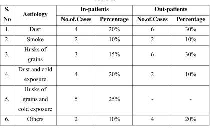
Noi Varum Vazhi (Etiology)
Yugi Vaidhya Chinthamani Says,
‘Ntfpd;w tjpfkhk; Gifap dhYk;
kPWfpd;w ghzj;jhy; kpf;Fe; jhNd”
- 690
‘ghdj;jhy; gukhf;fpdp kpFf;if ahYk;
ghukh khkprq;fs; Grpf;if ahYk;
jhzj;jhw; rQ;rhue; jtph;f;if ahYk;
rhpglhg; gjhh;j;jq;fs; Grpj;j yhYk;
jPzj;jhw; Grpahk ypUf;if ahYk;
Nrapio ahh;Nkypd;gQ; rpijt jhYk;
khdj;jhw; khJf;f kilj yhYk;
kjj;jhYQ; RthrkJ kUTq; fhNz”
Diet and habits
Excessive smoking
Excessive intake of cold water
Increased body heat
Excessive intake of non-vegetarian diet
Lack of Exercise
Taking improperly cooked food.
Starving on hunger
Mental stress
‘fhzNt Njtijf;Fg; gphpj;j gz;lk;
fsthbj; jpd;whYq; fztd;wd;idj;
NjhzNt epe;ijiar; nrhy;YtjhYQ;
Rrpahd gjhh;j;j nkr;rpy; gz;zpdhYk;
NtzNt XUth; nra;j ed;wp jd;id
kpf kwe;J nfhLikflhd; tpsk;GNthh;f;Fk;
NgzNt rigjdpNy nrhd;dg; Ngr;Rg;
Guz;Nlhh;f;Fq; fhrkJ gpwf;Fe; jhNd”
Character and Behaviours
Excessive Coitus
Over Stress
Stealing foods which were prepared for god
Cursing life partner
Tasting other’s foods
Forgetting one’s help
Madhava Nidhanam @ Roga Vrichayam says,
Gifapdhy; Rthr khh;f;fk; milgLjy;> Mkurk; Rthrhraj;jpy;
NrUjy;> mjpf tpahahkk; Urpahd md;dj;ijg; Grpj;jy;> tpKq;Fk; NghJ
czT mjd; ghijia tpl;L NtW topapy; gpuNtrpg;gJ> Ntfq;fis
mlf;FtJ> mt;tpjNk Jk;kiy mlf;FtJ.
Excessive smoke
Excessive gastric secretion and regurgitation
Taking improperly cooked food
Food enters into the larynx while swallowing
Controlling reflexes like sneezing.
Roga Nirnaya Saram Says,
Njfj;jpy; mf;fpdp mjpfupj;J neQ;R Gz;zhfp thA mNjhKfkhf
Nky;nrd;W khu;gpy; jq;fp cz;lhFk.;
Due to excessive body heat, gas ascends to the lung field, thus causes
the disease.
Siddha Maruthuvam (Pothu) Says,
,e;Neha; Fsph;fhw;wpyPLgly;> ntapypy; kpFjpAk; miyjy;> kpf;f
Fsph;r;rpiaj; jUk; nghUisAk;> #l;ilj; jUk; nghUisAk; cz;zy;>
ngupJk; cuj;Jg; NgRjy;> kpf;f fhug;nghUs;> ed;kzk;> jP kzk; Mfpatw;iw
Kfh;tjhYk; gpwf;Fk;.
Exposure to cold weather
Over strain in hot climate
Taking cold and hot foods
Singing in high pitched voice
Due to irritants like dust, mud, lime etc.,
Thanvandri Vaidhyam Says,
‘murNuh fe;jdf;Nf aikr;ruha; fhrNuhfk;
jiu kpir khe;jh; jk;ikr; rhh;e;jpLk; tifNah jd;dp
Yukpir fpNyre; jq;F KWJa uhY khjh;
jUkayhYe; J}kQ; rhh;Jfs; Kfh;e;jjhYk;.”
Over stress
Excessive Coitus
Inhalation of dusts, pollens etc.,
Anubava Vaitheya Deva Ragaseyam says,
thj fgq;fspd; tpUj;jp> mrPuz Ngjp> the;jp, tp\g;ghz;L> tplhr;Ruk;>
Gif> fhw;W> jhdpar;Rid> mjprPjsf;fgk;> kh;k jhdq;fspy; mbgLjy;
Kjypa fhuzq;fspdhy; cz;lhFk;.
Excessive Vadha and Kaba.
Diarrhoea due to indigestion.
Toxic anaemia.
Persistance fever.
Excessive cold.
Outer coat of the grains and pulse.
Trauma in the genital organs.
Murkurigal (Preliminary Signs)
njhz;il Gz;gl;lJ Nghy Nehjy;> gpd; njhz;il rptj;jy;>
njhz;ilapy; Ks;shy; Fj;JtJ Nghd;w czh;r;rp Vw;gly;> FuNyhir
Fiwjy;> %f;F ePh; gha;jy;> khh;G Nehjy;> #Ls;s nghUspy; tpUg;gk;
Kjypad cz;lhFk;.
Sore throat
Pricking sensation in the throat
Decreasing the pitch of voice
Running nose
Pain in the chest
Desire towards hot foods
Noi Enn (Classification)
Yugi Vaidhya Chindamani says,
Swasakasam is described as one of the twelve types of Kasam.
The twelve types are,
1. Mandhara kasam
2. Pakka Mandhara kasam
3. Sudar kasam
4. Vadha Kasam
5. Pitha Kasam
6. Swasa Kasam
7. Ratha Kasam
8. Silethuma Kasam
9. Peenisa Kasam
10. Vadhapitha kasam
Sikitcha Rathna Dheepam @ Vaidhya Chinthamani Says
There are twelve types of Kasam. They are
1. Mandhara Kasam
2. Patcha Mandhara Kasam
3. Sudar Kasam
4. Vadha Kasam
5. Pitha Kasam
6. Swasa Kasam
7. Ratha Kasam
8. Silethuma Kasam
9. Peenisa Kasam
10. Vadha pitha Kasam
11. Pitha Silethuma Kasam
12. Thontha Kasam
Raja Vaidhya Bodhini - Part I says
There are twelve types. Those are
1. Vadha Kasam
2. Pitha Kasam
3. Sethuma Kasam
4. Vadha Pitha Kasam
5. Pitha Sethuma Kasam
6. Mandhara Kasam
7. Swasa Kasam
8. Shaya Kasam
9. Sudar Kasam
10.Peenisa Kasam
Jeeva Rakshamirtham Says,
There are five types of Kasam. These are,
1. Vadha Kasam
2. Pitha Kasam
3. Silethma Kasam
4. Ratha Kasam
5. Shaya Kasam
Agasthiyar – 2000 Says
There are eight types. Those are
1. Vadha kasam
2. Pitha kasam
3. Kaba mandhara kasam
4. Pakka mandhara kasam
5. Mandhara kasam
6. Sudhika kasam
7. Marundheedu kasam
8. Kasam
Anubava Vaidhya Deva Ragasiyam Says,
There are five types. Those are,
1. Vadha Kasam
2. Pitha Kasam
3. Silethuma Kasam
4. Ratha Kasam
Tamilaga Siddha Vaitheya Gurugulam says,
There are twelve types. Those are.
1. Vadha kasam
2. Pitha kasam
3. Kaba kasam
4. Vadha pitha kasam
5. Pitha kaba kasam
6. Thontha kasam
7. Mandhara kasam
8. Patcha mandhara kasam
9. Ratha kasam
10. Peenisa kasam
11. Sudar kasam
12.Swasa kasam.
SWASA KASAM
Kuri Gunangal
The signs and symptoms are described in many siddha literatures. They
are described as follows,
Yugi Vaidhya Chintamani Says,
‘tz;ikaha; Nfhio fl;o ,UkP tPOk; khehfk; NghyNtthq; FQ;R thrk;
jpz;ikaha; nrUkYz;lh kbf;f bf;Fr;
rPuzapy; yhkNyt apW CJk;
ed;ikaha; ehrpaJ jdy;Ngh yhFk;
espe;J lk;Gtw;wptUk; FuYq; fk;Kk;
According to Yugi Vaidhya Chinthamani the characteristic features of
Swasakasam are cough with expectoration, breath sound like hissing of snake,
throat irritation, indigestion, flatulence, redness of the nose, low pitched voice,
excessive salivation.
Uyir Kakkum Siddha Maruthuvam @ Athma Ratchamirtham says,
cly; cyh;e;J tUk;> Ruk;> Fsph;> ,Uky;> Mahrk;> jiytyp fhZk;.
tapW nghUkp the;jp gz;Zk,; kyk; fl;b tpah;it> jhfk; kpFk;> Gwe;jhs;
mijf;Fk;.
Dryness of the skin, fever, rigor, cough, malaise, head ache, vomiting
because of indigestion, sweating due to constipation, excessive thirst, pedal
oedema present in Swasakasam.
Raja Vaidhya Bodhini - Part I says
thj ehbAk;> gpj;j ehbAk; xUq;F Nrh;e;J Mikiag; Nghy; nky;y Cwp
elf;fpy;> tapW ,iur;ry;> md;dQ; nrhpahik> Nfhio fl;ly;> ,Uky;> ehrp
tul;ly;> Fuw; fk;ky;> KJnfYk;G vhpr;ry;> Nkw;Rthrk;.
While the vadha pulsation and pitha pulsation is felt like the movement
of tortoise, Swasakasam characters are flatulence, indigestion, mucoid sputum,
cough, dryness of nose, low pitched voice, burning sensation along the
vertebral column, dyspnea.
Tamilaga Siddha Vaitheya Gurugulam says,
mjpfkhd NfhioAld; ,Uky;> ehfg;ghk;gpd; rPwiynahj;j
rg;jj;Jld; Rthrj;jpy; rPw;wk;> rPuzkpd;ik> tapw;Wg;gprk;> %f;fpy; mdNyhL
twl;rp> cly; tw;wy;> Fuw;f;fk;ky;> %r;R jpzwy;> cs;ehf;fpy; totog;ghd
eP&wy;> msTf;F kPwpa ,Og;G> ,rpT.
Cough with expectoration of large quantity of sputum, breath sound like
Mukkutra Verupadugal (Pathology)
In siddha system of medicine, the manifestation of all the diseases are
the result of derangement of Doshas i e., Vadham, Pitham, Kabam. The prime
factor which is involved in Swasakasam is Kaba, which is accompanied with
vitiated Vadha or Pitha so and produce clinical symptoms of Swasakasam. This
is clearly indicated by Theraiyar as ,
‘fgj;jpid ad;wp fhr Rthrk; fhzhJ”
- Njiuau;
1. Excess of Kaba in the respiratory organs affects the Melnokku kal and
Uyir kal and so the vayu is not able to reach the terminal points of
respiration which producing gasping and laboured breathing.
2. Some authors says that the disease is caused by deranged Vadha. This
thought is also acceptable because the destruction of Vayu in the
respiratory tract is abnormally present.
3. Excessive intake of Pitha prompting diet induces Pitha Kutram. This
type of Pitha produce more heat and this heat goes to head resulting in
running nose, heaviness of head and neck, sneezing and also induces
formation of water vapours in the lungs and causing narrowing of air
passage, which leads to the onset of the disease. This is indicated as,
‘gpj;jNk kpFe;jh yPis
apUkYk; ngyj;J epw;Fk;”
- Neha; ehly; Neha; Kjy; ehly;
So the changes in the diet and habits which increase Vadha and Kaba
produces the clinical symptoms of Swasa Kasam.
When Pranan, the primary vayu is affected it leads to difficulty in
breathing and involvement of Udhanan leads to cough and sneezing.
Involvement of Kirugaran leads to running nose, cough, sneezing.
Involvement of Devathathan leads to tiredness. Involvement of Samanan
causes inability to control the other Vayus and causes loss of appetite.
Involvement of Sadhaga pitham leads to sluggishness. In Kaba, the
derangement of Avalambagam leads to dyspnea, cough, wheezing. In the seven
Udal Thathus, Saaram, Senneer are affected which leads to lethargy and
depression. In severe cases Oon and Kozhuppu are also affected leads to
symptoms of emaciation and body pain.
Piniyari Muraimai (Diagnosis)
The way of diagnosis is very important by which a physician can deal
the disease, then only he will rule out the cause of the disease which is the main
thing to be treated. Thiruvalluvar said,
‘Neha;ehb Neha;Kj dhbaJ jzpf;Fk; tha;ehb tha;g;gr; nray;”
- jpUf;Fws;
The diagnosis is based on four criterias
1. Poriyal arithal
2. Pulanal arithal
3. Vinathal
4. En Vagai Thervugal
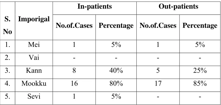
1. Poriyal Arithal
Porigal are the five organs of perception. They are Eyes, Ears,
Nose, Tongue, Skin. Poriyal Arithal is examining the Pori ofthe patient
Mei (Skin) : Normal
Vai (Tongue) : Excessive salivation
Kann (Eye) : Some times affected (redness)
Mookku (Nose) : Running nose
Sevi (Ear) : Normal
2. Pulanal Arithal
Pulangal are the five objects of senses.
Ooru (Sensation) : Warmth
Oosai (Sound) : Normal
Ozhi (Vision) : Normal
Suvai (Taste) : Normal
Naatram (Smell) : Altered or absent due to
running nose and inflammation of
nasal mucosa.
3. Vinadhal
By Vinadhal, the physician knows about the patient’s Name, Age,
Occupation, Native place(Thinai), Family history, Socio - economic status,
Diet habits, Prone to any allergens, (ex: dust, smoke, pollens) His complaints,
History of previous episodes, Frequency of attacks by changes in season,
aggravates factors relevant history of treatment and Habits etc.,
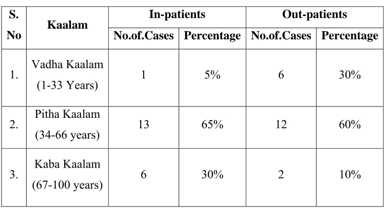
Kaalam (Age Distribution)
The period of human life is totally 100 years. This is divided into three
stages, according to the domination of three humours. As per this
1. Vadha Kaalam - 1 to 33 years
2. Pitha Kaalam - 34 to 66 years
Even though in each of these stages, the other humours are also involved
a particular humour is dominating more. According to this, Kaba types of
diseases are more prone in the later stage (Kaba Kaalam).
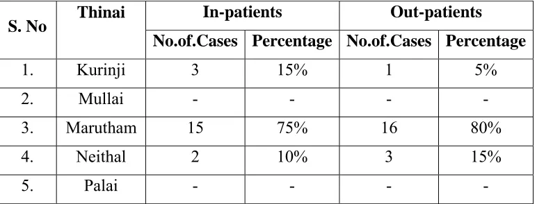
Ivagai Nilangal
Study of Ivagai Nilangal is very important and useful because there may
be possibility of the disease in some area. Ivagai Nilangal are,
A. Kurinchi - Mountains and its surroundings
B. Mullai - Forests and its surroundings
C. Marutham - plains and its surroundings
D. Neithal - Seas and its surroundings
E. Palai - Deserts and its surroundings
Kurinchi
‘FwpQ;rp tU epyj;jpw; nfhw;wKz;b uj;jk;
cwpQ;rp tU RuK Kz;lhk;- mwpQiuf;
ifaNk jq;Fjuj; jhikty;iy Aq;fjpf;Fk;
IaNk jq;F kwp.”
- gjhHj;j Fz rpe;jhkzp
In Kurinchi Nilam, people are affected by fever that reduces blood level
in the body, diseases related to spleen and liver and mainly by Kaba diseases.
Mullai:
’Ky;iy epyj;j ika Ke;epiu NktpDkt;
nty;iy epiyj;j gpj;j nka;JWq;fhz;- my;yntdpd;
thjnkhop ahjjDz ;kd;D kittop Neha;g;
Ngjnkhop ahjiwag;; gpd;G.”
- gjhHj;j Fz rpe;jhkzp
Though Mullai Nilam is the place of cattles, it is the place of increasing
Marutham
‘kUjepy ed;dPH tsnkhd;iwf; nfhz;Nl
nghUjepy khjpaNeha; Nghf;Fq; - fUjepyj;
jhwpujQ; #o tUe;Jtnud; whw;gpzpnay;
VwpujQ; #o;tpf;F kpy;”
- gjhHj;j Fz rpe;jhkzp
Marutha Nilam, due to its water sources, cures all the three Vatha, Pitha
and Kaba diseases.
Neithal
‘nea;;jdpy NkYth;g;ig ePq;fh JwpDkJ
nta;jdpy Nkjq;F tPlhFk; - neha;jPd;
kUq;Fliy Kf;fhf;fp ty;YWg;ig tPf;Fq;
fUq;Fliyf; fPopwf;Fq; fhz;”
- gjhHj;j Fz rpe;jhkzp
Through Neithal Nilam has the dominant taste of Uvarppu (Salty), it is
the place of Pitha Vayu. The people who dwell here are susceptible to odema
due to Kaba, Silipatha Rogam (Filariasis), Kudalanda Viruthi (Hernia).
Palai
‘ghiy epyk;Nghw; gliug; gpwg;gpf;f Nkiyepy kPahJ tphpj;jw;F - Ntiyepy
Kg;gpzpf;F kpy;yhk; KiwNa atw;whyhk;
vg;gpzpf;F kpy;yh k‡njz;”
- gjhHj;j Fz rpe;jhkzp
The Palai Nilam is the place for grief and place of deadly Vadha, Pitha
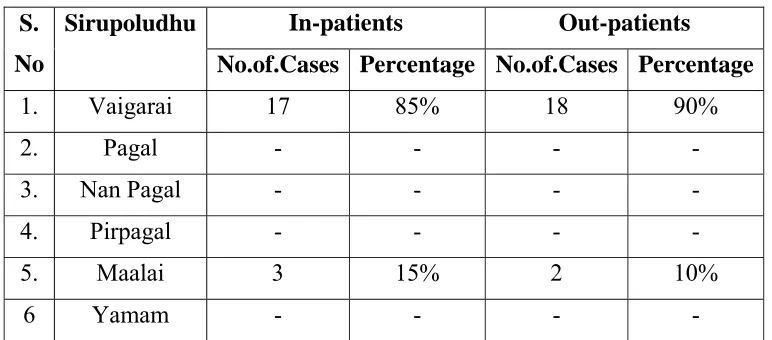
PARUVA KAALAM (Season)
As the earth revolves around the sun it gets sunlight at various positions.
With reference to the position of the earth towards the sun, year is divided into
six seasons.
They are,
1. Karkaalam (Avani & Purattasi) : August & September
2. Koothirkaalam (Iyppasi & Karthigai) : October & November
3. Munpanikaalam (Margazhi & Thai) : December & January
4. Pinpanikaalam (Masi & Panguni) : February & March
5. Elavenilkaalam (Chithirai & Vaigasi) : April & May
6. Mudhuvenilkaalam (Aani & Aadi ) : June & July
According to literature, Swasakasam comes during rainy season
(Karkaalam). In Koothirkalam also due to cold wind there is increased
incidence of disease.
Swasakasam mainly occurs due to vitiation of Kaba. Kabam thannilai
sirappurum Kaalam - Karthigai to masi.
‘%tU kPwp KdpT nfhshky;
jj;jk; epiyapy; jd;durpaYk;
fhytiujid fpsuf; Nfz;kpd; Mbahjpaha; Ig;grp <uha; Mdpykjw;Nfh uhrpay; fhyk;
kPd; Kjyhsp tPWnfhs; ke;jphp Njs; Kjd; khrp Nrdhgjpf;Nf”
Njs; - fhHj;jpif
- Neha; ehly; Neha; Kjdhly;
Hence the disease can occur in the later part of Koothir Kaalam to early
part of Pinpani Kaalam, (i.e,) from the last two weeks of October to the first
7. MUKKUTRA NILAIGAL
VADHAM
Pranan
It is responsible for respiration.
In Swasakasam, Vayu is affected leading to difficulty in breathing.
Abanan
It helps in excretion of urine and motion.
In Swasakasam, some patients had constipation.
Viyanan
It’s main function is distribution of saaram.
Samanan
Samanan is the vayu that controls other vayus and digestion.
In Swasakasam, this vayu is affected since it cannot control the other
vayus.
Udhanan
Its main function is inspiration and expiration and distributes the saaram
equally to all tissues.
In Swasakasam, this vayu is affected due to difficulty in breathing.
Nagan
This vayu maintains opening and closure of eye lids and is not affected
in Swasakasam.
Koorman
This vayu is responsible for vision and yawning.
Kirugaran
In Swasakasam, this vayu is deranged causing running nose, sneeze,
cough and loss of appetite.
Devathathan
It is responsible for tiredness, anger and emotional expression.
In Swasakasam, this vayu is deranged causing emotional stress and
insomnia.
Dhananjeyan
It produces swelling of the body after death and escapes through the
scalp after the third day of death.
PITHAM
Anal pitham
This lives in the stomach and helps in digestion.
In Swasakasam, most of the patients complained loss of appetite and
indigestion.
Ranjagam
This is residing in stomach and gives colour to the blood.
Sadhagam
It resides in the heart and executes the day to day activities with the help
of mind and brain.
In this disease, restlessness, breathlessness present.
Aalosagam
It resides in both eyes and is responsible for clear vision.
Prasagam
It resides in skin and gives complexion.
In Swasakasam, some patients may have eczema with blackish
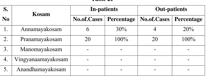
KABAM
Avalambagam
It is residing in lungs and helps other four types of Kaba to function and
also helps in the function of heart.
It is deranged in Swasakasam patients, since the presence of tightness of
chest, cough, wheezing, and dyspnea.
Kilethagam
It is present in the stomach and gives moistures to the food materials and
also helps in digestion.
In this disease, some patients have indigestion.
Pothagam
Living in the tongue and responsible for taste sensation, is not affected
in Swasakasam patients.
Tharpagam
Living in the head and keep the eyes cooling.
In Swasakasam, there may be redness of eyes.
Sandhigam
It resides in the joint and helps for free movements.
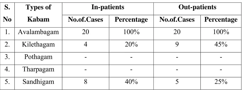
UDAL KATTUGAL
Saaram
It is the energy part of end product of digestion.
It strengthens the body and mind. It is deranged in Swasakasam due to
loss of appetite causing tiredness in the body and mind.
Senneer
Oon
It gives the structure to the body and is responsible for the movement of
the body and is not affected in Swasa kasam.
Enbu
It gives the shape to the body and is responsible for motion of the body
is not affected in Swasa kasam.
Kozhuppu
When the organs are doing their work this gives lubrication and
facilitates their work and is not affected in Swasa kasam.
Moolai
It is present in the core of the bone which strengthens and maintains the
normal condition of the bone, is not affected in Swasakasam.
Sukkilam / Suronitham
It is responsible for reproduction.
When the seven Udal Katukal increase or decrease from the normal
level, the normal functioning of the body is affected.
4. EN VAGAI THERVUGAL
It is the basic diagnostic principle and the uniqueness of the Siddha
system of Medicine. The following lines reveal this as follows.
‘ehb];ghprk; ehepwk; nkhoptpop
kyk; %j;jpukpit kUj;Jt uhAjk.;”
- Neha; ehly; Neha; Kjdhly;
And,
‘nka;f;Fwp epwe;njhdp tpop ehtpUkyk; iff;Fwp”
The diagnostic value of EN VAGAI THERVUGAL is specific to Siddha
system of Medicine and presumes the vitiated doshas in the patients.
En Vagai Thervugal are,
a. Naa (Tongue)
b. Niram ( Colour of the skin)
c. Mozhi ( Speech)
d. Vizhi ( Eye)
e. Malam ( Motion)
f. Moothiram (Urine)
g. Sparisam ( Palpation)
h. Naadi ( Pulse)
a. Naa
It is noted for its colour, ulcer, growth, coating, colour and consistency
of the sputum that is spitted from mouth.
In Swasakasam, patients have the sputum scanty and mucoid.
b. Niram
Colour of skin, conjunctiva, and teeth.
In Swasakasam, the colour of the skin, conjunctiva, may be pale. In
some patients, the conjunctiva may be red due to conjunctivitis.
c. Mozhi
Generally, speech is generated from the voice box. Abnormalities are
low pitched speech, lalling, diplegia, monotonous speech, jerky,
scanning, hot potato, indistinct, lisping.
In Swasa kasam mode of speech may be emotional or difficulity in
d. Vizhi
Type of eye - redness, ulcer, pallor, protrusion, tears, shedding of eye
lashes, excreta of eye, diseases of eyes are noted.
In Swasa kasam, the eyes may be red.
e. Malam
Consistency hard or semisolid or diarrhoea, undigested food, fluid
resembling the water used to clean meat, colour, frothy, dysentery,
bloody, pus, mucous, smell, frequency of defaecation, constipation,
reduced or increased stool content, lower abdominal pain during
defaecation are noted.
In Swasa kasam, the patients may be constipated.
f. Neer @ Moothiram
Colour - yellow, black, white copper coloured, mixed colour, colour of
fumes, smell-smell of fire, honey, sweet odours, fragrance of flowers,
fruity odour, odour of deer flesh, frothy or not, frequency and quantity
are noted.
In Swasa kasam it may be transparent and frothy.
g. Sparisam
Heat or coldness of the body.
It may be cold due to sweating in this disease.
h. Naadi
Naadi is the very important helpful observation for diagnosis and
prognosis.
In Noi Naadal Noi Mudhal Naadal Text, Naadi is defined as,
‘clypy; capH jhpj;jpUg;gjw;Ff; fhuzkhd rPtrf;jp
Genesis of Naadi
The three Uyir Thathukkal are formed by the combination of three
Naadis and three Vayus .
Idakalai + Abanan = Vatha
Pinkalai + Pranan = Pitha
Suzhumunai + Samanan = Kaba
This can be felt one inch below the wrist on the radial side by means of
palpation by the three fingers - index, middle and ring fingers corresponding to
vadha, pitha and kaba respectively.
‘fhpKfdbia tho;j;jpf; ifjdpy; ehbghHf;fpy;
ngUtpuyq;Fyj;jpy; gpbj;jb eLNt njhl;lhy;
xU tpuNyhby; thjKaH eLtpuypw; gpj;jk;
jpUtpuy; %d;wpNyhby; Nrj;Jk ehbjhNd”
- mfj;jpaH ehb.
Naadi Nadai in Swasa Kasam
Vatha Kaba Naadi
‘ghq;fhd thjj;jpy; Nrj;Jk ehbg;
ghprpj;;jhy; jpkpH NkT Kisr;ryhFk;
jPq;fhd ,UkYld; re;jp Njhlk;
NrHe;j tplk; ntb#iy apUj;Nuhfk;
thq;fhj <is ke;jhu fhrk;
typAlNd GwtPr;RAs; tPr;R tPf;fk;
Xq;fhZQ;Ru KlNd Rthr fhrk;
cz;lhFk; ntF Neha;f;F KWjpjhNd”
Iya Naadi
‘jhdKs;s Nrj;J ke;jhdpsfpy; ntg;G>
rakPis apUky; ke;jhu fhrk;
<dKWQ; re;eptpl Njhlk; tpf;fy;
apUj;Nuhfq; fug;ghd; tpuz Njhlk;
khdidaPH #iyjpus; tpahjp tPf;fk;
tUQ;rf;jp Rthrk; neQ;rilg;G J}f;fk;
VdKWq; fhkhiy ghz;L Nrhig
VORuq;fs; gyJf;fk; tpl Kz;lhNk”
- rjf ehb
Kaba Pitha Naadi
‘,lkhd Nrj;Jkj;jpy; gpj;j ehb
vOe;jZfpy; tplKlNd tPf;fKz;lhk;
jplkhd FspH fha;r;ry; kQ;rs; NehTe;
Njfj;jp Yisr;rypisg; gpUky; the;jp
Tplkhd neQ;rilg;G Rthrk; tpf;fy;
ntF RuKk; ehtwl;rp ghz;LNuhfk;
mlkhd Ftisuj;j kjprhue;jhd;
mZfp ntFgy Neha;f;Fe; jlq;fz;lhNa”
- rjfehb
Iya Ushnam
‘fjpg;ghd Nrj;Jkj;jpYl;bzq; $by;
fye;j FzQ;rakpUky; Rthrfhrk;
kjpg;ghd Nfhiouj;jk; tpg;GUjpAlNd
tsHehrpfh gPlkpUj; Nuhfk;
nfhjpg;ghd rpq;qitahf; fpuhzthA
nfhl;lhtp tpf;fy; ke;jhufhrk;
Jjpg;ghd tPuyj;jpf; fha;Tuj;jk;
Iya Vayu
‘njhe;jpj;j Nrj;Jkj;jpy; thA$bj; njhlHe;j
Fd;kk; neQ;rilg;G Rthrfhfk; te;jpj;j Fuy;jdpNy cWj;j yPis
tOtOg;G ePUwy; kyj;jpy; rPjk; nte;jpj;jy; nfhOj;jy; Fj;Je; jpkpHtpahjp
tPr;RlNd typnal;Le; jpul;;rp ghz;l me;jpj;j fpWfpWg;G kaf;fk; tpf;fy;
Mdgy gpzpfSNk te;jl Ue;jhNd”
- rjf ehb
Hence the Naadi Nadai in Swasa kasam is Kaba, Vadha kaba, Kaba
Pitha, Iya Ushna, and Iya Vayu Naadis.
Nei Kuri
This urine examination is unique in Siddha system of Medicine.
For this examination urine is collected in the early morning in a pure glass
vessel. Patient is advised to take a balanced diet and avoid excessive diet, and
in take of diet during irregular timings on the previous day of examination.
‘mUe;Jkhwp ujKk; mtpNuhjkjha; mfy; myHjy; mfhyT+d; jtpHe;jow;
Fw;wstUe;jp cwq;fp itfiw Mbf;fyrj; jhtpNa fhJ nga;
njhUK$Hj;jf; fiyf;Fl;gL ePhpd; epwf;Fwp nea;f;Fwp epUkpj;jy; flNd”
- rpj;j kUe;Jthq;fr; RUf;fk;
‘epwf;Fwpf; Fiuj;j epUkhz ePhpw;
rpwf;f ntz;nza;NahH rpWJsp eLtpLj; njd;Wwj; jpwe;njhyp Nafhjikj;jjp
A drop of gingelly oil is dropped on a wide glass vessel containing the
urine to be tested which is kept under sunlight in a calm place. The
derangement of the three dhoshas can be diagnosed by the mode of spread of
gingelly oil on the surface of urine.
‘muntd ePz;bd; m/Nj thjk;”
‘MopNghw; gutpd; m/Nj gpj;jk;”
‘Kj;njhj;J epw;fpd; nkhoptnjd; fgNk”
- Neha; ehly; Neha; Kjdhly;
Oil spreading like a snake indicates Vatha.
Oil spreading like a ring indicates Pitha
Oil spreading like a pearl indicates Kaba
‘mutpyhopAk; Mopapy; muTk;
mutpy; Kj;Jk; Mopapy; Kj;Jk;
Njhw;wpy; njhe;j Njhlq;fshNk”
- Neha; ehly; Neha; Kjdhly;
Oil spreading like snake and ring, ring and snake, snake and pearl, ring
and pearl all comes under Dhondha Dhosham.
In Swasa kasam, most of the Nei Kuri findings result- pearl like oil
DIFFERENTIAL DIAGNOSIS
DISEASES SIMILAR TO SWASAKASAM are,
Mandara Kasam
'jhdhd J}aNjhH ehrp jd;dpy;
ryNeha; ePHjhd; tpOe;j Jk;kYz;lhk;
khdhd khHGneQ; rilj;J %r;R
tYthf ghk;GNghy; rPw yhFk;
fhdhd fz;lNkhL KfKq; fhJk;
fhakJq; frpthfp tpaHit ahFk;
Vdhd ,UkNyhL Nfhio fk;ky;
,iug; ghF ke;jhufhr khNk”
- A+fp itj;jpa rpe;jhkzp
In Mandara kasam there is running nose, sneezing, tightness of chest,
breath sound like hissing of snake, sweating all over the body, cough,
expectoration, dyspnea, etc.,
In Swasa kasam there is no sweating all over the body.
Kandakiragam
‘tifahd Fuyjidg; gw;wp nehe;J
khHNghL gplhpapdpy; typ Az;lhfp
Efuhd rhPunky;yhk; nehe;j ohw;wp
EZf;fkha;r; RthrkJ Gwg; glhky;
Kifahd ehthNt %r;R khwp
Kfj;jpNy tpaHthfp tpyhNeh Tz;lhk;
Gifahd td;dj;ijg; gUnfhl; lhJ
ghpafz;l fpufj;jpd; gz;G jhNd”
In Kandakiragam, there is difficulty in speech, pain in the chest and
occipital region, pain all over the body, breathlessness, buccal respiration,
sweating in face, pain in the ribs, anorexia.
In Swasa kasam, there is no pain in the occipital region.
Swasa Pitham
‘fUj;jhfr; RthrkJ kpfTz; lhFq;
fdkhf tapWNk Cjpf; fhZk;
cUj;jhf clyJjhd; kpft ypf;F
%WNk Nfzp Nghy; tha;ePH jhDk;
kUj;jhf kaq;fpNa fz;k iwf;Fk;
khHgpNy typNahL ,Uk Yz;lhe;
JUj;jhf tapwjdpd; grpNah tpy;iy
Rthrkhk; gpj;jj;jpd; #l;re; jhNd”
- A+fp itj;jpa rpe;jhkzp
In Swasa Pitham, there is increased respiration (tachypnoea), flatulence,
pain all over the body, brushing, loss of consciousness, pain in the chest
followed by cough, loss of appetite etc.,
In Swasa kasam there is no loss of consciousness.
Swasa Silethumam
‘jpwikaha; neQ;Rjdpw; Nfhio fl;LQ; rpf;nfd;W jhdpUkp %f;f ilf;Fq;
FWikaha; Fwl;nld;W Rthrq; fhZq;
FspNuhL RuKz;lha; kaf;f khFk;
kwikaha; khHNghL neQ;r ilf;Fk;
tha;twz;L %f;fjdpy; ePNu ghAk;
ntWikaha; kpfj;jz;zPH jhg Kz;lha;
tpLRthr rpNyl;Lkj;jpd; tptue; jhNd”
In Swasa Silethumam, there is congestion in lungs, nasal congestion,
cough, dyspnea, fever with rigor, syncope, tightness of chest, dryness of mouth,
running nose, excessive thirst etc.,
In Swasa kasam, there is no fever with rigor, excessive thirst etc.,
Silethuma Vadha Suronitham
‘gz;ghf Tly;FspHe;J tapW tPq;fpg;
gijg;ghd tple;njhl;lhw; Nghy; Nehthe;
jpz;ghd rpuR new;wp Nehf;fh Lz;lhQ;
rpNyl;Lkkha;f; NfhioNahL RthrkhFk;
kz;ghf kaf;fnkhL fdT Kz;lhk;
tha;twz;l Urpapy;yh tUj;;j khFk;
ez;ghf ehbANk glg; glf;Fk;
ew;rpNyl;k RNuhzpjkhk; ehLq; fhNy”
- A+fp itj;jpa rpe;jhkzp
In Silethuma Vadha Suronitham, there is chillness of body, abdominal
distension tenderness in abdomen, headache, expectoration, dyspnea, fainting,
dreaming, decreased salivation, loss of taste, rapid pulse etc.,
In Swasakasam, there is no abdominal distention and decreased
salivation.
LINE OF TREATMENT
The line of treatment of SwasaKasam consists of the following.
1. Kalichal Maruthuvam - To bring the dhoshas in equilibrium
2. Internal Medicine - Mainly anti-spasmodic, expectorant, to relieve the
spasm and to expel the sputum.
3. Diet - To maintain tridhoshas and energy in equilibrium.
5. Yoga therapy - To maintain dhasa vayukkal and to improve mental and
physical health.
1. Kalichal Maruthuvam (Purgation)
Patients are given laxative Nilavagai Chooranam 5 gm with hot water at
the bed time on the previous day, after starting the internal medicine.
2. Administration of internal medicine
For the treatment of the disease Swasakasam, Kasa Chooranam – 1 gm
and Anna Pavala Chenduram – 100mg thrice daily with honey was given
before meals.
3. Diet
Siddhars advice the diet regiments for Kaba patients and they are
explained below.
‘fj;jhp Nga;Gly; tiu apUghfy; gUq;fhsh fz;lfhhp
mj;jpf; fha;fSk; tUf;ifkhgaw;iw fiuahy; gPHf;fUk; gpQ;RNtH
nkha;j;j #uzq; fjypj; jz;Lfisg; G+Ksq;fp KUf;fUk;Gk;
mj;jp G+rzpf; fhaUs;sp ts;spAq; fgj;NjhHf; fhzkhNk”
-gjhHj;j Fz rpe;jhkzp
‘Ntis kzj;jf;fhsp nkd;rPij rf;futHj;jp
gPis triy Rf;F ngz;fzq;fs; - Ntisapy;
nre;jspH fisf;fPiu nra;tH fgNjfH epjk;
te;jspAzj;jhd; kfpo;e;J”
- gjhHj;j Fz rpe;jhkzp Vegetables to be added
fj;jhp (Solanum melongena)
Nga;Gly; (Trichosanthes cucumerina)
mj;jp (Ficus glomavata)
gPHf;F (Luffa acutaugula)
khtL ( Mangifera indica)
thiof;fha; (Musa paradisica)
KUq;if ( Moringa tinctoria)
Rz;il (Solanum torvum)
Tubers to be added
Ks;sq;fp (Raphanus sativus)
<Us;sp ( Allium sativum, Allium cepa)
,Q;rp (Zingiber officinale)
fUizj;jz;L ( Amorphophallus companulatus)
Greens to be added
kzj;jf;fhsp (Solanum Nigrum)
fhprhiy (Eclipta alba)
gPis(Aerva lanata)
triy ( Bascella alba)
rpWfPiu ( Amaranthus gangeticus)
kzypf;fPiu(Gisekia pharmacoides)
gul;ilf;fPiu(Justicia madurensis)
Gspahiuf;fPiu (Oxalis corniculata)
Diet Restriction
Siddhars advice to avoid certain food items during diseased conditions.
They are,
‘fLF ew;wpyj; njz;nza; $o;ghz;lq; fliy
tLt jhfpa njq;Fkh tUf;if ew;fha
Mustard Gingelly oil
Bengal gram Coconut
Mango Jack fruit
Garlic Horse gram
Tobacco Alcohol
Bitter guard Sesban
Asafoetida
And also, they are advised to avoid coitus
These are general diet and habitual restrictions for all diseases.
Kaba patients should restrict the followings also.
Onion
Jaggery
Curd
Butter
Ghee
Fish
Dry Fish.
Prevention
1. Avoid chill and cold weather
2. Avoid working in dust, cement, cotton mills and in husks.
3. Avoid smoking
4. To sleep in phoenix mat, prevent Kaba diseases.
‘rpw;wPr;Rg; ghapw; wpdKk; gLg;gtUf; Fw;wpLNk fhe;j Ylk;GtUQ;-Rw;wpaNjhH thATWk; gpj;jkW kw;Wq; fge;jPUe;
jhafkh kpf;Fzj;ijr; rhw;W”
PRANAYAMAM (Breathing Exercise)
Pranayamam or breathing exercise mainly consists of Pooragam
(inhalation of air by deep inspiration), Kumbagam (holding the breath as far as
possible) and Resagam (exhalation of air by expiration)
During breathing exercise, the lungs filled with fresh air in its
anatomical dead space also and expand well and get proper supply of oxygen
by proper expansion of chest. So, Pranayama practice is one of the prevention
for SwasaKasam.
By this exercise, the duration of Kumbagam is increased. This results in
proper gaseous exchange which provides increased oxygen supply to the cells.
By the regular practice of Pranayamam, one can get rid mental and
physical stress and enjoy pleasure. It provides good concentration and
meditation. This practice also gives good appetite, strength, enthusiasm, rigor
and vitality.
‘ehnshd;Wf;F ,Ugj;Njhuhapuj;J mWE}W
eykhd Rthre;jhNd Oe;jpUf;Fk;
Nfhnshd;wpg; gjpdhyhapuj;J ehD}W
Ftpe;j %yhjhuj;Js; nshLq;Fk;
ghnshd;wp Naohapuj;jpUE}W Rthrk;
ghopdpw; gha;e;jpLnkd; wwpfg;gpd;id
Vnshd;wp apjidNa Al;rhjpj;jhy;
vg;nghOJk; ghyuh apUf;fyhNk”
YOGA THERAPY
Yogasana is one of the most spiritual legacies gifted by our ancient
sages. The practice of asanas strengthens the body and mind.
Asanas strengthen the muscles of respiration and diaphragm as well as
regulate respiration. So, practising asanas is more helpful in asthmatic patients
as supportive therapies. The following asanas are helpful in Asthma.
Bujankasanam
Chakrasanam
Machasanam
Mayurasanam
Patha hasthasanam
Arai machayendhirasanam
Trikonasanam
Savasanam.
MODERN ASPECTS
ANATOMY & PHYSIOLOGY OF RESPIRATORY
SYSTEM
The respiratory system brings air in close relationship with the mixed
venous blood enabling tissue respiration by uptake of oxygen into the
circulation and elimination of carbon dioxide.
The organs of the respiration are
Nose
Pharynx
Larynx
Trachea
Two bronchi
Bronchioles and small air passages
Muscles of respiration - the inter costal muscles and the
diaphragm
Nose and Nasal cavity
Nose is lined by ciliated columnar epithelium which contains mucus
secreting goblet cells. The anterior nares or nostril are the openings from
exterior into the nasal cavity. The posterior nares are the openings from nasal
cavity into the pharynx.
The Nasal cavity is the first of the respiratory organs and consists of a
large irregular cavity divided into two equal passages by a septum. The
posterior bony part of the septum is formed by the perpendicular plate of
ethmoid, sphenoid, frontal and nasal bones. The floor is formed by the roof of
mouth consists of soft palate and hard palate.
The para nasal sinuses are air filled cavities in certain of the skull bones,
lined by mucous membrane and communicating with the nasal cavity. The
main sinuses are maxillary sinuses, frontal, sphenoidal and ethmoidal sinuses.
Respiratory Functions of Nose
The function of the nose is to begin the process by which the air is
warmed, moistened and filtered. The projecting choncha increases the surface
area and cause turbulance, spreading inspired air over the whole of the nasal
cavity. Warming is due to immense vascularity of the mucosa. Filtering and
cleaning of air occurs on hairs at the anterior nares traps layer particles.
Mucous protects the underlying epithelium from irritation and prevents drying.
Humidification occurs as air travels over moist mucosa and becomes saturated
with water vapour. Irritation of the nasal mucosa results in sneezing a reflex
action that forcibly expects an irritant.
Pharynx
Pharynx is the passage extending from the base of skull to the level of
6th cervical vertebra where it is continous with the oesophagus. 13 cm length,
35 cm width.
Pharynx is divided into three parts. Naso Pharynx , Oro Pharynx,
Laryngo Pharnx.
Naso pharynx is the nasal part of the pharynx is situated behind the nasal
cavity and above the level of the soft palate. Oro pharynx extends from the
level of the soft palate to the level of the upper border of the epiglottis, Laryngo
pharynx extends from the upper border of the epiglottis to the lower border of
Functions
Passage of air and food, warming and humidifying of air, taste, hearing
protection.
Larynx
The larynx is the voice box and serves as an air passages. Extends from
the root of the tongue at the inlet of the larynx to the commencement of the
trachea at the level of the 6th cervical vertebra. 4.3 cm length.
Functions
Production of sound, speech occurs during expiration when the sound
produced by the vocal cords in manipulated by the tongue, cheeks, lips.
Protection of lower respiratory tract from the swallowed food from
mouth. It is the passage for air between pharynx and trachea.
Humidifying, filtering and warming continue as the air travels through
the larynx.
Trachea
Trachea is the wind pipe. It starts at the lower border of the cricoid
cartilage and ends at the level of the upper border of the 5th thoracic vertebra by
dividing into two bronchi right and left. 11 – 12 cm length.
Functions
Support and potency. The arrangement of the cartilage and elastic tissue
events linking and obstruction of the airway on the head and the neck moves.
The cartilage prevents collapse of the tube, when the internal pressure is less
than intra thoracic pressure; get at the end of forced expiration.
Mucociliary escalator, this is synchronous movement of the cilia that
Cough reflex
Nerve endings in the larynx, trachea and bronchi are sensitive to
irritation that generates nerve impulse which is induced by the vagus nerve to
the respiratory centre in the brain stem. The reflex motor response is deep
inspiration followed with closed glottis. So the intra pleural pressures rises.
Then glottis is suddenly opened with explosive out flow of air at a higher
velocity. Irritates may be rapelled out of the respiratory tract.
Bronchi and Smaller Air Passage
The two bronchi are formed when the trachea divides at the level of 5th
thorcic vertebra. The right bronchus is a wider, shorter tube than the left
bronchus and it lies in a more vertical position. It is approximately 2.5 cm long.
After entering the right lung at the hilum it divides into three branches. Each
branch divides into numerous smaller branches.
The left bronchus is about 5 cm long and is narrower than the right.
After entering the lungs at the hilum, it divides into two branches one for each
lobe. Each lobe branch then sub divides into progressively smaller tubes with
in the lung substance.
Bronchi are composed of the same tissue the trachea. They are lined
ciliated columnar epithelium. The bronchus progressively subdivides into
bronchioles, terminal bronchioles respiratory bronchioles, alveolar duct and
finally alveoli.
Functions of air passage not involved in gas exchange
Control of air entry
The diameter of the respiratory passage may be altered by contraction
and relaxation of the involuntary muscles of their walls, thus regulating the
volume of air entering the lungs. These changes are controlled by the
The following functions continue as in the upper airways.
Warming and humidifying
Support and potency
Removal of particulate matter
Cough reflex
Respiratory Bronchioles and Alveoli
Lobules are blind ends of the respiratory tract, distal to the terminal
bronchus consist of respiratory bronchioles, alveolar duct and alveoli. The
walls gradually thinner until muscle and connective tissue fade out leaving a
single layer of simple squamous epithelial cells in the alveolar duct and alveoli.
These distal respiratory passages are supported by a loose network of
capillaries. The exchange of gases during respiration takes place across two
membranes - alveolar and capillary membrane.
Interspersed between the squamous cells are other cells that secrete
surfactant, a phospholipid, fluid which prevents the alveoli from drying out. In
addition, surfactant reduces the surface tension and prevents alveolar walls
collapsing during expiration.
Functions of respiratory bronchioles and alveoli
1. External respiration
2. Defence against microbes
Cells in connective tissue protect against infection and inhaled foreign
particles not trapped by mucous. Lymphocytes and plasma cells produce
antibodies in the presence of antigen and macrophages and poly morpho
nuclear lymphocytes are phagocytic. These cells are most active in the distal
Warming and humidifying continue as in the upper airways. Inhalation
of dry or inadequately humidified air over a period of time causes initiation of
the mucosa and facilitates the establishment of pathogenic microbes.
Lungs
Lungs are paired organs of respiration. They are situated one on each
side of the mediastinum with the thoracic cavity. Each lung resembles a half
cone. It has an apex, a base, medial surface and costal surface.
Right lung is broader than the left lung and weight 220zs,and is divided
in to three lobes, where as the left lung weight 200zs and is divided in to two
lobes.The apex is rounded and rises into the roof of the neck about 25mm
above the level of middle third of the clavicle. The base is concave and
semilunar in shape and is closely associated with the thoracic surface of the
diaphragm.
The costal surface in convex and in closely associated with the costal
cartilages, the ribs and the inter costal muscles. The medial surface in concave
and has roughly triangular shaped area, called hilum at the level of 5th, 6th, 7th
thoracic vertebra. Structures that enter and leave at the hilum are 1 bronchus, 1
pulmonary artery, 2 pulmonary veins, 1 bronchial artery, 1 bronchial vein,
lymph vessels, parasympathetic and sympathetic nerves. The area between the
lungs is the mediastinum. It is occupied by heart, great vessels, trachea, right
and left bronchi, oesophagus, lymph nodes, lymph vessels and nerves.
Pleura and Pleural Cavity
Each lung is covered by the pleural cavity. The pleura consist of a
closed sac of serous membrane, which contain small amount of serous fluid.
The visceral pleura is adherent to the lungs covering each lobe and posses into
the fissures which separates them. The parietal pleura is adherent to the inside
RESPIRATION
Inflation and deflation of the lungs ensues that regular exchange of gases
takes place between alveoli and external air. This is dependent upon the
arrangement of pleura and the contraction and relaxation of muscles of
respiration and the elastic connective tissue.
Muscles of Respiration
The expansion of the chest during inspiration occurs partly voluntary
and partly involuntary. The muscles of normal quiet breathing are the inter
costal muscles and the diaphragm. During difficult breathing they are assisted
by the muscles of the neck, shoulder and abdomen.
Cycles of Respiration
This occurs 12-15 times per minute and consists of three phases.
Inspiration
Expiration
Pause
Inspiration
The capacity of the thoracic cavity is increased by simultaneous
contraction of the inter costal muscles and the diaphragm. The parietal pleura
move with the walls of thorax and the diaphragm. This reduces the pressure in
the pleural cavity to the level considerably lower than the atmospheric pressure.
The visual pleura follow the parietal pleura. During the process, the lungs are
stretched, the pressure within the alveoli and the air passage reduced drawing
air into the lungs in an attempt to equalize the atmospheric and alveolar air
The process of inspiration is active as it requires expenditure of energy
for muscle contraction. The negative pressure created in the thoracic cavity aids
venous return to the heart and is known as respiratory pump.
Expiration
Relaxation of inter costal muscles and the diaphragm results in the
downward and inward movement of the rib cage and the elastic recoil of the
lungs. As this occurs, the pressure of the gases inside the thorax exceeds the
atmospheric pressure and therefore air is expelled from the respiratory tract.
The lungs still contain some air and are prevented from complete collapse by
the intact pleura. The process is passive as it does not require the expenditure
of energy.
After expiration there is a pause, before the next cycle begins.
Physiology Variables Affects Respiration
Elasticity
Loss of elasticity of the connective tissue in the lungs necessitates forced
expiration and increased effort of inspiration.
Compliance
This is the measure of distensibility of the lungs, I. e., the effort required
to inflate the alveoli when compliance is low, the effort needed to inflate the
lungs is greater than normal e.g. in some diseases where elasticity in reduced or
when surfactant is present insufficiently.
Air flow resistance
When this is increased e.g. in broncho constriction, more respiratory
effort is required to inflate the lungs.
Lung volumes and capacity
gases take place only across the wall of the alveolar ducts and alveolar. The
remaining capacity of the respiratory passages is called the anatomical dead
space (about 150ml).
Tidal Volume
It is the amount of air which passes into and out of the lungs during each
of quite breathing about 500ml.
Inspiratory Reserve Volume
It is the extra volume of air that can he inhaled into the lungs during
maximal inspiration.
Inspiratory Capacity
It is the sum of Tidal Volume and Inspiratory Reserve Volume.
Functional Residual Capacity
It is the amount of air remaining in the air passages and alveolai at the
end of quiet respiration. The functional residual volume also prevents the
collapse of the alveoli on expiration.
Expiratory Reserve Volume
It is the largest volume of air which can be expelled from the lungs
during maximal expiration.
Vital Capacity (VC)
It is the maximum volume of air which can be moved into and out of the
lungs.
Residual Volume
It cannot be measured directly but, it is the volume of air remaining in
the lungs after forced expiration.
Alveolar Ventilation
This is the volume of air that moves into and out of the alveoli per
minute. It is the tidal volume minus the anatomical dead space, multiplied by
the respiratory rate.
Alveolar ventilation = (TV-anatomical dead space) respiratory rate
= (500-150) ml x 15 per minute
= 5.25 liters / minute.
Lungs function tests are carried out to determine respiratory function
and are based on the parameters out lined above.
External Respiration
This is the exchange between alveoli and blood. Total area of gas
exchange in the lungs is 70-80 square meters. CO2 diffuses from venous blood
along the contraction gradient into the alveoli until equilibrium with alveolar
air is reached. By the same process O2 diffuses from alveoli to the blood.
Internal Respiration
This is the exchange of air between the tissue and blood. When there is
difference in partial pressures, oxygen diffuses outward from the blood to extra
cellular fluid then into the cell walls. The process involved is diffusion.
Control of Respiration
Control of respiration is normally involuntary. Voluntary control is
exerted during activities such as speaking, singing but is over ridded if
homostasis of arterial PO2 and PCO2 is threatened i.e. if this is high arterial
THE RESPIRATORY CENTRE
This is formed by group of nerve cells that control the rate and depth of
respiration. They are situated in
Brainstem
Medulla oblongata
Pons varoli
In the medulla there are inspiratory neurons and expiratory neurons.
Neurons in the pneumotoxic and apneutic centre situated in the pons influence
the inspiratory and expiratory neurons of the medulla.
Motor impulses leaving the respiratory centre pass in the phrenic nerves
and inter costal nerves top the diaphragm and inter costal nerves.
Chemoreceptor
These are the receptors that respond to the changes in PO2 and PCO2.
They are located centrally and peripherally.
Central receptor - present on the surface of medulla oblongata and
bathed in CSF. When PCO2 is raised even slightly, the central receptors
respond by stimulating respiratory centre, by increasing ventilation and
reducing PCO2. The sensitivity to raised PCO2 is the most important factor in
maintaining hemostasis of blood gases in health.
Peripheral chemoreceptors are situated in the arch of aorta and in the
carotid bodies.
An increase in H+ concentration stimulates the peripheral
chemoreceptors resulting in increased ventilation, increased CO2 excretion and
Speech, singing
Emotional displays
Drugs e.g. - sedatives, alcohol
Sleep
Temperature influences breathing. In fever, respiration in increased due
to increased metabolic rate while in hypothermia it is decreased. It is depressed
as in metabolism, temporary changes in respiration occur in swallowing,
BRONCHIAL ASTHMA
Bronchial Asthma is a disease of airways that is characterised by
increased responsiveness of the tracheo bronchial tree to a variety of stimuli,
resulting in widespread spasmodic narrowing of the air passages which may be
relieved spontaneously or by therapy.
Asthma is an episodic disease manifested clinically by paroxysms of
dyspnea, cough, polyphonic wheeze. However, a severe and unremitting form
of the disease termed status asthmaticus may prove fatal.
Prevalence
Asthma is common and prevalent world wide. It occurs at all ages but
nearly 50% of cases develop before the age of 10 years. In adults, both sexes
are affected equally, but in children there is 2:1 male – female ratio.
Aetiology
From the aetiological point of view, asthma is heterogenous disease. It is
useful for epidemiological and clinical purposes to classify asthma by the
principle stimuli. There are two types of asthma.
Early onset asthma (atopic, allergic, extrinsic)
Late onset asthma (Non-atopic, Idiosyncratic, Intrinsic)
Atopic Asthma
This is the most common type of asthma usually begins in childhood.
The disease is triggered by environmental antigens, such as dust, pollens,
animals dander, fungal spores and food. A positive family history of atopy is
common, and asthmatic attacks are often preceded by allergic rhinitis, utricaria
or eczema. Serum IgE levels are usually elevated. A skin test with antigen
Non – Atopic Asthma
This type of asthma develops later in adult life with negative personal
(or) Family history of allergy, negative skin test and normal serum levels of
IgE. Most of these patients develop typical symptom-complex after an upper
respiratory tract infection by viruses (e.g. rhinovirus, para influenza virus).
Associated nasal polyp and chronic bronchitis are commonly present. About
10% of patients become hypersensitive to drugs, most notably to small doses of
aspirin.
Pathogenesis of Asthma
The common denominator underlying the asthmatic diathesis is a
nonspecific hyper irritability of tracheo-bronchial tree. When airway reactivity
is high, symptoms are more severe and persistent and the magnitude diurnal
fluctuation in lung functions is greater. The patients tend to awaken at night or
in the early morning with breathlessness.
In both normal and asthmatic patients, air reactivity rises following vital
infections of the respiratory tract and exposure to oxidants such as ozone and
nitrogendioxide. Allergen can cause airway responsiveness to rise within
minutes and remain elevated for weeks.
A number of causes have been postulated for the increased airway
activity of asthma, but the basic mechanism remains unknown. The most
popular hypothesis at present is that of airway inflammation. Increased
numbers of mast cell, epithelial cells, neutrophils, eosinophils and lymphocytes
have been found in the broncho alveolar lavage fluid of patients with asthma &
have number of mediators.
The airway can be oedematous and infiltrated with eosinophils,
finding is a generalised increase in cellularity associated with an elevated
capillary density.
Although, the translation of this histological observation into the disease
is still incomplete, it is widely believed that the physiological and clinical
features of asthma derived from interaction among the residence and
infiltrating inflammatory cells in the airway and the surface epithelium. The
cells that play more important role are mast cells, eosinophils, macrophages,
neutrophils and lymphocytes. The mediators released are histamine,
bradykinin, the leukotrienes C, D, & E, platelet activating factor (PAF) and
prostaglandins (PGs) E2, F2a and D2 produce an intense, immediate
inflammatory reaction involving broncho constriction, vascular congestion and
oedema formation. In addition to their ability to evoke prolonged constriction
of airway smooth muscles and mucosal edema, the leukotrienes may also
account for some of other patho physiological features of asthma such as
increased mucous production and impaired mucociliary transport.
Chemotactic factors elaborated bring eosinophil, platelets and
polymorphic nuclear leukocytes to the site of reaction. These infiltrating cells
and resident macrophages and airway epithelial cells themselves potentially are
an additional source of mediators to enhance immediate and the cellular phase.
Like mast cells in the early reaction the eosinophils play an important
role in the infiltrative components. The granular protein in thin cell major basic
protein and the eosinophilic cationic protein are capable of destroying the
airway epithelium, which then sloughed into the bronchial lumen in the form of
creak bodies. Besides resulting in a loss of barrier and secretary function, such