FUNCTIONING OF DEEP LOBE OF THE
PAROTID GLAND AFTER SUPERFICIAL
PAROTIDECTOMY
A DISSERTATION SUBMITTED IN PARTIAL FULFILLMENT OF THE M.S. BRANCH 1 (GENERAL SURGERY) EXAMINATION
OF THE
TAMILNADU DR.M.G.R UNIVERSITY, CHENNAI,
CERTIFICATE
This is to certify that the dissertation entitled
“FUNCTIONING OF DEEP LOBE OF THE PAROTID GLAND AFTER SUPERFICIAL PAROTIDECTOMY”
is the bonafide original work of Dr. T. Joseph Victor Gnanadurai
during his academic term in Christian Medical College & Hospital, Vellore in partial fulfillment of the M.S Branch 1 (General Surgery) examination of the
Tamilnadu Dr.M.G.R University, Chennai to be held in March 2008.
Dr. John. C. Muthusami MS, Dr. Venkatramani Sitaram MS, FRCS(G), Professor & Head, Professor & Head,
Acknowledgements:
Firstly, I thank God for giving me the opportunity and strength to
complete this study. I would like to express my gratitude and thankfulness to
Dr. John C. Muthusami and all my teachers for their constant support,
encouragement and suggestions. I would like to specially thank Dr. Regi
Ommen, Professor and Head, Department of Nuclear Medicine, Faculty and
all the staff members of the department for their great help. I wish to thank
my friends and family members for their encouragement and support.
Table of contents: Page numbers
1. Introduction --- 2
2. Aims and objectives --- 4
3. Materials and methods --- 6
4. Review of literature --- 19
5. Observation and results --- 68
6. Discussion --- 90
7. Conclusions --- 93
8. Statement of limitations --- 94
9. Comments --- 95
10. Bibliography --- 97
1. Introduction:
Salivary fistula rarely occurs after superficial parotidectomy, an incidence of 0.02%. This is an unusually low incidence considering the surgical procedure of cutting across the gland. It is postulated that:
1. The innervation of the deep lobe is through the superficial lobe, branches from the auriculo-temporal nerve. While removing the superficial part these nerves are severed and the deep lobe loses its innervation. As a result of this there is no secretion of saliva and the deep part gradually atrophies.
2. The duct arises mainly from the deep lobe and the superficial lobe secretions drain into the main duct via small ductules that cross the facio-venous plane of Patey to enter the deep part. Hence there is preferential flow which remains intact.
3. Fistula formation is based on multiple branching pattern of the Stenson’s duct.
There are no definite studies or trials on the above postulates. This study attempts to focus on the reduction in function of the deep part of the parotid gland following superficial parotidectomy, proving hypothesis number one.
Hypothesis:
2. Aims and objectives:
To assess the functioning of the remnant deep lobe of the parotid gland after
3. Materials and methods:
Based on this background knowledge, a prospective analytical study
was designed to assess the function of the remnant deep lobe of the parotid
gland after superficial parotidectomy in the Department of General Surgery ,
Christian Medical College and Hospital, Vellore. Lesions were confirmed
clinically and all of them had Fine needle aspiration Cytology done prior to
surgery. The same surgical technique of superficial was adapted in all the
cases. During the period , May 2006 to July 2007, men and women
undergoing superficial parotidectomy for all diseases of the parotid gland
fulfilling the inclusion criteria were subjected to a prospective trial to assess
the functioning of the remnant deep lobe of the parotid gland as compared to
the opposite non-operated side deep lobe after superficial parotidectomy by
Technetium - 99 scan on the tenth day and sixth week after surgery and on
the on the same days, salivary secretion from the remnant gland was
quantified by modified Saxon’s test. All patients were included in the trial
after obtaining written informed consent. Details were documented in the
Exclusion criteria:
• Recurrent disease
• Patients requiring post operative radiation
• Patients who had previous radiation in the head and neck region
• Deep lobe involvement
• Malignancy – intermediate and high grade
• Pregnancy
• Subtotal superficial parotidectomy
• Tuberculosis of the parotid gland
Sample size:
Based on the criteria set out, 20 patients were enrolled in the
study after obtaining written informed consent.
Saxon’s test:
This test involves chewing on a folded sterile sponge for 2 minutes. Saliva
production is measured by weighing the sponge before and after chewing.
Normal control subjects produce greater than or equal to 2.75 gm of saliva in 2
Procedure:
1. Clinical test (Modified Saxon’s test):
On the days of the first and second scans, the salivary secretions from the
glands on both sides were quantified. A Vacutainer® was used with a small cut
gauze piece instead of the sponge originally described (Fig. 1). The
weight of the container with the gauze was measured using a standard chemical
balance (Fig. 2). At a time two such containers were used, one for each side. The
oral cavity was mopped dry with cotton and then the floor of the mouth was
packed with gauze to absorb the secretion from the sub-mandibular and sub-lingual
salivary glands. The Vacutainers were then opened and the gauze was packed in
the upper gingivo-buccal sulcus in the region of the parotid duct opening on both
sides for a period of 2 minutes. Salivation was stimulated by placing 2-3 drops of
concentrated lemon juice over the dorsum of the tongue. After 2 minutes, the
gauze pieces from the two sites were placed back into the respective containers and
now the post test weight was measured. The net saliva secretion was the difference
Figure 1. Vacutainers
2. Technetium 99 m pertechnetate scan:
The function of the parotid glands were also measured by parotid
scintigraphy using radioactive Technetium 99m on the tenth day and at six
weeks after the surgery. Technetium 99m is trapped and excreted by the
salivary glands via the Na-K-Cl transport system in the basement membrane
of the parotid acinar cells.
Parotid scintigraphy:
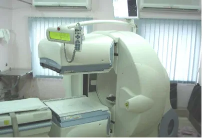
Following intravenous administration of 370 MBq of 99m Tc sodium pertechnetate, a scintigram (Fig. 5) was taken with a digital large
field gamma camera and data analysis system using a low energy, high
sensitivity, parallel hole collimator (Fig. 3 & 4). The images were digitally
recorded in a 128 x 128 matrix. Duration of the scan was 30 to 45 minutes
and the images of the salivary glands were acquired sequentially. Secretion
from the salivary gland was stimulated with 2ml of concentrated lemon juice
placed over the dorsum of the tongue for 2 minutes prior to the scan. Patients
were also instructed not to swallow during imaging.
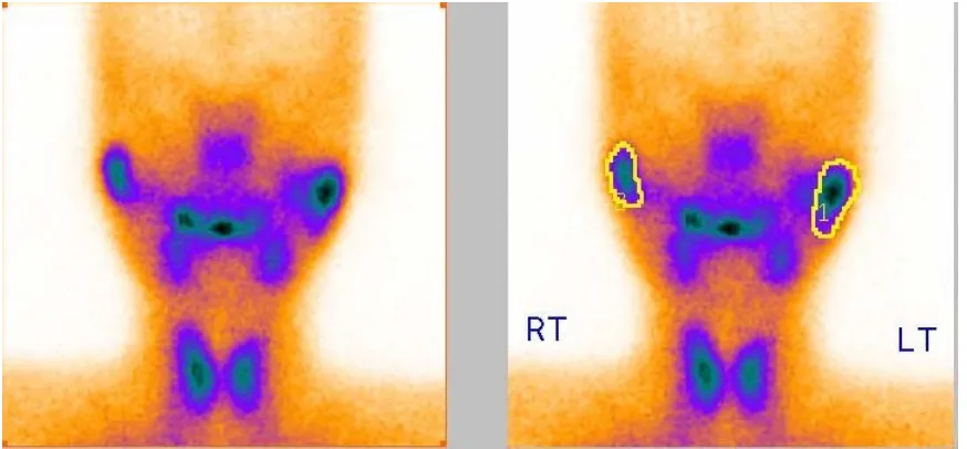
Semi-quantitative analysis was done by the same radiologist who
was blinded to the patient’s clinical information. Oval shaped regions of
interest were drawn over the salivary glands (Fig. 6). Time activity curves
were generated by background subtraction. For each salivary gland the
The function of the gland was calculated using the following expression:
.
Function of the gland = (Maximum uptake – background)/background.
Percentage function of the affected gland = (Function on the affected [image:15.612.92.497.309.592.2]side/function on the normal side) x 100%
Figure 4: Gamma camera
Figure 6: Measuring the uptake
Surgical technique:
Sistrunk’s incision was made, raising a skin flap as required and leaving as much of the superficial musculo- aponeurotic system attached to the skin.
The gland was separated from the cartilaginous external auditory canal and
the anterior border of the sternocleidomastoid muscle. After identifying the
facial nerve exiting at the stylo-mandibular foramen, the superficial lobe was
lifted off by dissecting lateral to the branches. After achieving hemostasis,
the skin flap was closed over a suction drain. Attempt was made to
Post operative studies:
Patients were reviewed on the tenth day and six weeks later
after surgery and each had modified Saxon’s test and parotid scintigraphy at
these times.
Statistical methods:
The data was described using summary statistics such as mean,
median, range and standard deviation. Univariate and bivariate graphs were
plotted. Mann – Whitney test was used for analyzing unpaired groups and
Wilcoxon signed rank test was used for paired data analysis. A ‘p’ value of
less than 0.05 at 95% confidence intervals was considered significant. The
data analysis was performed using SPSS 11.0 for windows.
Ethical issues:
All the patients were explained about the safety of the radiation
dose used for the scintigraphy. The dose was 370 mBq which was a safety
4. Review of literature:
Anatomy of the parotid gland:
Parotid glands are the largest salivary glands. Each has an average
weight of 25 gm and is an irregular, lobulated, yellowish mass. It lies
largely below the external auditory meatus between the mandible and
sternomastoid muscle. The gland projects forwards on the surface of the
masseter muscle where usually a detached part, the pars accessoria or socia
parotidis, lies between the zygomatic arch above and parotid duct below.
The parotid consists almost entirely of serous glandular tissue.
The capsule of the gland is derived from the deep cervical fascia; its
superficial layer is dense, closely adherent and sends fibrous septa into the
gland, it is attached to the zygomatic arch. Medial to the gland, it is attached
to the styloid process, mandible and tympanic plate blending with the
fibrous sheaths of the related muscles. The fascia extending from the styloid
process to the mandibular angle formsthe stylo-mandibular ligament which
intervenes between the parotid and submandibular glands.
The parotid gland is like an inverted, flat, three-sided pyramid,
presenting a small superior surface and superficial, antero-medial and
postero-medial surfaces; it tapers inferiorly to a blunt apex. The superior,
meatus and posterior aspect of the temporo-mandibular joint; here the
auriculo-temporal nerve curves round the neck of the mandible, embedded in
the gland's capsule. The apex overlaps the posterior belly of the digastric
muscle and carotid triangle to a variable extent.
The superficial surface is covered by skin and superficial fascia, the
latter contains the facial branches of the great auricular nerve, superficial
parotid lymph nodes and posterior border of platysma muscle. It extends
upward to the zygomatic arch, backward to overlap the sternomastoid,
downward to its apex postero-inferior to the mandibular angle and forward
superficial to the masseter below the parotid duct.
The antero-medial surface is grooved by the posterior border of the
ramus of the mandible. It covers the postero-inferior part of the masseter,
the lateral aspect of the temporo-mandibular joint and the adjoining part of
the mandibular ramus, passing forward medial to the ramus to reach the
medial pterygoid muscle. Branches of the facial nerve emerge on the face
from the anterior margin of this surface.
The postero-medial surface is moulded to the mastoid process,
sternomastoid, posterior belly of the digastric and the styloid process and its
muscles. The external carotid artery grooves this surface before entering the
from the gland by the styloid process and its muscles. The antero-medial
and postero-medial surfaces meet at a medial margin, which may project so
deeply as to be in contact with the lateral wall of the pharynx.
Several structures traverse the gland partly or wholly and even
branch within it. The external carotid artery enters the postero-medial
surface, dividing into the internal maxillary artery, which emerges from the
antero-medial surface and the superficial temporal artery, which gives off its
transverse facial branch in the gland and ascends to leave its upper limit.
The posterior auricular artery may also branch from the external carotid
within the gland, leaving by its postero-medial surface. The retro-
mandibular vein, formed by the union of the maxillary and superficial
temporal vein, is superficial to the external carotid artery; its posterior
division emerges behind the gland's apex to join the posterior auricular vein,
forming the external jugular and its anterior division emerges anterior to the
apex to join the anterior facial vein, forming the common facial vein. Most
superficial is the facial nerve, entering high on the postero-medial surface
and passing forward and down behind the mandibular ramus in two main
divisions, from which its terminal branches diverge to leave by the antero-
medial surface, passing medial to its anterior margin.
spreading back toward the ear and covering the facial nerve, prolongations
of the gland penetrate medially between the branches of the nerve to form its
deep part; the largest part being between the nerves main temporal and
cervical divisions (Bailey 1947; McKenzie 1948)2, 3. These processes finally
engulf the nerve and its branches, which are sometimes considered to divide
Parotid duct:
About 5 cm long, this begins by the confluence of two main
tributaries within the anterior part of the gland, then crosses the masseter and
at its anterior border turns medially at almost a right angle, traversing the
buccal pad of fat and buccinator muscle. It then runs obliquely forward for a
short distance between the buccinator and the oral mucosa to open upon a
small papilla opposite the second upper molar crown. While crossing the
masseter, it receives the accessory parotid duct and here it lies between the
upper and lower buccal branches of the facial nerve; the accessory part of
the gland and the transverse facial artery are above it. The buccal branch of
the mandibular nerve, emerging from beneath the temporalis and buccinator,
is just below the duct at the masseter's anterior border.
The parotid duct, as seen in lateral sialograms, is formed near the
centre of the posterior border of the mandibular ramus by the union of two
ducts, which respectively, ascend and descend at right angles to the main
duct. The intraglandular part of the main duct receives an alternating series
of descending and ascending tributaries, each formed from an arborization of
fine ductules receiving acini. The acini usually do not show as dilatations in
sialograms but are represented by the free endings of the smallest ducts. As
it crosses the face, it also receives from above five or six ductules from the
Blood supply:
The parotid arterial supply is from the external carotid and its
branches within and near the gland. The veins drain into the external
jugular, through local tributaries. The lymphatics end in the superficial and
deep cervical lymph nodes, interrupted by two or three lymph nodes lying on
and within the gland.
Innervation:
The efferent innervation is autonomic, which consists of the
sympathetic fibres from the external carotid plexus while the
parasympathetic fibres reach it via the tympanic branch of the glosso-
pharyngeal nerve relaying in the otic ganglion and thence traveling along the
auriculo-temporal nerve. The gland also receives secretomotor fibres
through the chorda tympani nerve (Reichert and Poth 1933; Diamant and
WiIberg 1965) 4, 5. In dogs, secretomotor fibres pass to the parotid gland
from the maxillary plexus and the facial and auriculo-temporal nerves, but
this type of distribution of secreto-motor fibres is not confirmed in man
(Holmberg 1972)6. The termination of these supplies is still controversial.
In cats, both parasympathetic and sympathetic fibres end in relation to
glandular cells (Genis-Galvez et al. 1966)7. Most salivary glands, except
secretion. The nerves involved are cholinergic (parasympathetic) and
adrenergic (sympathetic) (Garrett 1976)8. Cholinergic nerves often
accompany the ducts and arborize freely around thesecretory end pieces, but
adrenergic nerves usually enter the gland along thearteries and ramify with
them. Although there are separate sympathetic axons for secretion and
vasoconstriction (Emmelin and Engstrom 1960)9, the cholinergic nerves may
also induce myoepitheliocyte contraction. Infact, a single parasympathetic
axon may induce vasodilatation, secretion and myoepitheliocyte contraction
(Emmelin,1972) 10. Secretory end pieces usually have the most innervation,
cholinergic and adrenergic, individual cells often having both. Cholinergic
axons have long been accepted as the secretomotor innervation; however, in
the parotid gland of the rat, at least, sympathetic nerves are also
secretomotor (Harrop and Garrett 1974; Hodgson and Spiers 1974)11, 12. The
innervation of myoepitheliocyte is sympathetic and parasympathetic
(Kagayama and Nishiyama 1972; Garrett 1975)13, 14. Salivary arterioles are
supplied by both adrenergic (vasoconstriction) and cholinergic
(vasodilatation) axons (Young and van Lennep 1978)15.
In parotid surgery, the facial nerve is reliably found between the
mastoid process and the bony part of the external auditory meatus, where it
the meatus above the upper border of the posterior belly of the digastric
muscle. It can then be traced forward following all its branches (Shaheen
1984)16.
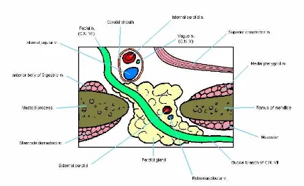
Figure 13: Cross-section of the parotid gland
Coverings:
The gland is invested by an inner true and outer false capsule.
True capsule is formed by condensation of the fibrous stroma of the gland.
False capsule or parotid sheath is formed by the splitting of the investing
layer of the deep cervical fascia. The superficial lamella of the sheath is
strong, attached to the lower border of the zygomatic arch and blends with
the epimysium of the masseter muscle to form a thick parotido-masseteric
Presenting parts:
The gland presents an apex or lower end, base or upper surface,
three surfaces, superficial, antero-medial and postero-medial, and three
margins, anterior, posterior and medial. Apex is directed below, overlaps
the posterior belly of the digastric muscle and extends to thecarotid triangle.
Structures emerging at the apex are cervical branch of the facial nerve,
anterior division of the retro-mandibular vein, posterior division of the retro-
mandibular vein. Base is concave and is related to the external auditory
meatus and back of the temporo-mandibular joint. Structures emerging at
the base are temporal branch of the facial nerve, superficial temporal vessels,
and auriculo-temporal nerve.
Superficial (lateral) surface is covered by the skin, superficial
fascia, posterior fibres of platysma and superficial lamella of the parotid
sheath. The superficial fascia contains the superficial group of parotid
lymph nodes and branches of great auricular nerve, which supply the skin
over angle of the mandible.
Antero-medial surface of the gland is grooved by ramus of the
mandible and presents the following relations. Postero-inferior part of the
masseter, posterior border of the mandibular ramus, capsule of the temporo-
transmitting branches of the facial nerve, inner lip of the groove transmitting
maxillary artery medial to neck of the mandible.
Postero-medial surface of the gland is extensive and is related to
mastoid process, sternomastoid, posterior belly of the digastric, styloid
process, styloid group of muscles, facial nerve entering the gland at the
upper part of the surface, external carotid artery lodged on this surface
before it enters the gland at this surface.
Anterior border of the gland is thin, rests on the masseter and
separates the superficial from antero-medial surface. Structures radiating
deep to this border are zygomatic branch of the facial nerve, transverse facial
vessels, upper buccal branch of the facial nerve, parotid duct, lower buccal
branch of the facial nerve, and marginal mandibular branch of the facial
nerve.
Posterior border of the gland rests on the sternomastoid and
separates the superficial from the postero-medial surface. Structures passing
upward and backward deep to this surface are posterior auricular branch of
Processes of the gland:
Facial process is a triangular projection extending forward superficial to the
masseter along the parotid duct.
Pterygoid process is a triangular process sometimes extending forward
from the deep part between the mandibular ramus and medial pterygoid
muscle.
Glenoid process extends upward between the external auditory meatus and
capsule of the temporo-mandibular joint.
Pre-styloid process is the gland in front of the groove for styloid process
and is related to the internal carotid artery.
Post-styloid process is behind the groove for styloid process and may be
Embryology of the parotid gland:
Understanding of the development of the parotid gland revealed that
the primordium from which the main duct develops is always medial to the
facial nerve rami. Also the auriculo-temporal branches are also closely
related to the facial nerve plane. There is some evidence to suggest this,
According to Raymond F Gasser in the article,“The Early Development
of the Parotid Gland around the Facial Nerve and Its Branches in Man”
, the development of the parotid gland centers and the related facial
nerve branches were studied with the aid of reconstructions in 15 human
embryos and fetuses 18 to 80 mm, crown-rump length, 7 + to 13.5 weeks
(w), gestational age 17. Peripheral branches of the nerve terminate in the
cervico-mandibular region at 18 mm (7 + w), when the unbranched- parotid
bud is farther rostral. By 22 mm (8w), a small nerve branch approaches the
buccal region superficial to the bud that extends toward the pre-auricular
region. At 26 mm (8.5w), several nerve branches course superficial to the
parotid primordium which has first order ductules and is adjacent to the
masseter muscle. Second order ductules form quickly (27 mm. 8.5w) as the
primordium approaches the superficial aspect of the lower buccal, marginal
mandibular and cervical nerve branches. The primordium enters the parotid
has fourth order ductules and the buccal branches are superficial to the main
duct. Nerve branches of the temporo-facial ramus (temporal, zygomatic and
upper buccal) occupy a superficial position in the primordium whereas
branches of the cervicofacial ramus (lower buccal, marginal mandibular and
cervical) are deeper. A similar arrangement is evident at 56-80 mm (11.5-
13.5w) when the complex primordium has connections with its superficial
and deep portions between many nerve branches. Tumors that occur in the
human parotid gland often require removal by partial or total parotidectomy.
Such an operation is hazardous because of the intimate relationship the gland
has with the facial nerve that supplies the muscles of expression. In order to
define this relationship, many authors have reported studies on dissected
adult and infant specimens. (e.g., Gregoire, 18; McWhorter, 19; Mc-Cormack
Cauldwell and Anson, 20; Mc-Kenzie, 21; Davis et al., 26; Patey and Ranger, 27;
Youssef, Talaat and El-Malt28). However, these studies summarize the
results by the time the gland –nerve relationship has already become
intricate and very complex. There is consistent lack of uniformity in the
conclusions drawn with regard to whether the gland is uni or bilobed,
whether or not a cleavage plane exists in the gland where the nerve passes
through and where and how many areas of communication (isthmi) exists
McWhorter, McKenzie, and Sammarco, Ryan and Longenecker here tried to
clarify the relationship with reports on newborns and late fetuses, but even in
these specimens, the relationship is too complex 18, 19, 21, 29.
A better understanding of the relationship is gained with
information on the early development of the gland around the nerve. The
relationship is easier to define at this time because the morphology of the
gland is simple. Such information however, at best is incomplete and, in
some instances, questionable. Bujard briefly mentioned a relationship in
one 35 mm fetus 24. The earliest fetus examined by Gregoire was 80 mm
when the arrangement is already quite complicated 18. Shulte did not discuss
the nerve in his detailed account of early salivary gland development 33. The
first significant contribution on the early relationship was made by Rouviere
and Cordier, who studied two specimens (31 and 31 mm) 30. Additional
specimens were examined by Winsten and Ward and Brunner but they
reported no pattern in the formation of the relationship and incompletely
defined the relationship between the gland and the peripheral branches of the
facial nerve 31, 23. The investigations of Cody and Samengo, insufficiently
covered the early arrangement and some of their statements together with
some of Winsten’s and Ward’s are disputed 25, 32, 31.
parotid gland around the extra cranial branches of the facial nerve and the
related blood vessels in man. The manner in which the gland-nerve
relationship develops with the pattern that it follows is given. It is hoped
that such basic information will prove helpful to those who treat conditions
involving the pre-auricular and buccal regions. A better understanding of
the early arrangement should also help explain variations that are sometimes
encountered at the operating and dissecting tables.
The sequence of development of the parotid gland around the facial
nerve and its branches, the related parts of the retro-mandibular vein, and the
external carotid artery is divided into four stages based on the location of the
parotid gland primordium as it grows into the parotid space and on the
degree of branching it exhibits.
Stage 1: The unbalanced parotid bud (18 to 22 mm, 7 + to 8 weeks)
The primordium of the parotid gland at 18mm is a simple, solid, short
but broad, epithelial bud that extends into moderately dense mesenchyme
(Fig. 14). It arises from the lateral most part of the oral epithelium, cranial
to the angle of the mouth and grows dorsally and slightly laterally toward the
first visceral groove. By 21-22 mm the bud is longer but narrower, remains
unbranched and extends farther dorsally toward the pre-auricular region.
Farther dorsally at 18 mm, the facial nerve terminates in the cervico-
mandibular region, deep to the mandibular (pre muscle) lamina after
coursing ventrally in the vanishing second visceral arch. Four fascicles are
discernable in the distal part of the nerve. Definitive peripheral branches do
not begin to form until 21-22 mm when the temporo-facial and cervicofacial
rami are evident. The parotid bud is separated from the facial nerve
terminals at 18 mm by the following structures from rostral to dorsal; the
buccal nerve, inferior alveolar nerve, Meckel’s cartilage and myelo-hyoid
nerve. Both the bud and the facial nerve approach one another on a plane
that is lateral to these structures. At 21-22 mm an upper buccal branch from
the temporo-facial ramus lies lateral to the bud.
[image:41.612.108.529.429.664.2]Caudal end Cranial end
Fig.14. The arrangement of the facial nerve and parotid primordium
Caudal end Cranial end
Fig.15. The arrangement of the facial nerve and parotid primordium
at 22 mm. The outline of the auricle is indicated by a stippled line.
This stage is characterized by dorsal growth of the solid, unbranched
bud that approaches the facial nerve deep to the upper buccal branch of the
temporo-facial ramus (Fig.18,10arrow). The temporo-facial ramus and its
branches are on a plane that is superficial to the bud while the cervicofacial
Stage II: First and second order branching of the parotid primordium
(26 to 27 mm, 8.5 weeks)
By 26 mm, the primordium can be sub divided into a long, narrow,
proximal segment that is continuous with the oral epithelium and a large,
bulbous, irregular shaped, distal segment that lies opposite the middle one-
third of the masseter muscle (Fig. 16). The proximal segment will become
the main parotid duct; lumen is developing in its middle part. The distal
segment is a solid, epithelial mass that has knob-like projections extending
from it that represent the first branch or ductules of the primordium. It
undergoes secondary branching very quickly. Ducts that extend dorsally and
laterally are not prominent. Moderately dense mesenchyme surrounds the
primordium and separate it medially from the masseter muscle. Accessory
parotid tissue is sometimes present as one or more isolated buds arise from
the main duct near the mid of the masseter muscle.
Caudal end Cranial end
Fig.16. The arrangement of the facial nerve and parotid primordium at
26 mm, 8.5 weeks.
Definitive relationships are established during this stage between the
peripheral branches of the nerve and the larger drainage ductules of the
gland. Buccal branches are closely related to the primordium. Buccal
branches were never observed medial to the proximal segment of the
primordium that forms the main duct.
At 26 mm, a large venous plexus is in the superficial part of the
cervico-mandibular region near the dorsolateral aspect of the masseter
medial to the facial nerve and lateral to the external carotid artery.
In this stage, those branches that arise from the temporo-facial ramus
(temporal, zygomatic and upper buccal) are usually on a plane that is
superficial to the primordium (Fig. 18, 20 arrows). Those branches that arise
from the cervicofacial ramus (lower buccal, marginal mandibular and
cervical) are usually on a plane that is deep to the primordium. Dorsolateral
growth of the primordium superficial to the facial nerve and its branches
begins in this stage.
Stage III: Third and fourth order branching of the parotid primordium
as it enters the parotid space (32 to 44 mm, 9 to 10.5 weeks)
The primordium undergoes third order branching and begins to enter
the parotid space by 32 mm. Most of the branching is dorsal to the masseter
muscle. Fourth order ductules are present at 37 mm, when the primordium
is mostly within the parotid space (Fig.17). Dorsolateral growth increases
considerably and extends cranially and caudally. Moderately dense
mesenchymal tissue continues to surround the expanding primordium. A
distinct lumen is present in the main duct and many of its tributaries.
A comparatively small portion of the primordium lies deep to the
nerve at the stage. At 32 mm, small ductules in the caudal part of the
buccal branch. However, even at 41 mm, the only part of the primordium
that is actually deep to the nerve is opposite the temporo-facial ramus and its
upper buccal branch. This portion of the primordium attains its position by
[image:46.612.93.520.234.492.2]growing cranially from the main duct.
Fig.17. The relationships of the facial nerve, parotid primordium,
retromandibular vein and external carotid artery at 37 mm, ten weeks.
Notice the deeper position of the cervicofacial branches compared to the
temporofacial branches.
The main feature in this stage is dorso-lateral growth of the
nerve and its branches (Fig.18, 30 arrows). The deep portion is primarily
opposite the temporo-facial ramus and proximal part of its buccal branches.
Ductules of the superficial portion begin to grow medially, dorsal to the
cervicofacial ramus and between this ramus and its lower buccal branch to
contribute to the deep portion (Fig. 17, 40 arrows).
Stage IV: Multiple branching of the parotid primordium superficial and
deep to the facial nerve (49 to 80 mm, 11 to 13.5 weeks)
The superficial portion grows rapidly and extends dorsally, laterally,
cranially and caudally. Progressive enlargement of the superficial portion is
apparent at 80 mm.
Additional ductules accumulate deep to the nerve and grow medially
past the ramus of the mandible especially in the cranial part of the
primordium. That segment of the deep portion that began during stage II
enlarges and grows medial to the retro-mandibular vein. Its ductules unite as
they pass caudally to join the main duct between the upper and lower buccal
branches (Fig.18). Contributions to the deep portion are also made by the
medial growth of ductules in the superficial portion. Such growth is
especially evident dorsal to the temporo-facial and cervico-facial rami, but it
also occurs between other branches of the facial nerve. Very few ductules in
The facial nerve and its branches in the parotid space are almost
completely surrounded by the primordium. Superficial ductules grow
cranially and caudally by 49 mm covering the lateral aspect of the proximal
portion of the upper buccal branch (Fig.18). A lesser quantity of the
primordium is apparent deep to the cervicofacial ramus and its branches.
Ductules appear on the medial aspect of the undivided part of the facial
nerve by 62 mm. The communication between the temporal branch of the
facial nerve and the auriculo-temporal nerve occurs within the primordium.
Ductules surround the retro-mandibular vein earlier than the external
carotid artery since the vein is more superficial. The ductules attain this
medial position by growing rostral and dorsal to the vein. This stage is
characterized by medial growth of ductules in the superficial portion
between and around branches of the facial nerve especially dorsal to the
temporo-facial and cervicofacial rami (Fig.18, 4 0 arrows).
Many conflicting reports exist on early relationship of the parotid
gland and the facial nerve. Winsten and Ward observed the bulk of the
gland primordium within the crotch of the main rami of the facial nerve at
27 mm 31. Cody noticed the primordium completely separate from the nerve
at 30 mm 25. According to Rouviere and Cordier the nerve is entirely deep at
at 35 mm 24. Brunner found the nerve mainly deep to the primordium at 40
mm 23. The study by Gasser however, shows that the temporo-facial
branches on a plane which is superficial to the primordium at 22 mm (fig.
15). The upper buccal branches are entirely superficial the primordium at
26-32 mm, but the cervico-facial ramus and its branches are either deep to
the primordium or on a plane that is deep (Fig. 16). Between 32 and 44 mm,
most of the primordium expands superficial to the facial nerve branches with
only a small parotid that is opposite the temporo-facial rami proximal part of
the upper buccal branches (Fig. 17).
Fig.18. General pattern of development. Arrows indicate the direction
of growth of the primordium around the facial nerve and its branches.
The present thought is that, in most instances, the growth of the
parotid gland primordium around the facial nerve and its branches follows a
pattern (Fig. 18). The findings of Winsten and Ward during early
development of the oblique alignment of the nerve branches with the more
medial position of the marginal mandibular and cervical branches are
confirmed in the study by Gasser 31. The latter author, disagrees with
Winsten and Ward in that the continued growth of the gland does not always
take place entirely lateral to the cervical branch. A considerable quantity of
the primordium is medial or deep to this branch at 56 mm. Bailey’s
conclusion that apparently the main ductules connect only with the
superficial portion is not supported by present findings 22. Most of the main
ductules during early development do connect to the superficial portion, but
some are also continuous with that part of the deep portion that is opposite
the temporo-facial ramus and the proximal part of its branches.
Brunner found the deep portion of the parotid gland beginning as a
medial sprout at 40 mm 23. According to Gregoire the deep portion is absent
in the 80 mm fetus (3 months) and does not begin to develop until six to
seven months 18. Cody indicated that the primordium does not begin to
extend medially through the branches of the facial nerve until 200 mm 25.
begins as early as 32-37 mm (Figs. 17). A considerable quantity of gland is
deep to the nerve by 80 mm.
Many different reports are also available of the manner in which the
deep portion of the parotid gland develops. Gregoire concluded that it
formed by migration of the superior pole of the gland cranial to the facial
nerve 18. Rouviere and Cordier observed at 51 mm projections which passed
from the superficial portion over the nerve cranially as well as some which
passed between the two main rami 30. McWhorter and many others observed
the location of a prominent area of communication between the two main
rami 19. Winsten and Ward found the largest area of communication in the
region of the lower branches of the nerve around the cervicofacial trunk 31.
Samengo, studying 80 and 100 mm fetuses and adults, believed the
two portions of the gland meet at their dorsal border 32. Sammarco, Ryan
and Longenecker observed several connections between the two portions in
third trimester fetuses and stillborn infants 29. Winsten and Ward stated that
as the gland grew all the interstices of the branches of the nerve are filled
with proliferating buds 31. In the study by Gasser, connections between the
superficial and deep portions are present at 56 mm dorsal to and between the
temporofacial and cervicofacial rami and between some of their branches.
through any gap in the plexus. Because of the manner in which the
primordium forms around the facial nerve branches, a more prominent
communication necessarily exists between the two major rami of the nerve
(Fig. 18).
Bailey believed that no part of the facial nerve is actually within the
parotid gland substance and likened the nerve to the “meat” within a parotid
“sandwich” 22. This concept is an oversimplification of the relationship that
exists during early development. McCormack, Cauldwell and Anson and
Davis et al. reported a natural cleavage plane between the two portions of
the gland 20, 26. Davis et al. indicated that the cleavage plane contains tissue
that could be considered as an inward directed septal derivative of the
surrounding connective tissue 26. Winsten and Ward stated that cleavage
planes in which the nerve radicles transverse the gland do not exist 31.
Between 49 and 80 mm, Gasser found the area immediately surrounding the
facial nerve and its branches to be devoid occasionally of parotid gland
ductules, but this arrangement cannot be described as a cleavage plane. A
complete cleavage plane through the gland primordium with a single mass of
communicating ductules (isthmus) was never observed. Many ductules that
are superficial to the facial nerve contribute to the deep portion by growing
rare that ductules in the deep portion of the gland grow laterally and
contribute to the superficial portion.
McCormack. Cauldwell and Anson, Davis et al. and many other
believed that the parotid gland is bilobed 20, 26. Winsten and Ward and Patey
and Ranger showed the gland to be unilobar 31, 27. Traditionally, lobes are
demarked by fissures, sulci, connective tissue or shape. 34. None of these
demarcations are clear during early development but only the position of the
primordium with reference to the position of the nerve. In light of this, the
terms, superficial and deep portions of the parotid gland, which were
suggested by McKenzie and later adopted by Nomina Anatomica, are more
appropriate than the terms, superficial and deep lobes 21, 35.
Auriculo-temporal nerve:
The trigeminal nerve is the first branchial arch nerve. It develops in the
intra-uterine weeks 5 and 6. The auriculo-temporal nerve breaks up into
branches in front of the external auditory pit at 7 weeks. In the 26 mm length
stage of fetal development, it is connected to the facial nerve. Interruption
or external effects within this period of development may lead to deviations
from normal development. It originates from the posterior trunk of the
passes between them. This nerve runs through the deep lateral pterygoid
muscle and supplies fibres to the parotid gland. It contains somatosensory
and secretomotor fibres of third division of the fifth cranial nerve and the
ninth cranial nerve. The auriculo-temporal nerve crosses medial to the neck
of the mandible and changes its direction upward in the parotid gland
between the temporo-mandibular joint and external auditory meatus. The
auriculo-temporal nerve has communicating branches to the facial nerve and
inferior alveolar nerve at the posterior border of the mandibular ramus in its
course.
There are also variations in the anatomy of the nerve. Nadir
Gulekon et al. concluded that, on 32 dissections in majority of the cases the
auriculo-temporal nerve with two roots, the upper root was lateral to the
middle meningeal artery and the lower root was medial. In the cases with
one root, the number of those with roots lateral to the artery was almost
equal to those with roots medial to the artery 36. The variations are in the
number of roots by which the nerve arises and the nerve was always medial
to the facial nerve.
In the article, “The surgical anatomy of the parotid duct with
emphasis on the major tributaries forming the duct and relationship of the
facial nerve to the duct” by Richard’s et al dissection was made in cadaver
right and left sides. Parotid duct in 31% of the head hemi sections presented
as a single discernible duct from the parotid papilla to within the gland. In
62.1% of the head hemi sections, the ducts were formed by a branching
pattern within the gland. In the ducts with a branching pattern, 48.3%
displayed a bifurcated pattern, 6.9% were trifurcated, and 6.9% had multiple
branches. In 6.9%, of the head hemisections, the parotid ducts bifurcated
distal to the parotid gland. In all cases, the deep lobe of the gland enveloped
the parotid duct; only small tributaries connected the superficial lobe with
the duct. The facial nerve and its branches were always observed lateral to
the parotid duct. Because one dissects lateral to the facial nerve during a
superficial parotidectomy, generally, the parotid duct remains intact and
potential complications such as sialoceles and fistulizations are, thereby,
Physiology of the parotid gland:
For their size the salivary glands produce a large volume of saliva;
the maximal rate in humans is about 1 ml/min/g of glandular tissue. The rate
of metabolism of salivary glands is also high, accompanied by a high blood
flow-both proportional to the rate of saliva formation. The flow of blood to
maximally secreting salivary glands is approximately 10 times that of an
equal mass of actively contracting skeletal muscle.
Receptors:
The receptors are moieties that interact with ligands involving recognition and transfer of information (Lefkowitz et al., 1984a) 38. The interaction initiates a biologic response or sequence of responses. The quality of the biologic response resides in the receptor-effector and not in the ligand. The binding of a ligand to a specific receptor is the first step in the regulation of cell function by extra-cellular factors. The majority of regulatory receptors are found in the plasma membrane (proteins or glycoproteins); a few (hormonal) are present within the cell. Once occupied by its specific regulatory molecule, the receptor complex activates an effector process to bring about the cellular response. Activation is either direct or by a second messenger. A receptor may be a single or composite membrane protein and may contain subunits with special functions.
polypeptide hormones and neurotransmitters are on the plasma membrane, and for exocrine glands they are on the basal or lateral membranes (Stump, 1984) 39. For some cells, receptor occupancy and biologic effects follow saturation kinetics; for other cells, regulation does not require full occupancy (Lefkowitz et al., 1984a) 38.
Salivary function and regulatory molecules*
Salivary
gland Regulator
Second messenger Effect Parotid Acetylcholine (ACh), x-adrenergic, substance P
Ca++ Production of saliva, limited enzyme secretion, increased cell metabolism
B-adrenergic, vipadrenergic intestinal peptide (VIP)
AMP Secretion of enzymes, increased cell metabolism
*Data from Williams JA: Ann Rev Physiol 46:361, 1984. 40
Secretion:
Saliva consists of two components; macromolecules and fluid. The fluid component is derived from perfusing blood vessels; the macromolecules are primarily derived from secretory granules of the acinar cells. The fluid is produced at the secretory end-pieces and is currently thought to occur via an osmotic coupling (solute-solvent coupling) of transepithelial fluxes of sodium chloride and water (Izutse, 1989) 45. It is likely that the water and electrolyte fluxes occurs transcellularly through acinar cells. Movement of sodium and chloride through the cell and into the acinar lumen provides an osmotic gradient to establish an accompanying water flow across the cell. The currently favored hypothesis to explain how sodium and chloride cross the basal cell membrane is that a co-transport of the two ions occurs with the movement of sodium down its electrochemical gradient driving the accumulation of chloride against its electrochemical gradient (Izutsu, 1989) 45.
potassium, or calcium ions open after a depolarization of the plasma membrane (Findlay, 1984; Petersen, 1980) 47, 41. The sodium pump exists in all cells, and since more sodium ions are actively pumped out than potassium is taken up, it contributes directly to the membrane potential. The pump is primarly activated by an increase in sodium. Calcium-sensitive ion channels, permeable to sodium, potassium, or chloride ions, open when calcium increases (Petersen, 1980) 41. This is in addition to a calcium-activated channel. Possibly, cyclic nucleotides (AMP, GMP) activate ion channels or pumps.
Despite the many gaps in the knowledge of the electro-physiology of salivary acinar cells, the action of the major secretagogues can be summarized as follows. In mammalian salivary glands, acetylcholine and epinephrine and/or nor-epinephrine acting on alpha-receptors cause an increase in potassium and sodium ion permeability of the plasma membrane. This results in a pronounced reduction of the surface membrane resistance and a loss of potassium from the cells, balanced by an uptake of sodium. An active electrogenic extrusion of sodium and accumulation of potassium follow. An increase in calcium permeability probably mediates the permeability of sodium and potassium. Epinephrine and/or norepinephrine, acting on receptors, produce only small potential and resistance changes. The most important effect of beta-adrenergic activation is an increase in intracellular c-AMP, stimulating enzyme secretion, Cholinergic or alpha-adrenergic stimulation causes a marked fluid and some calcium-dependent enzyme secretion.
chloride-sodium is the only one with a pattern of very low concentrations at lowest flow rates to high concentrations at the highest rates of flow. Sodium is also the primary contributor to the increasing osmolality of the fluid as the level of secretion increases (Shannon et al., 1974) 42. In human parotid saliva, sodium is the key in the secretion of fluid because local osmotic effects across the secretory luminal membrane influence the generation of the fluid (Shannon et al., 1974) 42.
Acinar secretion:
Secretory cells such as the acinar cells of the parotid gland discharge their products by a process of exocytosis, wherein fusion of secretory granules with a delimited portion of the plasma lemma at the apex of the acinar cell occurs. The membrane fusion is the last of a series required for the transfer of export proteins from their synthesis in the rough endoplasmic reticulum (RER) to the extracellular environment. Using the model of Palade (1975), the secretory process can be divided into six successive steps: (1) synthesis, (2) Segregation, (3) intracellular transport, (4) concentration, (5) intracellular storage, and (6) discharge.
Saliva:
Figure 19: Saliva secretion process:
Table 1: Contribution to saliva and relative viscosities of saliva by salivary gland*
Gland Percent of total
saliva (24 hours)
Relative viscosity
Submandibular 71 3.4
Parotid 25 1.5
Sublingual 3 to 4 13.4
Minor (oral, labial) Trace amounts --
Table 2: Saliva composition in normal adults:
Mean valves Parotid gland Submandibular
gland
Flow rate (ml/min/gland: stimulated) 0.7 0.6
Inorganic analytes (mEq/L)
K+ 20
17
Na+ 23 21
C1- 23
20
HCO-3 20
18
Ca++ 2 3.6
Mg++ 0.2
0.3
HPO4-1 6
4.5
Organic analytes (mg/dl)
Urea 15
7
Ammonia 0.3 0.2
Uric acid 3 2
Glucose <1
<1
Cholesterol <1 ?
Fatty acids 1 ?
Total lipids 2 to 6
2 to 6
Amino acids 1.5 ?
Whole or mixed saliva is composed of approximately 99.5% water and has a specific gravity between 1.002 and 1.012. In humans the amount of saliva secreted in 24 hours is between 1000 and 1500 ml (Arglebe, 1981; Mandel, 1980) 46, 44. The secretory rate is highest during meals; during sleep or in the absence of stimulation the secretary rate is low or nearly absent. In human mixed saliva the pH varied from 5.75 to 7.05 (Arglebe, 1981) 46. If data on flow and composition are to be meaningful, the saliva must be collected under standardized conditions (Arglebe, 1981; Mandel, 1980) 46, 44. Resting and stimulated secretions should be evaluated, and because many constituents of saliva are circadian, the times of collection should be uniform.
The flow rate in nonstimulated parotid glands is about 0.04 ml/min/gland; submandibular saliva has a somewhat higher resting flow rate, 0.05 ml/min/gland (Mandel, 1980) 44. This decreases markedly during sleep; the sleeping flow rate from the parotid gland is nearly nil.
Flow rates after stimulation vary with the stimulus. With usual gustatory stimulation a 0.6 ml/min/gland flow rate is obtained. A range of 0.4 to 1.0 is not uncommon. Discomfort from dryness is usually not a complaint until the rate of flow is below 0.2 ml/min/gland, (Mandel, 1980)
44
Neoplasms of the parotid gland:
Salivary gland neoplasms are rare, constituting 3% to 4% of head and neck neoplasms. The majority of the neoplasms arise in the parotid gland (70%), whereas tumours of the submandibular gland (22%), and sublingual gland and minor salivary glands (8%), are less common. The ratio of malignant to benign tumours varies by site as well: parotid gland 80% benign, 20% malignant; submandibular gland and sublingual gland 50% benign, 50% malignant; minor salivary glands 25% benign, 75% malignant. The incidence of the various tumours in the parotid gland is as follows,
Animal studies:
There are a few trials that have assessed the function of the remaining part of the gland after various described surgeries of the parotid like functional superficial parotidectomy and deep lobe parotidectomy.
Zhao K et al., designed a study in rats to clinically
test a new type of parotid surgery termed ‘functional superficial
parotidectomy (FSP)' to preserve the function of the residual gland 48.
The results showed that 91.4% of the main parotid ducts were deep to facial nerve. Physical examination, sialography and scintigraphy showed that function of the residual gland was well preserved by FSP.
Zheng G et al, had done a stereological study of
the remaining parotid gland of rat after duct-preserved partial parotidectomy.
The remaining parotid glands of rats after duct-preserved partial
parotidectomy were sectioned and examined randomly by stereological methods, which determine the acinar area and proportional volume (PV) of
the component tissues 49. The volumes of the remaining glands were measured at the same time.
1) There was no difference of the above quota 1 – 3 days after the operation
(p>0.05).
the PV of acini decreasing (p<0.05).
3) 4 – 7 weeks after the operation, the acinar area increased (p<0.05), the
PV of acini and gland volume regained (p<0.05). All the results indicated that with the duct preserved, the remaining parotid glands, after partial
parotidectomy, do have the regenerating ability and significant function.
The conclusion drawn here was, the acinar cells have
regenerating capabilities with function which are statistically significant.
The study mentioned below deals with the proliferative capacity of the
acinar cells.
Burford Mason et al., developed an animal model of
potential progenitor cells on the grounds of their putative limited cycling capacity.
In the article “Experimental study on salivary gland
scintigraph” by Funds Masayuki et al., serial salivary scintigraphy
was done after ligating the parotid ducts in rabbits 53. There was progressive
significant decrease in the uptake function from third day to 4 weeks after
ligation.
There are no human studies demonstrating regeneration of the acinar cells.
Clinical trials:
In a trial by Giuseppe Colella et al., in the article
“Parotid function after selective deep lobe parotidectomy”, selective deep lobe parotidectomy was done on fourteen patients who had a
mass involving the deep lobe of the parotid seen between January 2001 and
March 2004 51. Evaluation of postoperative function of the superficial lobe of
the parotid after selective resection of the deep lobe was done. At 6 months
follow-up all patients had scintigraphy of both parotid glands.
After scintigraphy the maximum uptake value and function of the gland
were evaluated with the concentration index (CI) and the CI percentage
ratio. The concentration function of the gland in the resected side of the
study group had a mean (S.D.) CI index of 5.5 (3.6) and a CI percentage
ratio of 84%. They concluded that selective deep lobe
parotidectomy has the following advantages: it minimizes the impact of
treatment on the facial contour, it does not increase postoperative morbidity
and it preserves the function of the gland.
Tumors in the parotid gland may affect salivary flow. The
effects of tumor on glandular function and postoperative changes in both
resected gland and contra-lateral gland were not formerly reported. In this
trial named, Salivary flow dynamics after parotid surgery
,
flow rate and composition dynamics before and after parotidectomy were
studied 52. The Stenson’s duct was routinely ligated in total parotidectomy
only.In this study, the group prospectively evaluated salivary flow rates
and composition in patients undergoing parotidectomy preoperatively and
postoperatively. Stimulated parotid saliva from patients undergoing
parotidectomy was collected bilaterally preoperatively and postoperatively
by using a parotid cup. Subjective complaints were recorded. Salivary flow
rates, sodium, potassium, and amylase levels were evaluated.
Thepre-operative flow rates of the non-involved glands were
significantly higher. No post-operative changes in flow rate were noticed in
the un-involved glands, whereas involved glands presented with statistically
significant post operative reduction. Analysis of the individual results
revealed three patterns of pre operative and post operative response. They
were as follows,
Group 1: Post operative compensatory group.
The mean results of this group are similar pre-operatively and
close to mean (0.7 ml/minute). The mean post-operative compensatory
80% of the normal mean value. The mean flow rate is 0.04 ml/min/gland in un-stimulated state and 0.6ml/min/gland in the stimulated state.
Group 2: Pre operative compensatory group.
The mean pre operative value was much higher in the non -involved glands (0.92 ml/minute Vs 0.23 ml/minute, p=0.001). The pre-operative compensatory increase was about half (31%) of the decline in the involved gland (67%). The total mean parotid salivary flow rate of both glands was 1.15 ml/minute, approximately 80% of the normal mean value.
Group 3: Non responders.
The mean pre operative and post operative results in this
group were lower compared with groups 1 and 2 (p=0.014) and no saliva
was secreted from the involved glands post operatively.
Pre operative salivary secretion was detected in all the patients
undergoing superficial parotidectomy. The mean pre-operative flow
rate of the involved gland was 0.46 +_ 0.3 ml/minute. The mean post
operative flow rate was 0.05 +- 0.05 ml/minute which shows a reduction
of 89% (p=0.041).
None of the patients complained of “dry mouth” before or after surgery.
Analysis of the individual results revealed 3 patterns of preoperative and
postoperative compensatory mechanism in the contra-lateral gland. The
postoperative decrease in flow rate corresponds with the amount of gland
removed. Salivary electrolyte composition was unchanged. This study is the
first to demonstrate the effects of parotid tumors and their surgery on
salivary flow and a compensatory response and its different patterns in
human parotid glands after their excision.
In the article “Scintigraphic assessment of early and late
parotid gland function after radiotherapy for head-and-neck cancer” by
Judith M. Roesink M.D et al., parotid scintigraphywasperformed before radiation therapy and 6 weeks and 1 year after radiation therapy 54. The
uptake, excretion fraction of the saliva from the parotid gland to the oral
cavity (SEF), and the ratios of uptake and SEF after and before treatment
were calculated. The SEF decreased to 18.7% at 6 weeks, but recovered to a
Table 3. Parotid uptake functions
Serial
no. Sex Age
Operated side
Duct status
Technetium scan 1 – Right side uptake (%)
Technetium scan 1 – Left side uptake (%)
Technetium scan 2 – Right side uptake (%) Technetium scan 2– Left side uptake (%)
1 Male 22 R Preserved 35 65 34 66
2 Female 38 L Preserved 84 16 76 24
3 Female 84 L Ligated 75 25 88 12
4 Male 49 L Ligated 80 20 95 5
5 Male 15 L Preserved 72 28 63 37
6 Male 18 R Preserved 25 75 24 76
7 Male 36 R Preserved 51 49 26 74
8 Male 45 R Preserved 11 89 0 100
9 Female 40 L Preserved 63 37 90 10
10 Male 51 R Preserved 27 73 20 80
11 Male 39 R Not Known 49 51 46 54
12 Female 38 L Preserved 75 25 75 25
13 Female 27 R Not Known 42 58 39 61
14 Male 58 L Preserved 61 39 73 27
15 Female 43 L Preserved 91 9 98 2
16 Female 24 L Preserved 68 32 71 29
17 Male 60 L Preserved 79 21 89 11
18 Female 41 R Preserved 25 75 38 62
19 Male 35 R Preserved 52 48 42 58
Table 3 shows the uptake function of the glands of 20 cases. The operated
side uptake is written in green. In 16 patients there was decrease in the
function by 10.68% in the second scan compared to the first one. In one case
(no. 12) there was no change in the function. In 3 cases (no. 2, 5, 18) there
was increase by 10% in the uptake function. In 2 cases (no. 19, 20) the
uptake function in the operated gland was more than the normal gland in the
first scan with subsequent reversal in the second scan. In one case (no. 8)
the second scan function was 0%. In the cases (no. 2, 3) where duct was
Serial
no. Sex Age Side Duct status
Saxon’s 1 -Right side(mg)
Saxon’s 1 – Left side(mg)
Saxon’s 2 – Right side(mg)
Saxon’s 2 – Left side(mg)
1 Male 22 R Preserved 15 110 5 41
2 Female 38 L Preserved 200 152 Not done Not done
3 Female 84 L Ligated 156 946 22 11
4 Male 49 L Ligated 71 2 109 27
5 Male 15 L Preserved 132 85 194 157
6 Male 18 R Preserved 26 51 29 59
7 Male 36 R Preserved 19 255 28 61
8 Male 45 R Preserved 15 78 35 94
9 Female 40 L Preserved 155 32 184 114
10 Male 51 R Preserved 44 48 22 32
11 Male 39 R Not Known 12 59 18 32
12 Female 38 L Preserved 67 28 52 16
13 Female 27 R Not Known 210 114 90 102
14 Male 58 L Preserved 194 117 114 28
15 Female 43 L Preserved 28 18 48 33
16 Female 24 L Preserved 88 18 356 296
17 Male 60 L Preserved 14 16 36 21
18 Female 41 R Preserved 22 28 34 202
19 Male 35 R Preserved 12 20 12 22
[image:75.612.61.566.107.602.2]20 Male 28 L Preserved 28 25 45 16
Table 4. Saliva secretion
In table 4, modified Saxon’s test was done on the
first scan day in all the 20 patients. In one case the second modified Saxon’s
test was not done as the scan was done in the native place. In case (no.3),
the saliva weight was erroneously high due to practical difficulty, as she had
no tooth leading to saliva contamination from the floor of the mouth. In 11
cases, there was actually increase in the saliva weight on the second
measurement. In 5 cases (10, 12, 13, 14, 20), there was decrease in the saliva
secreted. In on case (no. 19) there was no change in the weight.
From the above data percentage function and
percentage saliva secretion was calculated for the operated gland compared
to the non-operated gland. Only in 7 cases (35%) both scan and Saxon’s test
Sex distribution: (Fig. 20) n=20
8(40%)
12(60%)
Male Female
The male to female ratio in the present study of 20 patients was 2:3 with 8
males and 12 females ( 40 % and 60 % respectively). The age of the 20
patients enrolled in the present study rangedfrom 18 to 84 years with mean
age of 40 years. The mean age for the males was 38 years while it was 42