Copyright © 2000, American Society for Microbiology. All Rights Reserved.
The Role of CD8
⫹
T Cells and Major Histocompatibility Complex
Class I Expression in the Central Nervous System of Mice
Infected with Neurovirulent Sindbis Virus
TAKASHI KIMURA
ANDDIANE E. GRIFFIN*
W. Harry Feinstone Department of Molecular Microbiology and Immunology, School of Hygiene and Public Health,
Johns Hopkins University, Baltimore, Maryland 21205
Received 3 December 1999/Accepted 29 March 2000
Little is known about the role of CD8
ⴙT cells infiltrating the neural parenchyma during encephalitis
induced by neurovirulent Sindbis virus (NSV). NSV preferentially infects neurons in the mouse brain and
spinal cord; however, it is generally accepted that neurons can express few if any major histocompatibility
complex (MHC) class I molecules. We evaluated the possible roles and interactions of CD8
ⴙT cells during
NSV encephalitis and demonstrated that MHC class I antigen (H2K/D) was expressed on endothelial cells,
inflammatory cells, and ependymal cells after intracerebral inoculation of NSV. No immunoreactivity was
observed in neurons. On the other hand, in situ hybridization with probes for MHC class I heavy chain,

2
microglobulin, and TAP1 and TAP2 mRNAs revealed increased expression in a majority of neurons, as well as
in inflammatory cells, endothelial cells, and ependymal cells in the central nervous system of infected mice.
NSV-infected neurons may fail to express MHC class I molecules due to a posttranscriptional block or may
express only nonclassical MHC class I genes. To better understand the role CD8
ⴙT cells play during fatal
encephalitis induced by NSV, mice lacking functional CD8
ⴙT cells were studied. The presence or absence of
CD8 did not alter outcome, but absence of

2 microglobulin improved survival. Interestingly, the intracellular
levels of viral RNA decreased more rapidly in immunocompetent mice than in mice without functional CD8
ⴙT cells. These observations suggest that CD8
ⴙT cells may act indirectly, possibly via cytokines, to contribute
to the clearance of viral RNA in neurons.
Sindbis virus
(SV), an alphavirus in the family
Togaviridae
,
causes acute encephalomyelitis in mice. Neuroadapted Sindbis
virus (NSV) is a neurovirulent strain of SV that can elicit fatal
encephalitis in adult mice as well as in suckling mice and
provides a model system for examining the factors that
deter-mine outcome (11). Antibodies play a crucial role in the
clear-ance of infectious virus from the central nervous system (CNS)
of SV-infected mice (23), but infection also induces a brisk
mononuclear inflammatory response that is immunologically
specific and includes both CD4
⫹and CD8
⫹lymphocytes (13,
26, 28). Little is known about the role of T cells infiltrating the
neural parenchyma in the pathogenesis of NSV-induced
en-cephalomyelitis. By adoptive transfer, virus-specific antibody
can protect mice from fatal infection with NSV when given
before or after infection, while T cells are not protective (11,
12, 43). However, preimmunization with the nonstructural
pro-teins of SV protects mice from fatal NSV encephalitis by a
mechanism that appears to be dependent on T cells (8).
The primary target cells for NSV infection in the CNS are
neurons (15, 16). If neurons could express functional major
histocompatibility complex (MHC) class I molecules, then
in-fected neurons could be recognized by CD8
⫹T cells and be
targets for cytotoxic processes. Such a mechanism for neuronal
damage could be involved in fatal disease induced by
neuro-tropic virus infection. The ability of neurons to express class I
molecules remains controversial. Tissues of the nervous system
and primary cultures of neurons do not express detectable
levels of MHC class I molecules normally (20, 21, 30, 48).
However, MHC class I expression can be induced in neuronal
cell lines (5, 19) and cultured neurons (32, 33, 39, 50, 51) by
gamma interferon treatment. Recent studies have described
neuronal class I expression in vivo. In rats, constitutive
expres-sion of class I antigen has been detected in motoneurons, and
this expression was increased following axotomy (24). In the
developing cat brain, expression of class I mRNA and protein
in neurons of the lateral geniculate nucleus correlates closely
with synaptic remodelling of the visual system (2).
In this study, we first investigated whether NSV-infected
neurons expressed MHC class I as determined by
immunohis-tochemistry and in situ hybridization. Subsequently, we
ana-lyzed the disease course in mice that genetically lack functional
CD8
⫹cytolytic T lymphocytes to determine whether CD8
⫹T
cells have a functional role in disease pathogenesis.
MATERIALS AND METHODS
Virus and mice.NSV (11) was used in this study. Stock virus was harvested from infected BHK-21 cells. Mice homozygous for a targeted disruption of the
2 microglobulin gene (C57BL/6J-B2mtm1Unc) and the CD8␣gene
(C57BL/6-Cd8atm1Mak) and syngeneic C57BL/6J (B6) mice were purchased from the
Jack-son Laboratory (Bar Harbor, Maine).
Infection of mice and tissue processing.Eleven-week-old female mice under anesthesia were inoculated intracerebrally with 1,000 PFU of NSV in 30l of Hanks balanced salt solution. At 1, 3, 5, 7, and 10 days after infection, groups of mice were anesthetized and perfused with phosphate-buffered saline (PBS). For virus titration and RNA extraction, brain, spinal cord, and spleen were quickly removed and frozen in liquid nitrogen. For histology, mice were further perfused with 0.5% periodate-lysine-paraformaldehyde (27). Fixed tissue was incubated in 0.5 M sucrose overnight at 4°C and then snap frozen in dry-ice-cooled isopen-tane.
Virus titration.Brains and spinal cords were thawed, and 33% homogenates were prepared with PBS as a diluent. Virus content in each homogenate was determined by plaque formation of serial 10-fold dilutions on BHK-21 cells. Values from the tissues of three mice were averaged for each time point.
* Corresponding author. Mailing address: Department of Molecular
Microbiology and Immunology, School of Hygiene and Public Health,
Johns Hopkins University, 615 N. Wolfe St., Baltimore, MD 21205.
Phone: (410) 955-3459. Fax: (410) 955-0105. E-mail: dgriffin@jhsph
.edu.
6117
on November 9, 2019 by guest
http://jvi.asm.org/
Antibody measurement.At 1, 3, 5, 7, and 10 days after infection, sera were collected by cardiac puncture under anesthesia and were pooled from groups of four mice. Neutralizing antibody was measured by the 50% plaque reduction test in BHK-21 cells.
Double immunofluorescence.Immunofluorescence was performed by using the indirect streptavidin-biotin method with tyramide signal amplification (TSA) (NEN Life Science Products, Boston, Mass.). Frozen tissues were embedded in Tissue-Tek OCT compound (Sakura Finetek U.S.A., Torrance, Calif.) and were cryosectioned. Sections were treated with 0.03% H2O2in PBS to block
endog-enous peroxidase activity and then blocked with an Avidin-Biotin blocking kit (Zymed, South San Francisco, Calif.) and a solution containing 0.1 M Tris-HCl (pH 7.5), 0.15 M NaCl, and 2% blocking reagent before each primary antibody. All incubations with the antibodies were for 30 min. For simultaneous detection of MHC class I antigen and SV antigen, sections were first stained for MHC class I and then for virus. MHC class I antigen was detected with biotinylated anti-H-2Kb/H-2Db monoclonal antibody (clone 28-8-6; PharMingen, San Diego, Cal-if.) and TSA. Virus-antigen-positive cells were detected by using rabbit anti-SV immunoglobulin G (IgG), followed by a biotinylated goat anti-rabbit IgG and Texas Red-avidin D (Vector Laboratories, Burlingame, Calif.). For simultaneous detection of F4/80 antigen (macrophage-lineage cells) and SV antigen, sections were stained with biotinylated anti-mouse F4/80 monoclonal antibody (Serotec, Kidlington, Oxford, United Kingdom) and TSA and were subsequently stained for SV as described above. For simultaneous detection of MHC class I and F4/80 antigens, sections stained with the biotinylated anti-H-2Kb/H-2Db plus TSA were subsequently incubated with rat anti-mouse F4/80 and then with a biotin-ylated rabbit anti-rat IgG and Texas Red-avidin D (Vector). Sections were mounted in Permafluor, and fluorescence was viewed with a Nikon Eclipse E800 microscope. Images were scanned and imported into Adobe Photoshop 5.0.
cDNA cloning and preparation of RNA probes.Complementary DNA frag-ments of SV E2 RNA and mouse MHC class I heavy chain,2 microglobulin, TAP1, TAP2, and glyceraldehyde-3-phosphate dehydrogenase (GAPDH) mRNAs were cloned by reverse transcriptase PCR by using the method of Wesselingh et al. (49) with modifications. For the mouse clones, cDNA was synthesized from C57BL/6J mouse spleen total RNA. For the SV clone, a DNA fragment was amplified from the molecular clone of SV 633 (E2 Q55G172) (46). The PCR primers used for amplification were as follows: for SV, 5⬘-GGCGAA TTCTTGACGACTTTACCCTGACC-3⬘and 5⬘-ACTCAAGCTTAAGCCTTC TACACGGTCCTG-3⬘; for heavy chain, 5⬘-GGCGAATTCGGCTCTCACACT ATTCAGG-3⬘and 5⬘-ACTCAAGCTTGCGTTCCCGTTCTTCAGGTA-3⬘; for
2 microglobulin, 5⬘-ACTCAAGCTTGTCTTTCTGGTGCTTGTCTC-3⬘ and 5⬘-GGCGAATTCGGCGTATGTATCAGTCTCAG-3⬘; for TAP1, 5⬘-GGCGA ATTCGATGTCTCTTTTGCCTACCC-3⬘and 5⬘-TTCCCAGTCTCACCTACC TC-3⬘; for TAP2, 5⬘-GGCGAATTCAGCTCTGACACCTCTCTGAT-3⬘and 5⬘ -CACGTCTTTTTCCAGGTCTC-3⬘; for GAPDH, 5⬘-TGAAGGTCGGTGTGA ACGGATTTGGC-3⬘and 5⬘-GGCGAATTCATGTAGGCCATGAGGTCCAC CAC-3⬘. PCR products were digested with restriction enzymes EcoRI and HindIII and then cloned into pGEM-3Z vector (Promega, Madison, Wis.). Re-sultant recombinant plasmids were sequenced to verify their identity. The SV clone was 99.6% identical to residues 8638 to 8912 of GenBank entry J02363. The heavy chain clone was 100% identical to residues 333 to 593 of GenBank entry U47328. The2 microglobulin clone was 99.6% identical to residues 74 to 349 of GenBank entry X01838. The TAP1 clone was 100% identical to residues 1447 to 1712 of GenBank entry U60019. The TAP2 clone was 99.6% identical to residues 760 to 1034 of GenBank entry U60087. The GAPDH clone was con-firmed by partial sequencing.
Strand-specific RNA probes were prepared by using a digoxigenin (DIG) RNA labeling kit (SP6/T7) (Boehringer Mannheim, Indianapolis, Ind.). To ob-tain templates for RNA transcription, the plasmid DNA conob-taining the cloned cDNA was linearized with restriction enzymeEcoRI orHindIII. Each linearized cDNA was labeled with the SP6/T7 transcription runoff method by incorporating DIG-11-UTP into the single-stranded specific RNA probe. The labeled probes generated from 1g of the plasmid DNA were precipitated with ethanol and then dissolved in 50l of RNase-free water. RNA probes were stored at⫺80°C. In situ hybridization. Contamination with RNase was carefully avoided throughout. OCT-compound-embedded frozen tissues were sectioned at 10m and were mounted on silane-coated glass slides. The slides were fixed in 4% paraformaldehyde for 10 min and were washed two times with 0.1 M phosphate buffer, pH 7.4. Then the slides were digested with 5g of proteinase K per ml at room temperature for 10 min, were washed with phosphate buffer, were acety-lated for 10 min in 0.1 M triethanolamine, pH 8.0, containing 0.25% acetic anhydride and were washed, dehydrated in an ascending series of ethanol, and then air dried. DIG-labeled RNA probes were diluted 1:400 to 1,000 in a preheated hybridization solution consisting of 50% formamide, 10 mM Tris-HCl (pH 7.6), 200g of tRNA per ml, 500g of fragmented salmon sperm DNA per ml, 1⫻Denhardt’s solution, 10% dextran sulfate, 600 mM NaCl, 1 mM EDTA, and 0.25% sodium dodecyl sulfate (SDS). A volume of 40l of the hybridization solution was placed on each section and covered with a siliconized coverglass. The slides were incubated at 44°C for 16 h in a moist chamber. After hybridiza-tion, the cover glasses were removed in 5⫻SSC (0.75 M NaCl plus 75 mM sodium citrate) at 44°C. The slides were washed once for 30 min at 44°C with 50% formamide and 2⫻SSC and were washed twice for 20 min each wash at 44°C with 0.2⫻SSC. Hybridized probes were detected with anti-DIG antibodies
coupled to alkaline phosphatase and were developed according to the manufac-turer’s instruction (DIG Nucleic Acid Detection Kit; Boehringer Mannheim). Hybridization with DIG-labeled sense probes was used as a negative control.
Dot blot analysis and Northern hybridization.Total RNA was extracted from brains and spinal cords by using TRIzol reagent (GIBCO BRL). RNA samples were treated with RQ1-DNase I (Promega) and were diluted to a concentration of 2.5g/l. Hybridization was performed by the method of Shifman and Stein (40) with minor modifications. Briefly, 1l of each sample was spotted onto a dry Hybond-N⫹nylon membrane (Amersham). After air drying, the RNAs were fixed to the membrane by GS Gene Linker (Bio-Rad). Membranes were prehy-bridized in 0.25 M Na2HPO4(pH 7.2), 10% SDS, 1 mM EDTA, and 2% blocking
reagent at 68°C for 3 h. Hybridization was carried out in the same buffer containing 20 ng of the DIG-labeled cRNA probe per ml at 68°C for 16 h. After hybridization, membranes were washed three times for 20 min for each wash in 25 mM Na2HPO4(pH 7.2), 1% SDS, and 1 mM EDTA at 68°C. The
hy-bridization signal was detected on X-ray film by using alkaline phosphatase-conjugated anti-DIG antibody and disodium 3-(4-methoxyspiro{1,2-dioxetane-3,2⬘-(5⬘-chloro)tricyclo[3.3.1.13,7]decan}-4-yl)phenyl phosphate (CSPD)
chemiluminescent substrate (Boehringer Mannheim). Quantitative analysis of the autoradiograms was performed using NIH Image 1.61 software. The signal intensity was normalized by probing the filters for transcripts of the cellular housekeeping gene encoding mouse GAPDH. The relative amount of RNA was calculated by dividing the intensity of the signal for SV RNA by the intensity of the signal for GAPDH mRNA. Values from the tissues of three mice were averaged for each time point.
For Northern hybridization, RNA samples (2.25l) were electrophoresed through a 1.2% agarose–2.2 M formaldehyde gel and were transferred to nylon membrane. Hybridization was done as described above.
RESULTS
Changes in the expression of MHC class I antigens in the
CNS after NSV infection.
For CD8
⫹T lymphocytes to
recog-nize virus-infected cells, viral antigens must be presented as
peptides complexed with MHC class I molecules. To
deter-mine if cells infected with NSV expressed class I molecules,
tissue sections from NSV-infected brains and spinal cords were
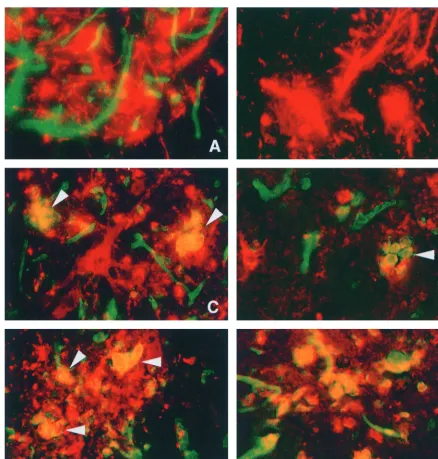
stained to detect both H-2Kb/H-2Db and SV antigen (Fig. 1).
The highest level of SV antigen was observed between 3 and 5
days after infection, predominantly in neurons in the
hip-pocampus, thalamus, brain stem, and ventral horn of the
lum-bar spinal cord. MHC class I antigen was lum-barely detectable in
endothelial and choroid plexus cells of uninfected B6 mice. At
1 day after infection, no change in the MHC class I
immuno-reactivity levels could be detected (data not shown). At 3 days
after infection, endothelial and ependymal cell class I
expres-sion was greatly intensified. A few inflammatory mononuclear
cells with class I staining were seen in the areas with
NSV-antigen-positive cells. No infected neurons showed
immunore-activity for MHC class I (Fig. 1A).
At 5 days after infection, the number of MHC class
I-posi-tive mononuclear cells increased conspicuously in
NSV-in-fected foci, perivascular areas, and the meninges. Areas of
tissue without foci of NSV-infected cells rarely contained
MHC class I-positive mononuclear cells. Class I-positive
mononuclear cells accumulated in NSV-infected foci (Fig. 1C
and D), making it difficult to determine if the neurons also
displayed a surface expression of MHC class I, although no
cells with neuronal morphology were class I-antigen positive.
Double labeling for SV and F4/80 demonstrated that the
virus-positive mononuclear cells were macrophages and microglia
surrounding and engulfing the infected neurons (Fig. 1E and
F). Even higher levels of MHC class I immunoreactivity with a
similar distribution were seen at both days 7 and 10 after
infection. Controls in which primary antibody was omitted
were consistently negative.

2 microglobulin knockout
(B2m-KO) mice infected with NSV also had no MHC class I
expres-sion (Fig. 1B).
Northern blot analysis for MHC class I.
The effect of NSV
infection on transcription of MHC class I mRNA was
investi-gated by Northern blot hybridization of RNA extracted from
6118
KIMURA AND GRIFFIN
J. V
IROL.
on November 9, 2019 by guest
http://jvi.asm.org/
C57BL/6 mice during the course of acute NSV infection (Fig.
2). Expression of mRNA for the MHC class I heavy chain,

2
microglobulin, TAP1, and TAP2 was barely detectable in the
brains of uninfected C57BL/6 mice. Infection with NSV
re-sulted in a marked increase in expression of heavy chain (1.8
kb) and

2 microglobulin (0.9 kb) mRNAs. The levels of
mRNA for TAP1 (2.6 kb) and TAP2 (2.4 kb) also increased
after infection. The levels of all of these mRNAs peaked
be-tween 5 and 7 days after infection. The expression of

2
mi-croglobulin mRNA was not detected in the brains of B2m-KO
mice.
In situ hybridization for MHC class I mRNA after infection
with NSV.
In situ hybridization with a probe for MHC class I
heavy chain mRNA revealed a low constitutive expression in
neurons of uninfected mice (Fig. 3A). Hybridization signals
were higher in large spinal cord motor neurons than in brain
neurons. Ependymal cells, glial cells in white matter, and
me-ninges also expressed heavy chain mRNA in low, but
detect-FIG. 1. Double immunofluorescence microscopy in the CNS of C57BL/6 mice (A, C, D, E, and F) and B2m-KO mice (B) after infection with NSV. Panels A, B, C, and D show double staining for MHC class I (H-2Kb/H-2Db) antigen (green) and SV antigen (red). Panel E shows double staining for F4/80 antigen (green) and SV antigen (red). Panel F shows double staining for H-2Kb/H-2Db antigen (green) and F4/80 antigen (red). At 3 days after infection, H-2Kb/H-2Db antigen was detected in endothelial cells of C57BL/6 mice (A) but not in B2m-KO mice (B). SV-antigen-positive neurons shown in panel A (red) did not demonstrate MHC class I immunoreactivity. At 5 days after infection (C and D), the numbers of MHC class I immunoreactive cells increased, and cells that were positive for MHC class I and SV (arrowheads) were detected. Note that SV-antigen-positive cells with neuronal morphology do not show immunoreactivity for MHC class I. At 5 days after infection (E), F4/80-antigen-positive cells (green) with engulfed SV antigen (red) accumulated. Most F4/80-positive cells (red) detected in NSV-infected foci show MHC class I (green) immunoreactivity (F). (A, B, C, E, and F) Ventral horn of lumbar spinal cords; (D) thalamus. (A, B, C, D, and F) Magnification,⫻322; (E) magnification,
⫻403.
on November 9, 2019 by guest
http://jvi.asm.org/
[image:3.612.83.521.68.527.2]able, levels. The mRNA expression of

2 microglobulin (Fig.
3B), TAP1 (Fig. 3C), and TAP2 (Fig. 3D) was barely
detect-able in the CNS of uninfected mice. At 1 day after NSV
infection, no clear change in the mRNA signals was detected.
The number of cells expressing heavy chain,

2
microglobu-lin, TAP1, and TAP2 increased in infected mice from days 3 to
7. The signals were confluent and generally elevated in both
gray and white matter. A majority of neurons hybridized with
the probes (Fig. 3F, H, J, and K), and levels of mRNA
expres-sion in neurons in infected mice were higher than in uninfected
mice (Fig. 3A, B, C, and D). Signals were more intense in
perivascular (Fig. 3E, G, and I) and meningeal inflammatory
mononuclear cells than in neurons.
[image:4.612.144.460.255.695.2]Similar neuronal staining was observed in spinal cord
neu-rons 5 days after NSV infection, and no staining was observed
with identically labeled sense probes (Fig. 4).
FIG. 2. Northern blot analysis of mRNAs for the MHC class I molecules and peptide transporters in the brains of C57BL/6 and B2m-KO mice at 1, 3, 5, 7, and 10 days after intracerebral inoculation of 1,000 PFU of NSV. C, control unin-fected mice; HC, MHC class I heavy chain.
FIG. 3. In situ hybridization for MHC class I and peptide transporter mRNA in the cerebral cortex of C57BL/6 mice infected with NSV. Heavy chain mRNA was expressed in neurons in uninfected mice (A) at very low levels.2 microglobulin (B), TAP1 (C), and TAP2 (D) mRNAs were barely detectable. Expression of heavy chain (E and F),2 microglobulin (G and H), TAP1 (I and J), and TAP2 (K) mRNA was increased at 5 days after NSV infection. Signals were detected in the cytoplasms of neurons, glial cells, and inflammatory cells. (A, B, C, D, E, G, and I) Magnification,⫻77; (F, H, J, and K) magnification,⫻155.
6120
KIMURA AND GRIFFIN
J. V
IROL.
on November 9, 2019 by guest
http://jvi.asm.org/
Infection of B2m-KO mice and CD8-KO mice with NSV.
To
determine whether a deficiency in CD8
⫹MHC class
I-re-stricted T cells affects the susceptibility of mice to
NSV-in-duced encephalitis, adult B2m-KO mice and CD8
␣
chain
knockout (CD8-KO) mice on a C57BL/6 background and
con-trol B6 mice were inoculated intracerebrally with NSV and
were observed for mortality (Fig. 5). Immunocompetent B6
mice showed a high mortality (72 to 90%), but only 20% (4 of
20) B2m-KO mice died by 11 days after infection (Fisher’s
exact probability test;
P
⬍
0.001), although 70% developed
hind limb paralysis. In contrast, 63% (12 of 19) of CD8-KO
mice died by 10 days after infection, similar to B6 mice.
Virus replication in vivo.
To determine whether lack of
CD8
⫹T cells affected virus clearance, both infectious virus and
viral RNA in the CNS of infected mice was quantitated. There
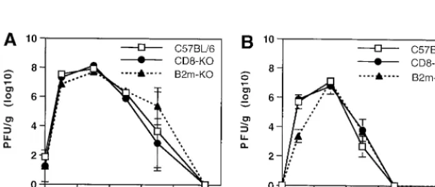
were no significant differences in the amounts of infectious
virus in the brains or spinal cords of CD8-KO mice and B6
mice at any time after infection (Fig. 6). At day 1, the amount
of infectious virus was lower in the spinal cords of B2m-KO
mice than in those of B6 mice and CD8-KO mice (Student’s
t
test;
P
⬍
0.05).
To compare the level of viral RNA in the CNS of B6 mice
with the levels in B2m-KO and CD8-KO mice, we performed
dot blot hybridization with a DIG-labeled probe for SV. At 1
day after infection, the level of viral RNA in the brains of B6
mice was higher than the levels in the brains of B2m-KO and
CD8-KO mice (
P
⬍
0.01) (Fig. 7A). At 3 days after infection,
the time of peak virus titer (Fig. 6A) and peak virus antigen,
the level of viral RNA in the brains of B6 mice was higher than
the viral RNA level in the brains of B2m-KO mice (
P
⬍
0.01)
(Fig. 7A). The viral RNA level in the spinal cord of CD8-KO
mice was lower than that of B6 mice at day 3 (
P
⬍
0.01) (Fig.
7B). On days 5 to 10, during the period of virus clearance, the
viral RNA levels in the brain and spinal cord decreased more
FIG. 4. In situ hybridization with strand-specific probes. The lumbar spinal cords of uninfected C57BL/6 mice (A, D, G, and J) and C57BL/6 mice at 5 days after infection with NSV (B, C, E, F, H, I, K, and L) was stained with heavy chain probes (A, B, and C),2 microglobulin probes (D, E, and F), TAP1 probes (G, H, and I), and TAP2 probes (J, K, and L). (A, B, D, E, G, H, J, and K) Antisense probe; (C, F, I, and L) sense probe. Magnification in all panels,⫻97.
on November 9, 2019 by guest
http://jvi.asm.org/
quickly in B6 mice than in B2m-KO and CD8-KO mice. This
difference was significant (
P
⬍
0.05) between B6 mice and both
knockout mice at days 5 and 7 in brain (Fig. 7A) and at day 7
in spinal cord (Fig. 7B).
Antibody response to NSV infection.
To exclude the
possi-bility that a difference in the antibody response accounted for
differences in virus replication and virus clearance and
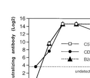
mortal-ity, levels of neutralizing antibody in the serum of infected B6,
B2m-KO, and CD8-KO mice were compared (Fig. 8). No
differences were identified.
DISCUSSION
Studies showed that mRNAs for the heavy and light chains
of MHC class I molecules and for the peptide transporters
necessary for loading peptide onto class I molecules before
transport to the cell surface were expressed, but mature
im-munologically reactive protein was not expressed, by neurons
after infection with NSV. Mice deficient in CD8
⫹T cells due
to lack of expression of the CD8
␣
chain (CD8-KO) or MHC
class I light chain (B2m-KO) cleared infectious virus as quickly
as normal mice, but cleared viral RNA more slowly. Mortality
in B2m-KO mice was lower than in mice without a defect in
MHC class I. Therefore, these studies suggest that CD8
⫹T
cells play a role in clearance of viral RNA, but are unlikely to
interact directly with infected neurons, and that expression of

2 microglobulin is involved either directly or indirectly in fatal
NSV-induced encephalomyelitis.
It is generally acknowledged that the CNS is an
immuno-suppressive and immune-privileged microenvironment. CNS
resident cells normally express few or no MHC class I
mole-cules (20, 21, 30, 48). In the present study, we demonstrated
that MHC class I expression was induced in the CNS after
NSV infection. By immunofluorescence, MHC class I antigen
expression in the CNS of B6 mice peaked between 5 and 10
days after infection and was found on resident endothelial,
meningeal, and macrophage and microglial cells as well as on
inflammatory cells. Peak titers of virus in the CNS of infected
mice occurred 3 days after infection, and infectious virus was
rapidly cleared between days 5 and 10. At all time points, viral
antigen was localized while expression of MHC class I was
widespread. Therefore, the upregulation of class I expression is
probably induced by factors produced by infected neurons or
associated with the immune response induced by infection.
Cells labeled with both anti-SV and anti-class I antibodies
were detected at days 5 and 7 but not at day 3 in infected foci.
These cells were almost exclusively macrophages and
micro-glia. SV does not replicate in mononuclear cells in vitro or in
vivo (10, 17). The distribution of SV-antigen-positive
macro-phages was restricted to areas around infected neurons.
Nei-ther SV antigen nor SV RNA could be detected in resting
microglia. These findings suggest that the viral antigens
de-tected in macrophages and microglia were the result of
phago-cytosis. Macrophages and microglia, however, may be able to
process viral antigen and interact with locally infiltrating T cells
(47).
[image:6.612.144.465.73.202.2]The paucity of MHC class I antigens in infected neurons
suggested that the transcription of class I molecules or the
FIG. 5. Survival of B2m-KO (solid triangle), CD8-KO (solid circle), and immunocompetent (open box) C57BL/6 mice after infection with NSV. Eleven-week-old mice were inoculated intracerebrally with 1,000 PFU of NSV in 0.03 ml of Hanks balanced salt solution. Groups of 20 mice (A) or 19 mice (B) were examined.
FIG. 6. Replication of NSV in the CNS of B2m-KO (solid triangle), CD8-KO (solid circle), and immunocompetent (open box) C57BL/6 mice after infection with NSV. (A) Amount of infectious virus in brain; (B) amount of infectious virus in spinal cord. Each time point represents the geometric mean and standard deviation for three mice.
6122
KIMURA AND GRIFFIN
J. V
IROL.
on November 9, 2019 by guest
http://jvi.asm.org/
[image:6.612.146.462.563.699.2]molecules that were required for surface expression of
peptide-MHC class I complexes was suppressed specifically in neurons.
Functional cell surface expression of MHC class I molecules
depends on the production of both MHC class I heavy chain
and

2 microglobulin, which noncovalently bind to each other
(1, 41). Antigenic peptide presentation also requires the
het-erodimer of TAP1 and TAP2 proteins that transports short
peptides from the cytosol into the endoplasmic reticulum
lu-men for loading onto assembled MHC class I molecules (3, 29,
42). In the present study, mRNAs for all of the molecules
required for class I antigen presentation (heavy chain,

2
mi-croglobulin, TAP1, and TAP2) were expressed in neurons as
well as in cells that expressed detectable levels of these
pro-teins. Heavy chain,

2 microglobulin, TAP1, and TAP2 mRNA
levels were transiently increased after infection with NSV,
sug-gesting that transcription of these mRNAs was coordinately
regulated in neurons, as well as in cells that expressed
detect-able levels of protein.
Expression of MHC class I mRNAs in neurons suggests that
failure of NSV-infected neurons to express MHC class I
pro-tein was due to a posttranscriptional block in propro-tein
expres-sion rather than a deficiency in either

2 microglobulin or
peptide transporter mRNAs. Posttranscriptional block for

2
microglobulin, TAP1, or TAP2 protein expression also could
result in failure to detect MHC class I protein. This may be a
neuron-specific block or a block in translation of cellular
mRNAs due to SV infection since SV shuts down host protein
synthesis. An alternative explanation is that the class I mRNAs
detected in neurons may be associated with expression of
non-classical class I genes. In addition to the non-classical polymorphic
MHC class I molecules, multiple genes code for heavy chains
of “nonclassical” nonpolymorphic class I (class Ib) molecules
(25). The heavy chains of nonclassical class I molecules share
sequence homology with classical class I molecules, and both
kinds of molecules use

2 microglobulin as the invariant light
chain (44). Therefore, the possibility exists for
cross-hybridiza-tion with nonclassical class I genes. An inability to detect the
class I protein in neurons might result from the lack of a
suitable, high-affinity antibody for nonclassical MHC class I
proteins, and not due to the absence of expression of these
molecules in vivo. Our data are consistent with the findings of
Pereira et al. that primary sensory neurons upregulate MHC
class I mRNA, but not class I proteins, in response to acute
herpes simplex virus infection (36). However, in a subsequent
study, low-density classical class I proteins were demonstrated
by flow cytometry on the cell surface of dissociated primary
sensory neurons recovered from mice infected with herpes
simplex virus (35). Upregulation of MHC class I mRNA has
also been detected in neurons of mice infected with Theiler’s
murine encephalomyelitis virus (34), measles virus (7), and
rabies virus (14). Characterization of the class I heavy chain
transcripts in neurons remains to be investigated.
Both B2m-KO mice and CD8-KO mice were expected to
show comparable defects in MHC class I-restricted
T-cell-dependent immune responses. However, a lack of expression
of

2 microglobulin, but not CD8
␣
, decreased mortality from
NSV infection. This differs from the observations of mice
in-fected with lymphocytic choriomeningitis virus, where fatal
disease still occurs in B2m-KO mice because CD4 T cells
substitute functionally for CD8 T cells to mediate cytotoxic
damage in the CNS (4, 6, 22, 31, 37). B2m-KO mice also have
impaired NK cell function (38), but a role for NK cells in fatal
NSV-induced encephalitis is unlikely since NK-cell-deficient
B6 mice with the beige mutation died faster than B6 mice after
NSV infection (data not shown). It also seems unlikely that
protection from fatal NSV encephalitis is due to lower levels of
early viral replication in the CNS of B2m-KO mice since these
were not consistently different than levels in B6 or CD8-KO
mice. Differences in mortality could in some way be related to
the lack of expression of nonclassical MHC molecules which
results in cytotoxic damage to neurons since this will be
defi-cient in B2m-KO, but not CD8-KO, mice.
[image:7.612.131.473.73.191.2]Although no differences in the amounts of infectious virus in
brain were detected between B6, CD8-KO, and B2m-KO mice,
there were differences in the levels of viral RNA. B6 mice had
more viral RNA early after infection and more rapid clearance
of viral RNA from the CNS. The SV-specific probe we used in
this study hybridized with both full-length and subgenomic
RNAs, so it is possible that more subgenomic RNA, not
re-flected in progeny virions, was produced by B6 mice early after
FIG. 7. Graph of viral RNA in the CNS of B2m-KO, CD8-KO, and immunocompetent C57BL/6 mice after infection with NSV. (A) Relative amount of viral RNA in brain; (B) relative amount of viral RNA in spinal cord. Each time point represents the geometric mean and standard deviation for three mice.
FIG. 8. Graph of neutralizing antibody response of B2m-KO, CD8-KO, and immunocompetent C57BL/6 mice after infection with NSV.
on November 9, 2019 by guest
http://jvi.asm.org/
[image:7.612.355.504.578.708.2]infection. Antibody is the primary mechanism for clearance of
SV from the CNS (23), but more rapid clearance of viral RNA
from the brains and spinal cords of B6 mice than from CD8
T-cell-deficient B2m-KO or CD8-KO mice suggests an
auxil-iary role for CD8 T cells in alphavirus clearance from the CNS.
The paucity of MHC class I protein in infected neurons
sug-gests that CD8 T cells probably act indirectly to help clear
intracellular virus RNA. Possible cytokines participating in
viral RNA clearance (e.g., gamma interferon and tumor
ne-crosis factor alpha, since they have antiviral activity) are
ex-pressed in the CNS after infection with SV, and expression
coincides with mononuclear infiltration and virus clearance
(49). However, CD8 T cells are not required for clearance
since RNA, as detected by dot blot hybridization, is eventually
cleared from the CNS of CD8 T-cell-deficient mice either
through the effects of CD4 T cells with overlapping functions
or through the effects of antibody.
ACKNOWLEDGMENTS
This work was supported in part by grant NS18596 from the
Na-tional Institutes of Health (D.E.G.) and by Hokkaido University
(T.K.).
REFERENCES
1.Bjorkman, P. J., M. A. Saper, B. Samraoui, W. S. Bennett, J. L. Strominger, and D. C. Wiley.1987. Structure of the human class I histocompatibility antigen, HLA-A2. Nature329:506–512.
2.Corriveau, R. A., G. S. Huh, and C. J. Shatz.1998. Regulation of class I MHC gene expression in the developing and mature CNS by neural activity. Neuron21:505–520.
3.Deverson, E. V., I. Gow, W. J. Coadwell, J. J. Monaco, G. B. Butcher, and J. C. Howard.1991. MHC class II region encoding proteins related to the multidrug resistance family of transmembrane transporters. Nature348:738– 741.
4.Doherty, P. C., S. Hou, and P. J. Southern.1993. Lymphocytic choriomen-ingitis virus induces a chronic wasting disease in mice lacking class I major histocompatibility complex glycoproteins. J. Neuroimmunol.46:11–17. 5.Drew, P. D., M. Lonergan, M. E. Goldstein, L. A. Lampson, K. Ozato, and
D. E. McFarlin.1993. Regulation of MHC class I and beta 2-microglobulin gene expression in human neuronal cells. Factor binding to conserved cis-acting regulatory sequences correlates with expression of the genes. J. Im-munol.150:3300–3310.
6.Fung-Leung, W. P., T. M. Kundig, R. M. Zinkernagel, and T. W. Mak.1991. Immune response against lymphocytic choriomeningitis virus infection in mice without CD8 expression. J. Exp. Med.174:1425–1429.
7.Gogate, N., P. Swoveland, T. Yamabe, L. Verma, J. Woyciechowska, E. Tarnowska-Dziduszko, J. Dymecki, and S. Dhib-Jalbut.1996. Major histo-compatibility complex class I expression on neurons in subacute sclerosing panencephalitis and experimental subacute measles encephalitis. J. Neuro-pathol. Exp. Neurol.55:435–443.
8.Gorrell, M. D., J. A. Lemm, C. M. Rice, and D. E. Griffin.1997. Immuniza-tion with nonstructural proteins promotes funcImmuniza-tional recovery of alphavirus-infected neurons. J. Virol.71:3415–3419.
9.Griffin, D. E.1976. Role of the immune response in age-dependent resis-tance of mice to encephalitis due to Sindbis virus. J. Infect. Dis.133:456–464. 10. Griffin, D. E., and R. T. Johnson.1973. Cellular immune response to viral infection: in vitro studies of lymphocytes from mice infected with Sindbis virus. Cell. Immunol.9:426–434.
11. Griffin, D. E., and R. T. Johnson.1977. Role of the immune response in recovery from Sindbis virus encephalitis in mice. J. Immunol.118:1070–1075. 12. Hirsch, R. L., D. E. Griffin, and R. T. Johnson.1979. Interactions between immune cells and antibody in protection from fatal Sindbis virus encepha-litis. Infect. Immun.23:320–324.
13. Irani, D. N., and D. E. Griffin.1991. Isolation of brain parenchymal lym-phocytes for flow cytometric analysis: application to acute viral encephalitis. J. Immunol. Methods139:223–231.
14. Irwin, D. J., W. H. Wunner, H. C. Ertl, and A. C. Jackson.1999. Basis of rabies virus neurovirulence in mice: expression of major histocompatibility complex class I and class II mRNAs. J. Neurovirol.5:485–494.
15. Jackson, A. C., T. R. Moench, D. E. Griffin, and R. T. Johnson.1987. The pathogenesis of spinal cord involvement in the encephalomyelitis of mice caused by neuroadapted Sindbis virus infection. Lab. Investig.56:418–423. 16. Jackson, A. C., T. R. Moench, B. D. Trapp, and D. E. Griffin.1988. Basis of
neurovirulence in Sindbis virus encephalomyelitis of mice. Lab. Investig. 58:503–509.
17. Johnson, R. T.1965. Virus invasion of the central nervous system. Am. J. Pathol.46:929–943.
18. Johnson, R. T., H. F. McFarland, and S. E. Levy.1972. Age-dependent resistance to viral encephalitis: studies of infections due to Sindbis virus in mice. J. Infect. Dis.125:257–262.
19. Joly, E., and M. B. Oldstone.1992. Neuronal cells are deficient in loading peptides onto MHC class I molecules. Neuron8:1185–1190.
20. Lampson, L. A.1995. Interpreting MHC class I expression and class I/class II reciprocity in the CNS: reconciling divergent findings. Microsc. Res. Tech. 32:267–285.
21. Lampson, L. A., and W. F. Hickey.1986. Monoclonal antibody analysis of MHC expression in human brain biopsies: tissue ranging from “histologically normal” to that showing different levels of glial tumor involvement. J. Im-munol.136:4054–4062.
22. Lehmann-Grube, F., J. Lohler, O. Utermohlen, and C. Gegin.1993. Antiviral immune responses of lymphocytic choriomeningitis virus-infected mice lack-ing CD8⫹T lymphocytes because of disruption of the beta 2-microglobulin
gene. J. Virol.67:332–339.
23.Levine, B., J. M. Hardwick, B. D. Trapp, T. O. Crawford, R. C. Bollinger, and D. E. Griffin.1991. Antibody-mediated clearance of alphavirus infection from neurons. Science254:856–860.
24. Linda, H., H. Hammarberg, S. Cullheim, A. Levinovitz, M. Khademi, and T. Olsson.1998. Expression of MHC class I and beta2-microglobulin in rat spinal motoneurons: regulatory influences by IFN-gamma and axotomy. Exp. Neurol.150:282–295.
25. Lindahl, K. F.1997. On naming H2 haplotypes: functional significance of MHC class Ib alleles. Immunogenetics46:53–62.
26. McFarland, H. F., D. E. Griffin, and R. T. Johnson.1972. Specificity of the inflammatory response in viral encephalitis. I. Adoptive immunization of immunosuppressed mice infected with Sindbis virus. J. Exp. Med.136:216– 226.
27. McLean, I. W., and P. K. Nakane.1974. Periodate-lysine-paraformaldehyde fixative. A new fixation for immunoelectron microscopy. J. Histochem. Cy-tochem.22:1077–1083.
28. Moench, T. R., and D. E. Griffin.1984. Immunocytochemical identification and quantitation of the mononuclear cells in the cerebrospinal fluid, menin-ges, and brain during acute viral meningoencephalitis. J. Exp. Med.159:77– 88.
29. Monaco, J. J., S. Cho, and M. Attaya.1990. Transport protein genes in the murine MHC: possible implications for antigen processing. Science250: 1723–1726.
30. Mucke, L., and M. B. Oldstone.1992. The expression of major histocom-patibility complex (MHC) class I antigens in the brain differs markedly in acute and persistent infections with lymphocytic choriomeningitis virus (LCMV). J. Neuroimmunol.36:193–198.
31. Muller, D., B. H. Koller, J. L. Whitton, K. E. LaPan, K. K. Brigman, and J. A. Frelinger.1992. LCMV-specific, class II-restricted cytotoxic T cells in beta 2-microglobulin-deficient mice. Science255:1576–1578.
32. Neumann, H., A. Cavalie, D. E. Jenne, and H. Wekerle.1995. Induction of MHC class I genes in neurons. Science269:549–551.
33. Neumann, H., H. Schmidt, A. Cavalie, D. Jenne, and H. Wekerle.1997. Major histocompatibility complex (MHC) class I gene expression in single neurons of the central nervous system: differential regulation by interferon (IFN)-gamma and tumor necrosis factor (TNF)-alpha. J. Exp. Med.185:305– 316.
34. Njenga, M. K., L. R. Pease, P. Wettstein, T. Mak, and M. Rodriguez.1997. Interferon alpha/beta mediates early virus-induced expression of H-2D and H-2K in the central nervous system. Lab. Investig.77:71–84.
35. Pereira, R. A., and A. Simmons.1999. Cell surface expression of H2 antigens on primary sensory neurons in response to acute but not latent herpes simplex virus infection in vivo. J. Virol.73:6484–6489.
36. Pereira, R. A., D. C. Tscharke, and A. Simmons.1994. Upregulation of class I major histocompatibility complex gene expression in primary sensory neu-rons, satellite cells, and Schwann cells of mice in response to acute but not latent herpes simplex virus infection in vivo. J. Exp. Med.180:841–850. 37. Quinn, D. G., A. J. Zajac, J. A. Frelinger, and D. Muller.1993. Transfer of
lymphocytic choriomeningitis disease in beta 2-microglobulin-deficient mice by CD4⫹T cells. Int. Immunol.5:1193–1198.
38. Raulet, D. H.1994. MHC class I-deficient mice. Adv. Immunol.55:381–421. 39. Rensing, E. A., U. Malipiero, M. Irmler, J. Tschopp, D. Constam, and A. Fontana.1996. Neurons induced to express major histocompatibility com-plex class I antigen are killed via the perforin and not the Fas (APO-1/CD95) pathway. Eur. J. Immunol.26:2271–2274.
40. Shifman, M. I., and D. G. Stein.1995. A reliable and sensitive method for non-radioactive Northern blot analysis of nerve growth factor mRNA from brain tissues. J. Neurosci. Methods59:205–208.
41. Silver, M. L., K. C. Parker, and D. C. Wiley.1991. Reconstitution by MHC-restricted peptides of HLA-A2 heavy chain with beta-2 microglobulin. Na-ture350:619–622.
42. Spies, T., M. Bresnahan, S. Bahram, D. Arnold, G. Blanck, E. Mellins, D. Pious, and R. DeMars.1990. A gene in the human major histocompatibility complex class II region controlling the class I antigen presentation pathway. Nature348:744–747.
43. Stanley, J., S. J. Cooper, and D. E. Griffin.1986. Monoclonal antibody cure
6124
KIMURA AND GRIFFIN
J. V
IROL.
on November 9, 2019 by guest
http://jvi.asm.org/
and prophylaxis of lethal Sindbis virus encephalitis in mice. J. Virol.58:107– 115.
44.Strominger, J. L.1989. The gd T cell receptor and class Ib MHC-related proteins: enigmatic molecules of immune recognition. Cell57:895–898. 45. Taylor, R. M., H. S. Hurlbut, T. H. Work, J. R. Kingston, and T. E.
Froth-ingham.1955. Sindbis virus: a newly recognized arthropod-transmitted virus. Am. J. Trop. Med. Hyg.4:844–855.
46. Tucker, P. C., E. G. Strauss, R. J. Kuhn, J. H. Strauss, and D. E. Griffin. 1993. Viral determinants of age-dependent virulence of Sindbis virus for mice. J. Virol.67:4605–4610.
47. Tyor, W. R., G. Stoll, and D. E. Griffin.1990. The characterization of Ia expression during Sindbis virus encephalitis in normal and athymic nude mice. J. Neuropathol. Exp. Neurol.49:21–30.
48. Vass, K., and H. Lassmann.1990. Intrathecal application of interferon gamma. Progressive appearance of MHC antigens within the rat nervous system. Am. J. Pathol.137:789–800.
49. Wesselingh, S. L., B. Levine, R. J. Fox, S. Choi, and D. E. Griffin.1994. Intracerebral cytokine mRNA expression during fatal and nonfatal alphavi-rus encephalitis suggests a predominant type 2 T cell response. J. Immunol. 152:1289–1297.
50. Wong, G. H. W., P. F. Bartlett, L. I. Clark, F. Battye, and J. W. Schrader. 1984. Inducible expression of H-2 and Ia antigens on brain cells. Nature 310:688–691.
51. Wong, G. H. W., P. F. Bartlett, I. Clark-Lewis, J. L. McKimm-Breschkin, and J. W. Schrader.1985. Interferon gamma induces the expression of H-2 and Ia antigens on brain cells. J. Neuroimmunol.7:255–278.
on November 9, 2019 by guest
http://jvi.asm.org/