HEART RATE VARIABILITY IN HYPERTHYROID PATIENTS CORRELATED WITH THYROID FUNCTION TESTS
Dissertation submitted for
M.D. ( Branch – V Physiology)
THE TAMIL NADU DR. M.G.R. MEDICAL UNIVERSITY
Department of physiology
PSG Institute of Medical Sciences and Research Coimbatore – 641004
INTRODUCTION
The thyroid is a highly vascular, brownish-red structure located in the neck anteriorly extending from fifth cervical vertebra to the first thoracic vertebra 1. Thyroid gland is the largest gland which is essential to sustain normal life . Thyroid is a greek word meaning "shield", introduced by Thomas Wharton2. Thyroid gland consists of follicles lined by cuboidal epithelium. Each follicle contains clear proteinaceous colloid material. The colloid consists of
mainly thyroglobulin. Iodide plays a important role in thyroid synthesis. About 120 µg of iodide enter thyroid gland per day. The iodide pump or Na +/I- (sodium iodide symporter) reabsorbs iodide into thyrocytes against electrical gradient. Iodine is immediately bound to the tyrosine in the thyroglobulin3.
Thyroglobulin is iodinated to form monoiodothyrosine(MIT) and
diiodothyrosine (DIT). By coupling reactions, thyroxine(tetraiodothyronine) T4,
triiodothyronine T3 and reverse T3 are formed. Thyroid peroxidase helps in both
iodination and coupling reactions .
When the thyroid gland become overactive and synthesize excess of thyroid hormone ,it is called hyperthyroidism. The prevalence of hyperthyroidism has been estimated as 0.21 percent for men and 1.9 percent for women 4.
The most common cause of hyperthyroidism in younger age group is Graves' disease.
In graves the total daily disposal of T 3 is disproportionately increased
relative to that of T 4 , indicating that the production rate of T 3 is increased . it
could be due to increase in thyroid secretion of Triiodothyronine .Deiodinase -1
also causes increased peripheral conversion of T 4 to T3 .
Temporary viremia of thyroid gland, certain drugs, toxic nodules can also cause hyperthyroidism .
Thyroid stimulating immunoglobulins are the antibodies that activate the TSH-receptor, thereby stimulating thyroid hormone synthesis and results in diffusely enlarged goiter . This is the etiology for graves disease.
In most patients thyroid gland is massively enlarged or of normal size in a small fraction of patients.The consistency varies from soft to firm and rubbery. The surface may be lobular or smooth. Continous thrills and bruits at the upper or lower thyroid pole, due to increased blood flow ,which is suggestive of
Major emotional Stress and fright before a year or so can be a cause for the autoimmune activity in Graves’ disease patients 6 .
It is unusually seen in men . In females,it is more prevalent after puberty . The reason for this difference being gonadal steroids and estrogen . These
hormones attack the B lympocyte population. But androgens may suppress autoimmune activity in most of the men, so graves incidence in men is less. In female patients with hyperthyroidism, pregnancy and delivery are precipitating events for Graves’ disease7.
In patients with graves disease , the thyroid gland is characterised by a non homogenous infilteration with an absence of follicular destruction. The degree of infilteration can be reduced by proper antithyroid treatment8 .
Although the intrathyroidal lymphocyte population is mixed, most are T- lymphocytes. B cell germinal centres are less common than in autoimmune thyroiditis.
Symptoms:
perspiration, warm moist skin, lid retraction , frequent bowel movements, thinning of hair, hand tremors, heat insensitivity and pretibial myxedema9 .
Tachycardia is almost always present, and tachycardia during sleep pulse rate >90 beats per minute distinguish tachycardia of thyrotoxic origin from that of
psychogenic causes10.
Graves’ opthalmopathy presents with symptoms that include dry, gritty ocular sensation, photophobia, double vision and pressure sensation behind the eye .
Increase in connective tissue is due to the hydrophilic ground substance accumulation like hyaluronic acid and chondritin sulphate.
Digital swelling, clubbing, and sub periosteal reaction of toes and fingers is called Thyroid Acropachy.
Dermopathy is seen in 99% of graves opthalmopathy with increased content of hyaluronic acid and chondritin sulphate and lympokine activated fibroblasts.
Autoimmune thyroid diseases are due to antigen specificity in suppressor and regulatory T lymphocyte function.12
This disease is rare before ten years of age. Hashimoto’s disease and Graves’ disease are autoimmune disorder and females are mostly affected in both. There are many reasons for developing Graves’ disease . Genetic predisposition or family history may play a leading role in thyroid disorders. Stress can be one of the environmental factors for thyroid autoimmunity13.
Thyroid storm is also called thyrotoxic crisis usually occur in association with graves disease . It is a sudden incident when the thyroid hormones reach toxic levels with fever, perspiration, tachycardia and hypertension. The clinical picture is severe with hypermetabolism , accompanied by congestive heart failure,
pulmonary edema. Incidence of thyroid storm is about 10% in hospital admissions precipitated by trauma, surgical emergencies, infections . It is ten times more common in females. It has a mortality rate of 20 to 30%14.
Other causes for hyperthyroidism are,
Few nodular and spherical growths are seen in the thyroid lobes. These nodules cause increased synthesis of thyroxine and triiodothyronines 15.
Seen in patients over 50 yrs of age. Nodules become overactive after many years of thyroid enlargement.
Somatic mutations in the TSH receptor gene demonstrated in toxic adenomas in some cases of toxic multinodular goitre and appeared to be different from nodule to nodule.
Overmedication with Thyroid Hormone:
Levothyroxine given for hypothyroidism due to disease, radioactive iodine, can cause hyperthyroidism.
Excessive Iodine Ingestion:
High iodine content in cough syrup and amiodarone , an antiarrythmic drug
Postpartum Thyroiditis:
Pregnancy is a state of immunosupression due to increased steroid
harmone levels. when pregnancy proceeds from first to last trimester , the disease ameiliorates and after delivery the autoimmune processes flare up and may lead to the emergence of post partum thyroiditis 16
.
Thyroid stimulating antibody declines in the third trimester of pregnancy but passively transferred to foetus 17. In few cases mild to moderate postpartum hyperthyroidism is seen . Hyperthyroidism lasts for 1 to 2 months followed by several months of hypothyroidism. Most women will recover. In few cases hypothyroidism needs thyroid hormone replacement.
Subacute Thyroiditis:
It may follow a viral infection with painful thyroid gland. It is a self-limited condition associated with hyperthyroidism, hypothyroidism, and finally euthyroidism. Subacute thyroiditis accounts for 15-20% of thyrotoxicosis patients 18 .
Silent Thyroiditis:
Silent thyroiditis is thyroid gland inflammation, in which the person alternates
thyroiditis. It has both painful and painless variants and sometimes biopsy of the thyroid gland reveals the histo pathological changes of Hashimoto s thyroiditis 32.
Irradiation:
In the general population whom were previously exposed to radiation, thyroid auto antibodies are common. Transient clinical opthalmopathy can occur due to radioactive iodine.
Infection:
Y. enterocolitica and other infections of the thyroid gland like congenital rubella can be the etiology for hyperthyroidism and commonly associated with thyroid autoimmune phenomena19.
TSH, T
3and T
4Regulation:
Anterior pituitary gland produce thyroid-stimulating hormone (TSH),
TSH production itself is regulated by thyrotropin-releasing hormone (TRH), which is produced by the hypothalamus .
TSH synthesised by anterior pituitary is regulated by negative feedback mechanism. The TSH receptor is G –protein linked with seven trans membrane domains and employs cyclic adenosine monophosphate and phosphoinositol pathways for signal transduction .
When the T3 T4, levels are high , they act on anterior pituitary and rapidly
suppress TSH production. Low levels of thyroid hormones stimulate pituitary TSH Secretion and increase T3 T4, synthesis from the thyroid gland.
On exposure to cold, hypothalamus via TRH, increase TSH secretion from anterior pituitary and during warmth, reduce TSH secretion.
Emotional conditions like anxiety, excitement, fear arouse the sympathetic
nervous system and decrease TSH secretion. Both temperature modulations and emotional effects were not observed, after the transection of hypophysial stalk, since both these effects were mediated by the way of hypothalamus21.
Too little iodide, or too much iodide decreases iodide trapping and also thyroid hormone synthesis. Sodium –iodide symporter,( NIS) needs TSH for its positioning on plasma membrane. NIS loses its ability to transport iodide across the plasma membrane in the absence of TSH .
TSH production is decreased by growth hormone inhibiting hormone , rising levels of steroids secreted from adrenal cortex. Binding of Thyroid
stimulating hormone to its receptor on the thyrocytes increases cAMP levels. Cyclic AMP activates protein kinase which phosphorylates the cell and causes the growth of thyroid glandular tissue and increase thyroid hormone secretion 22. Cardiovascular and extra cardiovascular manifestations of hyperthyroidism
are due to hyperadrenergic state 23.
Thyroid hormones is vital for stimulating metabolic processes and it increases the demand for coenzymes and vitamins. Basal metabolic rate is increased by increasing the number of mitochondria and also by increasing the activity of sodium potassium ATPase pump.
Hyperthyroid patients usually have normal urinary and plasma epinephrine, norepinephrine levels and show normal response for epinephrine infusion and there is no evidence for increased catecholamine synthesis or metabolism at nerve endings.
Thyroid activity in foetus:
Thyroid hormone is important for fetal, as well as neonatal, brain
development and nerve myelination. Thyroid hormone production rate per unit area is more in neonates and children than adults.
Congenital hypothyroidism occurs in babies if thyroid gland does not produce adequate thyroid hormones. Mental retardation is prevented if the hypothyroid child is treated within a year of life.
Effect of Hyperthyroidism on Cardiovascular System:
There is increased circulatory demands which causes modifications in cardiac functions .Cardiovascular alterations also results from the
flow and the systolic of the heart. Heart has 2 myosin heavy chain isoforms, α MHC and β MHC encoded by two genes located on short arm of chromosome 17. In hyperthyroidism circulatory T3 enter cardiac myocytes combines with its receptors and enters the nucleus, causes enhanced transcription of α myosin genes- α MHC, β adrenergic receptors , G proteins, Na K ATPase and while it causes inhibition of transcription of β MHC, phospolamban, two types of adenylcyclase. Thus the cardiac contractility is increased24.
Excessive thyroid hormone production increase the tissue utilization of oxygen, thereby increasing the metabolites which cause vasodilatation. Therefore there is increase in the blood flow, cardiac output and heart rate. Increased thyroid hormones, by increasing the enzymatic activity, increases the strength of the heart25.
In our study , sympathetic noradrenergic system alterations with altered thyroid functions were studied in the subjects. Thyroid hormone variations on certain cellular mechanisms in the heart causes many cardiovascular
abnormalities.
It is well established hyper dynamic cardiovascular state with low
It is also associated with tachycardia, increased cardiac muscle oxygen consumption , increased left ventricular diastolic and systolic function, low systemic vascular resistance and increased prevalence of arrhythmias, decreased exercise performance, and increased risk of cardiovascular mortality. Pulse pressure is increased due to decrease in diastolic pressure and increase in
systolic pressure .
Heart Rate Variability as an index of Cardiac Autonomic activity:
Autonomic Nervous System can be assessed using HRV analysis. The cardiac rhythm influenced by sympathetic and parasympathetic dysfunction can be evaluated by the interbeat variations of R-R interval26.
The spectral variation of the heart rate in the lower (LF) and higher frequencies (HF) has a significant relationship to sympathetic and parasympathetic activity, respectively.
These derangements in autonomic nervous system can be found out by HRV, and the HRV indices help us to assess the disease severity in hyperthyroid patients.
Effect of Autonomic nervous system:
The thyroid glands are regulated by both neural and endocrine hormones. Many ganglia contribute to the innervation of this gland . There are two
divisions in the autonomic nervous system namely, sympathetic and vagal nervous systems. Both are antagonistic in their target actions. Cardiac inotrophic effects are partly due to adrenergic stimulation.
The increased sensitivity of atria to beta-adrenergic agonists is due to increased beta-adrenoceptor density and sympathetic stimulation on the β1
receptors in the heart causes increase in the heart rate and cardiac output of hyperthyroid patients .
II. AIM AND OBJECTIVES
PRIMARY AIM:
To investigate a possible relationshipbetween hyperthyroidism and autonomic imbalance in hyperthyroid patients usingspectral analysis of HRV.
SECONDARY AIM:
• To compare HRV of hyperthyroid and normal volunteers.
• To assess the cardiovascular risk in hyperthyroid patients using HRV.
OBJECTIVE:
III.REVIEW OF LITERATURE
Autonomic Nervous System
The autonomic system has afferent nerves which carries information from viscera and the central processes enter spinal cord or brainstem. Peripheral processes of afferents run with efferent nerves.
Word ” autonomous “ is taken from greek word the autos, meaning self and the memos , meaning control . Thus autonomic nervous system is an involuntary system which controls the vegetative functions, so it is also called vegetative system.
An important function of autonomic nervous system is to maintain homeostasis. i.e, constant internal environment by releasing
epinephrine, insulin and glucagon or renin, vasopressin Sympathetic and
parasympathetic divisions are complementary in nature rather than antagonistic.
ventral horn) and ventral horn cell which transmits the efferent impulses to skeletal muscle.
Sympathetic division can be considered as the accelerator i.e, it acts during fast responses flight or fight reactions and the parasympathetic
division acts as the brake and functions during resting conditions eg. "feed and breed" 27.
Both the divisions of autonomic nervous system carry visceral
afferent and sensation information like vasomotor, respiratory, and viscera somatic reflexes and interrelated visceral activities28 .
ANS is responsible for activities of the organs of digestion, circulation, excretion, respiration, reproduction as well as sweat, lacrimal, salivary glands and adrenal medulla29.
matter in brain stem and spinal cord. Preganglionic sympathetic fibres causes release of epinephrine and nor epinephrine from adrenal medulla. Preganglionic Parasympathetic nerves go directly to vicinity of viscera .
Cells of origin of sympathetic division of the autonomic nervous system are located in lateral horn spinal grey matter from eighth cervical and 1st thoracic through 3rd lumbar segmental levels.
Autonomic Nervous System and Hypothalamus:
Th nerves from the hypothalamus projects to the parasympathetic vagal nuclei and other neurons in the medulla. Medulla also has a group of neurons that descend to the sympathetic system in the spinal cord .Withall these connections, the hypothalamus can control rate of the heart, cardiac functions, and vasoconstriction, 30.
TRH is stored in median eminence from where it is released in to the hypothalamo-hypophyseal portal vessels which reach the anterior pituitary 31. TRH acts on the basophils via adenylcyclase - cAMP pathway and causes the release of TSH from the pituitary gland. Hypothalamus secretes TRH when the thyroid hormones secretion becomes low (negative feedback) and also by nervous stimuli like exposure to cold or stress 32.
Autonomic Nervous System and Thyroid Gland:
The hypothalamo pituitary thyroid axis and neural control of the thyroid gland are the essential regulators of thyroid function .The
autonomic nervous system derives from the neural crest which also gives rise to dorsal root ganglia.
The thyroid receive its sympathetic innervation mostly via superior cervical ganglion (SCGs). Jugular nodose ganglia and cervical dorsal root ganglia also supplies the thyroid gland .
The thyroid nerve ,which is a vagal branch , projects to the thyroid ganglion which has vasoactive intestinal peptide and neuropeptide Y expressing neurons 33.
Autonomic innervations to the thyroid gland may interfere with T3 ,
T4 concentrations by altering the thyroid responsiveness to TSH and also by
interfering the deiodination of Thyroxine to Triiodothyronineby the Deiodinase enzyme .
Nuclear effects of thyroid hormone are delayed for 1
hour,which is the time taken for transcription and translation of specific enzymes or contractile proteins. Extranuclear effects may influence plasma membrane transportof calcium, sugar, and amino acids in addition to directly influencing mitochondria and are very rapid, occurring within minutes. Itis possible that there exists an interaction between the adrenergicsystem and the thyroid hormone system, which may also contributeto the cardiac actions of thyroid hormone 34.
The relationship between graves opthalmopathy, hyperthyroidism and thyroid dermopathy was ascertained and it was showed that thyroid dermopathy was seen in 13% of patients with graves opthalmopathy 35.
Some of the manifestations of thyrotoxicosis like eyelid retraction, tremor, excessive sweating and tachycardia are atleast partly alleviated by adrenergic antagonists.
Graves disease was first named after Robert Graves in 1835.He observed thyroid gland enlargement in patients along with palpitations, exophthalmos, pretibial myxedema 36. Cardiovascular signs like increased heart rate, murmur, hyperdynamic precordium, abnormal heart sounds, ectopic beats and irregular rhythm were also associated.
Heart Rate Variability:
HRV has emerged as the common procedure to assess autonomic variations in humans for clinical studies from the last decade.
HRV is analysed from the interval between R-R waves. Many research articles have been published pertaining to HRV. Most commonly used spectral analysis method is fast fourier transformation( FFT) 38.
The same is used in our study. The inputs and frequency components are analysed rapidly and the computation time can be reduced using FFT. HRV can also be denoted as cycle length variability, RR Variability and RR interval tachogram. Cardiac function is good if the variability between RR interval is more. Cardiac mortality increases with sympathetic dominance and in parasympathetic inhibition. High-frequency domain decrease under acute pressure and emotional states 39 .
History of HRV:
HRV has a broad application in cardiology. Fifty years back , in Russia, the Science of heart rate variability was developed in one of the space
program 40.
In 1965 the clinical application of HRV was first brought out by Hon and
Lee 41 . He followed there is alterations in interbeat intervals before any appreciable change occurredin heart rate itself ,for example fetal distress. In diabetics, the autonomic neuropathy RR interval is usually reduced . this was evaluated by Ewing 42 using a number of bed side tests
Wolf et al 43, predicted that decreased heart rate variability is associated with increased risk of post infarction mortality in cardiac patients. Akselrod et al 44, developed heartrate variations analysis to investigate the interbeat differences in cardiovascular system.
Since the last five decades, different spectral analysis has been applied. Using Tachogram RR interval can be assessed . Power spectral density (PSD) analysis provides the basic information of how power (variance) distributes as a function of frequency .
Md Rasel Kabir, Noorzahan Begum 45, found out that
Hyperthyroidism is associated with altered cardiac autonomic nervous activity (CANA). Mean R-R interval was significantly lower but there is increased heart rate in untreated thyrotoxic patients. SDNN and RMSSD were
significantly lower in untreated hyperthyroids. He concluded that decreased
V Cacciatori, F Bellavere et al 46, conducted HRV analysis in
hyperthyroid patients to study the autonomic balance . In his study, heart rate variability was evaluated in different conditions. The power spectral analysis during standing, lying and rest were measured by analyzing the power spectral density of heart rate cyclic variations. While performing autonomic function tests, reflex response of heart rate during lying to standing was higher in controls than hyperthyroid patients. He also performed valsava maneuver and deep breathing tests, which showed no differences in subjects. After methimazole treatment the altered cardiovascular parameters were restored, with slight predominance of the vagal tone in the thyrotoxic patients. Thus concluding that thyroid hormone excess may be the root cause for sympathetic dominance and sub normal vagal component.
Karthik S, Pal GK, Nanda N et al 47, in his research, found autonomic imbalance in thyroid dysfunctions in various conditions like
hyperthyroid, hypothyroid, normal females. Thyroid profile, body mass index, basal heart rate , blood pressure and spectral indices of HRV (TP, LFnu, HFnu and LF-HF ratio, mean RR, SDNN and RMSSD) were studied in all the
In all the subjects, LF-HF ratio was correlated with thyroid profile and he emphazised that the Sympathovagal imbalance was detected due to both vagal withdrawal and sympathetic overactivity and stressed on , to improve vagal the component in thyrotoxic subjects to attain autonomic equilibrium and homeostasis in cardiovascular system.
Tobaldini E, Porta et al 48 , conducted a research on heart rate
variability ( in a short term) with conditional entropy indices in hyperthyroid and euthroid females. The study was conducted in different positions like standing, and rest. In both study groups, during standing, the mean heart period was reduced but there is no significant difference at rest. The low frequency power was elevated in thyrotoxic groups than the euthyroids. During standing, the respiratory rate and complexity was increased in the thyrotoxic group than the euthyroid groups. There is a significant complexity in cardiovascular function, which could be attributed to both sympathetic over activation and also to the increased metabolic effects of the elevated thyroid hormone levels.
in hyperthyroids is partly due to an increase in hepatic metabolism in liver cells.
*
Stuart Gordon recognised in his study that a patient may suffer from hyperthyroidism in the absence of a continually elevated metabolic rate, since the disease is characterised by remission and exacerbations, the basal metabolic rate can be normal or subnormal also and this occurs mainly in patients with nodular goitre and the degree of degenerations are more in nodular goiter 49.
Early in 1928, Clute suggested that there were patients with history , physical examination and investigations entirely consistent with
hyperthyroidism , yet the basal metabolic rate is normal or subnormal 50. During 1931, plummer has placed on record of hyperthyroid subject having a basal metabolic rate of -9 but the normal basal metabolic rate can vary from -10 to 15. Thus proved that hyperthyroidism can occur with least possible basal rate51.
Chen JL, Chiu HW et al 52,showed that autonomic imbalance state
analysed in the study groups. (LF/HF) Ratio an low frequency is increased in hyperthyroids than controls. After antithyroid treatment, HRV parameters were gradually brought back to normal levels like those of the controls. The study brought out relationship between HRVparameters and the thyroid function tests.
Burggraaf J, Tulen JH,et al 53, suggested that there is Sympathovagal imbalance in hyperthyroidism. In thyrotoxic cases, HF parameters are decreased along with SDNN parameters. This proves diminished resting parasympathetic inputs to the cardiovascular system . Raised urinary levels of epinephrine and norepinephrine clearly depicts the enhanced sympathetic activity in the cases. Thus both the craniosacral and thoraco lumbar outflow are deranged causing variations in heart rate.
Girard A, Hugues FCet al 54, computed the HRV analysis by studying
Barczy M, Tabor S, Thor P et al 55, studied the sympathovagal system with heart rate variability analysis in hyperthyroid patients . Reduction of RR intervals, increase of LF and HF ratio and in decrease of SDNN in
hyperthyroidism. After attaining pharmacological euthyroidism and surgical euthyroidism, there were increase in RR intervals, reduction of LF/HF ratio and increase of SDNN in comparison to hyperthyroidism .
Jin Long Chen et al56 , suggested that hyperthyroid patients and normal controls could be distinguished by correlation dimension of heart rate variability. He found that there is reductions in the R–R interval in cases than controls .Also there is reduced correlation dimension in hyperthyroid patients . This depicts the reduced complexity and decreased tolerance to cardiovascular stresses in thyrotoxic subjects. Thus this is the reason for exercise intolerance and anxiety, irritability in hyperthyroid patients.
1991, President George Bush had tachycardia and was diagnosed with atrial fibrillation ,which is usually seen in older individuals with high blood pressure, coronary artery disease, valves dysfunction. He was not known to have these problems, but after medical evaluation, he was found to have Graves’ disease. Atrial fibrillation was found in 25% of individuals who had developed hyperthyroidism over the age of sixty58.
Massimo Casu,et al 59,analyzed the sympathetic and
parasympathetic control of heart rate variability (HRV) in the population who had undergone thyroidectomy and later supplemented with Levo -thyroxine and I131 therapy for thyroid carcinoma. Systolic and mean Blood Pressure were elevated in hyperthroids. Decreased sympathetic tone with preserved vagal tone, was detectable in patients with hyperthyroxinemia after getting thyroxine treatment .Gradual restoration of increased systolic and mean blood pressure but not diastolic BP.
Frans Brandt 60 showed that severe hyperthyroidism was associated with coagulation disorder, rhythm disturbances, hemiplegia, and vascular
Marcus Dörr 61 studied the relationship between function of the
thyroid gland ,Left Ventricular hypertrophy, cardiac muscle mass . Decreased serum thyroid hormone levels and thyroid stimulating hormone levels predict cardiovascular mortality, which could be described by left ventricular hypertrophy (LVH).
Tremendous burden is imposed on the heart in hyperthyroidism. In such patients, even at rest the working capacity of the heart is so great as the normal heart, when undergoing the most strenuous exercise and if it is allowed to continue, myocardial reserve will decrease and myocardial damage will occur eg. Cardiac hypertrophy.
Subclinical hyperthyroidism could be the cause for increased risk of atrial fibrillation and cardiac mortality, osteoporosis in women of postmenopausal age group.
The study by Kaminski suggests that subclinical hyperthyroidism when compared to normal thyroid status may be associated with a statistically
Hartong R,Wang N et al 63, suggested that thyroid hormone T3 regulates gene transcription of the Ca-ATPase in the sarcoplasmic reticulum. So increased thyroid hormone responsiveness is pronounced .
Eric Oetter 64 conducted a Heart Rate Variability Research .He found out whenever body is confronted with stress conditions, there is decreased variation between beats. This is suggestive of overt activity of sympathetic nervous system .The “fight-or-flight” response come in to action to maintain homeostasis. When the body assumes resting condition, the heart rate variability increases, indicating the increase in the parasympathetic tone of the autonomic system.
Atrial fibrillation, cardiomyopathy, sinus arrhythmia, mitral stenosis, mitral valve prolapse , cardiac failure are the different types of cardiac pathology in hyperthyroid patients diagnosed from echocardiography, chest radiograph and electrocardiography.
Anne R. Cappola, M, showed that thyroid dysfunction is associated with increased prevalence of cardiovascular risk. The study was conducted in people above 65 years and TSH was tested. 82 % of the study population showed euthyroid status.15 % were known to have subclinical
subclinical hyperthyroidism . Subjects with subclinical hyperthyroidism had twice the risk of developing atrial fibrillation when compared to the people with normal thyroid function 65 .
Hyper active thyroid gland is linked with an increased risk for atrial fibrillation (a type of abnormal heart rhythm) . But the normal or under active thyroid gland has lower risk for cardiovascular problems or death66.
Simply the presence of high thyroxine levels in euthyroid patients of 60 years or older was known to have three times higher risk in developing atrial flutter or fibrillation 67.
Barczyński M et al 68 estimated sympathetic and parasympathetic
function in hyperthyroids by the spectral analysis of heart rate variability and evaluated the impact of pharmacological and surgical treatment on the ANS function. Statistical significance was found in reduction of range of RR intervals, in increase of LF/HF ratio and in decrease of SDNN in
hyperthyroidism in comparison to the control group .Pharmacological therapy with thyreostatics and surgical treatment can normalizes autonomic nervous system to the control group level.
provides significant benefit for the anatomical manifestations of Left ventricular hypertrophy. Incidence of left ventricular hypertrophy and cardiovascular morbidity were found to be different in the cases. This is because , the cardiovascular health is also assessed by glucose intolerance, high blood pressure, lipid profile alcohol and smoking 69.
Noh JY, Nakamura Y et al70 studied Sympathetic overactivity occurs in Graves' disease, but little is known about autonomic nervous function in their eyes They examined this function of the intraocular muscles in 12 patients with hyperthyroid Graves' disease and 12 controls. Pupil size, pupillary unrest, and The mean pupil size of the patients was not different from that of the controls. Pupillary unrest and
Accommodation in the patients was lower than that of the controls.; pupillary unrest and accommodation had improved in all five patients. These results indicate that intraocular muscles are sympathetically overactive in hyperthyroid patients .
Cavallo A71, in his research, discovered unusual cardiac manifestations in
severe cardiac morbidity occurred only in long standing cases ,because of the poor compliance with antithyroid treatment.
Thomas Seepal72 assessed the metabolic effects of untreated hyperthyroids and untreated hypothyroids by using Bioelectrical whole body resistance (R) and reactance (Xc) for calculating the lean body mass, body cell mass (BCM), extracellular mass (ECM) and body fat . ECM/BCM ratio was elevated in all hyperthyroid subjects due to a loss of muscle mass and due to an increase in extracellular fluids but in hypothyroidism there is decrease in ECM/BCM .Body composition variation could not be evaluated from the thyroid dysfunction . and he showed that BIA detects small metabolic manifestations in thyroid dysfunction. 105.
and weight can be increased significantly. Lozano HF73
, studied a case of Graves disease with congestive cardiac failure , tricuspid regurgitation with elevated pulmonary venous
pressure. All these complications slowly disappeared as soon as the treatment is initialised. Release of increased pulmonary vasodilators, high output failure and endothelial injury induced are all well known etiology for the emergence of autoimmune activity and pulmonary hypertension. Thus any unexplained cardiac failure and elevated pulmonary vascular resistance could be due to
IV
.
MATERIALS AND METHODOLOGY
This research study was carried out in our physiology laboratory, after getting ethical clearance from the institutional human ethics committee and informed consent from the subjects, in both hyperthyroid and control groups.
The period of study was nine months.
INCLUSION CRITERIA:
• Age between 30-50 yrs.
• Body Mass Index- Between 18.5 to 25.
• Both males & females who are Physically active.
• Newly diagnosed 30 hyperthyroid patients with TSH less than 0.27 micro IU/ml
EXCLUSION CRITERIA:
• Hypertensives.
• Diabetics.
• Patients on regular or irregular anti thyroid treatment.
• The subjects who were on drugs affecting Autonomic Nervous System like anticholinergics, digoxin, minoxidil, theophylline, sympathomimetics, phenobarbitone.
• Females who were psychiatric patients.
• Those who were having BMI more than 25 and less than 18.5
STUDY POPULATION
• Newly diagnosed hyperthyroid patients (before starting treatment) attending Medical Out Patient department and normal volunteers.
• After getting clearance from Institutional Human Ethics Committee (IHEC) and Department of Clinical Research and Bioethics ( DCRB) the subjects who fulfilled the criteria for study were taken for ECG recording and HRV analysis.
• A detailed history taking and a complete physical examination was done and a complete record was obtained for future verification.
ANTHROPOMETRY
Body Mass Index
All the subjects were measured for height and weight. From this BMI was obtained by dividing weight in kilogram by square of the height in meters.i.e,
BMI= weight in kilogram height in meter square
BMI of 18.5 to 24.9 kg/m2 were graded as normal, 25 to 29.9 kg/m2 were graded as overweight and more than 30 as obese. Only the participants with normal BMI were included in the study
It is defined as the rate of calorie consumption in the post absorptive state ,in the absence of any muscular activity, at a comfortable environmental temperature and
with the patient resting comfortably.
The thyroid hormone is one of the determinant of energy expenditure ,basal metabolic rate and thermogenesis . Many studies have showed the effects of thyroid hormone on cell metabolism ,involved with energy expenditure. But I t is not clear whether 3,3'-triiodothyronine dependent metabolism are most relevant for the evaluation of BMR .
BMR is affected by various factors like gender, age, degree of work , thyroid hormones, body temperature, nutritional state, pregnancy, caffeine and tobacco use, growth and development.
BMR can be responsible for burning up to 70% of the total calories expended.
Metric BMR Formula
Men: BMR = 66 + ( 13.7 x weight in kilos)+(5 x height in cm) - (6.8 x age in years)
THYROID FUNCTION TESTS:
In the recent days highly precise thyroid function tests are there to establish
a diagnosis of hyperthyroidism. The thyroid gland synthesise levothyroxine (T4) and
triiodothyronine (T3) which is based on the negative feed back loop. The thyroid
hormone dysfunction can be studied by TSH assay which is highly sensitive .
Immunoassay which we used for in vitro quantitative determination of T3, FT4,TSH, FT3 is ECLIA ( ELECTRO CHEMI LUMINESCENCE IMMUNO ASSAY) . Thismethod has been validated for determination of plasma thyroid
harmones in human samples and commonly are used in medical diagnostic Laboratories.
T3 and FT3 assay employs a competitive test principle with polyclonal antibodies. Direct measurement of FT4 , FT3 via equilibrium dialysis or
ultrafilteration is mainly used as a reference method for standardizing the immunological procedures used for routine diagnostic purposes.
disorders are suspected . TSH is a glycoprotein and consists of two subunits, the alpha and the beta subunit.
• The α (alpha) subunit is nearly identical to that of human chorionic gonadotropin (HCG), luteinizing hormone (LH), and follicle-stimulating hormone (FSH)74 . The α subunit is responsible for stimulation of adenyl cyclase and generation of Cyclic AMP.
• The β (beta) subunit determines its biological receptor specificity and it is unique to TSH.
TSH assay is a screening test and confirm thyroid disorders and to follow the
treatment response.
Reference range:
Cases and controls were selected based on this test .
Subjects whose TSH values less than 0.27 are taken as cases.
The volunteers whose thyroid function tests in the normal range are taken as controls.
HRV analysis is done before starting antithyroid treatment in the cases.
ECG RECORDING AND HRV ANALYSIS
HRV is a non invasive procedure. Recording electrocardiogram for 10 minutes in a computerized physiograph (NEVIQURE-Digital ECG recorder) in Lead II and analysis by using Finland software.
ECG Recording
ECG was recorded using the computerized physiograph Inco digital ECG recorder in lead II for a period for 10 minutes by placing disposable adhesion electrodes on the pattern of lead II configuration .
Positive electrode was connected to left arm, negative electrode to right arm, reference electrode on left foot and ground electrode on right foot. Baseline electro cardiogram were taken from the study population and the subjects with abnormal baseline ECG were excluded.
RR intervals were obtained after clearance of noise and baseline fluctuations by digital filters. Those who had ectopic beats were excluded from the study. Resting heart rate is also recorded. The datas were filtered using a digital notch filters with a sampling rate of 1000 samples/sec.
The inbuilt software selected the RR peaks and these RR intervals which were obtained as time points were then fed in to a Microsoft excel sheet and RR intervals were copied to a notepad file.
HRV analysis was done by feeding this RR intervals notepad file.
The resting autonomic activity was assessed by measuring the 10 minute heart rate variability and HRV indices determined are
TheTime domain parameters calculated are :
• Mean RR(NN)
This is the average of all NN intervals. At a given physiological state it varies inversely with mean heart rate, this also indicates sympatho vagal balance .
• SDNN
It is the standard deviation of all NN intervals.
It is a measure of total variability. When considered for a short time period, it reflects HRV in low and high frequency ranges.
• NN50
It is the number of pairs of adjacent NN intervals differing more than 50 ms.
• PNN50
It is the proportions of NN50 divided by total number of NNs. This is also an estimate of high frequency variation in heart rate.
• RMSSD
Frequency-domain measures:
It shows the HRV at few frequency ranges CORELATING WITH CERTAIN physiological processes. All the abnormal beat-beat variations and artifacts were analysed and removed. Tachogram of RR intervals must be sequentially taken to make it regularly sampled signal
A routine spectral analysis is applied to our modified recording and all the time and frequency domain parameters are analysed on 10-minutes time interval. Frequency domain parameters calculated are:
• Low frequency power(LF) –It is taken in the range from 0.04 to 0.15 Hz
.This power depicts both the divisions of autonomic nervous system .
• High frequency power- It is taken in the range from 0.15 to 0.4 Hz. This
power depicts vagal tone.
• LF norm (nu) - Low frequency power in normalized units. LF/(Total power – VLF) X100
• HF norm (nu) – high frequency power in normalized units.
• LF/HF power ratio- It is used to depict the sympathovagal imbalance
.increase in this ratio usually depicts vagal inactivity and sympathetic dominance .
HRV analysis was done by feeding this Harmonic components of RR interval
notepad file to HRV analysis software version 1.1 from Biomedical Signal Analysis group, Department of Applied Physics , University of Kuopio, Finland
Power spectral analysis was done by fast fourier transformation, discovered by Jean Baptiste Joseph Fourier.
Statistical analysis
The statistical analysis was done using SPSS software ( statistical package for the social science version- 19 ) By independent Students t’ test, Analysis was done between the study group and control group. Both HRV and Thyroid harmone levels were compared, which gave the exact relationship between HRV and increased T3, T4 and decreased TSH levels.
Values are expressed as Mean + SD.
POWER SPECTRUM OF HRV
PC display of the HRV analysis software version 1.1
The LF/HF ratio was expressed as the ratio of the normalized areas, calculating the ratio of the percentage power contained in the LF band to that in the HF band. Mean heart rate, LF/HF power ratio , individual LF and HF area power , and normalized area were individually measured in all subjects for all experimental conditions.
v
. RESULTS
The HRV parameters (Time domain measures and the frequency domain measures) were compared between the cases and controls.
Both HRV and thyroid hormone levels were compared, which gave the exact relationship between HRV and increased T3, T4 and decreased Thyroid Stimulating Harmone (TSH) levels.
ANALYSIS BETWEEN CASE AND CONTROLS.
1.
Baseline characteristics of the Hyperthyroids and normal
volunteers
Results are expressed as Mean + SD.
Age (in yrs)
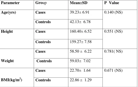
Mean age of the Hyperthyroids was 39.23 ± 6.91 and the Euthyroids was 42.13 ± 6.78 .There was no significant difference between ages with p value 0.140.
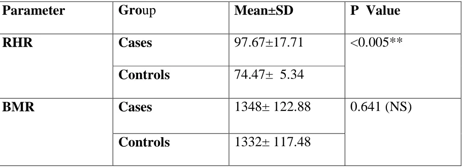
2. Resting Heart rate and BMR of the Hyperthyroids and normal
volunteers
Resting Heart rate (RHR per min)
Height of Hyperthyroids was 160.40 ± 6.52 and Euthyroids was 159.27 ± 7.58. There was no significant difference in weight between these groups with p value 0.551 .
Weight (kgs)
Weight of Hyperthyroids was 58.50±6.21 and Euthyroids was 59.03± 7.02. There was no significant difference in weight between these groups with p value 0.781.
BMI (kg/m2)
RHR of the hyperthyroids was 97.67 ±17.71 and euthyroids was
74.47 ±5.34 and the p value <0.001 ( extremely significant)
Basal Metabolic Rate ( BMR)
BMR of hyperthyroids was 1348± 122.88 and euthyroids was 1332 ± 117.48 and the p value 0.641.
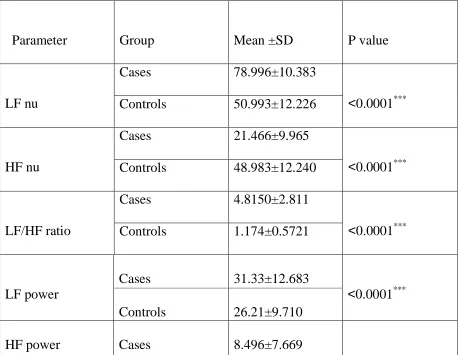
3. Comparison of frequency domain measures of the Hyperthyroids
and normal volunteers
Low power frequency in normalized units( LF n.u)
LF of the Hyperthyroids was 78.99. ± 10.38 and Euthyroids was 50.99± 12.23 and p value<0.0001 (extremely significant).
HF of the Hyperthyroids was 21.47±9.96 and Euthyroids was 48.98± 12.24 and p value<0.0001 (extremely significant)
LF/HF ratio
LF/HF of the Hyperthyroids was 4.81±2.81and Euthyroids was 1.17± 0.57 and p value<0.0001 (extremely significant).
Low power frequency ( LF ) power %
LF of the Hyperthyroids was 31.333 ± 12.68 and euthyroids was 26.213± 9.71 and p value of 0.070 (significant).
High power frequency (HF) power %
HF of the Hyperthyroids was 8.49±7.66 and euthyroids was 28.39± 13.99 and p value<0.0001 (extremely significant).
Very Low power frequency (LF) power %
4. Comparison of time domain measures of the of the Hyperthyroids
and normal volunteers
Mean RR(s)
Mean RR of the Hyperthyroids was 0.62±0.11 and the Euthyroids was 0.77±0.062. There was significant difference between the Mean RR of both the groups with p value<0.0001 (extremely significant).
RMSSD of the Hyperthyroids was 14.19± 7.69 and Euthyroids was 38.66 ± 7.66 and p value of <0.0001 (extremely significant).
NN 50(count)
NN 50 of the Hyperthyroids was 8.67± 8.07and Euthyroids was 79.67 ±17.34 and p value <0.0001 (extremely significant).
pNN50 (%)
pNN50 of the Hyperthyroids was 4.30± 4.02 and Euthyroids was 21.46 ±12.53 and p value < 0.0001(extremely significant)
SDNN( ms).
5.
Comparison of TFT (ELECSYS IMMUNOASSAY) of Hyperthyroid and
Euthyroid groups
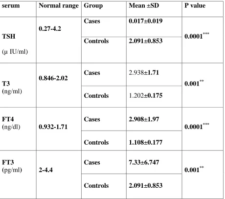
.SERUM TSH (in µ IU/ml)
Mean TSH of the Hyperthyroids was 0.017 ±0.019 and Euthyroids was2.0 9±0.062 with p value<0.0001 (extremely significant).
T3 (in ng/ml)
T 3 of the Hyperthyroids was 2.94± 1.71 and Euthyroids was 1.20± 0.175 and
p value <0. 001 (significant).
FT4 (in ng/ml)
FT4 of the Hyperthyroids was 2.91± 1.97 and Euthyroids was 1.11 ± 0.18 and
p value <0.0001 (extremely significant).
FT3 of the Hyperthyroids was 7.33± 6.75 and Euthyroids was 2.09±0.85 and p
1. Comparison of Baseline characteristics of Hyperthyroid and Euthyroid groups
Table-1
Parameter Group Mean±SD P Value
Age(yrs) Cases 39.23± 6.91 0.140 (NS)
Controls 42.13± 6.78
Height Cases 160.40± 6.52 0.551 (NS)
Controls 159.27± 7.58
Weight
Cases 58.50 ± 6.22 0.781( NS)
Controls 59.03± 7.02
BMI(kg/m2)
Cases 22.70± 1.64 0.671 (NS)
Controls 22.86 ± 1.29
NS- Not significant
*
**
Very significant
***
Extremely significant
2.
Comparison of Resting Heart rate and BMR of the Hyperthyroids
[image:58.595.68.524.428.593.2]and Euthyroid groups
Table-2
NS- Not significant * Significant ** Very significant
Parameter Group Mean±SD P Value
RHR Cases 97.67±17.71 <0.005**
Controls 74.47± 5.34
BMR Cases 1348± 122.88 0.641 (NS)
3.Comparison of Frequency domain measures of Hyperthyroids &Euthyroids
Table-3
Parameter Group Mean ±SD P value
LF nu
Cases 78.996±10.383
<0.0001*** Controls 50.993±12.226
HF nu
Cases 21.466±9.965
<0.0001*** Controls 48.983±12.240
LF/HF ratio
Cases 4.8150±2.811
<0.0001*** Controls 1.174±0.5721
LF power
Cases 31.33±12.683
<0.0001*** Controls 26.21±9.710
NS Not significant * Significant ** Very significant *** Extremely significant
4. Comparison of time domain measures of Hyperthyroids and
Euthyroids
Controls 28.396±13.985
<0.0001***
VLF power
Cases 59.114±21.098
<0.0001*** Controls 46.350±18.356
Table-4
NS Not significant * Significant ** Very significant *** Extremely significant
Mean RR(s)
Cases 0.621±0.106 <0.0001***
Controls 0.769±0.062
SDNN(ms)
Cases 24.297±12.36 <0.0001***
Controls 39.675±8.63
RMSSD
Cases 14.196±7.69 <0.0001***
Controls 38.656±7.66
NN50(count)
Cases 8.67±8.07 <0.0001***
Controls 79.67±17.35
pNN50(%)
Cases 4.30±4.0 <0.0001***
5.
Comparison of TFT (ELECSYS IMMUNOASSAY) of
[image:62.595.66.526.221.628.2]Hyperthyroid and Euthyroid groups
.Table-5.
NS - Not significant * Significant ** Very significant
*** Extremely significant
serum Normal range Group Mean ±SD P value
TSH
(µ IU/ml)
0.27-4.2
Cases 0.017±0.019
0.0001*** Controls 2.091±0.853
T3 (ng/ml)
0.846-2.02 Cases 2.938±1.71
0.001** Controls 1.202±0.175
FT4
(ng/dl) 0.932-1.71
Cases 2.908±1.97
0.0001*** Controls 1.108±0.177
FT3
(pg/ml) 2-4.4
Cases 7.33±6.747
VI. DISCUSSION
Thyroid hormones modulates the development, growth and metabolism
of each and every system in our body .Hyperthyroidism is due to hyperactive thyroid gland and increased production of thyroid hormones T3 , T4 and decreased serum TSH.
Hyperthyroidism frequently presents with tachycardia, palpitations and
widened pulse pressure. Cardiac contractility and cardiac output is immensely
elevated in hyperthyroids due to the additive effect of elevated ejection fraction,
resting heart rate, and blood volume with a decrease in diastolic function. Sympathetic nervous system activation and thyrotoxicosis manifestations are
mostly similar, especially in the ionotrophic effects
Recent studies in heart showed that thyroid hormones especially cause enhanced gene transcription of the calcium ATPase in sarcoplasmic reticulum and increase the pacemaker activity in cardiac cell
Thyroid hormone has its effect on duration of cardiac pacemaker
potential and repolarization currents. It alters both non genomic and genomic
actions and the net effect is to alter the heart function towards increased
contractility 76.
It is possible that there exists an interaction between the adrenergic hormones and the thyroid hormoneS, which may also contribute to the cardiac actions of thyroid hormone77.
A study done by Williams showed that thyroid hormones increase sensitivity to β-adrenergic agonists by increasing the β-adrenoceptor density and
Gs/Gi protein ratio with an excess activation of the adenylate cyclase 78
.
HRV is the good marker for identifying the cardiovascular risk and
severity in hyperthyroids. HRV denotes the individuals autonomic tone and
Increase in sympathetic tone in cardiac pacemakers induces tachycardia and reduce HRV, whereas increased parasympathetic activity causes bradycardia and increases beat to beat differences.
Higher HRV is always desirable and Lower Heart Rate variability is an established marker of cardiovascular deaths and complications . Abnormal variability also predicts the cardiovascular etiology for mortality, coronary atherosclerotic development and cardiac arrhythmias.
In this study population, cases showed decreased HRV parameters. Of the HRV parameters, frequency domain measures HF power, HF nu was less among patients than controls. This clearly depicts that parasympathetic activity is less among hyperthyroid patients.
Thyroxine hormone increase the body temperature and produce vasodilatation.This increase the cutaneous circulation. But decrease in the peripheral resistance with decrease in diastolic blood pressure.
LF nu, and LF power and LF/HF Ratio were high among hyperthyroids than controls. This shows that sympathetic activity is high in hyperthyroid patients and it is consistent with the study done by chiu HW and chen JL who emphasized that hyperthyroidism is characterized by a abnormal increase in sympathetic and decrease in vagal activity in heart.
So the sympathetic to vagal ratio may increase in Hyperthyroids. But few young Euthyroid patients (with normal TFT), have LF/HF ratio more than 1, showing little sympathetic dominance, which can be due to stress. However it is negligent when compared to hyperthyroid patients with enormous hike in LF/HF ratio.
In our study, basal metabolic rate is elevated in few hyperthyroid patients than the controls. By Calculating the BMR it is clear that BMR elevation is directly proportional to the increase in T3, T4 and LF. (Low frequency domain
measuring sympathetic activity) .This relationship is well brought out in our study than the previous studies. But there are few hyperthyroid patients, who had normal BMR. This depicts all hyperthyroid patients need not have elevated BMR.
Similarly SDNN and RMSSD were significantly lower in untreated
hyperthyroids than euthyroids. This shows that high frequency variations in heart
rate are less and vagal modulation of the autonomic nervous system is decreased. A certain number of cases showed NN50 and pNN50 equals 0. It
emphasizes that adjacent NN intervals differing by more than 50ms is zero .So the high frequency variations in heart rate in these patients were zero. Thus the vagal activity is least. These patients are high risk patients for cardiovascular
complication like atrial fibrillation and arrhythmias. Our study is of great relevance in this aspect.
Supraventricular arrhythmias , atrial fibrillation , cardiac failure are the known cardiovascular complications of thyrotoxicosis and the same was proved
to be the primary cause of death.
There is an association between thyroid gland function, heart muscle
mass, and ventricular hypertrophy. Hyperthyroidism is an independent risk factor for LVH.
In the geriatric age group, the TSH levels are low, and the
Left Ventricular Hypertrophy has emerged as a important marker of
progressing atheroscleroticprocesseswhich can be appreciated by ECG, ECHO or X-ray and Cardiovascular complications proportionately increase with increasing left ventricular muscle mass .
TSHvalues were significantly reduced in hyperthyroids. TSH has a linear relationship with parasympathetic activity.T3, T4 were immensely elevated, which is directly proportional with sympathetic activity and the degree of vagal
withdrawal. This relationship is well brought out in our study than any other previous studies.
Our study gives a solid and strong evidence of increased cardiac
autonomic activity –showcasing the sympathetic dominance as the culprit for all cardiac morbidity and mortality.
With the help of our HRV analysis in hyperthyroids, patients who are at risk for cardiac complications are found out using the time and frequency domains and early intervention can be done to prevent mortality rates.
obtained by radioactive iodine, antithyroid drugs and thyroidectomy. It all needs a brief period of time to bring it to normal cardiovascular status.
LIMITATIONS OF THE STUDY
Few limitations observed are,
1. Sample size of the study can be more.
2. It is difficult to get patients before starting anti thyroid treatment.
VII. Conclusion
To our knowledge , this is the first study to investigate the autonomic innervations of the heart in thyrotoxicosis subjects using HRV analysis and comparison of BMR between the case and control groups.
In the view of present study results, disturbances of sympathetic branch of Autonomic Nervous System can be observed in patients with thyrotoxicosis.
A predominance of sympathetic tone has cardio acceleratory effect and
reduced beat-to-beat variations and therefore causes positive ionotrophic effects. Reduced Heart Rate variability is most commonly linked with a risk of arrhythmic death and it is the independent predictor of cardiac mortality and morbidity, but recent data suggests that any abnormal variability also predicts circulatory dysfunction, progression of coronary atherosclerosis anddeath due to arrythmias.
The beta adrenergic blocking drugs reduce both cardiovascular and central
nervous symptoms. It helps to reduce the anxiety while preparing patients for
surgery.
Beta-adrenergic blockers slow the resting heart rate from 97 to 80 beats/min and bring the rate of diastolic deceleration to normal.
Beta blockers are usually prescribed to reduce the symptoms of
thyrotoxicosis such as tachycardia, palpitations, tremors, anxiety. Anti-thyroid treatment given to decrease the synthesis of thyroid hormones, particularly, in
We conclude that decreased vagal modulation on heart rate may occur in hyperthyroidism, which may be restored following adequate treatment of the disease by blocking beta receptors and there by inhibiting the adenylyl cyclase- cyclic AMP pathway.
This provides an attractive future option for management of arrhythmias and other cardiovascular complications due to thyrotoxicosis.
From our study it is obvious that cardiovascular risk in thyrotoxicosis patients can be evaluated by our HRV analysis before any appreciable change occurred in heart rate itself. Prevention is better than cure. So even before the appearance of cardiac complications, they can be assessed and halted.
Many randomised control trials can be done to bring out the unexplored effects of autonomic dysfunction on cardiovascular system. Thus cardiac
morbidity can be assessed and treated earlier and mortality can be prevented using
VIII. BIBLIOGRAPHY
1.Mohebati , Clinical Anatomy. Special Issue on Head and Neck . 2012 Jan;25(1) :19–31.
2. Cyril.keele, Samson Wrights, applied physiology. Thirteenth edition .vol 1,1982.537-538.
3. Kim E. Barrett,Susan M. Barman Ganong,S review of medical physiology. 24 th edition .341-342.
4. Turnbridge WM, Evered DC, Hall R. The spectrum of thyroid disease in a community: the Whickham survey. Clinical Endocrinology Oxford. 1977;7:481– 93.5 . Larry Jameson, Anthony P. Weetman .320 Disorders of the Thyroid Gland
march 2005;http:/www.anatomed.
6.Leclere J , Weryha S. Stress and autoimmune diseases. Horm Metab Res 1999;31:90-93.
7. Chan GW and Mandel SJ .Therapy Insight: management of Graves' disease during pregnancy. Nat Clin Pract Endocr Metab 2007 ;3: 470–478 .
8 .Livolsi VA. Surgical pathology of the thyroid . Philadelphia WB saunders, , 2nd edition, 2001.
10.P. Reed Larsen, Kronenberg. Williams Text Book Of Endocrinology.10th Edition :374-375.
11. John Michael Bernardi, Thyroid dermopathy. Journal of the American Academy of Dermatology.2011 june; 64( 6 ) 1219-1220.
12. Volpe R.The etiology of thyroid disorders.. Thyroid. 1994; 4: 373–377.
13. Mizokami T .Stress and thyroid autoimmunity. Thyroid .2004;1047–1055.
14. Nafisa KK, Goswami G. Thyroid thyrotoxic storm following Thyroidectomy. E Medicine Journal 2001; 2(7): Sec 1-9.
15. Toxic multinodular goiter; Plummer's disease A.D.A.M. Medical Encyclopedia. reviewed: 2012 June 4.
16.Weetman AP. The immunology of pregnancy. Thyroid 1999;9:643-646.
17.Volpé R. Endocrine practice office Journal of American college of
endocrinology.The pathogenesis of Graves' disease. 1995 Mar-Apr;1(2):103-15.
18. Stephanie L Lee. Subacute Thyroiditis. http:/www. Medscape . Oct 11, 2012. 19. Tommer Y Davies . Infection , Thyroid Disease and Autoimmunity .
20. P J Pringle, J S Barton . Thyrotrophin stimulating hormone (TSH) pulsatility in childhood: effects of a somatostatin analogue on pulsatility and thyroxine (t4) generation Pediatric Research 1993; 33: 26
21 .Arthur C. Guyton-guyton and hall textbook of medical physiology 12 th edition,914-915
22. Silvia Cantara Expression of the Ring Ligase PRAJA2 in Thyroid Cancer Journal of Clinical Endocrinology & Metabolism .2012september 4;2012-2360. 23 . Klein I, Levy GS. The cardiovascular system in thyrotoxicosis. The thyroid: a fundamental and clinical text.Philadelphia:lipincott Williams and wilkins, 8 th edition ,2000:596-604.
24. Kim E. Barrett,Susan M. Barman Ganong,S review of medical physiology 24 th edition, 348-349.
25. Arthur C. Guyton-guyton and hall textbook of medical physiology 12 th edition,912-913.
26. G.Kheder et al / Heart Rate Variability Analysis Using Threshold of Wavelet Package CoefficientsInternational Journal on Computer Science and Engineering 2009;1(3): 131-136.
27. Indukurana. Autonomic Nervous System .Text Book Of Physiology. 1st Edition.995-99
29. Indu khurana. Text book of medical physiology.1st edition.994-995. 30. Hypothalamus and autonomic nervous system http:/ O.U.C.H LINKS Washington School of Medicine; Neuroscience Tutorial, 1997
31. Larsen ,kronenberg .William textbook of endocrinology.10th edition. 104-105 32.A Rancibia S, tapia-Arancibia L .Direct evidence of short term cold-induced TRH release in the median eminence . Neuroendocrinology 1983;37:225-228. 33. Ahren B, Alumets J, Ericsson M. VIP occurs in intrathyroidal nerves and stimulates thyroid hormone secretion. Nature 287.1980;343-345.
34. Wolfgang H. Dillmann Cardiac function in thyroid disease: Clinical features and management considerations. Annual Thoraic Surg. 1993;56:S9-S15
35. Rebecca S. Bahn, M.D. Graves' Ophthalmopathy New England Journal of Medicine 2010 February 25; 362:726-738.
36. Daniel J. Toft and Susan Spinasanta Graves’ Disease Overview. What Is Graves’ Disease? Available from http:/www.endocrine web.com
37. G. Kheder, A. Kachouri, Heart Rate Variability Analysis Using Threshold of Wavelet Package Coefficients.computer science.cornell university. Submitted on 11 Dec 2009.
39. Nickel, P .Diagnosticity of Heart Rate Variability as an Indicator. Factors 45 (4): 575–590.
40. Eric Oetter .Heart Rate Variability Research Review Weeks Out
December 5, 2011.
41. Hon EH, Lee ST. Electronic evaluations of the fetal heart rate patterns preceding fetal death: further observations. American Journal of Obstet Gynecol. 1965;87:814-826.
42. Ewing DJ, Martin CN, Young RJ, Clarke BF. The value of cardiovascular autonomic function tests: 10 years’ experience in diabetes. Diabetes Care. 1985;8:491-492.
43. Wolf MM, Varigos GA, Hunt D, Sloman JG. Sinus arrhythmia in acute myocardial infarction. Med J Aust. 1978;2:52-53.
44. Akselrod S, Gordon D, Ubel FA, Shannon DC, Barger AC. Power spectrum analysis of heart rate fluctuation: a quantitative probe of beat to beat cardiovascular control. Science. 1981;213:220-222.
45. Md Rasel Kabir. Heart Rate Variability in Hyperthyroidism. Journal of
Bangladesh Society of Physiologist 2009 Dec;4(2): 51-57 .
47. Karthik S, Pal GK, Sympathovagal imbalance in thyroid dysfunctions in females: correlation with thyroid profile, heart rate and blood pressure. Indian Journal of Physiology Pharmacology. 2009 Jul-Sep;53(3):243-52.
48. Tolbaldini .Increased complexity of short-term heart rate variability in
hyperthyroid patients during orthostatic challenge . Department of Clinical Sciences, Internal Medicine . 2008;2008:1988-91.
49. Stuart Gordon And Roscoe R. Clinical Hyperthyroidism Associated with a normal Basal Metabolic Rate .Canadian Med Association Journal. 1985 February; 32(2): 162–165
50. Clute, H.M. Border line hyperthyroism . American journal of surgery 1929; 6:11
51.Plummer W A. Adenomatous goitre with hyperthyroidism accompanied by an unusually low metabolic rate. Proc. Staff meet. october 1938;13:641-646.
52. Chen JL, Chiu HWet al. Hyperthyroidism is characterized by both increased sympathetic and decreased vagal modulation of heart rate .Clinical Endocrinol (Oxf). 2006 Jun;64(6):611-612.
53. Burggraaf J, Tulen JH et al. Sympathovagal imbalance in hyperthyroidism. American Journal Physiology Endocrinology metabolism. 2001 Jul;281(1):190-195.
Jun;8(3):181-6.
55. Barczy M, Tabor S. Heart rate variability analysis in hyperthyroid patients and during euthyroidism after treatment . 1997;38(3-4):27-35.
56. Jin Long Chen .Nonlinear analysis of heart rate dynamics in hyperthyroidism
Physiological Measurement 2007; 28 (4 )333.
57. Faizel Osman .Heart rate variability and turbulenc