FINE NEEDLE ASPIRATION CYTOLOGY
TRUCUT BIOPSY AND
HISTOPATHOLOGICAL EXAMINATION IN
BREAST LUMPS
– A
Comparative Evaluation
Dissertation Submitted
for the Degree of
MASTER OF SURGERY
Branch I
(GENERAL SURGERY)
THE TAMIL NADU
Dr.M.G.R. Medical University
CHENNAI
2
COIMBATORE MEDICAL COLLEGE
COIMBATORE.
CERTIFICATE
Certified that this is the bonafide dissertation done by
Dr.S.SUJITH KUMAR
and submitted in partial fulfillment of the requirement for the
Degree of MASTER OF SURGERY
Branch I (GENERAL SURGERY)
of The Tamil Nadu Dr.M.G.R. Medical University, Chennai.
DATE : UNIT CHIEF
DATE : HEAD OF THE DEPARTMENT
DEPARTMENT OF SURGERY COIMBATORE MEDICAL COLLEGE
DATE : DEAN
DECLARATION
I solemnly declare that this Dissertation on “FINE NEEDLE
ASPIRATION CYTOLOGY, TRUCUT BIOPSY AND
HISTOPATHOLOGICAL EXAMINATION IN BREAST
LUMPS – A COMPARATIVE EVALUATION” was done by me
at Coimbatore Medical College Hospital, Coimbatore under the
guidance and supervision of Dr.B.Easwaran, M.S.
Place:
4
ACKNOWLEDGEMENT
I owe my great debt of gratitude to DR.P.ARUN KUMAR,
M.S., Professor and Head of the Department of Surgery, Coimbatore
Medical College and Hospital, Coimbatore for his excellent expert
advice and help in preparing this dissertation.
It is my proud privilege to express thanks and gratitude to my
Unit Chief DR.EASWARAN, M.S., for his help and guidance in the
course of study and preparation of this dissertation. Without his
guidance and encouragement this work wound not be fruitful and
complete.
I thank all the surgical unit chiefs DR.PERUMAL RAJAN,
M.S., DR.PREM THAMARAI SELVI, M.S., DR.RAMA
MOORTHY, M.S., DR.G.S.RAMACHANDRAN, M.S., for
permitting me to carryout the study in their unit.
I wound also like to extend my sincere thanks to DR.INDIRA
PRANESH, M.D., Path. DR.MOORTHY, M.D., Path and
DR.R.VIMALA, M.D., Path, Department of Pathology for their
guidance and assistance in pathological aspects.
My thanks to DR.KALANITHI, M.D., Dean, Coimbatore
Medical College and Hospital for permitting me to carry out the study
and utilizing the hospital facilities.
I would like to thank Mr.JOSHUA ALLAN SHEPHERD,
M.Sc., Statistical Analyst, for his participation in my study.
Lastly, but not the least, I express my gratitude to all those
CONTENTS
S.No TITLE PAGE NO
1 2 3 4 5 6 7 8 9 10 Abbreviations Introduction
Review of Literature
Aims and Objectives
Pathology of Breast
Cytologic appearances in mammary disease
Materials and Methods
A. Techniques of FNAC
B. Techniques of TCNB
Observation and Analysis
6
ABBREVIATIONS
ABC - Aspiration Biopsy Cytology
Acc - Accuracy
Ca - Carcinoma
CT - Computed Tomography
DNA - Deoxyribonucleic acid
EM - Electron Microscopy
FNAC - Fine Needle Aspiration Cytology
HPE - Histo Pathological Examination
MHZ - Mega Hertz
MRM - Modified Radical Mastectomy
N/C - Nucleo Cutoplasmic ratio
® - Right
(L) - Left
Sen - Sensitivity
Spe - Specificity
TCNB - Trucut Needle Biopsy
INTRODUCTION
A lump in the breast whether benign or malignant results in
anxiety for the patient and her family and the surgeon. Histological
tissue diagnosis is a universly accepted means of definitive diagnosis.
Fine Needle Aspiration Cytology (FNAC) is gaining a wide
acceptance as it gives a rapid diagnosis and can be carried out in out
patient services.
The trucut needle is a very handy instrument and it is almost
replacing the incision or excision biopsy in the breast lump, as it can
be carried out in the out patient services with minimal trauma.
In this study 65 patients having breast lumps, were subjected
to FNAC and Trucut Needle Biopsy as out patients and followed by
operative treatment with a histological diagnosis, which were
8
REVIEW OF LITERATURE
The clinical tissue cytology or non exfoliative cytology defined
by Banforth in 1966 as follows. “The examination of cells obtained
by needle or drill biopsy in solid organs or tissue masses or from cut
surface of such material freshly removed by surgical biopsy”.
The present day definition as given by S.Kline is that
“Fine needle aspiration cytology is the study of cells obtained
by small gauge Needle generally with vacuum system provided by an
air tight syringe”.
Initially clinical evidence was preferred to biopsy. If a lump
ulcerated, it was cancerous. In 1801, Adams2 observation on the used
excision biopsy and macroscopy. Both Paget31 and Erichsen13 were
pioneer in tumour microscopy and published cytological illustration.
The need for biopsy was recognized and emphasized by Laurence26 in
1855 stating the instance where a breast was amputated for a supposed
tumour which turned out after the operation to be only a chronic
abscess.
The necessity for the histological confirmation of the diagnosis
before contemplating complex and often distinguishing or mutilating
be over emphasized. Thus developed the preoperative diagnostic
procedures like frozen section and imprint smear cytology, or
preoperative biopsy which should be simple, not distinguishing and
carried out in the out patient department. The technique of
“TRUCUT” Needle biopsy and fine needle aspiration cytology
followed.
Investigation of a tumour by means of a needle was carried out
in St.Bartholomew’s Hospital in 1833 for a case of abscess of the liver
with Hydatid cyst. The patient improved after this. Needle biopsy
became established method of diagnosing a collection of pus.
Needle biopsy was first recorded by KUN25 in 1841 and was
adopted by others.
Erichsen in 1853 described as exploring needle to withdraw
cells from a tumour for microscopy. It is uncertain whether a syringe
was added for suction, but substantiated Needle aspiration biopsy was
introduced for the parasitological study of lymph nodes early this
century.
Menetrice in 1886 first used an aspiration needle to obtain
tissue from a carcinoma of the lung and described the microscopic
10
Mard in 1912 and Guthrie in 1921 used fine needle aspiration
cytology to examine enlarged lymph nodes in cases of reticulosis.
Subsequent exponents of this technique were martin and Ellis at
Memorial Hospital in New York, who in 1926 began examination of a
series of palpable malignant lesions, published the first series
involving aspiration of a wide variety of Neoplasms consisting of 65
malignancies including 6 breast cancers. Three years later in 1933,
Stewart reported the expanded experience in the same institution,
which then included 2500 malignancies, with 500 breast cancers.
Steward described in detail the pathological interpretation of the
material obtained by aspiration. Material was placed on a slide and
smeared out and he suggested that the pathologist might obtain
experience by smearing fragments from tumours obtained at operation
or autopsy.
Ferguson (1930) described the technique in prostate tumour ;
Coley, sharp and Ellis (1931) in bone tumours, Forster (1931) in CNS
tumours ; sharp in primary carcinoma of lung and Klinger and Burch
(1932) as aspiration technique for endometrial tumours. Graver and
Binkley (1939) revived the literature in aspiration biopsy and gave the
Despite the diagnostic success in these large series, there appear
to be very little interest in these procedure during the ensuring 25
years. Attention was paid on trucut and drill biopsies. In 1938,
Silverman described a device to trap, within the needle, a core which
was suitable for histological section. This was variously modified and
other devices developed for cutting off the end of the core retaining it.
Attempts were then made to improve the cutting edge and teeth and
bevels various angles were added to the needle. Thus there developed
method of drilling aimed at boring out a core of tissue rather than
aspirating a few loose cells. Initially these needles were rotated by
hand. But in 1934 and again in 1935 Kirschner described a hollow
drill which was rotated by an electric motor through a flexible device.
The technique of FNAC was revived in 1950’s by
Scandinavians and its application in the diagnosis of palpable breast
masses has become increasingly popular in recent years. The original
authors used an 18 gauge needle with air dried smear, and required
local anesthesia. The Scandinavians introduced the concept of fine
needle aspiration with a 23 gauge needle improved cytology fixation
and staining technique, and no need for local anesthesia. This
procedure was popularized in Scandinavia in 1968 for the diagnosis of
12
acceptance in the United States. These has resulted from a
combination of factors including the ease, rapidity, accuracy and lack
of morbidity of the technique, the increase in desire of female patient
to have the opportunity to adjust to a define malignant diagnosis
before consenting to surgery and relatively low cost of the procedure
compared to the open biopsy.
The fine needle biopsy was defined by Godwin17. Annals of
Newyork academy of Sciences, 63, 1348) as the withdrawal of cells or
small bits of tissue through a needle by means of a negative pressure.
Exfoliative cytology defers in aim from aspiration cytology in
that the former used primarily to detect a cancer clinically not yet
apparent, while the latter is used to determine the microscopic nature
of a clinically detectable tumour. In this respect aspiration cytology is
similar to histological examination of a surgical biopsy. The term
ABC – Aspiration Biopsy Cytology was used by Zajicek and
Lawhagel as a synonym of FNAB or FNAC. It (ABC) was chosen to
clearly distinguish aspiration from exfoliative cytology and to
emphasize its simplicity24.
Now at the Radium Hemet in Stockholm, about 12,000
aspiration are performed every year. Other centers using these
Moscow, the curie foundation in Paris and the Memorial Hospital in
Newyork.
In India10 these useful cost effective simple investigation has yet
to gain popularity though during the last 25 years or so reports have
tricked in on these subject, claiming the cells equivalent to those of
western counterpart.
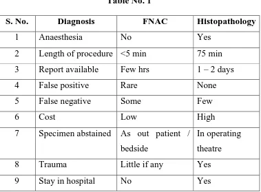
Trott and Raadal in 1979 summarized the relative merits of
FNAC compared to excision biopsy histopathology as given in the
[image:13.612.127.506.365.644.2]table.
Table No. 1
S. No. Diagnosis FNAC Histopathology
1 Anaesthesia No Yes
2 Length of procedure <5 min 75 min
3 Report available Few hrs 1 – 2 days
4 False positive Rare None
5 False negative Some Few
6 Cost Low High
7 Specimen abstained As out patient /
bedside
In operating
theatre
8 Trauma Little if any Yes
14
RECENT ADVANCES28
With advent of imaging with radiography, ultrasonography and
CT scanning, its usage has become still more wider in clinical
practice.
GUIDED FNAC
USG GUIDED
Aids in accurate localization of breast lumps. They also to
differentiate between solids and cyst lumps Lumps upto 2 mm can be
biopsied with the help of USG. 7 MHZ probe is used for better
discrimination. Cyst can be aspirated fully and followed up to direct
any recurrence.
MAMMOGRAM GUIDED
Mammography taken in two direction can be used to localize
non-palpable breast lumps and subject them to FNAC.
STERIOTACTIC GUIDED
This is mainly useful in core needle biopsies and erosion
AIMS AND OBJECTIVES
Out patient assessment of breast lumps
To compare the results of FNAC and trucut needle biopsy in
breast lumps with histopathological examination.
To assess the incidence of average age.
To compare the incidence – married Vs unmarried.
To know the average size of breast lump.
To evaluate the peak incidence of site of breast lump.
To compare the incidence – premenopausal Vs postmenopausal.
16
PATHOLOGY OF BREAST
BENIGN BREAST LUMPS35
1. Fibroadenosis (Fibrocystic Disease)
It occurs from adolescence through senescence, but particularly
during menarche. It generally produces a lumpy feeling rather than a
mass per se. It is due to an Aberrations of Normal Development and
Involution (ANDI). Areas sectioned with a knife may be white or
yellow but never present the grey tones of carcinoma.
MICROSCOPICAL FEATURES
a. Microcyst Formation – are long standing and vary much in
size. They contain dark – mucoid material.
b. Adenosis – an overall increase in Acinal material
c. Fibrosis – swelling of interstitial tissues and round cell
infiltration
d. Epitheliosis – hyperplasia of epithetium in the lining of acini
e. Papillomatosis – small branching papillomas inside the cysts
or small ducts.
f. Calcification – coarse irregular pattern chemically composed
18
Fibroadenosis with Epithelial hyperplasia may lead to a malignant
lesion. In a large breast the differential diagnosis between
fibroadenosis and carcinoma in the very early sage is difficult, when
an ill-defined lump is deeply situated.
2. MASTITIS3
Acute mastitis due to bacterial infection most commonly occurs
within the first few weeks of lactation. Infection usually results from
staphylococci or streptococci entering the breast through abraded or
lacerated nipple surfaces or by way of lactiferous ducts as they enter
the nipple. Lymphatic involvement results in either cellulitis or frank
abscess formation.
Streptococcal infection tend to produce a diffuse cellulitis often
with systemic toxic manifestations. Abscess formation is more
common in staphylococcal aureus infections. Usual cause of chronic
mastitis with abscess formation is tuberculosis, which is secondary to
pulmonary or chest wall diseases. Chronic mastitis when associated
with a thick fibrous wall cannot be differentiated clinically from a
carcinoma.
3. FAT NECROSIS
It occurs following trauma either direct or indirect (e.g.)
suggests the cause. The disease can situated carcinoma because of
skin retraction. On cut sections, a chalky white area of necrotic fat is
found resembling necrosis seen in subsiding acute pancreatitis.
4. DUCT PAPILLOMA
The papilloma projects in to a dilated duct, usually in the
vicinity of the nipple. Initially resembling a small raspberry but later
gets a smoother outline. Finally, distending the ducts it becomes a
solid, compact mass. It is the cause of a bright red, a dark blood
stained or rarely a serosanguinous discharge per nipple. A cystic mass
may be palpable behind the nipple.
5. FIBROADENOMA14
It is a slow growing benign Neoplasm with a predilection for
the young adult, majority of cases before the age of 63 years. It may
be caused by Hormonal Imbalance, when the concentration of
oestrogen is high they tend to grow faster (i.e.) during Adolescence
and pregnancy. Palpation reveals a dominant, discrete, mobile
rubbery mass, usually no greater than 3 cm in diameter. The cut
surface is solid, grayish white, and belonging, with a whorl like
pattern and slit like spaces. Necrosis, it may be hyalinised or calcified
20
Although mixed forms occurs, the encapsulated, predominantly
stromal tumour is of two varieties on histologic section. The
intracanalicular, fibroadenoma with broad, polypoid, loose branches of
connective tissue lined by cuboidal ductal cells, emerges from and
obliterates the duct. The pericanalicular, fibroadenoma encircles the
ducts with dense, concentric mesenchyma.
6. CYSTOSARCOMA PHYLLOIDES
This is a rare variant of fibroadenoma often referred to as a
giant fibroadenoma. When it was first described by Muller (1838), it
received its name because the tumour contained large cysts and was
fleshy, a connotation for the term “Sarcoma” at that time. Large
surface clefts were thought to resemble leaves in a book, this accounts
for the choice of the term “PHYLOON” most of these tumours are
benign (Leaf). But a few develop true sarcomatous potential.
7. GYNAECOMASTIA
It is the enlargement, probably endocrine related, of the male
breast and it most common in adolescent and elderly persons.
Although there is often generalized hypertrophy, there may be a
discrete tumour adjacent to the nipple. Microscopic examination
reveals ductal hyperplasia and dilatation, loose stromal proliferation
8. GRANULAR CELL MYOBLASTOMA
The origin of this tumour is obscure, perhaps in smooth muscle
or histocytic or neurogenic tissue. Clinically, both macroscopically
and microscopically, the tumour may appear poorly demarcated and
the overlying skin may show atrophy and retraction. The
non-capsulated tumour nests may be diffused dispersed in sheets or small
groups. However, the cells, with fine acidophilic granule are
morphologically benign.
9. MESENCHYMAL BENIGN NEOPLASMS
Rarely lipoma, Epidermal inclusion and Sebaceous cyst,
Fibroma, Keloid etc are like the same as in any other parts of body.
Their general pathological account are beyond the scope of this study.
MALIGNANT NEOPLASMS35
CLASSIFICATION
Foot and Stewart have stressed the fact that cancer of the breast
can arise from either, the lobules, the ducts or the nipple, with the
tumour arising from ductal epithelium in the majority of cases.
I. Carcinoma of nipple – Paget’s disease
22
A. Non infiltrating
1. Papillary
2. Comedo
B. Infiltrating
1. Papillary
2. Comedo
3. Adeno carcinoma with fibrosis (scirrhous carcinoma)
4. Medullary carcinoma with lymphoid infiltration
III. carcinoma of lobules
A. Non-infiltrating
B. Infiltrating
IV. Others
1. Mucinous carcinoma
2. Sweet gland carcinoma
3. Inflammatory carcinoma
NON INFILTRATING PAPILLARY CARCINOMA
It is a type of ductal carcinoma insitu and it is a rare variety,
arise from large, medium or small ducts. The papillary formation is
evident but a typical and distribution from benign – papillomatosis of
cystic hyperplastia may be difficult. The most valuable feature is loss
NON INFILTRATING COMEDO CARCINOMA
It arises in the smaller or intermediate sized ducts. The worm
like casts of comedo can be expressed from cut surface. The cells are
more anaplastic than papillary carcinoma. They completely occupy
and distend the ducts.
INFILTRATING ADENO CARCINOMA WITH FIBROSIS
This is the most common type of breast carcinoma accounting
for about 70-75% of the total cases, commonly known as scirrhous
adenocarcinoma. The tumour cells possess infiltrative properties and
have the ability to Evoke fibrous tissue profileration. Hence the
tumour becomes adherent to the surrounding tissues and the skin.
It presents an unyielding induration to the knife and gives a
sensation of cutting through an unripe pear. The cut surface is
depressed owing to the pull of fibrous tissue.
There is a great variation microscopically. Most frequently the
cells are spheroidal and arranged in small clumps and columns or in a
single file. In other cases the picture is more cellular with cells being
hyperchromatic and pleomorphic. Sometimes an almost acellular field
with the few cells compressed and narrowed by dense collagenous
MEDULLARY CARCINOMAS WITH LYMPHOID
INFILTRATION
It is bulky, soft and rounded. Haemorrhage and cyst formation
are common. The cells lack the invasive biological activity of the
scirrhous type, so the tumour is not adherent to the skin.
Microscopically the cells which are arranged in large masses
have abundant cytoplasm with large vesicular nuclei and many
mitoses. Infiltration with lymphocytes is a highly characteristic
feature.
LOBULAR CARCINOMA36
In this type the cells appear to arise with in the lobule itself. It
may be non infiltrating or infiltrating.
The non infiltrating type is carcinoma insitu. It cannot be
recognized in the gross and is an incidental finding. Microscopically,
it consists of large lobules confined to a limited area. The acinar cells
are piled up and are arranged in an irregular fashion. The appearance
of solid masses in enlarged lobules is characteristic. The infiltrating
type is indistinguishable in the gross firm scirrhous carcinoma.
Microscopically, it can be distinguished from that variety only by
finding examples of pre-invasive pattern in the neighbourhood. Both
PAGET’S DISEASE
It is found in women 40-60 years of age and commences as an
eruption of the nipple and areola. Following a variable period of 2-10
years a mass becomes palpable beneath the areola. The eczematous
area is bright red and inflamed with a moist and weeping or a dry,
scaly surface.
The microscopic picture presents, three features :
1. Epidermal hypertrophy
2. Paget’s cells
3. Sub-epidermal round cell
Epidermal hypertrophy is a constant feature before ulceration
occurs, papillae being increased in depth and width.
Pagets cells are large clear vacuolated cells with small pyknotic
nuclei. They look like clear spaces punched out of the epidermis. The
superficial part of the dermis shows round cell and plasma cell
infiltration. Proliferation changes in the epithelium of the breast are
also seen.
MUCINOUS CARCINOMA
This tumour has gelatinous materials within it and has sharply
delineated margins. Mucinous carcinomas are usually large, bulky
28
present on the cut surface. The cells have acquired the ability to form
mucin. Microscopically mucin filled cells surrounded cyst like
spaces. Some of the tumours show clumps of tumour cells in a sea of
mucoid material. The cells are often well differentiated and may even
have a signet ring appearance.
SWEAT GLAND CARCINOMA
The mammary and sweat glands have a common origin.
Structures which are apparently sweat gland tubules occur in the
normal breast and anastomose with the lacteal ducts. They are
distinguished by eosinophilia of the cytoplasm and an inner layer of
high columnar cells. Certain carcinoma of the breast especially
situated at the periphery may show these characteristics and hare
called sweat gland carcinoma. Their behaviour is the same as that of
ordinary breast carcinoma.
ACUTE INFLAMMATORY CARCINOMA
The term inflammatory carcinoma reflects the appearance of the
breast : hyperaemia, tenderness, skin retraction and oedema producing
the characteristic peau d’ orange. Although usually diffuse, the
malignancy may resemble a localised abcess. Histologic section
with nests of tumour in the dermal and epidermal lymphatics.
Blockage of vessels may cause the cardinal signs of inflammation.
MIXED DUCTAL AND LOBULAR CARCINOMA
This is a very rare carcinoma, it composed in part of a
component with definite features of invasive ductal carcinoma and in
part of a component with definite features of invasive lobular
carcinoma do occur.
This tumour has to be distinguished from tubular carcinoma and
from the cases in which two separate neoplasms of different
microscopic appearances are present in the same breast.
METAPLASTIC CARCINOMA
Metaplastic carcinomas in a genetic term for breast carcinoma
of ductal type in which the predominant component of the neoplasm
has an appearance other than epithelial and glandular and more in
keeping with another cell type.
1. SARCOMATOID CARCINOMA
(Carcinoma with sarcoma like stroma)
Grossly, it is well circumscribed. Microscopically, the
sarcomas like component may resemble malignant fibrous
histiocytoma, chondrosarcoma, osteosarcoma, rhabdomycosarcoma,
30 2. SPINDLE CELL CARCINOMA
The overt carcinomatous component of these tumours, entirely
squamous. The spindle cell component, which may be deceptively
bland, forms abundant fibrocollagenous stroma with featured, myxoid,
angioid and storiform patterns. The appearance may closely simulate
that of a fibro sarcoma or even fibromatosis.
3. Carcinoma with osteoclast like giant cells
4. Squamous cell Carcinoma
CYTOLOGICAL APPEARANCES IN
MAMMARY DISEASE
I. MALIGNANT NEOPLASMS35
a) CARCINOMA
Criteria for diagnosis
1. The most important criterion is the cellularity. Aspiration from
carcinomas are usually very cellular.
2. Due to loss of cohesiveness, cancers cells are frequently present
in small groups and also as single cells.
3. Overlapping of cells and crowding in seen
4. Nuclei are pleomorphic. Enlarged in size and of irregular
shape. Nucleoli are present.
5. High nucleocytoplasmic (N/C) ratio
6. Intranuclear variants are seen in some benign and malignant
breast tumours, hence not of much use in diagnosis.
7. Mitoses may or may not be seen and so it not a useful criterion
Cytological characteristics of benign and malignant aspirates are
compared
[image:33.612.124.512.199.636.2]FNAC OF BREAST – SMEAR CHARACTERISTICS
Table No. 2
Cytological
Findings
Benign Malignant
Pattern Epithelial cells in sheet and
clusters, monolayered,
regularly arranged, few
single cells
Epithelial cells in
clusters of varying
size, multi layered,
irregularly arranged.
Many single cells
Single Cells Monomorphic oval, naked Polymorphous round
with cytoplasm
Nuclei Small, uniform Enlarged,
pleomorphic
Chromatin Evenly distributed, light Coarse, heavy
Nucleoli Usually absent Present in moderate
34
Two most important criteria are cellularity and nuclear atypia. To
avoid over diagnosis which can lead to a mutilating surgery,
conservative approach while giving positive report of carcinoma in
essential. No diagnosis of malignancy should be given if only one of
the above mentioned criteria in identified.
It is also important to remember that the impression of
carcinoma should be recognized in several fields of the smears. When
the smears are extremely cellular, but lack sufficient atypicality for a
firm diagnosis of carcinoma. It is advisable to ask for a biopsy. In
most of these cases, the lesion will be a well differentiated duct
carcinoma. Repeat aspiration will be of no further help in such a
situation.
TYPING OF BREAST CARCINOMA
Typing of the carcinoma may help the clinician in
prognosticating the diseases and deciding the line of treatment. Ortel
and Galbum have come out with some criteria helpful in classifying
different types of breast carcinoma.
i) Medullary Carcinoma
It is soft of an aspiration. The smears are cellular with bubbly
back ground due to proteinaceous material and show varying
36
sizes and shapes with prominent nucleoli are seen. These may be
naked or with scanty cytoplasms. Few bizzare forms may also be
seen. Mitoses are often present.
ii) Mucinous Carcinoma
Like medullary carcinoma, this type of carcinoma in also soft to
aspirate. Aspirated sample consists of abundant bluish pink mucoid
material. Smears are thick and show tight groups of cells. Often
nuclei are regular. Metachromasia may be seen in Giemsa sainted
smears. A striking feature observed is the presence of many branching
blood vessels running through the mucus pools.
iii) Tubular Carcinoma
Cellular aspirates reveal groups of ductal cells with blunt
branching and tubular lamina. On low power these groups mimic
fibroadenoma, but naked nuclei are not present. Groups of epithelial
cells with branching are not as complex as seen in fibroadenoma.
iv) Adenoid cystic carcinoma
Large number of cells are seen in abundant pale pink mucoid
background in which bright pink dense globules are seen. Most of
these globules are surrounded by cells with small, round regular
v) Papillary carcinoma
Aspirate is usually haemorrhagic and thick. The smears are
cellular fragments of tissue with fibrovascular core and finger like
projections are present. Nuclei are usually enlarged but regular.
vi) Lobular carcinoma
This type of carcinoma, like infiltrating duct types, is fibrous
and gritty on aspiration. Aspirates are not usually cellular. Smears
reveal small cells in a small groups in short chains (Indian files) or as
scattered single cells. Not much variation in nuclear characters and
size is seen.
vii) Malignant cystosarcoma phylloides
smears are similar to those from fibroadenoma. Stromal
fragments show cellularity and nuclear atypia. Pleomorphism,
hyperchromasia and frequent mitoses suggest a malignant neoplasm.
II. BENIGN LESIONS
a. Fibrocystic diseases
Aspirates are not cellular. The material consists of metaplastic
apocrine cells, benign ductal epithelial cells in fluid back ground and
fragments of fibroadipose tissue. In addition, few foamy macrophages
often unsatisfactory. Presence of apocrine cells are necessary for the
diagnosis of fibrocystic disease.
b. Fibroadenoma
Fibroadenoma can be readily diagnosed cytologically as usually
these yield cellular aspirates. Smears are rich in large tight sheets of
benign ductal epithelial cells admixed with naked nuclei within the
clumps and also scattered single epithelial clusters reveal blunt
branching. Stroma can be myxoid.
c. Abscess
Aspirated material consists of thick yellowish pus. Smears are
thick and show numerous polymorphs, fibrin strands, foamy
macrophages, cellular debris and occassional groups ductal epithelial
cells. Inflammatory atypia when present create diagnostic problems.
However, carcinoma in usually not associated with such marked acute
inflammatory component.
d. Fat necrosis
Numerous lipid laden macrophages and epithelial cells are seen
in the back ground of acute and chronic inflammatory cells. Fatty
40
MATERIALS AND METHODS
Sixty five patients presenting to surgical out patient department
of Coimbatore Medical College during 2005 - 2006 period, were
subjected to Fine Needle Aspiration Cytology and Trucut Needle
Biopsy. All the patients underwent surgery depending upon the report
of the two methods and finally all the reports of the techniques were
42
A. TECHNIQUES OF FNAC
It need not be emphasized that the proper clinical examination
of the patient is to be carried out in detail and the disease process must
be localized and clearly defined. Simpler investigation should be done
routinely in every patient.
The case should be discussed with the pathologist before the
FNAC23 is being one regarding the feasibility and the likely
informative value of FNAC in the particular case concerned.
PREPARATION FOR FNAC
EQUIPMENTS REQUIRED
1. 23 gauge 0.6 – 1 mm, external diameter disposable needles 2.5
and 5 cm long.
2. 10 – 20 mm disposable syringe with leur lock tip
3. “CAMECO” syringe pistol
4. Microscopic glass slide with frosted ends
5. Fixative
6. Alcohol sponges
7. Sterile gauze pads
8. Sterile containers
44 NEEDLES
Standard disposable 25-22 gauge 25-50 mm long needles are
suitable for most superficial, palpable lesions.
Finest needles (25 gauge) are recommended for children and for
sensitive areas like orbit and eyelids.
Thicker needles offer no advantages instead cause more
bleeding and can be blocked by plug of tissue and carry the risk of
tumour implantation in the needle track.
The 22 gauge, 90 mm disposable lumbar puncture needles with
trocar are convenient for most deep lesions.
If still longer needles are required, then a 22 gauge 150 – 200
mm chible needle can be used. Franzen instrumentation provides
special long needles for biopsy of prostate and pelvic organs.
Rotex II screw needle (0.8 mm, 145-205 mm size) is used for
deep biopsy of lung, liver, kidney, lymphnodes etc. This is
particularly useful in fibrous lesions, soft tissue tumours and in richly
vascular lesions.
However the standard needles are less expensive, easier to use
and give a satisfactory yield in majority of cases if the technique is
46
Standard disposable plastic syringes of 10-20 ml are used. It should
be of good quality for strong rigid material and produce a good
negative pressure.
SYRINGE HOLDERS
The use of syringe holder is strongly recommended. Leaving
one hand free to immobilize and to feel the target lesion allows better
precision in placing the needle. Regularly used syringe holder is
cameco syringe pistol (Cameco AB, Taby, Sweden) made to fit either
10 mm or 20 ml syringe.
SLIDES
They should be dry and free of grease, those with frosted ends
are convenient for immediate labeling. A 0.1 mm haemocytometer
cover slip gives better control over pressure used in smearing. Air
dried slides are best transported in stainless steel carrier to avoid
contamination and scratching.
FIXATIVES
For wet fixation, 70-90% ethanol preferably in koplin jars (for
spongy fixatives) is used. Canroy’s fixative has the advantage of
lysing RBCS. Glutaraldehyde with 10% buffered formalin is used if
48 STERILE CONTAINERS
Those filled with physiological saline or Hank’s balanced salt
solution is used to rinse the needles and syringes to obtain material for
culture.
OTHERS
Skin disinfectants, sterile dressing, local anaesthetic, watch
glass. Thrombin powder, pencil, tongue depressor and sterile blades.
PREPARATION OF PATIENT
Clear explanation of the procedure to the patient will ensure the
patients consent and better cooperation.
Informed consent should be obtained.
Selective positioning of the patient is must for particular
anatomical areas.
Preparation of local area with sterile swabs are done
preliminarily. Local anesthetics are applied only when required. It is
not always indicated but if given if facilitates multiple passes more
acceptable by the patient.
PROCEDURE
INSERTION OF NEEDLE
Better control over needle is achieved by supporting the barrel
50
and allows better appreciation of depth. If needed, imaging techniques
may be used to localize the lesion for favouring correct insertion of
needle.
ASPIRATION1
This mechanism has been explained well by THOMBSON.
The function of the negative pressure used is not to tear cells from the
tissue but to merely fix the tissue against the sharp clothing edge of
the needle. The softer tissues protruding over the edge are cut off and
accumulate in the lumen of the needle as it advances through the
tissue (eg) tumour cells, glandular and epithelial elements. But the
stroma is poorly represented in the aspirate.
For greater yield, the needle should be moved back to forth
especially in fibrous lesion. This is termed as Jackhammer method.
Sometimes, multiple passes may be needed for obtaining satisfactory
number of cells. But in case of vascular tissues, it produces more
blood in the sample. Blood in the syringe means an unsatisfactory
aspirate.
The ideal aspirate has high cell content in a small amount of
It is necessary to release the negative pressure before the needle
is withdrawn. This prevents the aspirate to get into the syringe or
being contaminated with contents aspirated during withdrawal.
NEEDLING WITHOUT ASPIRATION27
It was introduced by Zajdela in the principle that the capillary
pressure of the fine needle is itself sufficient to keep the cells within
the lumen. Here the negative pressure and aspiration are not used.
Simple insertion and back to forth movement is applied while
simultaneously feeling the consistency of tissue concerned thereby
improving precision, lesser admixture of blood cells, it cell yield is
some what less but not significantly so.
A 25 gauge needle is preferably used this technique. It can be
used in all superficial lesions (except cystic and fibrotic ones) and
deep lesions (when more blood aspirated by regular technique).
After the procedure is over, application of gentle pressure over
the biopsy site is important to minimize bruising and to decrease the
chance of haematoma formation in case of highly vascular lesion.
CAUSES FOR UNSATISFACTORY YIELD32
1. Needle has missed the lesion tangentially.
2. Central cystic, necrotic or haemorrhagic area devoid of
52
3. Small malignant lesion adjacent to dominant benign mass
4. Fibrosclerotic target tissue poor in cells
5. Mislabeling or interchanging of specimen either during
collection or in laboratory
6. Deterioration of specimen because of delayed processing or
poor fixation
7. Imperfect staining
8. Contamination
9. Lack of an adequate history
PREPARATION OF ASPIRATE
DIRECT SMERING
An aspirate is said to be “Dry” if it consists of numerous cells
suspended in a small amount of tissue fluid and has a creamy
consistency and this is perfect one. In contrast, a ‘wet’ aspirate is a
one which consists of small number of cells suspended in fluid or
blood. A dry aspirate is best smeared with the flat of 0.4 mm.
Coverslip exerting a light pressure to achieve a thin even spread, the
firm pressure causes crush artifacts. So it should not be too thin or too
thick.
A wet aspirate should be smeared in a “two step” method. The
specimen slide, holding it at an obtuse angle which leave the fluid and
makes the cells follow the smearing slide like buffy coat. The second
step is same as that described under dry aspirate smearing.
The correct technique should be followed especially in air dried
smear good fixation depends on rapid drying.
If large amount of blood is aspirated it is expressed onto and
spread over a watch glass before clothing and minimal particles are
picked up for histological processing.
If the sample is large enough, several slides can be prepared
both air dried and wet fixed so that special staining can be carried out
if required.
FIXATION AND STAINING
We are using isopropyl alcohol as a fixative. Fixation does not
require more than a few minutes of thirty minutes to one hour is
advisable for proper adhesion of the smear to slide. Number of
fixatives are used in cytology. The common ones are modification of
95% ethyl alcohol can be used on its own with satisfactory results but
the addition of 3% glacial acetic acid increase the nucleoprotein fixing
properties.
This is the standard fixative and gives excellent nuclear and
54 STAINING PROCEDURES
Smears can be stained by pappanicolaou or by standard
hematoxylin and eosin methods. The basic constituent of both stains
is Harris hematoxylin.
Cytoplasms of cornified cells – reddish – pink. Cytoplasm of
non cornified cells green (deeper the younger cells, lighter in the
mature cells).
Nuclei are stained blue.
SPECIAL STAINS
1. PAS / Diastase or Alcian blue for mucin
2. Prussian blue for iron
3. Masson – Fontana for melanin
4. Grimelius for argyrophilic granules
5. Congo red for amyloid
6. Gram / PAS / Gomori silver stain for microorganisms
7. Ziehl – Neelson for AFB
8. Special stains for pneumocystis, Nocardia or Actinomycetes.
9. PAS for glycogen
10.Oil red – O – for fat
11.Fouchett’s reagent counter stained with Sirius red for bile
12.Formaldehyde induced fluorescence for amine, melanin
precursors.
Air dried smear are suitable for enzyme histochemistry (eg)
Acid phosphatase in carcinoma prostate.
PHASE CONTRAST MICROSCOPY
Phase contrast of unstained smears is an useful tool to check the
quality and representatives of smears to be used for
immunoperoxidase staining or for EM so that time and reagents are
not wasted on unsatisfactory samples.
ULTRASTRUCTURAL STUDIES
Aspirate obtained by FNAC are also suitable for
• Immuno cytochemistry
• Enzyme cytochemistry
• Electron microscopy
• Flow cytometric quantitation of DNA
COMPLICATIONS AND HAZARDS OF FNAC
FNAC is associated with relatively few complications. Possible
56 1. HAEMATOMAS
Bleeding from the puncture site and haematoma formation are
the commonest complications of the procedure. Firm pressure for 2-3
minutes immediately after the procedure greatly reduces this problem.
2. INFECTION
Introduction of infection is not a significant hazard in breast
FNAC. Transrectal aspiration in cases of acute prostatitis may result
in bacteraemia.
3. DISSEMINATION OF TUMOUR
Generally dissemination of malignant cells following FNAC is
a theoretical possibility. Local dissemination by seeding of malignant
B. TECHNIQUE OF TRUCUT NEEDLE
BIOPSY (TCNB)
REQUIREMENT FOR TCNB9
a. Needles
Disposable trucut needle 16 G or 18 G which can be used for
about 5 to 6 cases (or) metal trucut needles which can be used for
about 15 to 20 cases can be used. In this study 18 gauge disposable
trucut needle used.
b. Syringe
2ce disposable syringe for local anaesthesia
c. A local anaesthetic (2% xylocaine) cotton and spirits
TECHNIQUE OF TCNB22
The palpable lesion is fixed with two heads of assistant. The
skin in cleaned and local anaesthetic is infiltrated. The needle is
inserted and as soon as the lump in reached, the needle is advanced.
Once the inner needle is inside the mass the outer needle is pushed and
whole trucut withdrawn. The material inside the stillet is taken and
58 CAUSES OF FAILURE
(REF. GIBSON & SMITH 31, 1957)
1. TECHNICAL
a. Faulty aspiration – failure to insert the needle in to the tumour
especially when the tumour in small and breast in large and
fatty
b. Blocking the needle with fat
c. Local anaesthetic, if used, may dilute the specimen
d. Faulty fixation and staining
2. INTERPRETATION
For proper interpretation, adequate smear and expert
cytopathologist are essential.
Both the procedures was clearly explained to the patient and
informed written consent was obtained. The procedure was carried
out in the treatment room of the ward and in the supine portion with
the breast well exposed.
In this study, for FNAC we used 24 gauge needle and for trucut,
18 gauge needle.
The FNAC sample was usually reported within 24 hrs by our
All the reports were read by a single pathologist and HPE report
was also read by the same pathologist without revealing the FNAC
and TCNB reports.
CYTOLOGICAL REPORT
According to UK National Health Science screening35
programme. Cytological report divided into following categories.
1. Normal tissue / inadequate sample
2. Benign lesions – e.g.) Fibroadenoma / Fibrocystic disease
3. Lesion of uncertain malignant potential (e.g.) sclerosing
ductal lesions. Atypical ductal hyperplasia
4. Suspicious of malignancy
60
OBSERVATION AND ANALYSIS
Total number of patients in this study was 65. Out of a total 65
breast lump aspirations in 65 patients, final diagnosis was benign in 33
breast lumps and malignant in 32 breast lumps.
Analysis of results was done in benign and malignant disease
separately.
A. HISTORY
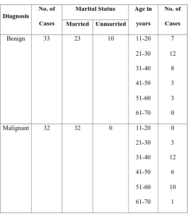
1. AGE, SEX AND MARITAL STATUS
Out of 33 cases with benign breast 23 (56%) were married.
Maximum incidence in this group was in 3rd decade (36%).
Where as, in 32 malignant breast lumps all were married
Table No. 3
AGE, SEX AND MARITAL STATUS
Marital Status Diagnosis
No. of
Cases Married Unmarried
Age in
years
No. of
Cases
Benign 33 23 10 11-20
21-30 31-40 41-50 51-60 61-70 7 12 8 3 3 0
Malignant 32 32 0 11-20
62
0 4 8 12 16
Diagnosis
11-20 21-30 31-40 41-50 51-60 61-70
AGE STATUS
Benign Malignant
0 4 8 12 16
Diagnosis
Married Unmarried
MARITAL STATUS
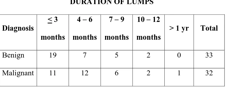
2. DURATION OF LUMP
Among the benign breast lesions, peak group was less than 3
months (19 cases out of 33) peak incidence of malignant lesions falls
[image:63.612.124.509.272.421.2]in the group for 4-6 months.
Table No. 4
DURATION OF LUMPS
Diagnosis
< 3
months
4 – 6
months
7 – 9
months
10 – 12
months
> 1 yr Total
Benign 19 7 5 2 0 33
64
0 3 6 9 12 15 18 21
Diagnosis
< 3 months 4-6 months 7-9 months 10-12 months
> 1 year
DURATION OF LUMPS
3. MENSTRUAL STATUS
Table No. 5
MENSTRUAL STATUS
Status of Menstruation Diagnosis No. of Cases
Premenopausal Postmenopausal
Benign 33 28 5
66
0 5 10 15 20 25 30
Premenopausal Postmenopausal
MENSTRUAL STATUS
B. EXAMINATION
1. NIPPLE CHANGES OF MALIGNANCY
Out of 32 malignant lesions of breast 6 cases showed changes in
the nipple suggestive of malignancy in the form of retraction. In the
series of benign lesions of 33 cases, no cases showed changes in the
nipple.
2. POSITION OF THE SWELLING IN BREAST
The following table was based on the occupancy of the lump
68 Table No. 6
SITE OF SWELLING
Maximum incidence of benign breast lesions and malignant
lesions were in upper inner quadrant.
Site Benign Malignant
Upper - Outer 6 6
Upper – Inner 16 7
Lower - Outer 8 3
Lower – Inner 3 3
Central 0 2
All Quadrants 0 2
Lower Inner & Central 0 2
Upper Outer & Central 0 3
Upper Inner & Central 0 1
Upper Outer & Inner 0 3
SITE OF SWELLING
0 4 8 12 16 20
Upper - outer Upper - inner Lower - outer Lower - inner
Central
All quadrant Lower inner and central Upper outer and central Upper inner and central Upper outer and inner
Diagnosis
70 3. SIZE OF SWELLING
Table No. 7
SIZE OF SWELLING
Maximum incidence of benign and malignant breast lumps were
3-5 cm in size.
Size Benign Malignant
< 2 cm 8 1
3 – 5 cm 23 20
6 – 10 cm 2 11
> 10 cm 0 0
0 5 10 15 20 25
Diagnosis
< 2 cm 3-5 cm 6-10 cm > 10 cm
SIZE OF SWELLING
72 Table No. 8
SHOWING CORRELATION BEWTEEN FNAC AND
EXCISIONAL BIOPSY HISTOLOGY
Sl. No. Excisional Biopsy Histology Correct Diagnosis False Positive False
Negative Insufficient
1 Fibroadenoma (28) 27 (96%) - - 1 (4%)
2 Ductal carcinoma (25) 22 (88%) - 3 (12%) -
3 Chronic nonspecific
mastitis (2)
2 (100%) - - -
4 Breast abcess (2) 2 (100%) - - -
5 Mixed carcinoma
(Both ductal and
lobular) (1)
0 - 1(100%) -
6 Malignant phylloides
tumour (1)
1 (100%) - - -
7 Benign Phylloides
tumour (1)
0 - 1(100%) -
8 Invasive squamous
cell carcinoma (1)
0 - 1(100%) -
9 Lobular carcinoma (2) 2 (100%) - - -
10 Mucinous adeno
carcinoma (1)
1(100%) - - -
11 Metaplastic
carcinoma (1)
1(100%) - -
Table No. 9
SHOWING CORRELATION BEWTEEN TRUCUT BIOPSY
AND HISTOPATHOLOGICAL EXAMINATION
Trucut Biopsy Sl.
No.
Histological
Diagnosis Correct
Diagnosis
False Positive
False
Negative Insufficient
1 Fibroadenoma (28) 25 (89%) - - 3 (11%)
2 Ductal carcinoma (25) 24 (96%) - - 1 (4%)
3 Chronic nonspecific
mastitis (2)
2 (100%) - - -
4 Breast abcess (2) 2 (100%) - - -
5 Mixed carcinoma
(Both ductal and
lobular) (1)
0 - 1(100%) -
6 Malignant phylloides
tumour (1)
1 (100%) - - -
7 Benign Phylloides
tumour (1)
0 - 1(100%) -
8 Invasive squamous
cell carcinoma (1)
1 (100%) - 1(100%) -
9 Lobular carcinoma (2) 2 (100%) - - -
10 Mucinous adeno
carcinoma (1)
1 (100%) - - -
11 Metaplastic
carcinoma (1)
1 (100%) - -
74
RESULTS
FNAC gave correct diagnosis in 89%, while in 6 cases the result
was false negative and in 1 case no opinion could be made.
The sensitivity of FNAC is 90% and specificity is 100%. The
positive predictive value is 100% while negative predictive value is
90%. In 1 patient, unsatisfactory smear obtained, which was not taken
for account for analysis. Overall accuracy of FNAC is 98% and that of
TCNB is 97%.
TCNB gave the correct diagnosis in 91%, 2 false negative cases
with 4 cases the biopsy was inadequate to give any diagnosis. The
sensitivity and specificity of TCNB was 96% and 100% respectively.
Similarly positive predictive value was 100% and 96% respectively.
DISCUSSION
All agree on the necessity for prompt diagnosis of any breast
lump. Hence workers all over the world are in search of a method
which can give an early as well as an accurate diagnosis. The incision
or excision biopsy in a well accepted diagnostic method for breast
lump, but both procedures are traumatic and require operation theatre
facilities. In the recent years, much of emphasis is laid on FNAC42.
Trucut needle is a simplified needle and needle biopsy can be
performed in out patient services.
FNAC is used extensively in the diagnosis of any lump. The
high rate of false negative diagnosis is early reports and seedling of
the cells along the needle track were the reasons that thought. Martin
and Ellis introduced the technique in 1934, it was not well accepted.
The visit of tumour dissemination has been shown to be more in
surgical biopsy as compared to FNAC. The false negative result in
cancer of the breast is 0-10 %. The present study had the same false
76
The reason for false negative diagnosis is due to number of
factors which include dense fibrosis of the tumour (failure to pierce
the tumour) and erroneous interpretation.
The correct diagnosis by FNAC5 can be achieved in 80-95%
cases. In the present series the correct diagnosis by FNAC in 89%
cases. There are many advantages of FNAC as it saves hospital
admission, saves preliminary biopsy, saves frozen section and the
patient known beforehand the type of operation.
Further, it allows rationale planning of operation list and avoid
unnecessary admissions. It can be carried out as an out patient
procedure with minimal trauma to the patient, can be repeated at ease
and mentally prepares the patient and surgeon.
Further, it provides and opportunity of follow up the patient
with clinically benign lesions without surgery.
In a busy out patient department and in busy operation list, the
surgical biopsy is a time consuming process. So cutting needle biopsy
provides an easier and time saving alternative.
The vim Silvermann’s needle was first used in 1960 in
diagnosis of the breast cancer. An excisional biopsy has several
disadvantages as it requires general anaesthesia and affects the choice
technique. The patients acceptance is high and apart from mild
brushing no complication has been encountered.
On positive diagnosis of malignancy by TCNB4, a definitive
surgery can be planned as no false positive results are reported by this
techniques. In the present study, there were 2 false negative cases and
in 4 cases, the biopsy material, inadequate to give any diagnosis. The
success rate of needle biopsy depends upon the size and consistency of
the lump and the type of needle used.
In fatty obese patients, where the breast is bulky, the needle
may miss the lesion. The trucut needle has the advantages on the
other needles as it quite handy, cuts a good core of tissue with least
trauma to the patient.
Both the technique have their own advantages and draw backs.
FNAC11 is the most simple techniques and does not require any
special instrument and the result can be obtained in a few hours time.
FNAC is associated with false positive and false negative results and
because of this, still it is controversial to decide the final surgery based
only on the results of FNAC.
The result of FNAC19 should be correlated with the clinical
impression. TCNB is a histological diagnosis while FNAC is
78
are no false positive results with TCNB so once a diagnosis of
malignancy is established. One can go for the definitive surgery.
TCNB44 is comparatively more traumatic than FNAC as it may
sometime bruise the breast.
On comparing the results of both techniques, it was found that
in benign, correct diagnosis was maximum of FNAC (94%) and
TCNB (88%), while in malignancy by FNAC and TCNB correct
diagnosis was 84% and 94% respectively. Taking all the techniques
together, diagnosis could be reached in 90% of cases.
[image:78.612.115.544.413.665.2]COMPARISON OF STUDY
Table No. 10
FNAC TCNB
Studies
No.
of
cases
Sen Spe Acc
P
Value
Sen Spe Acc P Value NS Yong (Singapore Medical College)
39 84
to
97.5
99 to
100
90 <0.02 90 100 67 <0.02
The accuracy of FNAC and TCNB are 98% and 97%
respectively. The difference beings statistically significant with a
p<0.02. P value of FNAC and TCNB are 0.016 and 0.031
respectively, using McNemar test for paired data, which shows both
the tests are significant with FNAC most significant compared to
80
CONCLUSION
This study has helped to correlate cytological report, trucut
needle biopsy and histopathology. Further out patient assessment of
breast lumps was done for the period of 2005 – 2006 in our hospital.
The results of this study showed almost equal detection rates by
FNAC (89%) and trucut biopsy (91%) when comparing with
histopathological examination. Trucut biopsy29, however was able to
give a histological diagnosis and results correlated 100% with the final
histology. However, in the setting of an out patient clinic, we would
like to recommend the use of FNAC for the diagnosis of suspicions
breast lumps. With the results we would be able to advise the patient
and recommend further treatment. However there is need for an
excision biopsy to obtain a definitive histology before proceeding to
definitive surgery as more have been cases of false positive results for
FNAC.
Considering both techniques, it can be concluded that if
FNAC37 can find a diagnosis one can go ahead with a definitive
operation. But, if in a clinically suspected case, FNAC is negative then
ideal for getting the histological report. Even if TCNB report comes
out to be negative, one should proceed with excisional or incisional
biopsy and according histopathological report, patient can be planned
82
BIBLIOGRAPHY
1. Abele, J.S., Miller, T.R., Goodson III, W.H., Hunt, T.K., and
Hohn, D.C. Fine needle aspiration of palpable breast masses. A
program for stages implementation. Archieve of Surgery, 1983,
118 (7) : 859-863.
2. Adams J, (1801) Observation on the cancerous breast, London,
Longman.
3. Arisio R., Curorese C, AcineliG, Mano MP, Bordon R, Fessia
L. Role of fine needle aspiration biopsy in breast Lesions :
Analysis of series of 4, 110 cases. Diagn. Cytopathol 1998, 18 ;
1493-98.
4. Ballo MS, Sneige N, Can Core needle biopsy replace fine
needle aspiration cytology in the diagnosis of palpable breast
carcinoma? Cancer 1996 ; 78 : 773-7.
5. Berner A, Davidson B, Sigstal.E, Risbey B. FNAC Vs core
biopsy in diagnosis of breast lesions. Diagn. Cytopathol. 2003
Dec : 29(6) : 344-8
6. Bondeson L : Aspiration cytology of radiation induced changes
7. Chuo CB, Corder AP Core biopsy Vs FNAC in a symptomatic
breast clinic Eur J Surg Oncol. 2003 May : 29(4) : 374-8.
8. Coleman, D.V. Aspiration cytology in preoperative
management of breast cancer (Letter to the Editor). The Lancet
1980, 2: 1083.
9. Cusick JD, Dotan J The role of trucut needle biopsy in
diagnosis of CA breast. Surg. Gynacol obstet 1990 ; 170 :
407-10
10.Das DK FNAC – Its origin, development and present status
with special reference to developing country, India. Diag.
Cytopathol 2003 JUNE ; 28(6) : 345-51 Review.
11.Dennison G, Anand R, Makar SH, Pain JA A prospective study
of role of FNAC and core biopsy in diagnosis of breast
carcinoma. Breast J. 2003 Nov-Dec ; 9(6) : 491-3.
12.Elston CW, Cotton RE, Davies CJ, Blamey RW – A
comparison of the use of the “trucut Needle” and Fine Needle
aspiration cytology in the preoperative diagnosis of carcinoma
of the breast. Histopathology 1978 ; 2 : 239-54
13.Erichsen J.E. (1853) : The science and art of surgery, London,
84
14.Essential surgical practice, 3rd Edition, Cuschieri, Giles, Moosa
978-909.
15.Franzen S, Zajicek J – Aspiration Biopsy in diagnosis of
palpable lesions of the breast ; critical review of 3479
consecutive biopsies, Acta Radiol (Ther) (Stockholm) 1968 ; 7 :
241-62.
16.Gelabert HA, Hsiu JG prospective evaluation of role of FNAC
in diagnosis and management of patients with palpable solid
breast lesions AM J Surg 1990 ; 56 : 263-7.
17.Godwin DT (1955-56) Annals of New York academy of
Sciences, 63, 1348.
18.Gupta, S.K., Ghosh, A.K., Choudhary T, Dutta TK, Talwar BL
and Dutta BN. Aspiration cytology in diagnosis of breast
cancer. The Indian Journal of Cancer 1979 : 16(1) : 1-8.
19.Homesh NA, ISSA MA, El-sofiani HA. The diagnostic
accuracy of FNAC Vs core needle biopsy for palpable breast
lump. Saudi Med J 2005 Jan ; 26(1) : 42-6.
20.Horgan PG, Waldron D The role of aspiration cytologic
examination in diagnosis of carcinoma breast. Surg Gynaecol
21.Joffe SN, Hughes HW, Primrose JN, Williamson BWA,
Aspiration cytology and out patient excision of breast lumps.
The Lancet 1979 ; 2 : 294-295
22.Kernohan R, Logan H Trucut needle biopsy in breast lumps
ulster Med J 1983 ; 52 : 142-4.
23.Kline TS & Neal HS Role of Needle aspiration biopsy in
diagnosis of carcinoma of breast. Obstertrics and Gynaecology
1979 : 46 : 89-92.
24.Kline TS, Joshi LP, Neal HS Fine Needle aspiration of the
breast ; Diagnosis and pitfalls. Cancer 1979, 44 : 1458-1464
25.Kun M (1847) Monthly Journal of Medical Science, 7 ; 953.
26.Laurence J.Z. (1855) The diagnosis of surgical cancer, London,
Churchill.
27.Ljung BM, Chero K, Deng G, Matsumura K, Waldman F,
Smith H Fine Needle aspiration techniques for the
characterization of breast cancer. Cancer 1994, 74 : 1000-1005
28.Melcher DH, Linchan JJ and Smith RS Fine needle aspiration
cytology. Recent Advances in Histopathology, Vol.II
86
29.N.S. Yong, KH Chia, WT Poh, CY wong, Comparison of trucut
with FNAC in diagnosis of breast carcinoma, SMJ 1999
Volume 40(09).
30.Olszewki WI, Borowski.M, Fine needle aspiration cytology Eur
J Surg Oncol. 1993. Jun : 19(3) : 309-10
31.Paget J (1953), Lectures on surgical pathology, Vol. 2, London,
Longman.
32.Patell JJ, Gartell PC, FNAC of breast masses. An evaluation of
its accuracy and reasons for diagnostic failure. ANN. R. Coll.
Surg Engl 1987 ; 9 : 156-9
33.Patra AK, Mallik RN, Dash S FNAC as primary diagnostic
procedure to breast lumps. Indian J Pathol Microbiol 1991, Oct.
34(4) : 259-64.
34.Poole GH, Willsher PC, Pinder SE, Robertson JF, Elston CW,
Blamey RW, Diagnosis of breast cancer with core biopsy and
FNAC. Aust NZ J Surg 1996 Sep ; 66(9) : 592-4.
35.Rosai & Ackerman’s surgical pathology (1779-1817)
36.Sadler GP, Mogee S, Dallimore NS, Monypenny JJ, Douglas
Jones AG, LyonsK, Horgan K Role of FNAC and Needle core
biopsy in the diagnosis of lobular carcinoma of breast. Br J