1
The Roles of Hypoxia on Neuroblastoma Cell
Migration and Invasion
Submitted in accordance with the requirements of the University of
Liverpool for the degree of Master of Philosophy by:
Michael Rice
2
Acknowledgement
I would like to express my greatest thanks to all those people who supported me
throughout this challenging yet exciting year, without their help this project would
never have made it off the drawing board.
I feel truly privileged to have been offered the opportunity to work with such a
fantastic group of people on an intriguing and all engrossing subject. Firstly I must
thank my Primary Supervisor, Professor Paul Losty, for orchestrating the team and
guiding me through the intricate transition from working at the hospital bed to the
laboratory bench. His knowledge and expertise have shone throughout the duration
of this project, especially when submitting abstracts to conferences and journals. I
would also like to thank Dr. Diana Moss for permitting me to enter her lab in order to
work with chick embryos.
Dr. Violaine Sée deserves special mention as she wholly accepted me into her team
and provided the most excellent advice regarding the direction of this project. Aside
from impressive leadership skills Dr. Sée has an innate ability to encourage hard
work whilst maintaining a most pleasant atmosphere and an excellent spirit
throughout each member of the team. As such it has been an absolute pleasure to
work as a member of her team.
My greatest thanks go to Dr. Anne Herrmann. I entered the laboratory a complete
novice however her kind and patient style of teaching was the most effective I have
ever experience. As a result I now feel completely comfortable working
independently at the bench. Demonstrating a wealth of knowledge at every turn Dr.
Herrmann lead by example and her hard work most certainly helped to focus my
efforts. I truly believe I could not have wished for a better colleague or friend.
I would also like to thank the Royal College of Surgeons and Alder Hey Children’s
Hospital for generously supporting the work undertaken in this study.
Finally I would like to thank my family for the continuous support they have given me
3
Abstract
Solid tumours are predisposed to hypoxic conditions due to cellular proliferation
outpacing the rate of angiogenesis. Hypoxia is associated with enhanced tumour
aggression. We aimed to study how hypoxia influences the migration and invasion of
Neuroblastoma, a common and highly lethal, enigmatic childhood cancer.
Fluorescently labelled Neuroblastoma cell lines were cultivated under atmospheric (‘normoxic’) or hypoxic conditions for 3 days before implantation upon the
chorioallantoic membrane (CAM) of chick embryos and observed in-vivo for tumour formation and metastasis.
Tumourigenesis was successful regardless of oxygen levels. Normoxic cells failed to
metastasise, conversely hypoxic cells invaded the embryo forming tumour spheres in several organs. Interestingly, ‘normoxic’ cell invasion was observed when implanted
alongside hypoxic cells resulting in co-invasion and independent microtumour
deposition. These processes were later identified as ‘HIF dependent,’ following
pretreatment of cells with DMOG.
In conclusion we found an increase in metastatic phenotype following exposure to
hypoxia via stabilisation of the HIF transcription factor. This CAM model was also
utilised in order to begin to understand the molecular mechanisms underpinning the
4
Abbreviations
ARNT – Aryl Hydrocarbon Receptor Nuclear Translocators
BMPs – Bone Morphogenetic Proteins; BSA – Bovine Serum Albumin; β-III Tubulin – Neuronal Class III β-Tubulin Polyclonal Antibody
Ca9 – Anti-Carbonic Anhydrase IX; CAM – Chorioallantoic Membrane; CD 56 – Neural Cell Adhesion Marker; CHD5 – Chromatin Helicase-binding Domain 5; CISH - Chromogenic in-situ Hybridization; c-kit – Mast / Stem cell growth factor receptor; C-MYC – c-Myelocytomatosis Viral Oncogene Homolog; C-TAD – Carboxy-terminal Transcriptional Activation Domain.
dHAND – heart-and neural crest derivatives-expressed protein; DLX3 – (Distal-less homeobox 3); DLX5 – (Distal-less homeobox 5); DMEM – Dulbeco’s Minimum Essential Medium; DMOG – Dimethyloxalyl Glycine; DNA – Deoxyribonucleic acid
ECM – Extracellular Matrix; EMT – Epithelial to Mesenchymal Transition
FACs – Fluorescence Activated Cell sorting; FCS – Fetal Calf Serum; FIH - Factor Inhibiting HIF; FISH – Fluorescence in-situ Hybridization
GFAP – Anti-Glial Fibrillary Acidic Protein; GLUT-1 – Anti-Glucose Transport Receptor 1
H&E – Haematoxylin and Eosin; HASH-1 – human achaete-scute homolog-1; HIF-1α – (Hypoxia-inducible factor 1α); HIF-2α – (Hypoxia-(Hypoxia-inducible factor 2α); HRE – Hypoxia Response Elements ID2 – Inhibitor of Differentiation 2; ID3 – Inhibitor of DNA Binding 3, Dominant Negative Helix-Loop-Helix Protein; IHC-F – Immunohistochemical staining of Frozen sections; IHC-P – Immunohistochemical staining of Paraffin sections; IMS – Industrial Methylated Spirits; INPC - International Neuroblastoma Pathology Criteria; INRGSS - International Neuroblastoma Risk Group Staging System; INSS - International Neuroblastoma Staging System
KIF1β – Kinesin Family Member 1β LMO4 – Lim domain only protein 4
MASH-1 - Mammalian Achaete-Scute Homolog 1; MDM2 – Murine double minute 2 homolog; MEM – Minimum Essential Medium; MIBG – Iodine-131-Meta-iodobenzylguanidine; MKI - Mitosis-Karyorrhexis Index; MSX1 – (msh homeobox 1) and MSX2 – (msh homeobox 2)
NB – Neuroblastoma; NEAA - Non-essential Amino Acids; Notch-1 – Translocation-associated Notch homolog 1 {Drosophila}
O2 – Oxygen
PAS family bHLH-PAS – Period circadian protein; Aryl Hydrocarbon Receptor Nuclear Translocator; Single-minded protein; family basic helix-loop-helix proteins; PAX3 – (Paired Box Homeotic Gene 3);
PAX7 – (Paired Box Homeotic Gene 7); PHD’s – Prolyl Hydroxylase Domain Proteins; Phox2A – (Paired-like homeobox 2a); Phox2B – (Paired-like homeobox 2b); p21 / WAF1 – (cyclin-dependent kinase inhibitor 1); p53 / TP53 – (Tumour protein 53); p73 – (Tumour protein 73); p300 – E1A binding protein p300
RPMI – Roswell Park Memorial Institute Medium
SD – Standard Deviation; Slug – Zinc Homolog 2 {Drosophila}; Snail – Snail Homolog 1 {Drosophila}; Snail2 – Snail Homolog 2 {Drosophila}; SMA – Smooth Muscle Actin-α; SNS – Sympathetic Nervous System; SRC-1 – Steroid Receptor Coactivator 1
TBS – Tris Buffered Saline; TrkA – (neurotrophic tyrosine kinase receptor type A); TrkB – (neurotrophic tyrosine kinase receptor type B); TrkC – (neurotrophic tyrosine kinase receptor type C)
5
Table of Contents
Acknowledgements 2
Abstract 3
Abbreviations 4
1. Introduction 1.1 Neuroblastoma 7
1.1.1 Epidemiology & Screening for Neuroblastoma………...………...7
1.1.2. Clinical Presentation………..9
1.1.3. Staging, Risk Classification and Treatment……….12
1.1.4. Metastasis and Classifiction in Neuroblastoma….……….15
1.1.5. Embryology………...16
1.1.6. Pathology & Pathogenesis of Neuroblastoma……….18
1.1.7. Genetic Features………..21
1.2 Hypoxia 22
1.2.1. Hypoxia-inducible factor (HIF)………23
1.2.2. Physiology and Pathophysiology of Hypoxia………...27
1.2.3. Hypoxia and Solid Tumours………...28
1.2.4. Hypoxia and Neuroblastoma………..31
1.3 The Chorioallantoic Membrane and Tumour Cell Invasion 34
Introducing the Chick Chorioallantoic Membrane ………...34
1.3.1. Anatomy and Development of the CAM………...35
1.3.2. Chick embryo and CAM model in Cancer studies………....………..37
1.3.3. The Chick Embryo and Neuroblastoma….………...………...39
1.3.4. Tumour cell Migration...………..41
1.4. Study Aims 43
2. Materials and Methods 2.1.1. Overview of Cell lines………...44
2.1.2. Tissue Culture Protocol………...45
2.1.3. Culture and treatment of cells prior to CAM implantation………..45
2.2. Incubation and Fenestration of Chick embryos………...46
2.3. Implantation of Tumour cells upon the “CAM”………48
6
2.4. Paraffin embedding, Sectioning & Staining……….51
2.5. Freezing samples, Sectioning & Staining………52
2.6. Imaging……….55
3. Results 3.1. Introduction………...56
3.1.1. Pilot study investigating cell line survival upon the CAM………...56
3.1.2. Implantation of Normoxic Sk-N-AS………58
3.1.3. Normoxic Sk-N-AS Migration & Invasion……….61
3.1.4. MYCN amplified cell lines………...62
3.2.0. Implantation of Hypoxic Sk-N-AS cells………...64
3.2.1. Tumour differences – Normoxic Vs Hypoxic Preculture………67
3.2.2. Effects of DMOG on Sk-N-AS tumourigenesis………69
3.3.0. Co-application of Normoxic and Hypoxic Sk-N-AS……….71
3.3.1. Implantation of Normoxic – Hypoxic Mixtures……….72
3.3.2. Sk-N-AS Tumourigenesis and Invasion………76
3.3.3. Hypoxia unlocked tumourigenesis in MYCN amplified cell line………78
3.4.0. Paraffin staining of Sk-N-AS tumour samples……….80
3.4.1. Staining for markers downstream of HIF expression……….82
3.5.0. Tumour cell Proliferation and Intravasation - Normoxia Vs Hypoxia………...83
3.5.1. Frozen section tumour characterisation – Hypoxia Vs Normoxia………...88
4. Discussion 4.1. The Chick Embryo Model………...90
4.2. Preculture in Normoxia versus Hypoxia………..93
4.3. Hypoxia and Metastasis……….96
4.4. Future Work………100
4.5. Conclusions………101
7
1. Introduction
1.1 Neuroblastoma
Neuroblastoma (NB) is a highly dangerous often lethal, solid heterogenous tumour
which is regularly encountered in paediatric oncology. It is the most common cancer
diagnosed in infants accountable for nearly one third of all infant malignancies
(German Childhood Cancer Registry, Web Reference 1) and responsible for 15% of
mortality occurring in paediatric oncology (Maris et al. 2007).
1.1.1 Epidemiology & Screening for Neuroblastoma
Epidemiology
The global incidence of neuroblastoma is estimated at around 10.5 cases per million
children aged less than 15 years (Stiller et al. 1992). Age at diagnosis is a key
feature with the vast majority of patients presenting for the first time before 5 years.
Interestingly older patients (> 14 years) experience poorer survival (Castleberry.
1997) although < 10% are diagnosed beyond the age of 10.
Neuroblastoma can occur in adults, however incidence is low and steadily
decreasing; 1973-77 recorded 0.47 / 1,000,000; 1998-02 recorded 0.12 / 1,000,000
(Esiashvili et al. 2007). Overall survival in adults is poor (5-year survival 36.3%)
when compared to other age groups; infant 5-year survival 84.6%; 1-9 years old
47.8%; 10-19 years old 46.2% (Esiashvili et al. 2007). Due to the significant
difference in survival between infant and child, age has been included in the Children’s Oncology Group Prognostic Classification (Katzenstein et al. 1998).
Presenting such poor outcome data, adults might be expected to more commonly
express classic biological risk factors such as MYCN amplification, loss of
chromosome 1p, or DNA ploidy, however these markers of disease severity are
rarely encountered (Moody et al. 1996, Kushner et al. 2003). Regarding gender,
several studies (Spix et al. 2006; Park et al. 2008; Wiangnon et al. 2003) have
demonstrated a slight male preponderance for disease however this discrepancy is
8 In the USA, neuroblastoma is the most common solid tumour of childhood excepting
those occurring within the central nervous system (Reis et al. 1999). Asian / Pacific Islanders have also been identified as the groups most “at risk,” with 35% increased mortality (Johnson et al. 2011). A Children’s Oncology Group study found
inconsistencies in survival between African-American and White populations
(Henderson et al. 2011) however later studies suggest this could be due to
inequalities regarding access to health care (Pui et al. 2012). Internationally it is
generally regarded that no appreciable difference exists concerning race.
Among Europeans disease incidence is increasing. During the period 1978-82
disease occurred in 8.4 / 1,000,000 children whilst 1993-97 saw this figure increase
to 11.6 (Spix et al. 2006). Across European regions, the UK and Ireland experience
the lowest incidence (9.1) and also achieve some of the worst survival rates (49%
Confidence Interval: 45-52). However the overall trend shows increased survival in
Western Europe compared to the East which is worse-off by 20% (Spix et al. 2006).
Regardless, overall 5-year survival has almost doubled within the last 30 years. This
is most likely due to changes in treatment regimens following the identification of
biological indicators of disease severity.
Screening
High incidences of neuroblastoma have been observed in Japan due to the presence of a National Screening Program running from the 1970’s aimed at identifying cases in children ≤ 6 months old. Domestic and international evidence (Yamamoto et al.
2002, Woods et al. 2002, Schilling et al. 2002) halted all neuroblastoma screening
programs by 2004 having demonstrated screening increases incidence but failed to
significantly decrease disease mortality. It was found that the high incidences were
due to an increase in low risk cases whilst the number of intermediate / high risk
cases remained unchanged. In the past these low risk cases might have been
9 1.1.2. Clinical Presentation
Anatomic location
Well known for its unpredictable nature, neuroblastoma may present with any
number of highly variable symptoms. This is most likely due to its ability to develop
throughout the sympathetic nervous system making tumour formation possible in
most anatomic regions, see Figure 1.
10
Signs and Symptoms
The diagnosis of patients with low risk disease can quite commonly be made as a
result of an incidental discovery due to the asymptomatic patient undergoing
investigation for an unrelated problem. In the past however these patients were more
frequently encountered following identification by screening programs which have
now become redundant (Woods et al., Schilling et al. 2002).
Patients presenting with active symptoms commonly complain of pain and malaise.
Primary tumours develop chiefly upon the adrenal glands (Park et al. 2008) resulting
in abdominal bloating and constipation with severe cases developing liver
metastases resulting in abdominal compartment syndrome and subsequent
respiratory distress or anuria. Quantification of common anatomical regions is shown
in Figure 2.
Regular sites for disease dissemination are liver, bone and bone marrow which are
investigated when staging the tumour (Carlsen et al. 1985). Children presenting with metastasis are often very ill and may demonstrate “blueberry muffin” skin lesions, proptosis, and “raccoon eyes” known as periorbital ecchymosis. These classic
pathologies of the eye were historically believed to be due to a propensity for orbital
dissemination (Hutchison 1907) however this claim is yet to be tested in more recent
times.
Rare Presentations
Rare presentations include associated; “VIPoma,” a Vasoactive Intestinal Peptide
secreting tumour causing diarrhoea; or catecholamine secreting neoplasms such as
phaeochromocytoma resulting in facial flushing and excessive sweating. Tumour
formation in the thorax, head or neck can lead to complex problems such as spinal cord compression or Horner’s Syndrome, a triad of unilateral anhidrosis, miosis and
ptosis. Occurring in around 2% of neuroblastoma cases, an immune-mediated
paraneoplastic disease, opsoclonus-myoclonus ataxia, is a cerebellar pathology known to give patient’s “dancing eyes” (Rudnick et al. 2001, Mitchell et al. 2002).
However, of all recorded opsoclonus-myoclonus, up to 50% have been observed to
11
[image:11.595.74.525.138.646.2]12 1.1.3. Staging, Risk Classification and Treatment
Staging
As of the early 1990’s neuroblastoma were staged according to the International
Neuroblastoma Staging System (INSS) (Brodeur et al. 1993) shown in Table 1.
Table 1 The International Neuroblastoma Staging System (Brodeur et al. 1993).
Stage Definition
1 Localised tumour with complete gross excision, with or without microscopic residual disease; representative ipsilateral lymph nodes negative for tumour
microscopically (nodes attached to and removed with primary tumour may
be positive)
2A Localised tumour with incomplete gross excision; representative ipsilateral nonadherent lymph nodes negative for tumour microscopically
2B Localised tumour with or without complete gross excision, with ipsilateral nonadherent lymph nodes positive for tumour. Enlarged contralateral lymph
nodes must be negative microscopically.
3 Unresectable unilateral tumour infiltrating across the midline*, with or without regional lymph node involvement; or localized unilateral tumour with
contralateral regional lymph node involvement; or midline tumour with
bilateral extension by infiltration (unresectable) or by lymph node
involvement.
4 Any primary tumour with dissemination to distant lymph nodes, bone, bone marrow, liver, skin and/or other organs (except as defined for stage 4S)
4S Localised primary tumour (as defined for stage 1, 2A or 2B), with dissemination limited to skin, liver and/or bone marrow** (limited to infants <
1 year of age)
* The midline is defined as the vertebral column. Tumours originating on one side and crossing the midline must infiltrate to or beyond the opposite side of the vertebral column.
13 Much of the literature refers to tumours according to this system however it has
recently been replaced by the International Neuroblastoma Risk Group Staging
System (INRGSS) shown in Table 2 below (Monclair et al. 2009). This is due to
several practical issues such as the requirement to obtain tumour tissue which is no
longer necessary in the treatment of some patients with low risk disease.
Table 2. The International Neuroblastoma Risk Group Staging System is the current system replacing the INSS however most published literature quotes the older system.Patients with multifocal primary tumours should be staged according to the greatest extent of disease (Monclair et al. 2009).
Stage Definition
L1 Localised tumour not involving vital structures as defined by the list of image-defined risk factors and confined to one body compartment.
L2 Locoregional tumour with presence of one or more image-defined risk factors.
M Distant metastatic disease (except stage MS)
MS Metastatic disease in children younger than 18 months with metastases
confined to skin, liver, and/or bone marrow.
Risk Classification
It is necessary to classify neuroblastoma in terms of prognosis in order to utilise the
most appropriate methods of treatment. Originally the Shimada system quantified;
level of stromal development; neuroblastic differentiation; mitosis-karyorrhexis index
(MKI); and patient age in order to distinguish favourable from unfavourable cases
(Shimada et al. 1984). However the International Neuroblastoma Pathology Criteria
(INPC) later modified this system further subdividing disease subgroups, as shown in
14
Table 3. International Neuroblastoma Pathology Criteria (Shimada et al. 2001). FH: favorable histology; UH: unfavorable histology; MKI: mitosis-karyorrhexis index.
Category and Subtype Prognostic group
Neuroblastoma (Schwannian stroma-poor)
Undifferentiated
Poorly differentiated
Differentiating
FH or UH based upon:
Age
Grade of
Differentiation
MKI class
Ganglioneuroblastoma (Schwannian stroma-rich) FH
Ganglioneuroblastoma, nodular
(Schwannian stroma rich/dominant and poor)
UH
Ganglioneuroma (Schwannian stroma dominant)
Maturing
Mature
FH
Treatment
Treatment can be extensive or simple depending upon the biological features unique
to each tumour. Staging, tumour classification and the anatomic location of tumour
development are also important factors to consider before administering therapy.
Low risk cases such as asymptomatic Stage 4s disease may be left to undergo
spontaneous regression without the need for active management. However
intermediate to high risk patients often require an array of therapies such as:
chemotherapy, radiotherapy or extensive surgery, laparotomy, depending upon the
severity of their condition.
Research efforts are continuing to develop novel therapies for the treatment of
neuroblastoma patients. For instance, clinical trials are currently underway assessing
the therapeutic benefits of anti-GD2, an antibody which is known to react with
neuroblastoma cells (Mueller et al. 1990; Yu et al. 2010). Work is also continuing to
discern the role of antiangiogenesis in solid tumour therapy (Chen et al. 2011; Huang
et al. 2012). However, significantly more work must be undertaken if survival rates
[image:14.595.81.524.118.385.2]15 1.1.4. Metastasis and Classification in Neuroblastoma
Known as one of the most common malignancies identified in children, disseminated
neuroblastoma are classified as either Stage 4 (INSS) / Stage M (INRGSS) or Stage
4S (INSS) / Stage MS (INRGSS). Metastatic sites are widely variable and this leads
to difficulties in diagnosis as patients may be asymptomatic or present with a range
of life threatening symptoms. A large number of patients suffering this disease are
categorised as Stage 4 / M with some studies demonstrating up to 60% of
neuroblastoma present in this category (Juárez-Ocaña et al. 2009; Palma-Padilla et
al. 2010). Treatment protocols for this group are extremely severe and survival is
truly dismal, recorded at 20 - 25% (Philip et al. 1991; Li et al. 2012).
Stage 4S disease is widely different with metastasis limited to bone marrow, liver
and skin and overall survival rates approaching 90% (Katzenstein et al. 1998;
Nickerson et al. 2000). However this classification encompasses only a small
subsection of overall disease and the frequency of presentation decreases with age
with the majority of cases occurring in neonates (Iehara et al. 2012). The significant
difference between Stages 4 and 4S is the ability of the latter to undergo
spontaneous regression resulting in a surprising recovery often without relapse. As
such, some patients may receive no active treatment and instead are monitored until
all signs of the disease have been absent for a continuous period of time, usually up
to 10 years. This phenomenon is unique and distinguishes neuroblastoma as a
particularly interesting tumour among other cancers.
Although Stage 4S has a drastically improved survival rate compared to Stage 4,
patients still die. This is because they must remain alive for long enough for the
metastatic secondary tumours to regress and in this time the normal function of their
vital organs may be compromised as previously mentioned. MYCN status also has a
role, as non-amplified infants with Stage 4 and 4S disease have excellent survival,
with minimal or no treatment (De Bernardi et al. 2009). Finally a major problem
facing clinicians occurs when patients with disseminated neuroblastoma are initially
assessed. Here, there is a lack adequate tools to differentiate Stage 4S from Stage 4
disease, hence some patients may be subjected to severe and life-threatening
16 1.1.5. Embryology
Embryology
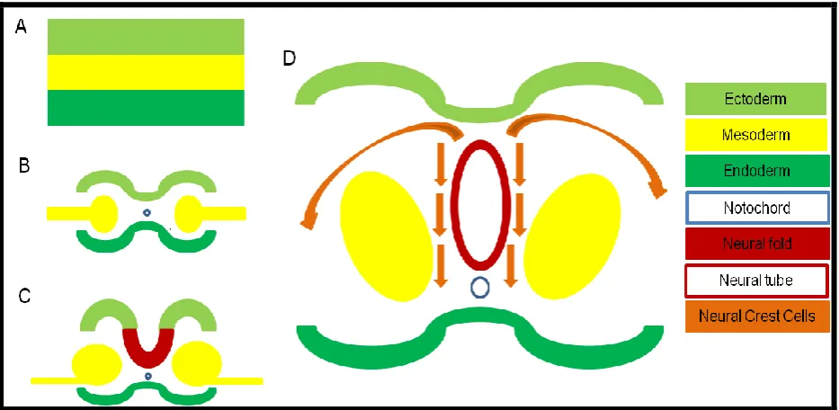
At the beginning of gestational week 3 human embryos initiate the process of
neurulation whereby the nervous system forms from embryonic ectoderm (Figure
3A). This process encompasses neural fold specification and delamination with latter
differentiation-migration-proliferation, which occur simultaneously. The first step is
neural plate formation, resulting in a three layer structure constituting neural
ectoderm, mesoderm and endoderm. Within days, plate edges thicken cranially
resulting in the creation of neural folds which grow toward each other and eventually
fuse to form the neural tube (Figure 3C). Inside this tube neuroepithelial cells are
modified becoming neuroblasts which later differentiate to form mature neurons.
Preceding fusion, neural crest cells become detached from neural folds, these cells
are versatile and undifferentiated. Crest cells migrate throughout the body whilst
simultaneously proliferating and differentiating in order to form mature adult
structures such as, the autonomic nervous system, catecholamine producing adrenal medulla “chromaffin” cells, melanocytes, and thyroid “C” cells (Le Douarin et al.
[image:16.595.73.540.468.696.2]1974; Le Douarin, Teillet. 1974; Dorsky et al. 1998).
17
Specification & Delamination
Synthesis and migration of neural crest cells begins with strategic specification of the
neural fold, as displayed in Figure 3C above. Key to this process are neural crest
specifier genes such as; C-MYC (C-Myelocytomatosis Viral Oncogene Homolog – Aoki et al. 2003); Snail (Snail Homolog 1 {Drosophila} – Essex et al. 1993); and Slug (Zinc Homolog 2 {Drosophila} – Mayor et al. 1995) which are present prior to and throughout migration (Meulemans et al. 2004). However activation of these genes is reliant upon previous
expression of PAX3 (Paired Box Homeotic Gene 3), PAX7 (Paired Box Homeotic Gene 7), DLX3 (Distal-less homeobox 3), DLX5 (Distal-less homeobox 5), MSX1 (msh homeobox 1) and MSX2 (msh homeobox 2) alongside many other neural plate border specifier genes (Bang et al. 1997; Luo et al. 2001; Tribulo et al. 2003).
On completion of specification, former neural fold cells must detach (delamination) to
be made available for migration. Transcription factor ID3 (Inhibitor Of DNA Binding 3, Dominant Negative Helix-Loop-Helix Protein) performs the role of stabilising crest cells and may also aid segregation from the neural fold (Kee et al. 2005). Under the influence
of Slug, cells undergo epithelial to mesenchymal transition (EMT), incidentally an
established mechanism for cancer cell invasion (Thierry 2002). Mitosis is halted
throughout this process via the action of Snail, permitting expression of Rho
GTPases and cadherins which enable alterations in morphology and adhesion
quality (Fukata et al. 2001).
Differentiation-Migration-Proliferation
Once free of the neural fold, crest cells migrate according to signalling pathways
whilst constantly proliferating. Early in migration an important distinction is made,
future melanocytes are forced to traverse dorsolaterally whilst future chromaffin /
sympathetic nervous system cells journey ventrally (Thiery et al. 1982). Key to both
dorsolateral and ventral pathway movement are ephrins which increase melanocytic
crest cell affinity for dorsolateral pathway fibronectin (Meulemans et al. 2004,
Santiago et al, 2002) whilst directing other crest cells ventrally (Krull et al. 1997,
18 Eventually extracellular interactions commence via integrin binding with extracellular
matrix (ECM), thus crest cells become available to migrate throughout the body
(Humphries et al. 2006).
In a normal foetus, crest cell differentiation requires a level of environmental
versatility (plasticity) provided by the strategic expression of specific genes. For
example, transition from neural crest cell to sympathetic neuron relies upon a
number of important transcription factors, notable key coordinators include: MYCN, Phox2A, Phox2B, HIF-1α and p73 (Nakagawara 2004). Overall the process of
differentiation is triggered by the presence of bone morphogenetic proteins (BMPs)
(Huber et al. 2002) however it is postulated that BMPs may also serve to inhibit later
phases of maturation. This complements independent findings that differentiation
potential is progressively inhibited in postnatal adrenal medullary cells implying that
there could be an overall restriction upon differentiation following initial triggers
(Mascorro et al. 1989). Finally it has also been observed that individual crest cell
diversifications may be reliant upon specific transcription factors, for instance it has
been suggested that chromaffin cell maturation is largely dependent upon MASH-1
expression (Huber et al. 2002).
1.1.6. Pathology & Pathogenesis of Neuroblastoma
Pathology
Described as a small, round, blue-cell tumour of childhood, neuroblastoma appear
histologically as a clutch of closely organised cells enclosed in neurofibrillary stroma
and partitioned by fibrovascular septa. They are believed to be neural crest cell
derivatives with a sympathoadrenal lineage classically defined histologically as
forming a pattern known as Homer-Wright rosettes (Figure 4). Each rosette consists
of a core resembling central nervous system neuropil (network of cytoplasmic
neuronal processes between cell bodies at synaptic connections) regarded as
neurofibrillary matrix, surrounded by small clustered rings of tumour cells (Owens,
Irwin 2012). Though regarded as the classic pattern of tumour formation,
undifferentiated neuroblastoma may demonstrate a complete absence of
19
Figure 4. This figure displays a diagram (A) and a histological picture (B) demonstrating neuroblastoma cell formation as characteristic Homer-Wright rosettes. Diagram A displays cells of different shapes, sizes and shading (pleomorphic hyperchromasia), representing microscopic findings suggestive of malignancy. The red circle in B highlights a core of neurofibrillary matrix at the centre of a rosette. This is an image produced by the US Armed Forces Institute of Pathology, it has been released into the public domain, see Web Reference 3.
Macroscopically, tumours tend to appear either as voluminous multinodular masses
or discrete spheroids and from time to time cystic neuroblastoma are reported
however they are often misdiagnosed as adrenal cyst or haematoma (Lack 1997;
Kozakewich et al 1998). Upon cross-section, chalky white calcification may be
witnessed however regions of necrosis and haemorrhage among coarse nodular
tissues are commonly observed (Lack 1997).
Pathogenesis
Solid tumours are regarded differently when they develop in children. An occurrence
unique to this cohort of patients is the formation of embryonal tumours which most
commonly appear as a remnant of foetal development, histologically resembling
immature tissues. As neuroblastoma emanate from neural crest cells they are a
prime example of an embryonal tumour and like all cancers, are believed to develop
as a consequence of genetic alteration or abnormality. Supporting claims linking
neuroblastoma to crest cells, expression profiling of favourable and unfavourable
tumours has shown that many possess the genes required to permit normal neural
crest cell differentiation (Ohira et al. 2003). From this it is sensible to suggest that
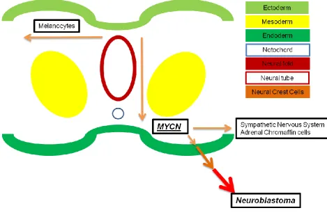
tumour cells appear during differentiation-migration-proliferation, see Figure 5,
because within this phase a key strategic advantage can be gained as a result of
environmental plasticity which encourages propagation of normal crest cells within
20 Related to neuroblastoma are two disease subtypes which exist in a more
differentiated state. Ganglioneuroblastoma and ganglioneuroma are more advanced
tumours identified histologically following recognition of spindle cell Schwannian
stroma (neuronal support network) and ganglion cells or their immediate precursors.
The International Neuroblastoma Pathology Classification (INPC) demarcated these
subtypes as follows; neuroblastoma – Schwannian stroma poor;
ganglioneuroblastoma – Schwannian stroma rich; ganglioneuroma – Schwannian
stroma dominant (Shimada et al. 2001). Further evidence of differentiation is
determined by assessing for neurotrophin receptors such as; synaptophysin; TrkC;
and TrkA with the latter inferring a favourable prognosis when highly expressed
[image:20.595.70.540.342.651.2](Nakagawara et al. 1998, Brodeur et al. 2009).
21 1.1.7. Genetic Features
Genetic Features
Neuroblastoma derived Myc proteins (MYCN) are important neural crest cell
transcription factors controlling cell maintenance and proliferation, Figure 5
(Pelengaris et al. 2002). Located at chromosome 2p24, gene amplification of >10
copies indicates rapid disease progression and poor prognosis. This can be
identified using: FISH - fluorescence in-situ hybridization, or more recently using
CISH - chromogenic in-situ hybridization (Squire et al. 1996; Bhargava et al. 2005).
The INRG Classification System (Cohn et al. 2009) encourages investigation of
MYCN status in all patients and further advises confirmation of DNA ploidy and
chromosome 11q status. Fortunately amplification occurs in a minority of cases;
5-10% in infants (Kaneko et al. 2006); 20-30% in childhood (Conte et al. 2006).
Survival is significantly improved in the absence of MYCN most notably in stage 4
disease where mortality rate of <10% has been demonstrated (Schmidt et al. 2000).
DNA-ploidy is investigated using flow cytometry with hyperdiploidy linked to TrkA
over-expression (Brodeur et al. 2009), a receptor found in favourable outcome
neuroblastoma (Nakagawara et al. 1993). However diploid tumours are associated
with segmental chromosomal changes indicating an unstable genome and
therapeutic resistance. Important segmental changes include deletions at 1p36 (30%
of cases) and 11q23 (40%), whilst 50% of cases express gains at 17q (Caron et al.
1993; Attiyeh et al. 2005; Brown et al. 1999). Commonly deleted regions encode
tumour suppressor genes, Eg. 1p36; chromatin helicase-binding domain 5 (CHD5); KIF1β and p73, this may explain increased tumour aggression in patients with
regional deletion. The most common chromosomal change, gain at 17q, is believed
to impart oncogenic tools for tumour survival however loss at 11q is more useful
clinically, indicating poor event-free survival (Attiyeh et al. 2005). Incidentally,
increased expression of TrkB has been noted in 11q deficient tumours, signifying
22
1.2 Hypoxia
Introduction
Oxygen (O2) is the vital component required to sustain mammalian life. It is such a
crucial molecule that must be supplied continuously throughout the course of an individual’s life. Appropriate oxygenation is crucial to maintain normal tissue function
however some tissues (bone marrow) may endure fluctuating oxygenation with
greater ease than others (brain parenchyma). In order to ensure adequate
oxygenation of all tissues, humans employ a series of mechanisms which constantly
monitor tissue oxygen tension and carry out adjustments according to supply and demand. E.g. baro / chemo – receptors exist close to O2 sensitive organs, constantly
testing the blood for potential changes in O2. These efforts ensure high level O2 is
decreased whilst low level O2 is boosted keeping tissue oxygen tension tightly
controlled within strict parameters.
Normal atmospheric oxygen constitutes 21% of air inhaled by humans however
levels decrease inside the body and what is more, different viscera are maintained at
a range of various oxygen concentrations. Upon inhalation, O2 concentrations are
significantly lower (14%) when measured at the terminal alveoli. Levels are further
reduced when molecules are exchanged into the blood and temporarily bound to
haemoglobin. Newly formed oxyhaemoglobin complexes traverse the body via the
vascular network and deliver fresh O2 to nearby tissues through the process of
diffusion. As a result, end-organ perfusion is variable but also much lower than
atmospheric conditions. An example of this can be appreciated in the brain which is
an extremely oxygen-sensitive collection of tissues. Overall, the mean cerebral
concentration of O2 is recorded at 3.2% however concentrations are widely altered
throughout different territories of the brain (Dings et al. 1998).
Given expected variance in oxygenation across different viscera, normal
physiological parameters are usually set between 2 – 9% O2, also known as
‘normoxia’ (Simon, Keith. 2008). Excluding several special exceptions, pathologically
decreased O2, ‘hypoxia,’ is regarded as less than 2% O2. Finally, extreme hypoxia
23 1.2.1. Hypoxia-inducible factor (HIF)
When studying erythropoietin gene expression in hypoxia a single DNA – protein
complex was found to bind the gene (Semenza and Wang, 1992). A resultant
increase in erythropoietin expression was observed expanding the vascular potential
for O2 carriage and directly linking hypoxia to erythropoiesis. The hypoxia dependent
transcription factor, hypoxia-inducible factor (HIF), was later described as a
heterodimer consisting of two PAS family basic helix-loop-helix proteins (bHLH-PAS)
(Wang et al. 1995). The complex constitutes two subunits existing as isoforms, alpha and beta; HIF-1α, 2α; and the latter (ARNT) aryl hydrocarbon receptor nuclear
translocators 1, 2, 3 (Brahimi-Horn et al. 2009).
HIF-1α and 2α are constitutively expressed in the cell however they are vulnerable to
hydroxylation by Prolyl Hydroxylases (PHDs) and Factor Inhibiting HIF (FIH) in the
presence of sufficient O2 levels. Normoxia predisposes alpha subunits to
proteasomal degradation by ubiquitination. This process requires hydroxylation of two proline residues residing within HIF-α subunits by PHD’s, principally PHD2
(Berra et al. 2003). Thereafter binding of tumour suppressor von Hippel-Lindau
protein (pVHL) linked to elongin C in an E3 ubiquitin-ligase complex (Maxwell et al 1999) facilitates bonding to ubiquitin leading to degradation of HIF-α, see Figure 6
(Jaakkola et al. 2001).
While PHDs hydroxylate HIF at proline residues, FIH hydroxylates asparagine
residue 803 in the carboxy-terminal transcriptional activation domain (C-TAD) of HIF-1α (Dayan et al. 2006). This interaction is important as it inhibits later coupling with
transcription coactivator p300, disrupting future expression of hypoxia associated
target genes (Lando et al. 2002). Incidentally this coactivator is also required during
24 In hypoxia these mechanisms are negated and as a consequence, HIF-α activity is no longer suppressed. At low levels of oxygenation, PHD’s are deactivated prior to
FIH, as both components avail of O2 during hydroxylation (Epstein et al. 2001; Lando
et al. 2002). PHD activity is the first to decrease, stabilising a large proportion of
α-subunits, the surplus avoid deactivation until O2 levels are further reduced. At which
point FIH activity is impaired (Koivunen et al. 2004). Consequently, non-ubiquitinated HIF-α translocates to within the nucleus via C-terminal interactions with nuclear pore
proteins.
Figure 6. The contrasting role of HIF-α under normoxic and hypoxic conditions. In normoxia, PHD activity results in proteasomal destruction via ubiquitination. In hypoxia, hydroxylation of prolyl residues (labelled Pro) no longer occurs due to the absence of hydroxylation substrates (O2). This
[image:24.595.76.542.299.678.2]25 Dimerisation of α to β subunits takes place within the nucleus prior to gene
transcription. Autonomous to O2, HIF-β isoform expression is regarded as
constitutive with ample supplies located within cell nuclei. Throughout the
dimerisation process, subunits strategically bind to ensure transcription activation
domains are impeded as they must remain available to bind with specific target gene
sequences, known as hypoxia response elements (HRE) (Wenger et al. 2005). HRE’s are domains of DNA stimulated by HIF to modify gene expression which in
turn stimulates proteins that promote cell survival and suppress factors inhibiting
adaptation. When initiating transcription, HIF targets necessitate the presence of a
coactivator such as SRC-1 or p300 (Ema et al. 1999; Carrero et al. 2000), these
have become therapeutic targets for tumour shrinkage (Kung et al. 2000).
Enabling HIF activity alters expression in hundreds of genes (Mole et al. 2011). This
indicates the grandeur of cellular adaptive responses associated with hypoxia. As
shown in Table 4, these genes are varied affecting a number of major physiological
26
Table 4. List of key pathways affected by HIF. Hypoxia results in the transcription of a wide range of genes across a broad spectrum of processes which occur throughout the body affecting the organism as a whole. This list of pathways is not exhaustive, rather it is meant to serve as an indicator of the extensive reach of the HIF transcription factor. References have been added to demonstrate some of the evidence currently available in the literature.
HIF Effected Process
Evidence
Angiogenesis Shweiki et al., 1992; Carmeliet et al.,1998;
Ryan et al., 1998
Apoptosis Bruick, 2000; Sowter et al., 2001; Leuenroth et
al.,2000
Cell Proliferation Carmeliet et al.,1998
Erythropoiesis Semenza et al., 1991; Wang and Semenza 1993
Extracellular matrix
synthesis
Pfander et al., 2003; Bruick and McKnight, 2001
Glucose metabolism Kim et al., 2006; Papandreou et al., 2006; Fukuda et al., 2007
Growth-factor signaling Zelzer et al., 1998; Caniggia et al., 2000 Iron transport Rolfs et al., 1997; Lee et al., 1997;
Mukhopadhyay et al., 2000
Metastasis Graeber et al., 1994; An et al., 1998; Blouw et al., 2003
27 1.2.2. Physiology and Pathophysiology of Hypoxia
Table 4 demonstrates the important role of hypoxia in human physiology. This is
most notable in foetal development whereby regions of hypoxia have been observed
in rat embryos with negative morphological connotations precipitated by altering
levels of oxygenation (Chen et al. 1999). Subsequent, mouse and chick studies also
identified hypoxia as essential to cardiogenesis with imposed oxygenation impeding
development of the heart and hindering the chances of survival to birth (Wikenheiser
et al. 2006; Krishnan et al. 2008). In the early phases of foetal development human
embryos grow rapidly in hypoxia. Not only does this ensure adequate protection from
O2 free-radicals, it also promotes cell proliferation and placentation (Genbacev et al.
1996). Therefore early hypoxic states are vital in the development process, however
hypoxia also occurs in normal healthy children and adults, moreover it can occur as
a cause or the result of pathology.
In health, hypoxic regions exist in areas where adult stem cells reside such as the
bone marrow and subventricular hippocampus (Mohyeldin et al. 2010). In disease
states the story is more complex. For example, children born with congenital defect
of the aorta / heart regularly demonstrate formation of accessory vasculature,
developed in order to ease tissue hypoxia due to obstructed throughput; follow-up of
adults surviving myocardial infarction or presenting with peripheral vascular disease
also highlight accessory development, easing the hypoxic burden upon otherwise
ostracised tissues. Further examples include, anaemia, which reduces vascular O2
carriage, leading to end-organ hypoxia; pulmonary disease, resulting in decreased
arterial pick-up of O2 with overall hypoxia as a consequence; or obstructive sleep
apnoea, a known indicator of cardiovascular disease (Khayat et al. 2009), emanating
in chronic hypoxia following regular interruptions in O2 supply.
For some time, angiogenesis - the formation of new blood vessels - has been an
area of intense interest, both independently and in association with hypoxia.
Vascular development is a highly regulated process which may cease almost as
instantly as it begins. It is integral to natural development and regarded as the basis
for wound healing. However, angiogenesis often occurs in the wake of pathological
change, such as neovascularisation in age related macular degeneration (wet
28 retinal layer must be reperfused immediately as starving these cells of O2 results in
apoptosis within a matter of hours.
The ‘angiogenic switch’ is understood to require among other things, vascular endothelial growth factor (VEGF), one of the main HIF-1α target genes (Shweiki et al. 1992; Forsythe et al. 1996). Not only does HIF-1α directly induce elements
essential for vascularisation (nitric oxide synthases, matrix metabolism regulators
etc.), it also regulates most other angiogenic instruments indirectly (Ryan et al.
1998). Nonetheless, VEGF alone has been seen to cause hypervascularity in mice
with upregulated gene expression (Thurston et al. 1999). However the same study
also found radical VEGF amplification responsible for the formation of hyperpermeable / ‘leaky’ vasculature. This fits evidence indicating that duration and
level of hypoxia have a vital influence upon HIF target gene expression, e.g. mild
hypoxia results in expression of HIF targets, while healthy cell functions are
maintained. However, severe hypoxia can lead to cellular impairment with
associated modifications to processes such as angiogenesis.
1.2.3. Hypoxia and Solid Tumours
Multiple genes regulated by HIF transcription code for products which are highly
expressed in tumours. For instance up-regulation of metabolic enzymes and
transporters (GLUT 1, etc.) are now identified as HIF targets (Yamamoto et al. 1990;
Ebert et al. 1996; Airley et al. 2001). Other HIF targets such as angiogenic growth
factors or genes enabling anti-apoptosis (Leuenroth et al. 2000), are linked to
tumourigenesis. Indeed 53-54% (Zhong et al. 1999; Talks et al. 2000) of malignant tumours stain positive with HIF-1α and HIF-2α antibodies while most normal tissues
appear negative. Groups have attempted to link HIF status with prognosis
uncovering a mixed message. Lung cancer studies concluded tumour aggression
decreased with HIF expression (Volm and Koomagi, 2000), the opposite was observed in cervical cancer with the presence of HIF-1α indicating poor prognosis
(Birner et al. 2000). This has been further complicated by demonstrations of hypoxia
increasing resistance to treatments such as radiotherapy and chemotherapy
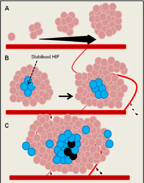
29 As solid tumours proliferate, they reach a critical mass whereby tumour size
becomes a threat to survival (Figure 7). At this juncture neoplasms become
dependent upon angiogenesis. Cells often proliferate rapidly and outstrip previous
metabolic requirements which were once satisfied by simple diffusion from local
vasculature (Folkman. 1990). Failed O2 supply initiates tumour hypoxia in territories
furthest from the blood vessel network, i.e. the tumour core. This drives the HIF
pathway, triggering angiogenesis, however tumour cells also die in anoxia as newly
constructed vasculature is inefficient and semi-permeable. It is thought that inhibition
of angiogenesis in solid tumours will boost anoxia or even HIF induced apoptosis.
However that is not necessarily the case. In fact some tumour cells accelerate the
rate of metastasis when angiogenesis is deterred (Ebos et al. 2009; Pàez-Ribes et
al. 2009).
Nonetheless, the role of anti-angiogenic therapy is currently under revision with the
aim of enhancing tumour specific immunotherapy (Huang et al. 2012) and
30
[image:30.595.72.532.71.654.2]31 Studies of apoptosis in hypoxia have revealed stabilisation of the tumour suppresser
protein, p53 (Graeber et al. 1994). This protein, once coined ‘guardian of the genome,’ targets multiple tumour suppressor genes in response to cellular stressors.
p53 target gene, WAF1, encodes p21, a protein regulating cell cycle arrest and
apoptosis (Waldman et al. 1995; Asada et al. 1999; Jin et al. 2000). Other genes
further regulate DNA repair, cellular senescence (Chen et al. 2005) and pluripotent
stem cell reprogramming (Marión et al. 2009). Non-mutant p53 is responsible for
maintaining a non-neoplastic phenotype, loss of p53 via TP53 mutation or protein
deactivation can have a devastating effect upon abolition of tumour cells (Petitjean et
al. 2007). Early investigation of p53 demonstrated autoregulation by murine double
minute 2 (MDM2), which is in turn downregulated by p53 (Wu et al. 1993). Hypoxic stability of p53 is dependent upon HIF1-α subunits (An et al. 1998), inferred by direct
and indirect interactions. MDM2 – p53 complexes bind and destroy HIF-1α (Ravi et al. 2000), while concurrently, local p53 and HIF-1α compete to interact with common
coactivator p300 (Blagosklonny et al. 1998). Reduced HIF transcription ensues
whilst p53 expression increases (Carmeliet et al. 1998), resulting in apoptosis.
1.2.4. Hypoxia and Neuroblastoma
In early foetal development HIF-2α is essential for formation of the sympathetic
nervous system (SNS) (Tian et al. 1998). At this point, the SNS is composed of
cellular subtypes, neurons and neuroendocrine cells, which have been found within
hypoxic regions of neuroblastoma. Here cells switch from one lineage to the other
(Gestblom et al. 1997), a differentiation which has been investigated in vitro and in vivo showing hypoxic neuroblastoma loose SNS markers such as ID2 (inhibitor of differentiation 2), HASH-1 (human achaete-scute homolog-1) and dHAND (heart- and neural crest derivatives-expressed protein). They also up-regulate markers characteristic of neural crest cells, c-kit and Notch-1, indicating cellular regression to an earlier
phenotype (Jögi et al 2002). Furthermore, previously expressed MYCN is no-longer
present 72 hours post-hypoxia. In consolidation of these findings, quantitative
polymerase chain reaction confirmed dedifferentiation with cellular regression to
stem-cell like states, enhanced survival, proliferation and induction of differentiation
32 (see Neuroblastoma, Pathology and Pathogenesis, section 1.1.5.), differentiated
tumours (e.g. ganglioneuroma) are perceived as lesser disease states and therefore
induced differentiation has been trialled clinically with reasonable success.
Dedifferentiation inhibitor, 13-cis-retinoic acid, improves overall survival in stage 3
neuroblastoma when administered after myeloablative therapy alongside bone
marrow transplant (Matthay et al. 2009).
Segments of neuroblastoma surrounded by vasculature, strongly express HIF-2α on immunohistochemical staining in contrast to HIF-1α which is rapidly accrued at the
onset of hypoxia but decreases significantly within 72 hours (Holmquist-Mengelbier
et al. 2006). In addition to altered subunit expression, different neuroblastoma cell
lines show widely varied transcription when cultured in hypoxia, even among cells with similar biology e.g. MYCN status (Fredlund et al. 2008). Moreover, both HIF-1α and HIF-2α stimulate VEGF transcription although each subunit maintains a degree
of subtlety in genetic expression.
For instance, HIF-2α does not exhibit the negative correlation with angiogenesis seen for HIF-1α (Noguera et al. 2009). Uniquely, normoxic neuroblastoma display inhibition of HIF-2α targets via microRNA-145 binding, which stifles tumour cell
dissemination and proliferation (Zhang et al. 2012).
The presence of HIF-2α is thought to infer poor prognosis, as opposed to HIF-1α,
which has been independently linked with improved survival and low tumour stages
(Holmquist-Mengelbier et al. 2006; Noguera et al. 2009). Controversially, other studies challenge the positive outlook for HIF-1α expression finding that the disease
presented at more advanced age with poor tumour biology and higher clinical staging when HIF-1α was expressed (Birner et al. 2000; Dungwa et al. 2012). This implies HIF-1 and 2α stabilisation is dissimilar in neuroblastoma however their
association with prognosis may be similar although this remains elusive.
Neuroblastoma is a life-threatening disease and chemotherapy is used extensively in
treating all disease stages. The link between hypoxia and resistance to
chemotherapy has been discussed, as has the predisposition of solid tumours,
neuroblastoma, to hypoxia. However, hypoxia induced chemoresistance is extremely
33 reduced levels of O2 (Hussein et al. 2006). This is in contrast to resistance in acute
hypoxia (< 23 hours) observed in colorectal cancer.
Finally, the issue of chemoresistance in neuroblastoma and other solid tumours
remains unresolved. In order to address the situation it is necessary to understand
the molecular mechanisms triggered by hypoxia in solid tumour formation. By doing
this, it should become possible to develop novel treatments inhibiting the proliferation
and migration of solid tumour cells. Furthermore this will also improve current clinical
34
1.3 The Chorioallantoic Membrane and Tumour Cell Invasion
Introducing the Chick Chorioallantoic Membrane
The molecular mechanisms underpinning cancer and cancer cell aggression remain
elusive. As such, all study models should be evaluated and modified in order to
develop a more robust investigative tool which can be used to uncover the key
aspects of tumour growth and migration. Promising results in vitro lay foundations for focused in vivo experiments providing an understanding of tumour cell behaviour in an environment similar to the human body. For years, murine models have been the
most commonly used species in vivo, however they present many disadvantages such as interaction of the host immune system and imaging limitations. Species from
zebra fish to chimpanzee inclusive have been examined and utilised in studies, for
example, to trial therapeutic interventions before beginning human testing.
Chick embryos are an extremely useful candidate for in vivo studies as chicken reproduction is associated with low husbandry, high throughput, straight forward
animal care in comparison with other in vivo models, non-invasive real-time and time-lapse imaging may be undertaken allowing short-term follow-up, interactions do
not occur between the embryonic immune system and tumour cells in early
gestational phases, and finally the CAM is intimately united with the embryo by a
common blood supply, therefore interventions may be examined both upon the CAM
and within the embryo avoiding insult to the foetus (Fuchs, Lindenbaum. 1988;
Cimpean, Ribatti. 2008).
Developing chicks exhibit characteristics ideal for the study of angiogenesis,
antiangiogenesis, tumourigenesis, tumour dissemination and metastasis (Chambers
et al. 1982; MacDonald et al. 1992; Auerbach et al. 2003). A key experimental
feature characteristic of the chick embryo model is the chorioallantoic membrane – CAM – a thin, transparent, easily accessible film which surrounds the embryo. Once
formed this surface is cohesive with the porous eggshell, acting as a placenta
presenting a large, heavily vascularised surface area for the provision of embryonic
35 Despite wide applicability and several advantages, the chick model has also some
disadvantages including, non-specific inflammatory reactions may occur, techniques
require practice, chick survival can be limited depending upon duration of
intervention, embryos are susceptible to infection, imaging potentially dries the
embryo resulting in chick death before the experiment is concluded, and lastly,
events witnessed in this model may not be of relevance to humans, although this is
the common risk associated with all in vivo study.
1.3.1. Anatomy and Development of the CAM
The chorioallantoic membrane, CAM, is a womb-like structure engulfing the chick
embryo and providing a boundary for gas exchange across the porous eggshell. The
structure constitutes three layers, ectoderm – mesoderm – endoderm, sandwiched
together to form a transparent collagenous matrix approximately 100 micrometers
thick. CAMs develop as an amalgamation of two embryonic structures, allantois and chorion, which fuse on embryonic day E5 – 6 surrounding the embryo completely 6 – 7 days later at E12 – 13 (Tufan and Satiroglu-Tufan. 2005).
In the chick embryo, chorionic ectoderm facilitates gas exchange, becoming
adhesive to porous eggshell at E10 whilst also forming a fine capillary plexus. Within
this plexus the vessel lumen can be minuscule allowing passage of erythrocytes in single file. The inner chorionic mesoderm fuses with the allantoic mesoderm at E5 –
6 forming a segment which will later become heavily vascularised by a host of
different calibre blood vessels (Fuchs and Lindenbaum. 1988). The allantois is
derived from embryonic hindgut which herniates into the extra-embryonic coelom,
before fusing with chorionic mesoderm to fulfill the CAM mesoderm and endoderm, a
border to the allantoic cavity (Romanoff. 1960). Figure 8 depicts a cross-section of
36
Figure 8. Cross-section of chick CAM. A is a diagram depicting a cross-section of the CAM and B is a microscopic photograph through the CAM, following immunohistochemical staining. Beginning superiorly, the initial layer – Ectoderm – constitutes 2 rows of epithelia and a fine capillary network. This surface sticks to the porous egg shell (E12-13). This gas exchange surface promotes rapid vascularisation and is ideal to support implanted cells. The middle layer – Mesoderm – is heavily vascularised and consists of collagen. Blood vessels run longitudinally and vertically as demonstrated by sectioned vessels containing erythrocytes in A. Finally, embryonic – Endoderm – borders the allantoic cavity. Brown cells (B) are neuroblastoma stained with a human specific antibody Ki-67, which indicates cellular proliferation.
CAM mesoderm contains extensive vasculature supporting arterioles and venules
which communicate within the ectodermal capillary plexus. Links with the embryo
itself are supplied by an umbilical artery which dichotomises into a pair of
chorioallantoic arteries and a single chorioallantoic vein (Fuchs and Lindenbaum.
1988). Early vessel walls span one single, undifferentiated endothelium in width
however by E8 they become embedded in mesenchymal cells sheathed in a basal
lamina (Ausprunk et al. 1974). Later cellular differentiation to smooth-muscle cells
has been observed as has expression of VEGFR-2 (Wilting et al. 1996) and
37 Chick lymphatic formation occurs after initial angiogenesis is well underway. Thus,
embryonic immunity lags behind that observed in early gestation murine models.
Evidence of differentiated immune cells indicates heterophil and monocyte formation detected around E10 – 15. The former, a major source of matrix-metalloproteinase 9,
act as early neutrophils (Zijlstra et al. 2006), whilst the latter express the angiogenic
promoter matrix-metalloproteinase 13 (Zijlstra et al. 2004). Late establishment of
lymph and mature immune cells is thought to explain why the chick embryo is
immunodeficient, making it an excellent model to study tumour cell proliferation and
metastasis.
1.3.2. Chick embryo and CAM model in Cancer studies
Initially shown to support murine sarcoma xenografts, the chick embryo has been
exploited for more than 100 years (Rous and Murphy, 1911). Further experiments
investigated compatibility with human tumour cells confirming proliferation of: adenocarcinoma, bronchial carcinoma, embryonal rhabdomyosarcoma, Hodgkin’s
lymphoma, melanoma and squamous-cell carcinoma (Karnofsky et al. 1952;
Korngold and Lipari. 1955; Harris. 1958). Tumour aggression and metastasis has
been evaluated by embryonic dissection and the effects of chemotherapy have also
been assessed.
Overall the chick embryo model is utilised according to three methods, each focused
on highlighting specific steps in tumour formation and the metastatic cascade. First,
the spontaneous metastasis model is used to demonstrate different features
concerning tumour growth and intravasation by implanting tumour cells upon the
CAM. Many different tumour cells are supported and proliferate to establish primary
tumours, although some cell lines have been identified as unsuitable for this model
(Deryugina, Quigley. 2008). Following survival and proliferation of a successful cell
line, individual cells segregate themselves from the tumour body and commit to
invasion of the CAM and local vasculature, a process known as intravasation. An
additional benefit of using the spontaneous model is that one may witness the overall metastatic process providing cells proliferate – intravasate – migrate – extravasate –
metastasise within experimental time constraints. In this sense the chick model
38 The experimental metastasis model involves introduction of tumour cells into the
embryonic environment, often via injection into the extra-embryonic vasculature
which is embedded within the CAM. This is undertaken in order to understand exactly how cells exit blood vessels, ‘extravasation,’ to later establish metastatic
secondaries. Tumour metastasis theory suggests disseminated cells traverse the
vascular tree and extravasate into tissues or organs, where they form secondary
tumours. It was once thought tumour cells with increased metastatic capabilities,
extravasate more effectively than cells with lower metastatic potential however
imaging of fluorescently-labelled tumour cells contradicted this theory (Koop et al.
1996). Indeed more recent mouse and human studies suggest normal and
pre-neoplastic cells may intravasate, migrate and extravasate to exist at locations other
than their origin (Podsypanina et al. 2008; Pantel et al. 2012).
Finally the on-plant model provides an opportunity to observe and extrapolate the
mechanisms behind angiogenesis stimulated by tumour cells. One group carried out
this model by lacerating the CAM before introducing a plastic ring containing 3 – 5
million human glioma cells (Hagedorn et al. 2005). Similarly, osteosarcoma cells
have been implanted upon the CAM via Thermanox™ plastic discs (Balke et al.
2010) whilst matrigel grafts have also been utilised to insert ovarian cancer cells
(Lokman et al. 2012). These methods are important as neovascularation is crucial to
both primary and metastatic solid tumour survival and dissemination (see Hypoxia,
Physiology and Pathophysiology of Hypoxia, section 1.2.2). Similar to the study of
tumour proliferation and metastasis, independent study of angiogenesis has made
great use of the avian model (Rabatti et al. 2000 and 2001; Tufan, Satiroglu-Tufan.
2005).
Consolidating the chick assay as a method used to study metastasis, human
epidermoid carcinoma cells were implanted upon the CAM with resultant formation of
metastatic foci in the heart and lung (Ossowski and Reich. 1980). Foci were
confirmed as implant disseminations by identifying the highly specific human
plasminogen activator. Another study verified the experimental metastasis model by
injecting melanoma cells into CAM vasculature demonstrating extravasation and the
foundation of secondary micrometastasis (MacDonald et al. 1992). Lastly, a range of