Investigating the renogenic potential of mesenchymal stem cells
Thesis submitted in accordance with the requirements of the University of Liverpool for the degree of Doctor in Philosophy
by
Maria Kuźma-Kuźniarska
Contents
Contents... ii
Acknowledgments ...x
Glossary of common abbreviations...xi
Abstract ... xii
Statement of authorship...xiv
Chapter 1: Introduction ...1
1.1. Overview of the anatomy and function of the adult kidney ...1
1.2. Kidney development ...6
1.2.1. Pronephros...7
1.2.2. Mesonephros ...8
1.2.3. Metanephros ...9
1.2.3.1. Collecting duct system development...9
1.2.3.2. Nephron development ...10
1.2.3.3. Development of the renal stroma ...12
1.2.3.5. Metanephric kidney as a model for in vitro nephrogenesis...22
1.3. Mesenchymal stem cells and their role in kidney repair and regeneration ...23
1.3.1. Characteristics of mesenchymal stem cells ...23
1.3.2. Mesenchymal stem cells in kidney disease ...24
1.3.3. Mesenchymal stem cells kidney specific kidney differentiation in vitro...29
1.3.4. Mesenchymal stem cells in nephrogenesis...31
1.3.5. Controversies regarding the renoprotective and renogenic action of mesenchymal stem cells...34
1.4. Other stem cells/progenitors in kidney repair and regeneration...37
1.4.1. Kidney progenitors ...38
1.4.2. Amniotic fluid stem cells ...42
1.4.3. Embryonic stem cells ...43
Aim of the study...46
Chapter 2: Material and methods ...49
2.1. Primary cells and stem cell lines ...49
2.1.2. Mouse embryonic fibroblasts ...49
2.1.3. Mouse embryonic stem cells ...49
2.1.4. Mouse neonatal kidney cells ...50
2.2. Cell culture ...50
2.2.1. Cell thawing protocol ...50
2.2.2. Cell freezing protocol...50
2.2.3. Cell passaging ...51
2.2.4. Routine mesenchymal stem cell culture...52
2.2.5. Preparation of mouse neonatal kidney cells ...52
2.2.6. Routine mouse neonatal kidney cell culture ...53
2.2.7. Gelatinization of culture dishes...53
2.2.8. Preparation of mouse embryonic fibroblasts...53
2.2.9. Routine mouse embryonic fibroblast culture ...54
2.2.10. Routine mouse embryonic stem cell culture ...54
2.3. Conditioned medium treatment ...55
2.3.2. Stimulation with conditioned medium ...56
2.4. In vitro multilineage differentiation protocols ...57
2.4.1. Adipogenesis assay ...57
2.4.2. Osteogenic assay ...58
2.4.3. Chondrogenic assay...59
2.5. Cell labelling ...60
2.5.1. Quantum dot labelling...60
2.5.2. CFDA SE labelling...61
2.5.3. Lentiviral transduction ...61
2.5.4. Immunostaining of human mesenchymal stem cells...62
2.6. Metanephric kidney culture...63
2.6.1. Dissection of mouse kidney rudiments ...63
2.6.2. Culture of mouse kidney rudiments in vitro...64
2.7. Chimeric kidney assay ...65
2.8. Immunostaining of intact kidney rudiments and kidney chimeras ...66
2.10. Molecular biology ...69
2.10.1. RNA extraction ...69
2.10.2. DNase treatment ...71
2.10.3. cDNA synthesis...71
2.10.4. Primers ...72
2.10.5. Polymerase chain reaction...73
2.10.6. Electrophoresis ...74
2.10.7. Real-time polymerase chain reaction ...75
2.10.8. Efficiency of the real-time polymerase chain reaction...76
2.10.9. Quantification of real-time polymerase chain reaction ...77
2.11. Statistical analysis ...78
2.12. Culture media and buffers ...78
2.12.1. Culture media ...78
2.12.2. Buffers and solutions...81
Chapter 3: Differentiation potential of mesenchymal stem cells and their labelling methods ...85
3.2. Results ...90
3.2.1. The multilineage differentiation potential of the mouse D1 MSC line ...90
The multilineage differentiation potential of human MSCs...94
3.2.2. Identification of suitable labelling method for tracking of MSCs...96
3.3. Discussion ...104
3.3.1. The multilineage differentiation potential of MSCs...104
3.3.2. The labelling methods of MSCs...105
Chapter 4: Potential of MSCs to contribute to metanephric development using the novel chimeric kidney system ...111
4.1. Introduction ...111
4.2. Results ...115
4.2.1. Chimeric kidney culture ...115
4.2.2. Renogenic potential of D1 cells ...120
4.2.3. Renogenic potential of human MSCs...125
4.2.4. Renogenic potential of other progenitors ...128
4.3.1. Expression profile of MSCs ...132
4.3.2. Renogenic potential of MSCs ...136
4.3.3. Renogenic potential of other stem cells ...140
Chapter 5: Potential of MSCs to contribute to metanephric development after stimulation with conditioned medium from neonatal kidney cells ...144
5.1. Introduction ...144
5.2. Results ...146
5.2.1. The multilineage differentiation potential of D1 cells stimulated with NKC CM...146
5.2.2. Renogenic potential of D1 cells stimulated with NKC CM...150
5.2.3. Renogenic potential of human MSCs stimulation with NKC CM...166
5.3. Discussion ...168
5.3.1. Characteristics of D1 cells stimulated with NKC CM ...168
5.3.2. Expression profile of D1 cells stimulated with NKC CM ...169
5.3.3. Renogenic potential of MSCs stimulated with NKC CM ...171
5.3.4. Analysis of integration potential of D1 cells stimulated with NKC CM ...173
cells...176
5.3.6. Mechanism of increased renogenic potential of D1 cells following preconditioning178 Chapter 6: Effects of MSCs on metanephric kidney development ...181
6.1. Introduction ...181
6.2. Results ...183
6.2.1. Effect of integration of D1 cells and human MSCs on metanephric development following chimeric kidney culture ...184
6.2.2. Effect of conditioned medium derived from D1 cells on the development of intact kidney rudiments ...192
6.3. Discussion ...195
6.3.1. Effects exerted by MSCs on kidney development ...196
6.3.2. Putative mechanism of detrimental action of D1 cell on kidney development...198
Chapter 7: Final discussion ...203
Acknowledgments
I would like to thank my supervisors Patricia Murray and David Edgar for their support during this project as well as all my collaborators in the KIDSTEM network. Especially, I would like to thank Christos Xinaris, Veronika Ganeva, Sandra Rak-Raszewska and Monika Wozińska for all the great scientific and not only scientific discussions.
I would like to thank Jarek Lipski for his support during the whole project and his help in the final stages of thesis preparation (for non-computer people like me).
Glossary of common abbreviations
AFSC amniotic fluid stem cellsBSA bovine serum albumin BMP bone morphogenetic protein cDNA complementary DNA
CFDA SE carboxyfluorescein diacetate succinimidyl ester DAPI 4',6-diamidino-2-phenylindole
DMEM Dulbecco's Modified Eagle Medium DNA deoxyribonucleic acid
E embryonic day
EDTA ethylenediaminetetraacetic acid ESC embryonic stem cells
FCS fetal calf serum
GAPDH glyceraldehyde 3-phosphate dehydrogenase GDNF glial cell-derived neurotrophic factor
GFP green fluorescent protein h hours
HBSS Hanks’s buffered saline solution HSC hematopoietic stem cell
IM intermediate mesoderm NKC neonatal kidney cells
MEF mouse embryonic fibroblast min minutes
MM metanephric mesenchyme MSC mesenchymal stem cells PBS phosphate buffered saline PCR polymerase chain reaction PFA paraformaldehyde
RNA ribonucleic acid sec seconds
QD quantum dot TAE tris-acetate EDTA
Abstract
Statement of authorship
Chapter 1: Introduction
1.1. Overview of the anatomy and function of the adult kidney
Kidneys remove waste products from blood. They also produce hormones, like erythropoietin and maintain homeostasis by regulating fluid balance, acid-base balance and blood pressure. The nephron is the structural and functional unit of the kidney. It consists of renal tubules, including the proximal tubule, limbs of the loop of Henle and the distal tubule, as well as the renal corpuscle formed by the Bowman’s capsule and the glomerulus. The role of the glomerulus is to produce an ultrafiltrate, in the process called glomerular filtration, which becomes urine once it is concentrated by the renal tubules, while the collecting duct system of the kidney is connecting the nephrons to the ureters. The kidneys, together with ureters, bladder and urethra, form the urinary system (Figure 1.1a) (Vize Peter 2003).
Accordingly, the urine flows from the medulla into the minor calyces, which open into the major calyces and subsequently into the renal pelvis. The renal pelvis is connected to the ureters and the urine flows into the bladder (Figure 1.1a) (Vize Peter 2003).
Figure 1.1 Schematic representation of the urinary system and kidney parenchyma. (a) The urinary system, including the minor and major calyx and renal pelvis. (b) The components of renal parenchyma, the cortex and medulla, with two types of nephron found in the kidneys.
renal pelvis
kidney
ureter
bladder
urethra major calyx
minor calyx a
cortex
outer medulla
inner medulla renal corpuscle
collecting duct connecting segment
distal tubule
loop of Henle proximal tubule
juxtamedullary nephron
loop of Henle
[image:17.595.86.486.109.612.2]Figure 1.2 A schematic representation of the renal corpuscle (a) and the filtration barrier found in the glomerulus (b).
vascular pole
urinary pole glomerular capillaries
proximal convoluted tubule
Bowman’s capsule Bowman's space a
b
lumen of the capillaries
podocyte
podocyte foot processes glomerular basement
membrane filtration slit
fenestration in the endothelial cells
[image:18.595.79.447.136.571.2]From the Bowman’s space, the ultrafiltrate enters the proximal tubule at the urinary pole (Figure 1.2a). The proximal tubule consists of a convoluted and a straight part. In the proximal tubules, reabsorption of water as well as amino acids, glucose, creatinine, bicarbonate, phosphate, potassium, calcium, sodium and chloride occurs. The epithelial cells of proximal tubules have a brush border of microvilli which enhance the reabsorption. They also express many proteins related to transport, such as aquaporin-1, a water channel protein responsible for water transport across the tubule. The proximal tubule is also the site of secretion of uric acid and the site of ammonia production (Vize Peter 2003).
The ultrafiltrate from the proximal tubule passes next through the loops of Henle which consist of the thin descending limb, permeable to water and ions, the thin ascending limb, permeable to salts but less permeable to water, and the thick ascending limb, impermeable to water but able to actively transport salts. The thick ascending limb is the largest site of sodium and chloride reabsorption after the proximal tubule. The loop of Henle is also the regulatory site of magnesium excretion. In addition there are two main populations of nephrons in the kidney depending on the length of the loop of Henle. Cortical nephrons have shorter loops in comparison to juxtamedullary nephrons (Vize Peter 2003).
characteristics of both distal tubule cells as well the collecting duct cells (Vize Peter 2003).
The collecting duct is divided into the following segments: cortical, outer medullar and inner medullary and papillary. These segments increase in size towards the papilla where the medulla empties urine into the minor calyx. The principal and intercalated cells are two cell types that are distinguished in the collecting duct. The principal cells, expressing aquaporin-2, are specialized in water transport, while the intercalated cells play a role in maintaining the acid-base balance by secreting bicarbonate into the collecting duct. In the medullary segment of the collecting duct, the reabsorption of sodium, chloride and water occurs (Vize Peter 2003).
1.2. Kidney development
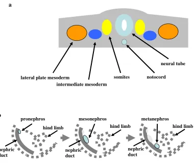
Figure 1.3 Schematic representation of kidney development in amniotes. (a) Localization of intermediate mesoderm within a mouse embryo before E8. (b) Sequential appearance of the pronephros, mesonephros and the metanephros in a mouse embryo.
1.2.1. Pronephros
The pronephric kidney (pronephros) is the most primitive kidney and contains a single nephron which filters blood. However, as there is no Bowman’s space, the filtrate from the glomus (glomerulus) is collected from an open cavity by ciliated tubules called nephrostomes which are connected to pronephric tubules. Pronephric tubules contain a proximal segment which resorbes solute, and a distal segment which resorbes water. Subsequently the urine passes through the nephric duct, also called the pronephric duct, to the cloaca. Pronephroi are present during the urinary tract development in mammals but they are rudimentary and undergo early degeneration
hind limb
nephric duct
nephric duct hind limb
pronephros
hind limb
mesonephros metanephros b
nephric duct
neural tube
lateral plate mesoderm
intermediate mesoderm
in comparison with well-developed pronephroi found in fish and amphibians. In mice, the pronephros appears around E8. Accordingly, the pronephros functions as an excretory organ in the larval stages of lower vertebrates. In adult fish the pronephros becomes a lymphoid organ, similar as in the amphibians, where it becomes the site of hematopoiesis (Saxen 1987; Vize Peter 2003).
1.2.2. Mesonephros
1.2.3. Metanephros
The metanephric kidney (metanephros) becomes the permanent kidney in mammals. It is the most complex kidney, and unlike the mesonephros, has a branched structure. The metanephros starts to develop in mice at E11 and its development continues postnatally for approximately 1 week. The development of the adult kidney starts with the invasion of the ureteric bud (UB) into the surrounding metanephric mesenchyme (MM). UB is an epithelial outgrowth of the posterior end of the Wolffian duct and is a precursor of the collecting duct system, while MM is involved in nephron formation. Aspects of metanephric development, which include the formation of the collecting duct, nephron and stroma in the metanephros, are discussed in the following sections.
1.2.3.1.Collecting duct system development
Figure 1.4 The branching morphogenesis of the UB.
1.2.3.2.Nephron development
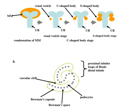
The interaction between UB and MM stimulates the uninduced MM to condense in the proximity of the UB tips and subsequently form nephrons. In the mouse, the MM starts to condense in the proximity of the UB tips around E11. Accordingly, the condensing MM undergoes epithelial conversion, generating renal vesicles. This transition is characterised by important changes in the expression of extracellular matrix proteins. The uninduced MM expresses collagen type I and III, as well as fibronectin, which are not found in condensing MM. Conversely, the condensation of MM is accompanied by expression of laminin, a component of the epithelial basement membrane (Horster et al. 1999; Dressler 2006). Furthermore it has been described that α1 chain of laminin-1 is highly expressed during early epithelial development and it is also the major chain found in adult kidneys (Ekblom et al. 2003). Subsequently, renal vesicles are converted into comma (C)-shaped bodies. At this stage the developing nephrons start to fuse with the UBs. Next, the C-shaped bodies develop into the S-C-shaped bodies (Horster et al. 1999; Dressler 2006). The
nephric duct
ureteric bud outgrowth nephric duct
uninduced mesenchyme
aggregating mesenchyme
ureteric bud initial branching of ureteric bud
invasion of ureteric bud condensation of mesenchyme dichotomous branching of the bud nephric duct nephric duct
Figure 1.5 Schematic representation of early nephron development. (a) Formation of the renal vesicle, C-shaped and S-C-shaped body (in orange). The ureteric bud is shown in blue. (b) A developing the S-C-shaped body. As demonstrated, the proximal end of the S-shaped body will develop into renal corpuscle and the distal into different renal tubules.
1.2.3.3.Development of the renal stroma
An important compartment in the developing metanephros, which is implicated in regulation of UB branching and mesenchymal-to-epithelial transition of the MM, is the renal stroma. The localization of the stromal compartment in a mouse metanephros is depicted in Figure 1.6. After the UB has invaded the MM and induced MM to condense around its tips, formation of two mesenchymal compartments can be determined in the metanephros. Accordingly, the condensing MM closely surrounding the bud tip will form the nephrons, and the peripheral cells will become
S-shaped body stage
condensation of MM
renal vesicle stage
C-shaped body stage UB
MM
UB UB UB
renal vesicle C-shaped body S-shaped body a
Bowman’s capsule
Bowman’s space
podocytes vascular cleft
the stromal cells (Figure 1.6). Later, the stromal cells are found surrounding the UB branches and forming nephrons (Figure 1.6). These cells are called the primary renal interstitium. As renal tubules develop, a secondary interstitium is formed; the cortical stroma and the medullary stroma (Cullen-McEwen et al. 2005). Earlier it has been proposed that renal stromal cells could be neural crest derivates, as the stromal cells were found to express neurofilaments (Sainio et al. 1994). However, recent results in chicken, presented by Guillaume et al., showed that the renal stroma mainly originates from paraxial mesoderm (Guillaume et al. 2009).
Figure 1.6 Localisation of stroma in developing mouse metanephros at the onset of metanephric development at E11 and later at E12.5.
1.2.3.4.Genes important during metanephric development
A number of genes are implicated in the development of the metanephric kidney (Dressler 2009). In order to ensure that the metanephros develops in the correct location at the posterior end of IM, metanephric development requires a tight regulation of key transcription factors along the mediolateral and anteroposterior axis (Dressler 2009). For example Eya1 (Sajithlal et al. 2005) and Hox11 paralogs (Mugford et al. 2008) play a crucial role in determining the region of IM that will become MM. Hox11 proteins, together with Eya1 and Pax2, form a transcriptional complex which activates expression of genes such as glial cell line-derived neurotrophic factor (Gdnf) in
UB MM
stroma UB
stroma
forming nephrons
(Gong et al. 2007). Other genes that positively regulate expression of Gdnf include Six2 and Sall1 (Saavedra et al. 2008). Metanephric development is initiated with the interaction between the UB and MM. Gdnf plays a crucial role in this interaction as the binding of Gdnf to the tyrosine kinase receptor, Ret, is responsible for UB outgrowth and branching (Vega et al. 1996; Sainio et al. 1997). Furthermore, some genes have been demonstrated to take part in later metanephric development. For instance, it has been shown that Notch2 signalling plays role in nephron patterning. Accordingly, kidneys of Notch2-deficient kidneys do not develop proximal tubules, although they form distal tubules (Cheng et al. 2007). Finally, it is important to note that some genes involved in metanephric development are not exclusively found in the metanephros. Genes such as Pax2 were also demonstrated to be expressed during development of the pronephros and mesonephros (Dressler et al. 1990). An overview of several important signalling pathways and genes involved in metanephric development is presented below.
Gdnf signalling
both UB and MM (Sainio et al. 1997). Gdnf-null mice do not develop a UB. The uninduced MM undergoes apoptosis, consequently leading to complete renal agenesis at birth (Sanchez et al. 1996). Similarly, in the Ret-deficient mice, the kidneys were also absent. However, occasionally some rudimentary dysplastic kidneys with large areas of undifferentiated mesenchyme were observed in the mutant animals (Schuchardt et al. 1994). The phenotype of GFR-α null mouse was consistent with Ret knockout, demonstrating lack of kidneys or renal dysgenesis (Enomoto et al. 1998).
Sall1
Lim1
Another essential gene in renal development is the LIM-class homeobox gene, Lim 1. Lim1 displays a broad expression profile during nephrogenesis. Its expression starts in the IM and is subsequently restricted to the nephric duct and UB. Lim1 expression is also present at different stages of nephron formation, including pretubular aggregates, comma-shaped and S-shaped bodies. Subsequently, postnatal Lim1 expression is found in collecting ducts and cortical tubules but remains downregulated in mature glomeruli (Karavanov et al. 1998; Kobayashi et al. 2005). Lim-/- mice lack head structures and die at E10. However, some stillborn Lim1-/- pups were
shown to not have developed kidneys (Shawlot and Behringer 1995). In addition, in E9.5 Lim1-null mice, the expression of Pax2, an important transcription factor during nephrogenesis, is only found in the posterior region of the IM in comparison with wild type mice where Pax2 expression was demonstrated along the entire length of the IM (Tsang et al. 2000).
Wnt signalling
develop small kidneys which are composed of undifferentiated MM and branches of collecting duct. Initial condensation of MM occurs in the Wnt4-deficient mice. However, no C-shaped and S-shaped bodies are detected in the E15 kidneys, while branching morphogenesis is preserved (Stark et al. 1994). The forced expression of Wnt4 in the NIH3T3 mouse embryonic fibroblast line was sufficient to elicit tubulogenesis in co-cultured MM. Accordingly, it has been shown that Wnt4 expression induces MM to condense, form epithelial structures and finally display
glomeruli, similar as the induction with the spinal cord (Kispert et al. 1998). Another member of the Wnt family is Wnt9b, inactivation of which leads to renal agenesis in mice. Wnt9b is expressed in collecting ducts. Further studies on Wnt9b mutant mice demonstrated that Wnt9b is important for establishing planar cell polarity in the renal tubules (Karner et al. 2009).
Pax2
Pax2, a pair box gene, is expressed in the UB, condensing mesenchyme and developing nephrons
Wt1
The Wilms' tumor gene, Wt1, encodes a transcription factor that plays a number of crucial roles throughout nephrogenesis, and ablation of Wt1 leads to renal agenesis (Kreidberg et al. 1993). Expression of Wt1 has been associated with both condensing mesenchyme and developing nephrons (Armstrong et al. 1993; Mundlos et al. 1993). Some Wt1 expression can be observed prior to the onset of nephrogenesis, both in the IM and uninduced MM (Armstrong et al. 1993). During kidney development, Wt1 expression becomes restricted to the precursors of podocytes in the S-shaped bodies in both the metanephros and mesonephros. Wt1 expression persists in the podocytes throughout adulthood (Mundlos et al. 1993). In Wt1-deficient mice, fewer mesonephric tubules are detected. Further, at E11.5, the UB is absent in the mutant mice and the MM is undergoing apoptosis. Ultimately, at E12, the MM is completely degenerated (Kreidberg et al. 1993). Pax2, Six2 and Gdnf are however expressed in the MM of mutant embryos, suggesting that Wt1-deficient MM acquired already features of the nephrogenic lineage and possibly for this initial process, Wt1 is not required. Nevertheless, Wt1-deficient MM could not be induced by UB to undergo tubulogenesis in vitro (Donovan et al. 1999).
Six2
Six2 expression is found in the MM before UB invasion as well as the induced MM surrounding
types within the metanephros (Kobayashi et al. 2008).
Osr1
Osr1 is odd-skipped related 1 gene encoding a transcriptional regulator. The Osr1-expressing
precursor population in the IM was shown to gives rise to most cells found within the developing metanephros, including the collecting duct epithelium, nephrons and interstitial mesenchyme, mesangial and smooth muscle cells. Still, it was demonstrated that Osr1-expressing precursors for nephron and interstitial mesenchyme separate before the start of metanephric development (Mugford et al. 2008). In E9.5 mice, Osr1 expression is present in intermediate and lateral plate mesoderm. Its expression is also found in undifferentiated mesonephric mesenchyme and tubules. At the onset of metanephric development, Osr1 is expressed in the MM but is absent in the UB. Later it is expressed in the condensing mesenchyme that surrounds the branching UB, but is down-regulated in pre-tubular aggregates, C-shaped and S-shaped bodies (James et al. 2006). Osr1-null mice lack kidneys as no MM and UB development occurs in the embryos (Wang et al.
2005). No expression of Six2, Eya1, Gdnf, Pax2 and Sall1 was found in the metanephric region in the mutant mice (James et al. 2006).
Eya1
Eya1 is a homologue of the Drosophila eyes absent gene. Eya1 expression is first observed at
mouse embryos, Ret expression is observed in the Wolffian duct but no UB outgrowth occurs (Sajithlal et al. 2005). Subsequently, in the absence of the UB, the MM undergoes apoptosis. Complete degeneration of the MM is observed by E12.5 (Xu et al. 1999).
Hox11 paralogs
Hox genes encode transcription factors that play an essential role in patterning of the body axes
during embryonic development. As Hox11 paralogous genes are redundant, the knockouts of Hox11 result in incompletely penetrant phenotypes. Accordingly, Hoxa11/Hoxd11-deficient mice
display hypoplastic kidneys, whereas Hoxa11/Hoxc11/Hoxd11-deficient mice completely lack kidneys. Accordingly, no UB formation is observed in triple mutants. This is accompanied by the lack of Six2 and Gdnf expression in the MM (Wellik et al. 2002). Hox11 paralogous proteins were demonstrated to form a complex with Pax2 and Eya1, which activates the expression of Six2 and Gdnf in the MM (Gong et al. 2007).
Notch2 signalling
Notch genes encode single-transmembrane receptors. It has been demonstrated that disruption of
Notch2 receptor expression in the metanephric mesenchyme disrupts nephrogenesis. Accordingly, Notch2-deficient mice at birth displayed smaller kidneys with no glomeruli or proximal tubules. The mutant mice demonstrated normal condensation of MM and subsequent transition to epithelium, however no segmentation of the nephron into proximal and distal tubules occurred in the kidneys (Cheng et al. 2007).
Bmp4
E12.5 Bmp4is expressed in the stromal mesenchymal cells. In addition, at E14.5, expression is seen in the S-shaped bodies. The Bmp receptor gene, Alk3, is ubiquitously expressed during kidney development, while Alk6 is expressed in the Wolffian duct and in UBs. Bmp4+/- mice display renal abnormalities similar to human congenital anomalies of the kidney and urinary tract (CAKUT), categorized as dysplastic kidneys, hydronephrosis or duplex kidney with bifid ureters (Miyazaki et al. 2000). In vitro it was demonstrated that Bmp4 promotes growth and elongation of UBs, but also inhibits condensation of MM and promotes expansion of the peripheral stromal compartment (Miyazaki et al. 2000; Raatikainen-Ahokas et al. 2000; Miyazaki et al. 2003).
Bf2
Brain factor 2, Bf2, (Foxd1) encodes a transcription factor expressed in stromal cells that in
mouse kidneys at E11.5 form a ring of stromal mesenchyme around the Pax2 positive nephrogenic mesenchyme. Later, Bf2 expression is found in cells surrounding the condensing MM and at the periphery of the kidney rudiment. Mice carrying a Bf2-null mutation show abnormalities in development of both the collecting duct system and nephrons. The number of nephrons is reduced, as large amounts of condensing mesenchyme are still present in the kidneys at birth. Further, the number of UB branches is diminished. The expression of Ret in mutant kidneys is not restricted to UB tips in the cortex, as it is in wild type E14.5 kidneys, but is found along the branches in both cortical and medullary regions (Hatini et al. 1996).
Rarβ2
Retinoid acid receptor β 2 (Rarβ2) expression is found in mouse metanephroi as early as E11.
Rarβ2 is associated with stromal cells surrounding the UB and MM, where its expression
expressed in stromal cells found in the developing cortex and medulla. It is also expressed in the subcapsular region, again co-localizing with Bf2 (Mendelsohn et al. 1999). In mice lacking Rarβ2 along with other retinoid acid receptors, Rarα1 and Rarα2, the kidneys at birth display diminished numbers of nephrons and UB branches (Mendelsohn et al. 1994). It has been demonstrated that in these mutant kidneys, starting from E12, Ret expression in the UB tips is downregulated, leading to reduced UB branching. Furthermore, the stromal cells are abnormally distributed, forming a thick peripheral stromal layer in the E14 mutant kidneys (Mendelsohn et al. 1999).
1.2.3.5.Metanephric kidney as a model for in vitro nephrogenesis
1.3. Mesenchymal stem cells and their role in kidney repair and regeneration
1.3.1. Characteristics of mesenchymal stem cells
al. 2006; Kern et al. 2006). Recently, it was shown that MSCs could be obtained from different foetal tissues. Accordingly, it was possible to establish human MSCs from foetal bone marrow, lung or liver (Campagnoli et al. 2001; in 't Anker et al. 2003).
It is important to note that for both mouse and human bone marrow-derived MSCs, subpopulations with higher plasticity have been isolated (Kucia et al. 2006; Anjos-Afonso and Bonnet 2007). Mouse bone marrow-derived MSCs capable of differentiation into all three germ-layer lineages are named very small embryonic-like (VSEL) stem cells. These cells resemble embryonic stem cells in their morphology and express embryonic stem cell markers, such as Oct4, SSEA-1 and Nanog (Kucia et al. 2006). A similar subpopulation of cells has been described by Anjos-Afonso and Bonnet, who called these cells the most primitive mesenchymal progenitors in the adult murine bone marrow compartment (Anjos-Afonso and Bonnet 2007). In humans, bone marrow-derived MSCs that maintain the ability to proliferate rapidly and differentiate into a wide range of lineages, among them neurons and pancreatic islet cells, have been named marrow-isolated adult multilineage inducible cells (MIAMI) (D'Ippolito et al. 2004).
1.3.2. Mesenchymal stem cells in kidney disease
labelled rat bone marrow was transplanted into irradiated recipient rats, and following, the induction of glomerulonephritis, the labelled bone marrow-derived cells were found in the glomeruli of injured rats and were described to provide structural support for glomerular capillaries (Ito et al. 2001). Others showed that wild type bone marrow cells injected into collagen type IV α3 knockout mice with progressive glomerulonephritis (a model of Alport syndrome), resulted in improved renal histology and function (Prodromidi et al. 2006). From this study, as well as from other studies, it was not entirely clear which bone marrow-derived population, i.e., the MSCs or the hematopoietic stem cells (HSCs), was responsible for the positive outcome. Subsequently, many authors concentrated on evaluating the contribution of bone marrow-derived MSCs to renal regeneration and it was demonstrated that MSCs can indeed protect mice from tubular damage and renal function deterioration in various experimental models of acute renal injury (Herrera et al. 2004; Morigi et al. 2004; Togel et al. 2005; Bi et al. 2007; Semedo et al. 2007; Togel et al. 2007; Qian et al. 2008; Li et al. 2010). Some of the data suggested also that MSCs are able, at least to some extent, to engraft into damaged kidneys and differentiate towards tubular cells (Herrera et al. 2004; Morigi et al. 2004; Qian et al. 2008; Li et al. 2010).
Lens culinaris lectin, which binds to the brush border of the tubular epithelium. Even 29 days
after the injury, the cells were still detected within the tubular epithelium. In the same study it was also demonstrated that administration of HSCs does not protect mice from renal function and tissue damage in the cisplatin-induced injury model. Nevertheless, occasional engraftment of HSCs into injured tubules was observed (Morigi et al. 2004). More recently, Morigi and the co-workers demonstrated the effectiveness of human cord blood-derived MSCs in ameliorating cisplatin-induced kidney injury. MSCs were injected into severe combined immunodeficiency (SCID) mice following cisplatin treatment. At the peak of injury the tubular damage was reduced in the kidneys of animals receiving MSCs. Importantly, the injection of the cells was able to improve the survival of cisplatin-treated mice. Consequently, MSCs were demonstrated to inhibit cisplatin-induced damage by reducing oxidative damage and apoptosis in the kidneys of cisplatin-treated animals. The injured kidneys receiving MSCs also demonstrated an increase in tubular cell proliferation, as well as having a high number of tubules positive for the serine/threonine protein kinase Akt, which mediates anti-apoptotic effects. Additionally, the injured kidneys treated with MSCs showed up-regulation in expression of hepatocyte growth factor, a factor responsible for anti-apoptotic effects during kidney injury. Nevertheless,
regarding the engraftment and differentiation potential of human MSCs in the injured renal tissue, the cells were mainly found in the peritubular areas and only rarely engrafted in the tubules or glomeruli (Morigi et al. 2010).
Accordingly, Herrera et al. investigated the effect of administration of mouse bone morrow-derived MSCs in acute injury induced by intramuscular injection of glycerol in the mice. Similarly as in the cisplatin model, MSCs were demonstrated to increase tubular cell proliferation. Furthermore, MSCs were detected in the tubular epithelium of glycerol injured kidneys expressing cytokeratin, suggesting that some of the MSCs were possibly differentiating into tubular epithelial cells (Herrera et al. 2004). Others demonstrated that human foetal bone marrow-derived MSCs administrated into rats with glycerol-induced acute renal failure enhanced tubular cell proliferation, which was accompanied by engraftment of MSCs into injured tubules. Consequently, the integrated cells acquired expression of aquaporin-1 and parathyroid hormone receptor 1 as well as stained positively for cytokeratin, suggesting epithelial differentiation of
engrafted MSCs (Qian et al. 2008).
localization of the cells to the injury site. Accordingly, CD44-positive MSCs were detected in the peritubular capillaries and interstitium in kidneys of glycerol-treated mice, whereas only rarely, CD44-negative MSCs were found in the renal tissue of injured animals (Herrera et al. 2007). In the same study it was demonstrated that CD44-positive MSCs were also present to some extent in glomeruli and within the tubular epithelium (Herrera et al. 2007).
Other reports have shown renoprotective effects of MSC administration in the ischemia-reperfusion model of acute kidney injury (Togel et al. 2005; Semedo et al. 2007; Togel et al. 2007; Li et al. 2010). For instance, Togel and co-workers demonstrated that injection of rat bone marrow-derived MSCs into the carotid artery in the ischemia-reperfusion model in rats improves renal function. In addition, the injured kidneys of animals receiving MSCs showed higher tubular cell proliferation and less apoptosis in comparison with injured kidneys of animals receiving no MSCs. After administration of rat MSCs, the injured kidneys also showed reduced expression of the pro-inflammatory cytokines, TNF-α and interleukin-1β, and an increase in expression of the anti-inflammatory molecule, interleukin-10. Furthermore, no MSCs were identified in the injured kidneys after 3 days from administration of the cells, strongly suggesting a differentiation-independent mechanism of MSC action (Togel et al. 2005).
mesangial marker, α-smooth muscle actin (α-SMA). In addition, MSCs were found in vitro to secrete high amounts of vascular endothelial growth factor and transforming growth factor-β1 (Kunter et al. 2006). Similar results were observed in a progressive rat model of glomerulonephritis. In this model, anti-Thy1.1 mesangioproliferative glomerulonephritis was induced after a right-sided uninephrectomy. Following the injury, rat MSCs were injected intra-arterially into the left kidney. After administration, MSCs localized to glomeruli and ameliorated acute renal failure by enhancing the functional recovery. In addition, more glomeruli were counted in kidneys of animals receiving MSCs following the injury (Kunter et al. 2007).
Ultimately, MSCs derived from adipose tissue were used in kidney injury models to test their contribution to regeneration. Mouse adipose-derived MSCs were injected in mice following cisplatin-induced renal injury. Subsequently, the cells were shown to increase functional and structural recovery and improve the survival of injured animals in a similar manner as bone marrow-derived MSCs. Interestingly, despite the renoprotective effect, no cells were found engrafted into the renal tissue in this study (Bi et al. 2007). On the other hand, human adipose-derived MSCs were demonstrated to engraft into the renal tubular epithelium, replacing dead tubular cells in mice with induced ischemia–reperfusion injury: in this study, it appeared that the MSCs, helped maintain the structural integrity of the damaged tubules, facilitating regeneration (Li et al. 2010).
1.3.3. Mesenchymal stem cells kidney specific kidney differentiation in vitro
(AQP1) following in vitro co-culture with glycerol-injured rat kidney tissue. MSCs were indirectly co-cultured for up to 7 days with injured kidney tissues obtained from rats that underwent glycerol-induced kidney injury 48 h before the co-culture. Accordingly, MSCs became more rounded, and thus morphologically, became more similar to renal tubular epithelial-like cells and started to express AQP1. The analysis showed also that MSCs were induced to express high levels of cytokeratin when incubated with injured tissue (Qian et al. 2008). Similarly, it was shown that co-culture with injured cortical tubular epithelial cells induced mouse MSCs to acquire a tubular epithelial-like phenotype and express AQP1 and kidney-specific cadherin (Singaravelu and Padanilam 2009). It has been also described that MSCs can acquire a
phenotype similar to juxtaglomerular cells which are specialized renal endocrine cells that express renin. As the expression of renin is regulated by the nuclear hormone receptor, liver X receptor-α (LXR-α), the treatment with 22-hydroxycholesterol or cyclic adenosine monophosphate (cAMP), which are the natural ligands for LXR-α, was demonstrated to increase the expression of renin in both mouse and human bone marrow-derived MSCs. In addition, stimulation with a synthetic ligand for LXR-α resulted in a significant increase in renin expression in the studied cells. Furthermore, mouse MSCs over-expressing the liver X receptor-α, were shown to produce, accumulate and subsequently release renin into the culture medium (Matsushita et al. 2010).
were shown to contract in response to simulation with angiotensin II. On the other hand, in the presence of all-trans retinoic acid, MSCs were demonstrated to express cytokeratin as well as podocyte-specific markers such as podocin, nephrin and synaptopodin (Bruno et al. 2009).
1.3.4. Mesenchymal stem cells in nephrogenesis
The expression of the following early kidney markers was recently confirmed in primary mouse bone marrow-derived MSC: Eya1, Six2, Osr1, cadherin 11, Gdnf, Wnt4 and Bf2 (Lusis et al. 2010). The first attempt to introduce MSCs into embryonic kidney in order to evaluate the contribution of MSCs to kidney development was made by Yokoo and co-workers (Yokoo et al. 2005). Human bone marrow-derived MSCs were injected into the IM of rat embryos at the site of nephrogenesis before the initiation of metanephric development. Subsequently, the embryos were cultured for 48 h ex utero, followed by the isolation of metanephroi and further in vitro culture of kidney rudiments harbouring MSCs for another 6 days. Figure 1.7 illustrates briefly the technique used by Yokoo and co-workers. Using this system, human MSCs were shown to contribute to the development of both the glomerular and tubular epithelium, as well as the interstitium of metanephric kidneys. Two genes expressed during early ureteric bud and nephron development, Kir6.1 and SUR2, were initially detected in human MSCs prior to their integration into kidney
rudiments (Yokoo et al. 2005). Integration of human MSCs into renal structures was accompanied by expression of the podocyte-specific genes, nephrin, podocin and glomerular epithelial protein 1, and tubular epithelial cell-specific markers, AQP1, 1α hydroxlyase,
parathyroid hormone receptor 1 and HCO3- co-transporter (Yokoo et al. 2005; Yokoo et al.
similar to urine (Yokoo et al. 2006). Importantly, in order to increase the number of MSC-derived renal structures, in all experiments, human MSCs were transduced with a virus encoding human GDNF (Yokoo et al. 2005; Yokoo et al. 2006). At the same time it has been shown that simple injection of human MSCs into an isolated kidney rudiment followed by in vitro culture for 6 days is insufficient to trigger differentiation of the cells, as the MSCs remained aggregated, did not disperse and consequently did not form any recognizable renal structures. MSCs also failed to express kidney specific genes (Yokoo et al. 2005). Experiments performed by Yokoo et al. led to the conclusion that MSCs can be reprogrammed towards nephron-specific cell types when put into an appropriate embryonic environment (Yokoo et al. 2005; Yokoo et al. 2006).
Figure 1.7 Technique used by Yokoo and co-workers to integrate successfully human MSCs into rodent metanephric kidneys
Efforts have also been made using a similar methodology described by Yokoo et al. to induce human MSCs to take part in the formation of the kidney collecting duct system (Fukui et al. 2009). Consequently, human bone marrow-derived MSCs expressing chicken Pax2 were transplanted into the region of the chicken embryo that was described to contain collecting duct progenitors. Human MSCs transfected with chicken Pax2 acquired expression of SALL1 and WNT4, and 24h following transplantation, were shown to migrate along the elongating Wolffian
Gdnf transduction of human MSC
injection of MSCs into IM of an embryo
duct. Subsequently, on the 2nd day following transplantation, integration of human cells into the duct epithelia occurred accompanied by the acquisition of LIM1 expression by the cells. In these experiments, although MSCs were demonstrated to integrate into the Wolffian duct, at the same time, it was shown that they were unable to incorporate into the ureteric bud, which gives rise directly to the collecting ducts of the adult kidney (Fukui et al. 2009).
wild-type animals, without any defects, did not result in the engraftment of the cells. This result highlights again the essential role of renal injury for homing and engraftment of MSCs in the kidneys (Guillot et al. 2008).
1.3.5. Controversies regarding the renoprotective and renogenic action of mesenchymal
stem cells
Observations made by several groups highlighted the potential role of bone marrow cells in renal regeneration (Ito et al. 2001; Poulsom et al. 2001; Gupta et al. 2002; LeBleu et al. 2009). However, at least two populations found in the bone marrow, namely the MSCs and HSCs, may be involved in the regeneration. According to data presented by Morigi et al., MSCs have the potential to engraft in the proximal and distal tubules and ameliorate kidney injury in the cisplatin-induced kidney injury, while HSCs demonstrate only occasional engraftment into injured tubules and no renoprotective capacity. Contradictory results were published by Fang et al., comparing the engraftment potential of MSCs and hematopoietic lineage marrow cells in
HgCl2-induced acute tubular injury in mice. Upon injury, mouse hematopoietic cells were found
in renal tubules, while mouse MSCs only occasionally engrafted into the interstitium of the injured kidneys (Fang et al. 2008). The differences discussed above could arise due to dissimilar approaches used to induce kidney injury, like cisplatin and HgCl2 (Fang et al. 2008). It is also
worth mentioning that different MSC populations were injected in both studies. Morigi et al. used short-term cultures of MSCs still harbouring CD45-positive contaminating hematopoietic cells, while Fang et al. administered cells obtained from a long-term culture of MSC containing no CD45-positive cells (Morigi et al. 2004; Fang et al. 2008).
not all authors could demonstrate the engraftment of MSCs into renal structures, as observed by Morigi et al. and Herrera et al. (Herrera et al. 2004; Morigi et al. 2004). In contrast, there is a vast body of evidence showing a differentiation-independent action of MSCs in experimental models of acute kidney injury and it appears that the paracrine activity of MSCs plays the major role in achieving the enhanced recovery from kidney injury (Togel et al. 2005; Bi et al. 2007; Imberti et al. 2007; Semedo et al. 2007; Togel et al. 2007; Togel et al. 2009). Accordingly, it has been demonstrated that injection of rat bone marrow-derived MSCs in the model of ischemia-reperfusion acute renal failure was able to improve renal function, despite that only a small number of MSCs was detected in the injured renal tissue (Togel et al. 2005). Furthermore, it has been shown that injection of conditioned medium derived from mouse MSCs is able, in a similar manner as the injection of the cells, to reduce kidney injury in cisplatin-induced acute renal failure. These results were also repeated in an in vitro model where conditioned medium derived from mouse MSCs was shown to increase the survival of immortalized mouse proximal tubule cells treated with cisplatin (Bi et al. 2007). Subsequently, some factors secreted by MSCs were identified that mediate the enhanced recovery, like the insulin-like growth factor-1 (IGF-1) and vascular endothelial growth factor (VEGF) (Imberti et al. 2007; Togel et al. 2009). Both were confirmed to be present in conditioned medium of MSCs (Kunter et al. 2006; Togel et al. 2007). Accordingly, the inhibition of VEGF expression in rat bone marrow-derived MSCs resulted in reduced functional renal recovery following ischemia-reperfusion injury (Togel et al. 2009). In addition, the inhibition of IGF-1 expression in mouse bone marrow-derived MSCs led to lower renoprotective potential of the cells following cisplatin-induced injury (Imberti et al. 2007).
bone marrow-derived MSC were injected intravenously into rats following ischemia-reperfusion injury. The analysis revealed that 1 h after injection, most of the cells were trapped in the lung or liver, and only very few cell could be detected in the injured kidney, where they were detected only in the tubulointerstitial areas. No beneficial effect of MSC administration on renal function was observed. Direct injection of MSCs into the renal parenchyma did result in higher numbers of cells detected in the injured kidneys and improved renal function in the injured animals. Still, MSCs were unable to repopulate the kidney in the longer term. In addition, many cells were found in the lungs or liver. The authors of this study conclude that the type of renal injury model might have an impact on the engraftment potential of the cells (Burst et al. 2010). Similar observations were made by Ninichuk et al. who found that while the administration of mouse bone-marrow MSCs reduced fibrosis, it did not protect mice lacking collagen type IV α-3 from renal failure (Ninichuk et al. 2006).
containing collagen types I, III, IV and cells expressing α-SMA as well as by some monocytes/macrophages (Kunter et al. 2007).
1.4. Other stem cells/progenitors in kidney repair and regeneration
high differentiation potential (Brignier and Gewirtz 2010). Nevertheless, it has been demonstrated that also iPSCs might have immunogenic potential in vivo (Zhao et al. 2011). Recently, a new population of stem cells has been identified, namely the amniotic fluid stem cells (AFSCs). AFSCs are a stem cell population that combines the features of embryonic and adult stem cells. The undifferentiated cells expand extensively and can be induced to differentiate towards several lineages, including the neuronal, hepatic and osteogenic lineages (De Coppi et al. 2007).Accordingly other stem cell/progenitor populations were demonstrated to be involved the enhanced recovery in models of acute renal injury, similarly as it has been described for MSCs (Bussolati et al. 2005; Dekel et al. 2006; Gupta et al. 2006; Sagrinati et al. 2006; Hauser et al. 2010; Lee et al. 2010). Similarly, other stem cell/progenitor types were shown to have potential to contribute to metanephric development following injection into kidney rudiments (Kim and Dressler 2005; Steenhard et al. 2005; Challen et al. 2006; Maeshima et al. 2006; Perin et al. 2007; Vigneau et al. 2007). In the following sections, a brief summary is given of the outcomes of administration of other stem cells and progenitors in the models of acute renal injury, and their in vitro differentiation potential towards renal cell types. Table 1.1 summarizes different
methodologies used to assess the renogenic potential of progenitors, including MSCs, in the metanephric environment.
1.4.1. Kidney progenitors
the epithelial cell marker, zona occludens-1 (ZO-1). In addition, when injected under the capsule of rat kidneys, undifferentiated MRPC were shown to form nodules and cyst-like structures, and integrated into the renal tubules and formed multiple tubular-like structures. It has also been attempted to differentiate MRPC toward a renal cell lineage using a combination of fibroblast growth factor 2, TGF-β and leukaemia inhibitory factor. Under these conditions, the cells started to grow in aggregates and express cytokeratin and ZO-1 (Gupta et al. 2006). Another renal progenitor population that was demonstrated to engraft into damaged kidneys was derived from the adult mouse. These cells were shown to express the stem cell antigen-1 (Sca-1) and lacked CD45 expression. Also, the Sca-1+CD45- population was demonstrated to incorporate into renal tubules in mice in the ischemia-reperfusion injury model (Dekel et al. 2006). Direct injection of other murine progenitors, such as mouse kidney progenitor cells (MKPC), was demonstrated to protect mice with ischemia-reperfusion renal injury from the renal function deterioration and to improve renal structure following the injury. In consequence, the mice with renal injury that received MKPC survived longer then untreated mice. Ultimately MKPC after injection into the medulla of normal mice were found incorporated into vessels and capillaries as well as into distal tubules and Henle’s loop expressing Tamm-Horsfall glycoprotein (THP) (Lee et al. 2010).
HGF, and towards the podocyte lineage using vitamin D3 and retinoic-acid. Accordingly, tubular differentiation resulted in the acquisition of binding of LTA as well as up-regulation of the expression proximal tubule-specific genes, including aminopeptidase A, 1, aquaporin-3, and thiazide-sensitive Na/Cl transporter. At the same time, following podocyte differentiation, the cells started to express podocyte markers like nephrin, WT1, synaptopodin and podocin (Ronconi et al. 2009). Finally, a similar population of cells was derived from human foetal kidneys. In the glycerol-induced kidney injury model in SCID mice, the cells incorporated into tubules stained with the proximal tubule marker, LTA, and the collecting duct marker, Dolichos biflorus agglutinin. The cells improved the function as well as the structural recovery of the
kidneys following the treatment with glycerol. Finally, following in vitro stimulation, the cells were shown to up-regulate expression of some important kidney genes, such as aminopepetidase A, aquaporin 1 and 3, thiazide-sensitive Na/Cl, megalin or THP (Lazzeri et al. 2007).
stained with Wt1 and Pax2. Importantly, it was shown that the kidney main population cells (i.e., the cells that were unable to efflux the Hoechst dye) were also able to engraft into UB and MM, albeit at a much lower percentage than the SP cells (Challen et al. 2006). Finally, human CD133/1+ kidney progenitors isolated from both the papilla and the cortex were injected into E12.5 mouse kidney rudiments. Accordingly, the cells demonstrated an ability to engraft into the tubular compartment of the metanephric kidney after 3 days of culture (Ward et al. 2011).
1.4.2. Amniotic fluid stem cells
Similar to MSCs and kidney progenitors, human AFSC were described to enhance functional and structural recovery of mouse kidneys following glycerol-induced acute renal failure. Furthermore, it has been demonstrated that following their administration, the AFSCs differentiated into renal tubules as they stained positively with peanut agglutinin and Dolichos biflorus agglutinin in the damaged kidneys (Perin et al. 2010). Interestingly, the intravenous
injection of human AFSCs was demonstrated to lead to a more rapid recovery of renal function in glycerol-induced acute kidney injury in comparison to MSCs. In addition, in these experiments, MSCs were demonstrated to induce more effectively the proliferation of tubular cells, while AFSC had a more pronounced anti-apoptotic effect on injured kidneys (Hauser et al. 2010). Ultimately, AFSCs were demonstrated to contribute to developing renal structures following injection into mouse metanephric kidneys. Accordingly, after 5 days from injection, human AFSCs were found integrated into stroma and the renal vesicle, C- and S-shaped bodies. This was accompanied by expression of some human kidney-associate genes, such as ZO-1, claudin and GDNF (Perin et al. 2007) (Table 1.1). Recently, another group confirmed these results by
bud structures during metanephric development (Siegel et al. 2010) (Table 1.1).
1.4.3. Embryonic stem cells
Regarding embryonic stem cells (ESCs) or ESC-derivatives, the cells were injected into developing metanephroi, similarly as for kidney progenitors and AFSCs, but were found primarily in tubular compartments (Kim and Dressler 2005; Steenhard et al. 2005; Vigneau et al. 2007) (Table 1.1). The injection of undifferentiated ESCs into E13 mouse kidney rudiments resulted after 5 days of culture, in the formation of large tubule-like structures consisting of cells displaying apical microvilli, junctional complexes and basal bodies, surrounded by a basement membrane. Furthermore, such ESC-derived structures stained with LTA, a marker for proximal tubules. It has been also observed that ESCs did not mix with native kidney cells to create chimeric tubules in the metanephroi. In addition, injected ESCs were rarely observed in developing glomeruli (Steenhard et al. 2005). Other groups injected pre-differentiated ESCs into kidney rudiments (Kim and Dressler 2005; Vigneau et al. 2007). For instance, Kim and Dressler used undifferentiated ESCs to form embryoid bodies (EBs), which after 5 days of culture, were further induced with retinoic acid, activin-A and bone morphogenic protein-7. Following the induction, the cells started to express genes involved in early kidney development, such as Pax2, Wt1, Wnt4, Lim1, Six2, Eya1 and Gdnf. These differentiated ESCs engrafted into tubules of E12.5
E11.5 kidney rudiment, the cells were found in pretubular aggregates after 4 days of culture. Accordingly, when injected into the kidneys of newborn mice, the cells incorporated into proximal tubules. The cells used in these experiments were derived from EBs, similarly as in the previous study, but with the difference that they were cultured in the presence of activin-A for 4 days before injection. This resulted in high expression levels of the nascent mesodermal marker, brachyury, which was subsequently used to divide the EB cell population in two separate
Table 1.1 Different techniques used to introduce stem cell/progenitor populations into the embryonic kidney environment with the aim of differentiating them towards a kidney-specific phenotype.
Stem cell/progenitor
No. of cells
Outcome Methodology Reference
Human MSCs 1000 Glomerular and
tubular epithelium
and interstitium
Injection into intermediate
mesoderm of a rat embryo
or E13 rat kidney
Yokoo et al.
2005; Yokoo
et al. 2006
Human MSCs 50-100 Wolffian duct Injection into intermediate
mesoderm of a chicken
embryo
Fukui et al.
2009
Human AFSCs 1000 Stroma, renal
vesicle, C- and
S-shaped bodies
Injection into E12.5-E18
mouse kidney rudiment
Perin et al.
2007
Human AFSCs 10 000 Developing
nephron, UB
Recombination of AFSCs
with E11.5 mouse kidney
cells (1:10)
Siegel et al.
2010
Human kidney
progenitors
4500 Integration into
tubules
Injection into E12.5 mouse
kidney rudiment
Ward et al.
2011
Mouse ESCs 1500 Proximal tubules Injection into E12-E13
mouse kidney rudiment
Steenhard et al. 2005 Mouse ESC-derived progenitors 1000-2000
Proximal tubules Injection into E12.5 mouse
kidney rudiment
Kim and
Dressler 2005
Mouse
ESC-derived progenitors
300 Pretubular
aggregates
Injection into E11.5 mouse
kidney rudiment
Vigneau et al.
2007
Mouse kidney
progenitors
100 Ureteric buds and
condensing
metanephric
mesenchyme
Injection into E12.5 mouse
kidney rudiment
Challen et al.
2006
Rat kidney
progenitors
200 Interstitium,
ureteric buds,
proximal tubules
Injection into E15 rat kidney
rudiment
Maeshima et
Aim of the study
of cells from different origins into kidney rudiments to form kidney chimeras and to determine the contribution of the cells to nephrogenesis in an in vitro environment, which mimics metanephric development. Furthermore, in this study an attempt will be made to enhance the potential of MSCs to engraft into structures of developing kidney chimeras by incubating the cells with conditioned medium from a kidney cell culture. The use of conditioned medium to induce differentiation of MSCs has been described before. It has been demonstrated that MSCs can adopt the characteristics of the cells from which the conditioned medium was derived (Rivera et al. 2006; Pan et al. 2008; Baer et al. 2009; Schittini et al. 2010). The conditioned medium from neonatal kidney cells will be used in this study to pre-condition both mouse and human MSCs and accordingly the renogenic potential of pre-conditioned MSC will be evaluated. Finally, the paracrine activity of MSCs was shown to play a major role in promoting recovery from kidney injury (Togel et al. 2005; Bi et al. 2007; Imberti et al. 2007; Semedo et al. 2007; Togel et al. 2007; Togel et al. 2009). However, it is not clear what effect factors secreted by MSCs might have on embryonic kidneys. Therefore the paracrine action of MSCs on metanephric kidneys will be additionally assessed in this study.
The following is a summary of the most important issues addressed in this study:
• the ability of the MSC to contribute to developing renal structures in a chimeric kidney
culture assay based on the protocol of Unbekandt and Davies
• the potential of other cells, such as embryonic stem cells and neonatal kidney cells, to
contribute to developing renal structures using the protocol of Unbekandt and Davies
neonatal kidney cells using the protocol of Unbekandt and Davies
Chapter 2: Material and methods
2.1. Primary cells and stem cell lines
2.1.1. Mesenchymal stem cells
Mouse mesenchymal stem cells
Mouse mesenchymal stem cells (MSCs) used in this study were the D1 MSC line, derived from bone marrow of BALB/c mice (Diduch et al. 1993). The D1 line was purchased from ATCC (CRL-12424).
Human mesenchymal stem cells
Primary human MSCs were obtained from bone marrow of healthy donors following immunodepletion of CD45 cells. Human MSCs were purchased from Lonza (Lonza Walkersville, Inc., USA)
2.1.2. Mouse embryonic fibroblasts
Mouse embryonic fibroblasts (MEFs) were isolated from mouse embryos at embryonic day (E) 11.5-12.5. The isolation protocol is described in section 2.2.8.
2.1.3. Mouse embryonic stem cells
2.1.4. Mouse neonatal kidney cells
Mouse neonatal kidney cells (NKCs) were derived from kidney of CD-1 mice by Cristina Fuente Mora at the University of Liverpool (Mora 2009). The isolation protocol is briefly described in section 2.2.5.
2.2. Cell culture
2.2.1. Cell thawing protocol
In order to thaw cells employed in this study, cryovials containing frozen cells were removed from the liquid nitrogen container and promptly transferred into a water bath at 37°C. As soon as the cells thawed, the cell suspension was transferred into a 15ml conical tube (Greiner Bio One, UK) filled with pre-warmed standard culture medium (see section 2.11.1). Subsequently the cells were centrifuged at 400g for 2.5 min and the supernatant discarded. Accordingly the cell pellet was resuspended in appropriate culture medium and placed in a humidified incubator (Thermo Fisher Scientific Inc., USA) at 37°C, 5% CO2 (v/v) in air.
2.2.2. Cell freezing protocol
In order to freeze cells used in this study, the medium was aspirated, the cell cultures were washed once with Dulbecco’s Phosphate Buffered Saline (PBS) without CaCl2 and MgCl2
(Sigma-Aldrich, USA) and subsequently incubated with 0.25% trypsin/EDTA (Sigma-Aldrich) solution at 37°C for 1-5 min. Depending on size of the culture dish, an appropriate volume of
suspension was transferred into a 15ml conical tube and centrifuged at 400g for 2.5 min. Following the centrifugation, the supernatant was discarded and the cell pellet resuspended in Recovery™ Cell Culture Freezing Medium (Invitrogen, USA). Accordingly, cells obtained form 3.5 cm dish were resuspended with 1ml of the Recovery™ Cell Culture Freezing Medium. The cell suspension was then divided between two cryovials (Corning, Holland), which were then left overnight in a freezing container (Nalgene, Denmark) filled with isopropanol (Sigma-Aldrich) at -80°C, to facilitate slow freezing of the cells. Next day the cryovials were transferred into a liquid
nitrogen container.
2.2.3. Cell passaging
All cells used in this study were adherent cells. In order to passage the cells, culture medium was aspirated and the cultures were washed once with PBS without CaCl2 and MgCl2. Next the
cultures were incubated with 0.25% trypsin/EDTA (Sigma-Aldrich) solution at 37°C for 1-5 min.
2.2.4. Routine mesenchymal stem cell culture
Mouse mesenchymal stem cells
D1 cells were cultured in standard culture medium (see section 2.11.1) on uncoated plastic culture dishes (Nunc, Denmark) in a humidified incubator at 37°C, 5% CO2. The cells were
passaged 1 to 3 or 1 to 4 every 2-3 days according to the protocol in section 2.2.3. D1 cells were used be between 10th and 30th passage.
Human mesenchymal stem cells
Human MSCs were cultured according to the manufacturer’s instructions (Lonza). In brief, the cells were maintained in Mesenchymal Stem Cell Growth Medium (MSCGM™) on uncoated plastic culture dishes (Nunc) in a humidified incubator at 37°C, 5% CO2. The medium was
changed every 3-4 days. The cells were passaged approximately once a week when the cultures reached around 90% confluency. Subsequently the cells were seeded, according to the instructions provided by Lonza, at 5,000-6,000 cells per cm2 of surface area. Human cells were used up to 8th passage.
2.2.5. Preparation of mouse neonatal kidney cells