JOURNAL OFVIROLOGY, Apr. 2006, p. 3912–3922 Vol. 80, No. 8 0022-538X/06/$08.00⫹0 doi:10.1128/JVI.80.8.3912–3922.2006
Copyright © 2006, American Society for Microbiology. All Rights Reserved.
Function of Bovine CD46 as a Cellular Receptor for Bovine Viral
Diarrhea Virus Is Determined by Complement Control Protein 1
Thomas Krey,
1† Anke Himmelreich,
1†‡ Manuela Heimann,
1Christian Menge,
2Heinz-Ju
¨rgen Thiel,
1Karin Maurer,
1and Till Ru
¨menapf
1*
Institut fu¨r Virologie, Fachbereich Veterina¨rmedizin, Justus-Liebig-Universita¨t, Frankfurter Str. 107, 35392 Giessen, Germany,1and
Institut fu¨r Hygiene und Infektionskrankheiten der Tiere, Fachbereich Veterina¨rmedizin, Justus-Liebig-Universita¨t, Frankfurter Str. 85-89, 35392 Giessen, Germany2
Received 25 November 2005/Accepted 5 January 2006
The pestivirus bovine viral diarrhea virus (BVDV) was shown to bind to the bovine CD46 molecule, which subsequently promotes entry of the virus. To assess the receptor usage of BVDV type 1 (BVDV-1) and BVDV-2, 30 BVDV isolates including clinical samples were assayed for their sensitivity to anti-CD46 antibodies. With a single exception the infectivity of all tested strains of BVDV-1 and BVDV-2 was inhibited by anti-CD46 antibodies, which indicates the general usage of CD46 as a BVDV receptor. Molecular analysis of the interaction between CD46 and the BVD virion was performed by mapping the virus binding site on the CD46 molecule. Single complement control protein modules (CCPs) within the bovine CD46 were either deleted or replaced by analogous CCPs of porcine CD46, which does not bind BVDV. While the epitopes recognized by anti-CD46 monoclonal antibodies which block BVDV infection were attributed to CCP1 and CCP2, in func-tional assays only CCP1 turned out to be essential for BVDV binding and infection. Within CCP1 two short peptides on antiparallel beta strands were identified as crucial for the binding of BVDV. Exchanges of these two peptide sequences were sufficient for a loss of function in bovine CD46 as well as a gain of function in porcine CD46. Determination of the size constraints of CD46 revealed that a minimum length of four CCPs is essential for receptor function. An increase of the distance between the virus binding domain and the plasma membrane by insertion of one to six CCPs of bovine C4 binding protein exhibited only a minor influence on susceptibility to BVDV.
The genusPestiviruscomprises bovine viral diarrhea viruses (BVDV type 1 [BVDV-1] and BVDV-2) as well as classical swine fever virus (CSFV) and border disease virus. Pestiviruses are small (40- to 60-nm) enveloped RNA viruses, which to-gether with members of the generaFlavivirusandHepacivirus
constitute the familyFlaviviridae (24). The enveloped virion consists of a message-sense single-stranded RNA of about 12,300 nucleotides and four structural proteins, namely, the capsid protein and the three glycoproteins Erns, E1, and E2
(38). The host range of pestiviruses is restricted to cloven-hoofed animals (Artiodactyla, e.g., ruminants and pigs) in vivo as well as in cell culture; however, certain cell lines from the rabbit and the domestic cat have been shown to be susceptible to BVDV (4). Within the group of cloven-hoofed animals BVDV is frequently observed to cross species barriers.
Recently increasing evidence has enlarged the understand-ing of the mechanism by which pestiviruses attach to their host cells. Heparan sulfate has been shown to act as a cellular receptor for tissue culture-adapted BVDV and CSFV (16, 17). A point mutation resulting in a basic amino acid (Arg476)
within the C-terminal domain of the glycoprotein Erns was
reported to account for an increased affinity to heparan sulfate (18). We have reported that bovine CD46 acts as a cellular receptor for BVDV (29). BVDV binding to CD46 and an increased susceptibility of bovine CD46-expressing porcine cells to BVDV were shown. After adsorption to the cellular receptor BVDV invasion proceeds by clathrin-dependent en-docytosis (22, 23). A peculiarity of BVDV entry is an activation step, which likely involves disulfide shuffling in the viral enve-lope proteins (22). This activation step occurs during invasion and is a prerequisite for acid-dependent fusion of the viral envelope with the endosomal membrane and thus the release of the RNA into the cytoplasm.
CD46 belongs to the regulator of complement activation family of proteins (RCA), which consist of various numbers of repetitive modules of 60 amino acids including two intramodu-lar disulfide bridges, which are termed short consensus repeat or complement control protein (CCP) (26, 27). The extracel-lular domain of CD46 is an antenna-like structure which con-sists of four CCPs (CCP1 to CCP4) of which CCP1 contains the N terminus and is located most distantly from the plasma membrane. Differential splicing determines the degree of O glycosylation in the STP region (rich in serine, threonine, and proline) that separates CCP4 from the transmembrane domain (29). At least three splice variants exist also for the cytosolic tail (25). Crystal structures have been solved for CCP1 and CCP2 of human CD46 as well as CD55 (decay-accelerating factor) and show CCPs as slender elongated bodies which span 3 nm in the longest extension (7). Physiologically, CD46 serves as a cofactor for plasma serine protease factor I to cleave
* Corresponding author. Mailing address: Institut fu¨r Virologie, Frankfurter Str. 107, D-35392 Giessen, Germany. Phone: 49-641-99-38356. Fax: 49-641-99-38359. E-mail: Till.H.Ruemenapf@vetmed .uni-giessen.de.
† These authors contributed equally to this work.
‡ Present address: Institut fu¨r Hygiene und Umwelt, Abteilung Mikro-biologischer Verbraucherschutz, Marckmannstr. 129a, 20539 Hamburg, Germany.
3912
on November 8, 2019 by guest
http://jvi.asm.org/
complement factors C3b and C4b and thereby protect the cell from complement attack (3, 36).
Several pathogens, including viruses and bacteria (measles virus, human herpesvirus 6, human group B and D adenovi-ruses, BVDV,Streptococcus pyogenes, and pathogenicNeisseria
species [reviewed in reference 8]) have been reported to use CD46 for invasion. Interestingly, the recognized physiological and microbial ligands attach to different regions on the CD46 molecule. Binding of complement factors C3b and C4b occurs at CCP2, CCP3, and CCP4 (1, 19) while measles virus interacts with CCP1 and CCP2 (5). Human herpesvirus 6 binds to CCP2 and CCP3 (31, 33), human adenoviruses interact with CCP2 (13), andNeisseriaspecies attach to the STP region (20). Here we describe experiments that locate the BVDV binding site within CCP1 of bovine CD46.
MATERIALS AND METHODS
Cells, viruses, and antibodies.SK6 cells (swine kidney) (21) were grown in Dulbecco’s modified Eagle medium (DMEM)-nonessential amino acids-5%
horse serum at 37°C in 5% CO2, and MDBK cells (Madin-Darby bovine kidney;
ATCC no. CCL-22) were grown in DMEM-nonessential amino acids-10% fetal
calf serum at 37°C in 5% CO2. BVDV strain NADL (ATCC no. VR-534) was
propagated on MDBK cells and stored at⫺70°C. The other viruses and the
clinical isolates were kindly provided by M. Ko¨nig and P. Becher, Giessen,
Germany. Hybridoma cells BVD CA 17, 26, and 27 (34) and 8.12.7 (10) were grown in DMEM-nonessential amino acids-15% fetal calf serum.
The anti-CD46 antiserum was raised in rabbits using 30g immune
affinity-purified bovine CD46 (29) as an immunogen in incomplete Freund’s adjuvant for primer immunization and successive boosting. Rabbits were boosted three times, and blood was taken from the ear vein.
Plasmids.For the generation of the CD46 deletion mutants PCR fragments were amplified from pKM6 (29) with oligonucleotides located at the ends of the joining CCPs. The fragments were phosphorylated and religated blunt ended, which resulted in CD46 expression plasmids lacking the respective CCPs. Sub-sequently a BamHI/BglII fragment was cloned into pTRE.
For generation of chimeric CD46 molecules total RNA from PK15 cells was prepared with the RNeasy kit (QIAGEN, Hilden, Germany). Porcine CD46 was obtained by reverse transcription-PCR using oligonucleotides KM1 and KM3. Single porcine CCPs were amplified by PCR and ligated into the fragments described above encoding CD46 with the respective bovine CCPs deleted. Sub-sequently a BamHI/BglII fragment was cloned into pTRE.
CD46 mutants with several amino acid exchanges were established by QuikChange mutagenesis. Introduction of amino acids GQVLAL into the por-cine sequence and ALPTFS into the bovine sequence was performed using two consecutive PCRs.
For generation of CD46 C4-binding protein (C4bp) chimeras total RNA was prepared from 1 g of cattle liver tissue with the RNeasy kit according to the manufacturer’s instructions. Bovine C4bp was obtained by reverse transcription-PCR using oligonucleotides BVTK68 and BVTK73. Different numbers of CCPs from C4bp were amplified by PCR and ligated blunt ended into a PCR fragment including the complete wild-type (wt) CD46 in pTRE. The design of the
oligo-nucleotides BVTK74 and SCR4b⫹for this vector facilitated the insertion of the
CCPs between CCP4 and the STP region.
A full list of oligonucleotides used in this study is available upon request.
Immunohistochemistry.Monoclonal antibodies (MAbs) BVD/CA 17, 26, and 27 (anti-CD46) or MAb 8.12.7 (anti-NS3) were used in a 1:10 dilution. Peroxidase-conjugated anti-mouse immunoglobulin G (IgG; Dianova, Hamburg, Germany) was used at a 1:10,000 dilution. Cells were washed once with phosphate-buffered
saline (PBS) and fixed with methanol-acetone, 1:1, for 20 min at⫺20°C. Primary
antibody was added for 1 h at room temperature, and the mixture was washed three times with PBS containing 0.1% Tween 20 for 5 min each and incubated with peroxidase-conjugated anti-mouse IgG for 1 h at room temperature. After washing, cells were exposed to AEC (3-amino-9-ethylcarbazole) reagent.
Generation of SK6 Tet-on cell lines that stably express CD46 mutants.For expression of the CD46 mutants in SK6 cells the Tet-on expression system was applied (14). To establish SK6 Tet-on cells (SK6T) expressing either wt or mutant CD46, the basic cell line SK6 Tet-on neo (15), which constitutively expresses the activator protein rtTa (reverse Tet-responsive transcriptional
ac-tivator), was transfected with 2g of wt or mutant CD46 DNA linearized by
HindIII using Metafectene reagent (Biontex, Mu¨nchen, Germany) according to
the manufacturer’s recommendations. pEF-PAC (32), which encodes puromycin acetyltransferase, was cotransfected as a dominant selectable marker. Stable
CD46-expressing cell lines were selected by addition of 2.5g/ml puromycin
(Alexis, Gru¨nberg, Germany) to the culture medium 24 h after transfection.
CD46-expressing cell colonies were identified by immunohistochemistry using either a mix of anti-CD46 MAbs (29) or the polyclonal rabbit anti-CD46 serum.
Binding assay using3H-labeled BVDV NADL.3
H-labeled BVDV NADL was
prepared as described before (29). For binding 104cpm of the virus was added
to 106
monolayer cells in PBS-2% horse serum for 2 h at 0°C. Virus inoculum was removed, and the cells were washed twice with ice-cold PBS. Cells were lysed in
300l PBS-1% Triton X-100 for 10 min at 25°C, the lysate as well as the
inoculum and washes was mixed with scintillation cocktail, and radioactivity was counted in a liquid scintillation counter. The amount of radioactivity contained in the cell lysate was correlated with the cpm from inoculum and washes. All binding experiments were repeated at least three times.
Quantitation of BVDV NADL infection on SK6T cells expressing CD46 mu-tants.106
SK6T CD46 cells and SK6T cells expressing either mutant of CD46 were grown in the absence and presence of 5 mg/ml doxycycline (Dox) for 24 h.
106
MDBK cells and SK6T cells were cultivated only in the absence of doxycy-cline for 24 h. Cells were inoculated with a serial dilution of BVDV NADL for 1 h at 37°C and washed with DMEM followed by incubation at 37°C. The numbers of infected cells were determined by light microscopy after immuno-histochemical detection with MAb D5 (anti-E2) 18 h postinfection. SK6T CD46 cells (MDBK cells for Fig. 2) in the absence of Dox served as a positive control, and the respective number of infected cells was taken as 100%. Numbers of infected cells expressing mutant CD46 molecules were correlated with the pos-itive control and expressed as susceptibility as a percentage of that of the control. All experiments were performed at least as triplicates.
Fluorescence-activated cell sorting analysis of CD46 surface exposition.For analysis of CD46 expression cells were grown for 24 h at 37°C in the presence and
absence of doxycycline (5g/l). Subsequently, cells were trypsinized,
thor-oughly resuspended, and counted by the use of a Neubauer chamber. 2⫻105
cells were then transferred to U-shaped microtiter plates (Greiner,
Fricken-hausen, Germany) for immunolabeling. Upon centrifugation (400⫻g, 3 min,
4°C), cells were washed once with PBS, pelleted again by centrifugation, and
resuspended in 200l of PBS as a negative control or in 200l of hybridoma
supernatant BVD/CA 17 (34). Cells were incubated for 30 min on ice in the dark
and then washed twice with PBS and resuspended in 50l of anti-mouse IgG–
fluorescein isothiocyanate conjugate (Dianova) diluted 1:400 in PBS. After 30 min on ice, the cells were washed twice with PBS and finally resuspended in 300
l of FACSlyse buffer (BD Biosciences, Heidelberg, Germany) and immediately
analyzed with a FACSCalibur (BD Biosciences). A total number of 10,000 events were acquired from each sample. Data analysis was performed by using the FCS Express Version 2 software (DeNovo Software) for those events that displayed forward and sideward scatter characteristics of viable cells. When electronic gates were set according to the negative control included in each test series defining less than 2% of the cells as positive, all cells analyzed were found to be positively stained with CD46. The mean fluorescence intensity of SK6T CD46 cells was set to 100%, and CD46 surface exposition was calculated as a percent-age of that of the control. All cell lines and conditions were tested in at least three independent experiments with duplicate determinations.
Inhibition of different BVDV strains and isolates by polyclonal rabbit anti-CD46 serum.MDBK cells (106
) were preincubated with either polyclonal rabbit anti-CD46 serum or preimmune serum (both 1:300) for 1 h at 0°C and washed twice with PBS. Subsequently cells were inoculated with a serial dilution (1:10) of the respective virus for 1 h at 0°C. The cells were washed once, incubated at 37°C, and fixed 22 h postinfection. The number of infectious cells was deter-mined by immunohistochemistry using anti-NS3 MAb 8.12.7. The FFU/ml de-termined for MDBK cells preincubated with preimmune serum was taken as 100%. All experiments were performed at least as triplicates.
RESULTS
Receptor usage of CD46 by BVDV strains and isolates.In a previous study (29) the interaction of BVDV with CD46 was determined only for the BVDV-1 strain NADL, which is the BVDV type virus. It was therefore important to determine the frequency of CD46 usage by other BVDV strains and pestivirus species. To assess the receptor usage, 13 strains of BVDV-1, four strains of BVDV-2, 13 clinical samples from
on November 8, 2019 by guest
http://jvi.asm.org/
persistently infected calves, and three other pestivirus species strains were investigated for CD46 dependence of infection. For this purpose MDBK cells were preincubated with either polyclonal rabbit anti-CD46bov serum or preimmune serum
prior to infection with the indicated pestivirus strains (Fig. 1). The polyclonal rabbit serum allowed circumvention of cross-reactivity between antibodies used for immunohistochemistry and antibodies used for the inhibition of infection. The num-bers of infected cells were determined 16 h postinfection after immunohistochemical detection using MAb 8.12.7, which rec-ognizes NS3 of all pestiviruses. The infection efficiency of all but one BVDV-1 strain and all BVDV-2 strains as well as clinical isolates was reduced by at least 70% after preincuba-tion with anti-CD46 serum (Fig. 1). This indicates that the majority of the tested BVDV-1 and BVDV-2 strains require CD46 for infection and therefore very likely use CD46 as a cellular receptor. The infection of MDBK cells with CSFV, BDV, and the giraffe isolate was not inhibited by anti-CD46 serum at all and thus occurs independently of CD46. Interest-ingly, BVDV-1 strain 519 also showed only a slight reduction of susceptibility after preincubation of MDBK cells with anti-CD46 antibodies. This suggests that BVDV-1 strain 519 does not exclusively use CD46 for infection.
Functional BVDV receptor CD46 requires the presence of four CCP domains.In a previous study BVDV receptor func-tion of bovine CD46 was demonstrated by overexpression in porcine PK15 cells, which are 500-fold less susceptible to BVDV infection than are MDBK cells (29). CD46 expression in PK15 cells resulted in elevated virus binding as well as 90-fold-increased susceptibility to BVDV infection. Mapping
[image:3.585.82.502.67.327.2]of the BVDV binding site on the bovine CD46 molecule was attempted by reverse genetics. To study the effect of mutations in CD46 on the receptor function, wt CD46 as well as CD46 mutants were expressed in tetracycline-inducible porcine SK6 Tet-on cells (SK6T), which are easier to handle than PK15
[image:3.585.328.511.479.661.2]FIG. 2. Expression of CD46 in porcine SK6T cells increases sus-ceptibility to BVDV. A tetracycline-inducible SK6T cell line expressing bovine CD46 was established (SK6T CD46). The susceptibility of MDBK, parental SK6T, and SK6T CD46 cells to BVDV was assayed by infection with a serial dilution of BVDV NADL. The columns represent mean values of triplicate experiments; error bars indicate maximum and minimum values.
FIG. 1. CD46 antibodies inhibit the infection of MDBK cells with BVDV-1 and -2 strains and BVDV isolates. The infection efficiencies of the indicated virus strains or isolates were assayed on MDBK cells preincubated either with polyclonal rabbit anti-CD46 serum or with preimmune serum. The number of infected cells after preincubation with preimmune serum was set as 100%, and the infection efficiency was calculated as a percentage of that of the control. The columns represent mean values of triplicate experiments; error bars indicate maximum and minimum values.
3914 KREY ET AL. J. VIROL.
on November 8, 2019 by guest
http://jvi.asm.org/
cells. For this purpose wt CD46 gene and mutant CD46 con-structs were cloned into an expression plasmid which carried an rtTa-responsive promoter and transfected into SK6T cells (15), which constitutively express the activator protein rtTa. A plasmid encoding puromycin acetyltransferase was cotrans-fected as a dominant selectable marker. CD46-expressing cells were selected with puromycin and identified by immunohisto-chemistry. Expression of CD46 in tetracycline-inducible SK6T
cells implies a higher stability of these cell lines than of the previously described PK15 CD46 cells (29). The construction of stable cell lines was also necessary because transient expression was not applicable for virus binding and infection studies due to poor efficiencies of transfection into PK15 and SK6 cells.
The susceptibilities of MDBK and SK6T control cells as well as of SK6T CD46 cells grown in the absence and presence of Dox were compared by infection of the cells with a serial
FIG. 3. Deletion of CCPs from bovine CD46 results in loss of BVDV binding and no increase of susceptibility to BVDV. SK6T cell lines that express CD46 with deletions (⌬) of one ore more CCPs were established. (a) Cells were grown for 24 h in the absence and presence of 5g/l Dox, and crude cell lysates were subjected to sodium dodecyl sulfate-polyacrylamide gel electrophoresis and immunoblot analysis using anti-CD46 serum. Observed differences in molecular weight are due to deletion of CCPs containing different numbers of potential N-linked glycosylation sites. (b and c) Binding of3H-labeled BVDV (b) and susceptibility to BVDV (c) were assayed in the absence and presence of Dox. The columns represent mean values of duplicate experiments; error bars indicate maximum and minimum values.
on November 8, 2019 by guest
http://jvi.asm.org/
dilution of BVDV NADL. Cells were fixed 18 h postinfection, and numbers of infected cells were revealed by immunohisto-chemistry. The susceptibility of MDBK cells was set to 100%, and for SK6T cells a susceptibility of 0.04% was observed. The susceptibility of SK6T CD46 cells (8.2%) was increased 200-fold over that of parental SK6T cells even in the absence of Dox. Interestingly, induction of CD46 expression resulted in a lower susceptibility of 2.9% (Fig. 2).
To determine the region of CD46 responsible for the bind-ing of BVDV, the codbind-ing regions for CCP1, CCP2, CCP1/2, or CCP3/4 were deleted from the CD46 gene and the respective plasmids (CCP1⌬, CCP2⌬, CCP⌬1/2, and CCP⌬3/4) were used
to establish inducible SK6T cells as described above. In all cell lines expression of a CD46 molecule with decreased apparent molecular mass was demonstrated by immunoblot analysis us-ing polyclonal rabbit anti-CD46 antiserum (Fig. 3a). The var-ious numbers of potential N-linked glycosylation sites (NGS) in the deleted bovine CCPs (one NGS in CCP1, two NGS in CCP2, and one NGS in CCP4) contributed to the observed differences in apparent molecular weight.
Functional analysis of the SK6T cell lines that were proven to express truncated CD46 at the cell surface (data not shown) revealed that neither of the deletion mutants was able to sig-nificantly bind3H-labeled BVDV (Fig. 3b). The susceptibility
FIG. 4. CCP1 of bovine CD46 is essential for virus binding and increased susceptibility to BVDV. SK6T cell lines were constructed that express chimeric CD46 molecules. CCPs of porcine CD46 were inserted at the analogous positions in bovine CD46. Binding of3H-labeled BVDV (a) and susceptibility to BVDV (b) were assayed in the absence and presence of Dox. The columns represent mean values of duplicate experiments; error bars indicate maximum and minimum values.
3916 KREY ET AL. J. VIROL.
on November 8, 2019 by guest
http://jvi.asm.org/
of cell lines expressing CD46 deletion mutants was analyzed in the absence and presence of Dox and compared to those of SK6T control cells and SK6T CD46 cells, whose susceptibility was set to 100%. While in the absence of doxycycline the susceptibility of the cell lines was similar to that of parental SK6T cells, induction of CD46 expression led to a decrease of susceptibility (Fig. 3c). This was most prominent with SK6T CD46 CCP1⌬.
CCP1 of bovine CD46 is essential for BVDV receptor func-tion. To investigate the binding properties of bovine CD46, chimeric molecules were generated by exchanging bovine CCPs for CCPs derived from porcine CD46. This approach was possible due to the finding that porcine CD46 is not in-volved in BVDV invasion (data not shown). Each CCP of CD46bovwas replaced by the analogous CCP from porcine CD46.
From these constructs (termed CD46p1, CD46p2, CD46p3, and
CD46p4) stable cell lines were established as described above.
All chimeric CD46 molecules were recognized by the poly-clonal anti-CD46bovserum in immunoblot experiments (data
not shown). Immunohistochemical analysis of these chimeric CD46 proteins using individual anti-CD46 MAbs indicated that the epitopes recognized by these MAbs are located in CCP1 (BVD CA 26) or CCP2 (BVD CA 17 and 27) of bovine origin. Binding analyses of the SK6T cell lines expressing the
CD46 chimeras revealed that 3H-labeled BVDV bound to
chimeric CD46 containing porcine CCP2 (CD46p2), CCP3
(CD46p3), and CCP4 (CD46p4) but not to chimeric CD46
which contained porcine CCP1 (CD46p1) (Fig. 4a). An
in-creased susceptibility to BVDV was also restricted to the pres-ence of bovine CCP1 in the chimeric CD46 molecule (Fig. 4b).
Localization of the binding domain within CCP1.The chi-meric CD46 molecules characterized so far displayed a fair correlation between increased susceptibility and virus binding. Because detection of an increased susceptibility to BVDV in-fection is more sensitive than analysis of virus binding, further CD46 mutants were assayed only for susceptibility to BVDV. The characterization of the chimeric CD46 molecules had highlighted a major role of CCP1 in the binding of BVDV. To map the binding domain, chimeric CD46 molecules were de-signed in which either the N-terminal (CD461p/b) or the
C-terminal (CD461b/p) half of bovine CCP1 was replaced by the
corresponding porcine amino acid sequence (Fig. 5a). For SK6T CD461p/b cells a 100-fold-increased susceptibility to
BVDV compared to that of SK6T cells was observed (Fig. 5b). In contrast, for SK6T CD461b/pcells a susceptibility to BVDV
similar to that of parental SK6T cells was detected, indicating that CD461b/pis not a functional BVDV receptor.
[image:6.585.81.503.66.384.2]Apparently the C-terminal 30 amino acids of CCP1 are
FIG. 5. The presence of the porcine hexapeptide A83LPTFS88within CCP1 of bovine CD46 results in a loss of receptor function. (a) Sequence alignment of CCP1 of both bovine and porcine origin. Shaded amino acids were replaced by the corresponding porcine sequences throughout this study. The N-terminal and C-terminal halves of CCP1 are indicated. (b) SK6T cell lines were constructed that express chimeric CD46 molecules in which parts of CCP1 from bovine CD46 were replaced by the analogous sequences from porcine CD46. The susceptibility to BVDV was assayed in the absence and presence of Dox. The columns represent mean values of duplicate experiments; error bars indicate maximum and minimum values.
on November 8, 2019 by guest
http://jvi.asm.org/
crucial for the function of CD46 as a BVDV receptor. Within this half porcine CCP1 contains three single amino acid ex-changes with respect to the bovine (b) sequence (bN
933D94p,
bS
983P99
p, and bG
1013A102
p), two double exchanges
(bHL
743RP75p and bVT803MV81p), and a mismatch
span-ning six amino acids (bGQVLAL
873ALPTFS88
p) (Fig. 5a). In
a loss-of-function approach two single and two double amino acid mismatches as well as the hexapeptide sequence were replaced by the analogous porcine sequences in the bovine CD46 molecule. The five resulting SK6T cell lines were as-sayed for susceptibility to BVDV. While an increased suscep-tibility was observed for four cell lines, the introduction of the hexapeptide ALPTFS88from porcine CD46 abolished receptor
function (Fig. 5b).
To study whether the presence of the bovine sequence GQVLAL87 was sufficient to gain receptor function in the
context of porcine CCP1, the bovine hexapeptide GQVLAL87
was introduced into either the CD461por the CD461b/p
mole-cule. The introduction of GQVLAL87into CD461b/p
contain-ing the N-terminal half of bovine CCP1 led to a 50-fold-in-creased susceptibility compared to that of parental SK6T cells. In contrast, expression of CD461pcontaining the bovine
hexa-peptide GQVLAL87in the entire porcine CCP1 did not result
in increased susceptibility to BVDV infection (Fig. 6). This indicated that further sequences within the N-terminal half of bovine CCP1 contributed to the interaction with BVDV. In
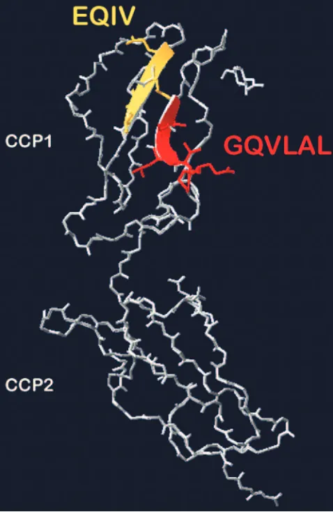
order to identify candidate sequences within the N-terminal half of bovine CCP1, bovine CCP1 and CCP2 were modeled using SWISS Model (35) on the basis of the deposited crystal structure of human CD46 CCP1 and CCP2 (7). The model revealed that the GQVLAL87sequence is exposed as a beta
sheet at the anterolateral side of CCP1 (Fig. 7). This stretch is situated antiparallel to the amino acids 66 to 69 (EQIV) lo-cated in the N-terminal half of bovine CCP1. At the analogous position in the porcine sequence an amino acid exchange span-ning four amino acids (DRVE70) is present.
The role of this tetrapeptide was analyzed by introduction of the bovine sequence EQIV69into the CD46 chimera that
al-ready contained the bovine hexapeptide GQVLAL87 in the
entire porcine CCP1 (CD461pGQVLAL87). The presence of
both bovine peptides in porcine CCP1 resulted in a 30-fold-increased susceptibility to BVDV and thus led to a functional BVDV receptor (Fig. 6). The introduction of the bovine se-quences EQIV69and GQVLAL87into the complete porcine
CD46 also resulted in an increased susceptibility of the respec-tive SK6T cell line (Fig. 6), which confirmed the important function of these amino acid stretches.
[image:7.585.98.483.70.369.2]Influence of the distance from CCP1 to the plasma mem-brane.In the case of measles virus a direct correlation between the distance from the binding domain to the plasma membrane and the inability to mediate membrane fusion was demon-strated (6, 9). In contrast to measles virus BVDV is taken up by
FIG. 6. The presence of tetrapeptide E66QIV69and hexapeptide G82QVLAL87from CCP1 of bovine CD46 leads to gain of receptor function of porcine CD46. SK6T cell lines were constructed that express the tetrapeptide and the hexapeptide in a chimeric CD46 molecule containing various parts of porcine CD46. The susceptibility to BVDV was assayed in the absence and presence of Dox. The columns represent mean values of duplicate experiments; error bars indicate maximum and minimum values.
3918 KREY ET AL. J. VIROL.
on November 8, 2019 by guest
http://jvi.asm.org/
clathrin-dependent endocytosis (22, 23) and does not fuse with the plasma membrane. An interesting question was whether the distance from the virus binding domain in CD46 to the plasma membrane is relevant for virus infection. For this pur-pose one to six spacer elements (CCPs derived from C4bp) were introduced into the CD46 molecule between the STP region and CCP4 and the respective SK6T cell lines were established. In all cell lines expression of a CD46 molecule with an increased apparent molecular mass was demonstrated by immunoblot analysis (Fig. 8a).
Likely, the appearance of the additional band observed for SK6T CD46 plus three CCPs is due to heterogeneous post-translational modifications. The CD46-C4bp chimeras bound equivalent amounts of virus regardless of the number of CCPs introduced in CD46 (data not shown). Determination of the susceptibilities revealed a 10-fold decrease in susceptibility to BVDV infection for the CD46-C4bp chimera containing six additional CCPs (Fig. 8b). Introduction of one to four addi-tional CCPs barely reduced susceptibility to BVDV.
The susceptibility of SK6T cells is reduced by expression of excess amounts of bovine CD46.Compared to a transient ex-pression system the use of stable cell lines poses the problem that each cell line originates from a single cell clone. A direct comparison between several cell lines is difficult because the expression levels considerably differ among the cell lines, e.g., due to the number of plasmid copies integrated into the ge-nome. This was further complicated by the fact that for SK6T cells expressing either wt or mutant CD46 considerable amounts of CD46 were detected even in the uninduced state. In this context it was necessary to analyze whether the observation of reduced susceptibility of SK6T cells upon the induction of CD46 expression (e.g., SK6T CD46) correlated with a certain level of protein on the cell surface. Based on the relationship of the susceptibility of uninduced cells to that of induced cells the cell lines were divided into three different phenotypes (Fig. 9). The phenotype A group comprised cell lines expressing functional CD46 whose susceptibility drops upon induction of CD46 expression. Phenotype B was constituted by cell lines which expressed nonfunctional CD46 but whose susceptibility was significantly decreased by induction. Cell lines expressing a functional receptor whose susceptibility is increased by induc-tion were referred to as phenotype C.
The relative amounts of CD46 molecules expressed on the cell surfaces were determined by flow cytometric analysis using anti-CD46 antibodies. For calibration the surface expression of uninduced SK6T CD46 cells was set to 100%. Induction of these cells for 24 h resulted in a mean fluorescence intensity of 154% (Fig. 9). For cell lines grouped in phenotype A or B in the absence of Dox a homogeneous basal surface expression of CD46 on similar levels was observed ranging between 73% (SK6T CD46 A82LPTFS88) and 106% (SK6T CD461b/p).
In-duction resulted in a further increased CD46 surface expres-sion of 98% (SK6T CD46 A82LPTFS88) up to 159% (SK6T
CD46 M80V81). In contrast, for cell lines that exhibited
phe-notype C only a minor percentage of cells in each sample expressed CD46 at the cell surface (0.4% for SK6T CD46 plus two CCPs up to 36.9% for SK6T CD461p/b). After induction a
homogeneous surface expression of CD46 was detected, and in addition the mean fluorescence intensity was clearly increased. However, even after induction the amount of cell surface-exposed CD46 remained below 100% (Fig. 9).
DISCUSSION
The finding that the infection of MDBK cells with a wide range of BVDV-1 and -2 strains can be blocked by anti-CD46 antibodies underscores the general use of CD46 as a cellular receptor for bovine pestiviruses. This clearly shows that CD46 usage by BVDV NADL is not a result of tissue culture adap-tation. For measles virus, which was initially shown to use CD46 as a cellular receptor (11), the usage of CD46 as a cellular receptor turned out to be restricted to a few vaccine or tissue culture-adapted strains (11, 28), while the common mea-sles virus receptor is CD150 (signaling lymphocytic activation molecule) (12, 37).
[image:8.585.43.282.72.439.2]For most BVDV strains, CD46 acts as the dominant recep-tor on the surface of MDBK cells. Virus entry independent of CD46 applies to BVDV 519, whose infection is reduced by
FIG. 7. Positions of E66QIV69and G82QVLAL87peptides in CCP1 and CCP2 of bovine CD46. Bovine CD46 was modeled using Swiss model (35) on the basis of human CD46 CCP1 and CCP2 crystal structure (7). Both peptides, E66QIV69 (yellow) and G82QVLAL87 (red), are located on antiparallel beta sheets in CCP1, forming a binding platform for BVDV.
on November 8, 2019 by guest
http://jvi.asm.org/
only 20% in the presence of anti-CD46 antibodies. Apparently an alternative cellular receptor facilitates infection by this strain. The existence of an alternative receptor is supported by previous observations in which excess amounts of anti-CD46 MAbs could not eliminate a residual susceptibility (1%) of MDBK cells by BVDV type strain NADL (29, 34). Two pos-sible candidates in addition to CD46 have been reported to act as a cellular receptor for BVDV, the LDL receptor (2) and heparan sulfate; the latter was shown to bind BVDV strain PE515 and tissue culture-adapted CSFV via the glycoprotein Erns (17, 18). Preliminary evidence suggests that BVDV-1
strain 519 interacts with glycosaminoglycans as well.
The attachment of a virus to its cellular receptor is the first step in the viral life cycle, and the clarification of this process is a basic requirement for understanding important aspects of the pathogenesis of the respective viral disease. Virus-receptor interactions have been elucidated for several viruses that use CD46 as a cellular receptor, including human group B and group D adenoviruses (13), human herpesvirus 6 (31, 33), and measles virus (5). The prime goal of this study was to identify the BVDV binding site within bovine CD46. This was dressed by systematic mutation of the CD46 molecule in ad-dition to epitope mapping of inhibitory monoclonal antibodies. Receptor function was analyzed in porcine cells, because a
90-fold-increased susceptibility to BVDV was observed after expression of bovine CD46 in porcine cells (29), hence provid-ing a sensitive assay to determine the receptor function. Poor transfection efficiencies of porcine cells excluded transient ex-pression as an experimental means to analyze virus binding and susceptibility. Therefore, a tetracycline-inducible expression system was chosen that provided regulated expression com-bined with a high stability of the resulting SK6T cell lines. With respect to susceptibility to BVDV, three phenotypic groups could be distinguished among the various cell lines established in this study. The susceptibility to BVDV infection as well as virus binding of several cell lines tested in this study was mas-sively increased by induction (phenotype C). In contrast, the susceptibility of a few cell lines decreased after induction with doxycycline, while significantly increased virus binding was ob-served (phenotype A). For these cell lines a remarkably higher level of basal expression and cell surface exposition in the absence of Dox was detected than for phenotype C, indicating that an accumulation of excess amounts of CD46 inhibited BVDV infection. A special case is represented by a few cell lines expressing nonfunctional CD46 chimeras (phenotype B). In the absence of Dox their susceptibility resembles that of negative-control SK6T cells; induction resulted in a significant reduction of susceptibility, which can be referred to as a
dom-FIG. 8. The distance between the virus binding domain and the plasma membrane influences the receptor function. SK6T cell lines were constructed that express chimeric CD46 molecules containing an increasing number of additional CCPs from C4bp between the STP region and CCP4. (a) Cells were grown for 24 h in the presence of 5g/l Dox, and crude cell lysates were subjected to sodium dodecyl sulfate-polyacrylamide gel electrophoresis and immunoblot analysis using anti-CD46 MAb BVD/CA 26. (b) Susceptibility to BVDV was assayed in the absence and presence of Dox. The columns represent mean values of duplicate experiments; error bars indicate maximum and minimum values.
3920 KREY ET AL. J. VIROL.
on November 8, 2019 by guest
http://jvi.asm.org/
[image:9.585.84.501.70.382.2]inant-negative effect. A possible explanation could be that a regulated distribution of CD46 is required for the infection process which is impaired by massive overexpression. Alterna-tively, CD46 itself interacts with cellular cofactors that are essential for virus invasion and overexpression thus results in a lack of unbound coreceptor. Future studies are required to clarify this mechanism in detail.
Systematic analysis of chimeric CD46 molecules including amino acid sequences from the porcine CD46 which does not bind BVDV indicated the importance of the two oligopeptides E66QIV69and G82QVLAL87within bovine CCP1. Mutation of
porcine CD46 at the positions of these two peptides resulted in a gain of receptor function for BVDV, confirming the crucial role of these two peptides for BVDV invasion. The modeling of CCP1 and CCP2 of bovine CD46 based on the crystal structure of CCP1 and CCP2 of human CD46 (7) illustrates that both peptides are located as adjacent beta sheets in CCP1 facing the small angle toward CCP2 (Fig. 7). From the results of this study we cannot exclude the possibility that amino acids conserved between bovine and porcine sequences contribute to virus binding located either in CCP2 or in neighboring E66QIV69 and G82QVLAL87. Nevertheless, this model
sug-gests that both peptides constitute a crucial part of a binding platform that interacts with BVD virions. The identification of this minimum essential virus binding site further underlines the role of CD46 as a BVDV receptor.
Chimeric CD46 molecules containing either four additional CCPs or four Ig-like domains have been shown to exert a dominant-negative effect on measles virus infection (6, 9) that is likely due to the elongated distance from the virus binding domain to the plasma membrane. Measles virus-induced fu-sion is completely abolished by the introduction of four Ig-like domains, which correspond to an elongation of the distance between the plasma membrane and the virus binding site by approximately 12.5 nm (6). In contrast, BVDV infection is reduced only 10-fold by the introduction of six CCP domains from C4bp corresponding to an elongation of the CD46 mol-ecule by approximately 18 nm. Introduction of four CCP do-mains, which doubles the length of CD46, barely affects the entry of BVDV. Previous studies have shown that heparan sulfate chains, which likely serve as alternative receptors for BVDV, averaged up to 105 kDa in apparent molecular mass (30). This roughly corresponds to 420 sugar residues per chain or 190 nm in length (39). This extensive variability of the
FIG. 9. Quantification of CD46 surface exposition on selected SK6T cell lines. Seven SK6T cell lines expressing either wt or chimeric CD46 were grown for 24 h in the absence or presence of 5g/l Dox. Trypsinized (2⫻105) cells were subjected to flow cytometric analysis using anti-CD46 MAb BVD/CA 17 and fluorescein isothiocyanate–anti-mouse IgG. Based on their susceptibilities in the absence and presence of Dox cells, cells were grouped into three phenotypes, A, B, and C. Susceptibility as a percentage of that of control cells (SK6T CD46 in the absence of Dox) is indicated beneath the graph. The mean fluorescence intensity of SK6T CD46 cells in the absence of Dox was set to 100%, and CD46 surface exposition was calculated as a percentage of that of the control. The graph includes the mean fluorescence intensity (leftyaxis) in light gray (⫺Dox) and dark gray (⫹Dox) as well as the percentage of positive cells (rightyaxis) in white bars. All cell lines and conditions were tested in at least three independent experiments with duplicate determinations.
on November 8, 2019 by guest
http://jvi.asm.org/
distance from the primary BVDV binding site to the plasma membrane confirms the minor influence of this distance.
Considering that BVDV was shown to use the clathrin-dependent endosomal pathway (22, 23), two alternative sce-narios of how BVD invasion proceeds are conceivable; either BVD virions are internalized as a cargo of the BVDV binding receptor or virions are moved from the binding receptor to a putative cellular cofactor, thereby minimizing the distance be-tween the bound virion and the plasma membrane. In this context the requirement for a cellular cofactor that was hy-pothesized in a previous study (29) has to be considered. Iden-tification of this putative cofactor as well as determination of the viral glycoprotein that acts as a ligand for CD46 will be a subject of future studies.
ACKNOWLEDGMENTS
This work was funded in part by the Deutsche Forschungsgemein-schaft, SFB 535. T.K. was a fellow of the Graduiertenkolleg Biochem-istry of Nucleoprotein Complexes and supported by the H. J. and G. Engemann Stiftung.
REFERENCES
1.Adams, E. M., M. C. Brown, M. Nunge, M. Krych, and J. P. Atkinson.1991. Contribution of the repeating domains of membrane cofactor protein (CD46) of the complement system to ligand binding and cofactor activity.
J. Immunol.147:3005–3011.
2.Agnello, V., G. Abel, M. Elfahal, G. B. Knight, and Q. X. Zhang.1999. Hepatitis C virus and other flaviviridae viruses enter cells via low density
lipoprotein receptor. Proc. Natl. Acad. Sci. USA96:12766–12771.
3.Barilla-LaBarca, M. L., M. K. Liszewski, J. D. Lambris, D. Hourcade, and J. P. Atkinson.2002. Role of membrane cofactor protein (CD46) in
regu-lation of C4b and C3b deposited on cells. J. Immunol.168:6298–6304.
4.Bolin, S. R., J. F. Ridpath, J. Black, M. Macy, and R. Roblin.1994. Survey of cell lines in the American Type Culture Collection for bovine viral
diar-rhea virus. J. Virol. Methods48:211–221.
5.Buchholz, C. J., D. Koller, P. Devaux, C. Mumenthaler, J. Schneider-Schaulies, W. Braun, D. Gerlier, and R. Cattaneo.1997. Mapping of the primary binding site of measles virus to its receptor CD46. J. Biol. Chem.
272:22072–22079.
6.Buchholz, C. J., U. Schneider, P. Devaux, D. Gerlier, and R. Cattaneo.1996. Cell entry by measles virus: long hybrid receptors uncouple binding from
membrane fusion. J. Virol.70:3716–3723.
7.Casasnovas, J. M., M. Larvie, and T. Stehle.1999. Crystal structure of two CD46 domains reveals an extended measles virus-binding surface. EMBO J.
18:2911–2922.
8.Cattaneo, R.2004. Four viruses, two bacteria, and one receptor: membrane
cofactor protein (CD46) as pathogens’ magnet. J. Virol.78:4385–4388.
9.Christiansen, D., E. R. De Sousa, B. Loveland, P. Kyriakou, M. Lanteri, F. T. Wild, and D. Gerlier.2002. A CD46CD[55-46] chimeric receptor, eight short consensus repeats long, acts as an inhibitor of both CD46 (MCP)- and CD150 (SLAM)-mediated cell-cell fusion induced by CD46-using measles
virus. J. Gen. Virol.83:1147–1155.
10.Corapi, W. V., R. O. Donis, and E. J. Dubovi.1990. Characterization of a panel of monoclonal antibodies and their use in the study of the antigenic
diversity of bovine viral diarrhea virus. Am. J. Vet. Res.51:1388–1394.
11.Dorig, R. E., A. Marcil, A. Chopra, and C. D. Richardson.1993. The human CD46 molecule is a receptor for measles virus (Edmonston strain). Cell
75:295–305.
12.Erlenhofer, C., W. P. Duprex, B. K. Rima, V. ter Meulen, and J. Schneider-Schaulies.2002. Analysis of receptor (CD46, CD150) usage by measles virus.
J. Gen. Virol.83:1431–1436.
13.Gaggar, A., D. M. Shayakhmetov, M. K. Liszewski, J. P. Atkinson, and A. Lieber.2005. Localization of regions in CD46 that interact with adenovirus.
J. Virol.79:7503–7513.
14.Gossen, M., S. Freundlieb, G. Bender, G. Muller, W. Hillen, and H. Bujard.
1995. Transcriptional activation by tetracyclines in mammalian cells. Science
268:1766–1769.
15.Heimann, M., G. Roman-Sosa, B. Martoglio, H.-J. Thiel, and T. Ru¨menapf.
2006. Core protein of pestiviruses is processed at the C terminus by signal
peptide peptidase. J. Virol.80:1915–1921.
16.Hulst, M. M., H. G. P. van Gennip, and R. J. Moormann.2000. Passage of classical swine fever virus in cultured swine kidney cells selects virus variants that bind to heparan sulfate due to a single amino acid change in envelope
protein Erns. J. Virol.74:9553–9561.
17.Iqbal, M., H. Flick-Smith, and J. W. McCauley.2000. Interactions of bovine
viral diarrhoea virus glycoprotein Erns
with cell surface glycosaminoglycans.
J. Gen. Virol.81:451–459.
18.Iqbal, M., and J. W. McCauley.2002. Identification of the
glycosaminogly-can-binding site on the glycoprotein Ernsof bovine viral diarrhoea virus by
site-directed mutagenesis. J. Gen. Virol.83:2153–2159.
19.Iwata, K., T. Seya, Y. Yanagi, J. M. Pesando, P. M. Johnson, M. Okabe, S. Ueda, H. Ariga, and S. Nagasawa.1995. Diversity of sites for measles virus binding and for inactivation of complement C3b and C4b on membrane
cofactor protein CD46. J. Biol. Chem.270:15148–15152.
20.Kallstrom, H., D. Blackmer Gill, B. Albiger, M. K. Liszewski, J. P. Atkinson, and A. B. Jonsson.2001. Attachment ofNeisseria gonorrhoeaeto the cellular pilus receptor CD46: identification of domains important for bacterial
ad-herence. Cell. Microbiol.3:133–143.
21.Kasza, L., J. A. Shadduck, and G. J. Christofinis.1972. Establishment, viral susceptibility and biological characteristics of a swine kidney cell line SK-6.
Res. Vet. Sci.13:46–51.
22.Krey, T., H. J. Thiel, and T. Ru¨menapf.2005. Acid-resistant bovine pestivirus
requires activation for pH-triggered fusion during entry. J. Virol.79:4191–
4200.
23.Lecot, S., S. Belouzard, J. Dubuisson, and Y. Rouille.2005. Bovine viral diarrhea virus entry is dependent on clathrin-mediated endocytosis. J. Virol.
79:10826–10829.
24.Lindenbach, B. D., and C. M. Rice.2000. Flaviviridae: the viruses and their
replication, p. 991–1042.InB. N. Fields, D. M. Knipe, and P. M. Howley
(ed.), Fields virology, 4th ed., vol. 1. Lippincott-Raven, Philadelphia, Pa. 25.Liszewski, M. K., and J. P. Atkinson.1992. Membrane cofactor protein.
Curr. Top. Microbiol. Immunol.178:45–60.
26.Liszewski, M. K., M. Leung, W. Cui, V. B. Subramanian, J. Parkinson, P. N. Barlow, M. Manchester, and J. P. Atkinson.2000. Dissecting sites important for complement regulatory activity in membrane cofactor protein (MCP;
CD46). J. Biol. Chem.275:37692–37701.
27.Liszewski, M. K., T. W. Post, and J. P. Atkinson.1991. Membrane cofactor protein (MCP or CD46): newest member of the regulators of complement
activation gene cluster. Annu. Rev. Immunol.9:431–455.
28.Manchester, M., D. S. Eto, A. Valsamakis, P. B. Liton, R. Fernandez-Munoz, P. A. Rota, W. J. Bellini, D. N. Forthal, and M. B. Oldstone.2000. Clinical
isolates of measles virus use CD46 as a cellular receptor. J. Virol.74:3967–
3974.
29.Maurer, K., T. Krey, V. Moennig, H.-J. Thiel, and T. Ru¨menapf.2004. CD46
is a cellular receptor for bovine viral diarrhea virus. J. Virol.78:1792–1799.
30.McClain, D. S., and A. O. Fuller.1994. Cell-specific kinetics and efficiency of herpes simplex virus type 1 entry are determined by two distinct phases of
attachment. Virology198:690–702.
31.Mori, Y., T. Seya, H. L. Huang, P. Akkapaiboon, P. Dhepakson, and K. Yamanishi.2002. Human herpesvirus 6 variant A but not variant B induces fusion from without in a variety of human cells through a human herpesvirus
6 entry receptor, CD46. J. Virol.76:6750–6761.
32.Rinck, G., C. Birghan, T. Harada, G. Meyers, H.-J. Thiel, and N. Tautz.
2001. A cellular J-domain protein modulates polyprotein processing and
cytopathogenicity of a pestivirus. J. Virol.75:9470–9482.
33.Santoro, F., H. L. Greenstone, A. Insinga, M. K. Liszewski, J. P. Atkinson, P. Lusso, and E. A. Berger.2003. Interaction of glycoprotein H of human
herpesvirus 6 with the cellular receptor CD46. J. Biol. Chem.278:25964–
25969.
34.Schelp, C., I. Greiser-Wilke, G. Wolf, M. Beer, V. Moennig, and B. Liess.
1995. Identification of cell membrane proteins linked to susceptibility to
bovine viral diarrhoea virus infection. Arch. Virol.140:1997–2009.
35.Schwede, T., J. Kopp, N. Guex, and M. C. Peitsch.2003. SWISS-MODEL: an
automated protein homology-modeling server. Nucleic Acids Res.31:3381–
3385.
36.Seya, T., J. R. Turner, and J. P. Atkinson.1986. Purification and character-ization of a membrane protein (gp45-70) that is a cofactor for cleavage of
C3b and C4b. J. Exp. Med.163:837–855.
37.Tatsuo, H., N. Ono, K. Tanaka, and Y. Yanagi.2000. SLAM (CDw150) is a
cellular receptor for measles virus. Nature406:893–897.
38.Thiel, H. J., R. Stark, E. Weiland, T. Ru¨menapf, and G. Meyers.1991. Hog cholera virus: molecular composition of virions from a pestivirus. J. Virol.
65:4705–4712.
39.Trybala, E., J. A. Liljeqvist, B. Svennerholm, and T. Bergstrom.2000. Her-pes simplex virus tyHer-pes 1 and 2 differ in their interaction with heparan sulfate.
J. Virol.74:9106–9114.
3922 KREY ET AL. J. VIROL.