A DISSERTATION ON COMPARATIVE STUDY OF SINGLE DOSE PROPHYLACTIC ANTIBIOTIC VERSUS EMPIRICAL
POSTOPERATIVE ANTIBIOTICS IN PREVENTION OF SSI
A PROSPECTIVE STUDY Dissertation Submitted to
THE TAMIL NADU
DR. M.G.R. MEDICAL UNIVERSITY CHENNAI
In partial fulfillment of the regulations for the award of the degree of
M.S. GENERAL SURGERY (Branch 1)
CHENGALPATTU MEDICAL COLLEGE
THE TAMILNADU Dr. M. G. R. MEDICAL UNIVERSITY CHENNAI, TAMILNADU
DECLARATION
I hereby declare that this dissertation entitled “COMPARATIVE STUDY OF SINGLE DOSE PROPHYLACTIC ANTIBIOTIC VERSUS EMPIRICAL POSTOPERATIVE ANTIBIOTCS IN PREVENTION OF SSI” was prepared by me under the direct guidance and supervision of Prof. DR.S. SURESH M.S., CHENGALPATTU MEDICAL COLLEGE. The dissertation is submitted to the Dr. M.G.R. Medical University in partial fulfillment of the University regulations for the award of MS degree in General Surgery, Examination to be held in April 2016.
CERTIFICATE
This is to certify that Dr.K. Alex Franklin post graduate student (2013-2016) in the department of General surgery, CHENGALPATTU MEDICAL COLLEGE has done this dissertation titled “COMPARATIVE STUDY OF SINGLE DOSE PROPHYLACTIC ANTIBIOTIC VERSUS EMPIRICAL POSTOPERATIVE ANTIBIOTCS IN PREVENTION OF SSI” under the direct guidance and supervision of guide Prof .DR.S.SURESH M.S., in partial fulfillment of the regulations laid down by the Tamilnadu Dr.M.G.R. Medical University, Chennai, for M.S., General Surgery Degree Examination.
Prof. DR.K. MUTHURAJ M.S., Prof. DR. K.MUTHURAJ M.S Professor & HOD DEAN
BONAFIED CERTIFICATE BY GUIDE
This is to certify that Dr. K.Alex Franklin post graduate student (2013-2016) in the department of General surgery, CHENGALPATTU MEDICAL COLLEGE has done this dissertation titled “COMPARATIVE STUDY OF SINGLE DOSE PROPHYLACTIC ANTIBIOTIC VERSUS EMPIRICAL POSTOPERATIVE ANTIBIOTCS IN PREVENTION OF SSI” under the direct guidance and supervision of guide Prof .DR.S.SURESH M.S., in partial fulfillment of the regulations laid down by the Tamilnadu Dr.M.G.R. Medical University, Chennai, for M.S., General Surgery Degree Examination.
Place : Chengalpattu Dr. S. SURESH M.S.,
Date : Professor of Surgery,
5
ACKNOWLEDGEMENT
At the outset, it is with a sense of accomplishment and deep gratitude that I dedicate this dissertation to all those who have been instrumental in its completion.
First and foremost I express my heartful thanks to my esteemed and respected HOD Department of General Surgery, CHENGALPATTU MEDICAL COLLEGE Prof. Dr. K. Muthuraj M.S., and my guide Prof.Dr.S.Suresh M.S., Had it not been for his whole hearted support throughout the period of this study, extending from his vast knowledge, invaluable advice and constant motivation, I truly would not have been able to complete this dissertation topic in its present form.
I sincerely thank my Professors, Dr. C.Srinivasan M.S., and Dr.T. Ragupathy M.S.,. I am greatly indebted to my Assistant Professors Dr. P. Sankarlingam M.S., and Dr. M. Sabrena M.S., for giving me practical suggestions and permitting me to carry out this study in their patients.
I dedicate this work to my parents and my wife Dr. R. Mary Stella for her constant encouragement and support. I am deeply indebted to all the teaching staff and my fellow postgraduates for their helpful attitude and valuable suggestions in every stage of my study.
10 CONTENTS
S.NO TITLE PAGE NO.
1 Introduction 1
2 Aim of the Study 2
3 Review of Literature 3
4 Materials and Methods 70
5 Results and Observations 72
6 Discussion 98
7 Conclusion 102
8 Recommendation 104
9 Bibliography 105
11
LIST OF TABLES
S.NO TITLE PAGE NO.
1 Organisms Causing SSI 61
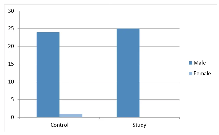
2 Age wise distribution of Cases in Class 1 Group 72 3 Sex wise distribution of cases and class 1 group 73
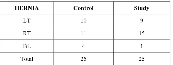
4 Side of hernia 75
5 Incidence of Fever in Class 1 76
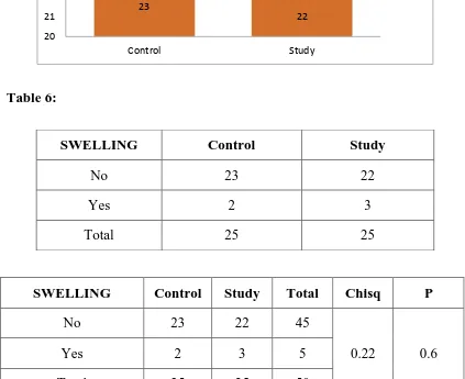
6 Incidence of swelling in Class 1 77
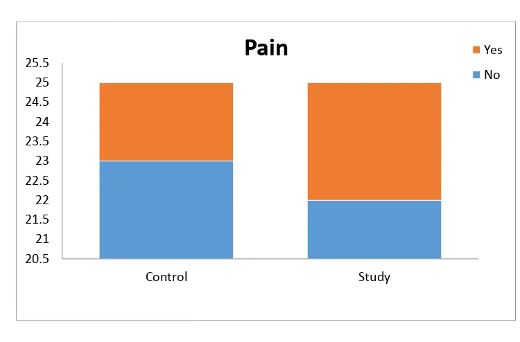
7 Incidence of Pain in Class 2 78
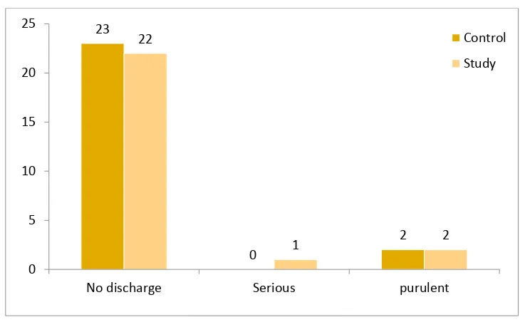
8 Incidence of Wound Discharge in Class 1 79 9 Incidence of Organisms Isolated in Class 1 81
10 Incidence of SSI in Class 1 82
11 Management of SSI in Class 1 83
12
17 Incidence of Pain in Class 2 91
18 Incidence of Wound Discharge in Class 2 92 19 Incidence of Organisms Isolated in Class 2 93
20 Incidence of SSI in Class 2 94
21 Management of SSI in Class 2 96
13
LIST OF GRAPHS
S.NO TITLE PAGE NO.
1 Age wise distribution of Cases in Class 1 Group 72 2 Sex wise distribution of cases and class 1 group 73
3 Side of hernia 74
4 Incidence of Fever in Class 1 76
5 Incidence of swelling in Class 1 77
6 Incidence of pain in Class 1 78
7 Incidence of Wound Discharge in Class 1 79 8 Incidence of Organisms Isolated in Class 1 80
9 Incidence of SSI in Class 1 82
10 Management of SSI in Class 1 83
11 Duration of Hospital Stay Class 1 84 12 Age wise distribution of Cases in Class 2 Group 86 13 Sex Wise Distribution of Cases and Class 2 Group 87 14 Incidence of Fever in Class 2 88 15 Incidence of Swelling in Class 2 89
14
17 Incidence of Wound Discharge in Class 2 91 18 Incidence of Organisms Isolated in Class 2 93
19 Incidence of SSI in Class 2 94
20 Management of SSI in Class 2 95
15
LIST OF ABBREVATIONS USED:
BT : Bleeding Time
CDC : Center for Disease Control CMV : Cytomegalovirus
CNS : Central Nervous System CO2 : Carbondioxide
CSF : Cerebro Spinal Fluid CT : Clotting Time
CVS : Cardiovascular System CXR : Chest X-ray
DC : Differential Count DM : Diabetes Mellitus DNA : Deoxyribonucleic Acid
ESR : Erythrocyte Sedimentation Rate FBS : Fasting Blood Sugar
Hb : Hemoglobin
HIV : Human Immuno Deficiency Virus Ht : Hypertension
IV : Intra Venous
LFT : Liver Function Test
16 RBS : Random Blood Sugar RR : Respiratory Rate RNA : Ribonucleic Acid RS : Respiratory System
SPO2 : Partial Pressure of Oxygen SSI : Surgical Site Infection TC : Total Count
URTI : Upper Respiratory Tract Infection USG : Ultrasound
17
LIST OF FIGURES
S.NO TITLE PAGE NO
1 Phases of Wound healing 16
ABSTRACT
TITLE:
A DISSERTATION ON COMPARATIVE STUDY OF SINGLEDOSPROPHYLACTIC ANTIBIOTIC VERSUS EMPIRICAL POST OPERATIVE ANTIBIOTICS IN PREVENTION OF SSI
AIM OF THE STUDY
To compare the efficacy of single dose antibiotic prophylaxis versus empirical post operative prophylaxis in prevention of SSI.
MATERIALS AND METHODS:
This study includes 100 clean and clean contaminated cases randomized to groups of 50 each. The study group will receive a single dose of antibiotic preoperatively while the control group will receive 3 to 5 days of empirical antibiotic therapy.
All the clean class 1 cases in the study group were given a single dose of 1gm of inj. Ceftriaxone at the time of induction or 30 minutes before skin incision in case the procedure is prolonged for more than 3 hrs a second dose was given.
They received no further antibiotics i.v or oral. All the cases in the control group received 5 days of inj. Cefotaxime 1Gm iv BD for 5 days. The incidence of SSI was noted and analysed.
inj. Metronidazole 500 mg i.v TDS for 5 days. In case of underweight or obese patients the dose was adjusted according to their body weight.
All the cases were followed up at 8th POD,15th POD, 30th POD and later at 3 months and 6 months. Any wound related complications noted and data obtained. The incidence of SSI in both the groups was calculated and results analysed.
CONCLUSION:
18
INTRODUCTION
Surgical site infections (SSI) are one of the most common causes of postoperative morbidity. The introduction of antibiotics in 20th century led to great improvement in surgical outcomes. From being a dreaded event surgeries have become an accepted part of modern day life due to the advent of antiseptic techniques and more importantly the advent of antibiotics.
But with rampant antibiotic use came its own set of problems like the rise in incidence of antibiotic resistant strains (MRSA) and rise in incidence of allergies and other complications of antibiotic use. The recent incidence of mass casualty due to tainted antibiotic in a mass sterilization camp being one of them.
19
In modern surgical care antibiotics are known to account for about 20% of total expenses during hospitalization. In our country where the proportion of health budget to GDP is one of the lowest in the world, the amount of savings that can be obtained by reducing our over reliance on antibiotics will be enormous.
AIM OF THE STUDY
20
REVIEW OF LITERATURE:
HISTORICAL CONTEXT:
The knowledge of infections and wound management have been known to man since time immemorial. The knowledge that the ancients learned has been recorded in their ancient writings. Egyptian Papyruses mention about use of salves and ointments in management of wounds.
In the bible references to wound care in the form of drainage of pus and application of local salves like vinegar and wine have been recorded in both the old and new testament. Among the Greeks Hippocrates taught the use of wine and vinegar in wound irrigation.
Later Roman surgeon Galen recognized that the localization of pus in wounds followed by drainage signaled the beginning of wound healing leading to his famous saying. Pus bonum et laudible. Sadly this was misunderstood in the dark ages of Europe and harmful practices like inoculation of feces in wounds to promote pus formation had developed.
Advances in wound healing in Europe had to wait until renaissance before rational thought and keen observation by figure like Ambroise Pare, Theodoric of cervia and others for the reintroduction of antisepsis and abandonment of harmful practices like cauterization and blood letting.
21
He was able to reduce puerperal sepsis from above 10 percent to less than 2 percent. Later Louis Pasteur with his germ theory and Anton Von Leeuwenhoek with his microscope Led to increase in knowledge about microbes and their role in wound infection.
Later Joseph Lister applied this knowledge and introduced the concept of antiseptic technique by introducing the concept of washing hands, instruments and sutures in carbolic acid. Later the discovery of steam sterilization by Earnrst von Bergmann in 1907 and its subsequent adoption all over the world led to improvement in surgical outcomes. Later the introduction of gloves and newer techniques by Halsted led to further improvement. But it was the discovery of antibiotics especially penicillin by Sir Alexander Fleming in 1928 and its subsequent use that surgery became truly modern and safe.
With the advent of modern anesthesia and newer antibiotics we are now performing surgeries that could only be dreamt of in the past. The days of certain death following fecal peritonitis are now past but challenges still remain. While the role of antibiotic prophylaxis in contaminated cases is accepted but the role of prolonged antibiotics in clean surgeries remain controversial.
Definition and classification of Surgical Site Infection
22
Even though study of Microbial density in tissue can be performed but for practical purposes the decision on starting antibiotics and the intended agents are decided based on clinical classification. The classification gives an idea of bacterial contamination during the procedure.
To be classified as Superficial incisional wound infection itmust meet the following criteria. Infection must occur at the incision site within 30 days after surgery and must involvethe skin or subcutaneous tissue abovefascial layers and any one of the following.13
1. There is purulent drainage from the incision or collection located above the fascial plane.
2. If the wound is opened deliberately by the surgeon, unless the exudate is culture negative.
3. If Organism is isolated from the culture of fluid aseptically obtained from the wound closed primarily.
To be classified as Deep surgical wound infection it must meet the following criteria. Infection must occurs at the operative site within 30 days after the surgery if no prosthesis was placed or within 6 months to l year if an implant had been placed, and the infection involves tissues or spaces at or beneath the fascial plane along with any of the following
1. The wound dehisces spontaneously or is opened deliberately by the surgeon when patient has persistent fever (>38o c) and or localized tenderness or pain, unless the wound is culture negative.
23
direct examination, during surgery or on histopathological examination. 3. The Surgeon declares infection.
Surgical wounds are classified based on the assumed magnitude of bacterial load at the time of operation as clean, clean contaminated, contaminated and dirty. 14
CLASSIFICATION 15:
Class I / Clean: This catergory includes those surgeries in which no contamination is present; only microflora from skin potentially contaminates the wound. An uninfected surgical wound in which no inflammation is encountered and the respiratory, alimentary, genital, or uninfected urinary tract is not entered. In addition, clean wounds are primarily closed and, if necessary, drained with closed drainage. Class ID wounds are similar except that a prosthetic device ( eg. Mesh or valve) is inserted.
Class II/Clean-Contaminated: It includes those surgeries in which a hollow viscus with indigenous bacterial flora is opened under controlled circumstances without significant spillage of the contents. A surgical wound in which the respiratory, alimentary, genital or urinary tracts are entered under controlled conditions and without unusual contamination, Specifically operations involving the billiary tract, appendix, vagina, and oropharynx are included in this category, provided no evidence of infection or major break in technique is encountered.
24 sterile area of the body.
Open, fresh, accidental wounds. In addition, operations with major breaks in sterile technique (e.g., open cardiac massage) or gross spillage from the gastrointestinal tract, and incisions in which acute, nonpurulent inflammation is encountered are included in this category.
Class IV/Dirty-Infected: Include traumatic wounds in which a significant delay in treatment has occurred and in which necrotic tissue is present, those created in presence of overt infection.
Old traumatic wounds with retained devitalized tissue and those that involve existing clinical infection or perforated viscera. This definition suggests that the organisms causing postoperative infection were present in the operative field before the operation.
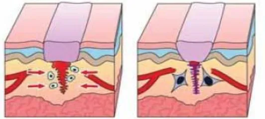
WOUND HEALING
Any injury surgical or otherwise starts a predictable sequence of events that follows a set pathway resulting in wound healing. Those phases are inflammatory phase, the fibroplastic phase and remodeling phase.
INFLAMMATORY PHASE
This phase prepares the area for healing and immobilizes the wound by causing the wound to swell and become painful, resulting in restriction of movement.
25
inflammatory response. The vast majority of specialized cells involved in this phase of healing process come from blood.17
Blood vessels that had traversed the wound before injury are severed at the time of injury and it results in exudation of whole blood into the wound, which on coagulation, seals the damaged vessels and lymphatic channels that drain the area in order to close the wound, and it prevents further bleeding. The release of histamine and other vasodilator agents by the injured tissues cause the undamaged vessels to dilate. Histamine causes short acting vasodilatation in nearby intact vessels and it is this combination of whole blood exudate from damaged blood vessels and serous transudate that creates a red, hot, swollen and painful local environment. Bradykinins, that are derived from plasma in the area of the injury, contribute to more prolonged vascular permeability. Certain types of prostaglandins further contribute to long-term vasodilatation. The fibrin plugs that clot in the wound also form in the lymphatic vessels. Blocking the lymphatic flow seals the wound and also helps to stop the spread of infection. The lymphatics remain closed until later in the healing process.18
The Mast cells also release hyaluronic acid and other proteoglycans into the mix of chemicals accumulating in the wound and these bind with the wound fluid to create a non-flowing gel that gradually slows down leakage and fluid loss. The inflammatory oedema that is formed as a result fills up the spaces in the wound and surrounds the damaged structures and binds them together.
26 reactant.
Secondary wound care is needed to address the changes made by induced vasodilatation, which varies in relation to the severity of the wound. This transudate can be diminished by following a regimen of rest, ice, compression and elevation. For healing to commence, the wound needs to be decontaminated by phagocytosis, and neovascularisation must then progress. Phagocytosis
Within blood vessels adjacent to the wound, WBC’s start to adhere to the capillary walls. Chemical changes in the wound attract these cells and induce them to slip through the enlarged capillary pores following which they migrate to the site of the injury. The main target of this phase is to prevent infection and to rid the wound of infective agents.
The first WBC’s to reach the wound are polymorphonuclear leucocytes. These cells begin the process of phagocytosis by attaching to bacteria and phagocytosing them.
27
many tissues, playing a critical role in wound healing. They responding to the stimulation secrete the precursors of all the components of the extracellular matrix, predominantly the ground substance and a variety of fibers.19
The macrophage influences repair by chemically recruiting the fibroblastic repair cells. It is the local platelet-derived growth factor that is released from the platelets during clotting and from macrophages that signals the fibroblasts.
Neovascularisation is growth of new blood vessels, as healing will not proceed without new functioning blood vessels to supply oxygen and nourishment to the injured tissue. It is likely that it is the macrophages that signal this vascular regeneration to start. Patent blood vessels in the wound area develop small ‘sprouts’ that grow into the wound area and it is from these buds that eventually come into contact and anastamose with other arteriolar or venous buds to form a functioning capillary loop. The capillary sprouts, during the early stages, lack full thickness walls, which renders them delicate and easily disrupted, this neccesitates immobilization during this phase to permit the vascular regrowth and prevent the formation of microhaemorrhages. At the conclusion of this phase, fibrinolysin in blood vessels is formed to assist in dissolving clots and the lymphatic channels open in to assist in resolving the wound oedema.
28 FIBROPLASTIC PHASE
When the inflammatory phase is completed, rebuilding can commence. This phase is named after the primary cell that aides scar production, the fibroblast. Many different cells are also involved in the inflammatory phase, but fewer cell types operate during the fibroplastic phase, which lasts for about three weeks. During this phase the wound is resurfaced resulting in strength being imparted to the wound. The fibroblasts originate from the mesenchymal cells located in connective tissue around blood vessels and fat. In response to chemotactic signals, fibroblast precursors transform into cells with migratory ability.
These fibroblasts follow the fibrin meshwork created earlier in the wound fluid environment, which enveloped all injured structures, and thus they have access to all depths of the wound. Once in place, the fibroblasts initiates synthesis of the collagen molecule. During this phase, three processes occur concurrently to achieve coalescence and closure. These processes are epithelialisation, wound contraction and collagen production.
Epithelialisation
29
of the wound. These new cells are true epithelial cells and therefore this multiplication represents a regeneration process. The surviving epidermal structures like hair shafts and sweat glands also give rise to epithelial mitoses. Once the wound bed is viable and a decent blood supply is available, the migration of these new cells begins, with those coming from the periphery moving in and those from appendages moving out. These migratory cells continue to remain attached to the parent cells and this movement causes tension on the normal skin around the wound edge. The advancing edge of the epithelium seeks out oxygen-rich tissue, moist tissue.
If the epithelial edge meets eschar, foreign material like sutures or blood clots, it then plunges under it to maintain contact with the vascular loop network in the wound. If the necrotic tissue or the wound is too extensive or if oxygen availability is poor, then epithelial migration cannot proceed. If sufficient capillary circulation does not exist to maintain epithelial integrity then wound dehiscence can occur.
Dehiscence is the phenomenon of premature bursting or splitting of a wound along natural or surgical suture lines. This is a complication of surgery that occurs secondary to poor wound healing.
30 Wound Contraction
Epithelialisation closes the wound surface, but it is contraction that pulls the entire wound together, thereby shrinking the defect. A successful contraction results in a smaller wound that can be repaired by scar formation. Minimizing the area to be healed is highly beneficial in certain tissues with fixed, deep structures that are covered by mobile, loose skin.
Wound contraction may be harmful in those areas that require every millimeter of skin and tissue length, like the hands and face. Allowing uncontrolled contraction here is a potential problem as it will distort the topography of the skin and cause the tissues to be drawn abnormally towards the site of healing, causing disfigurement and discomfort.
A specialized cell called the myofibroblast is involved in the contraction process. In terms of differentiation, myofibroblast lies between a fibroblast and a smooth muscle cell. Myofibroblasts attach to the skin margins, pull the entire epidermal layer inward, and are a feature in the hypertrophic scars. Control of wound contraction and scar formation at the time of wound formation is useful in order to control the direction of wound contraction and thus prevent the distortion.
Collagen Production
31
cofactors such as zinc, iron and copper are needed to help create the proper background for fibroplasia, which is the production of fibrous tissue, usually implying an abnormal increase of non-neoplastic fibrous tissue.20
The fibroblast synthesizes three polypeptide chains that coil to form a right-handed helix. These spiraled chains (procollagen) are then extruded from the fibroblast into extracellular space. Once extruded, the triple-helical molecule undergoes cleavage at specific terminal sites to become tropocollagen. Tropocollagens then associate spontaneously in an overlapping array to eventually convolve with other tropocollagen molecules to form a collagen fibril. These filaments lay disorganized in the wound and are in a gelatinous state. There is little strength inherent in this collagen mass, which requires cross links and other bonding to be formed before the wound durability or tensile strength can be achieved.
32 REMODELLING PHASE
Synthesis-lysis Balance
Despite the fact that collagen synthesis continues to occur at a high rate, no further increase in the scar mass occurs. At this point, new collagen is created and the old collagen is broken down in a balanced fashion as a result of the action of the action of enzyme collagenase. Collagen turnover is then accelerated as old fibrous tissue is removed and new fibrous tissue is formed. This process continues until the remodelling phase ends at completion of six months to a year, depending on the state of the injury. It should be remembered at this point that the speed of collagen synthesis and laying down of new collagen is age-related and its speed decreases with advancing years.
Collagen fiber orientation
During the remodelling phase, collagen turnover allows the randomly deposited scar tissue to be arranged, in both lateral and linear orientation. Scar tissue is non-elastic and it attempts to mimic the characteristics of the tissue that is being healed. The tissue structure induces the collagen weave so that dense tissues induce a dense, highly cross-linked scar, while more pliable tissues induce a loose, coiled, less crosslinked scar. A scar can adapt through remodelling forces of synthesis and lysis.
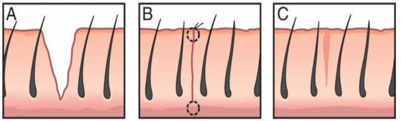
33 Types of Wound Healing :
Surgical wounds may heal by either primary intention, secondary intention or tertiary intention (delayed primary).
Healing by Primary intention
Most heal by primary intention, here the wound edges are brought together (apposed) and then held in together by mechanical means (adhesive strips, staples or sutures), allowing the wound time to heal and then develop enough tensile strength to withstand stress without support. The goal of surgery is to achieve healing by such means so that there is minimal oedema, no serous discharge or infection, no separation of the wound edges and with very minimal scar formation.
34 FibroplasticPhase :
[image:36.595.190.456.135.255.2]RemodellingPhase :
Fig 1: Phases of Wound Healing Healing by Secondary intention
35 Healing by Tertiary intention
On occasions surgical incisions are allowed to heal by delayed primary intention where non-viable tissue are removed and the wound is initially left open. Wound edges are then brought together at about 4-6 days, before granulation tissue is visible. This method is often used after traumatic injury or dirty surgery.22
Healing by Primary intention
[image:37.595.120.528.316.439.2]Healing by Secondary intention
36 Factors Affecting Wound Healing23
The factors that may adversely affect the wound healing can be conveniently considered in two categories: factors, which locally affect wound repair (local factor) and the systematic abnormalities which have remote effects on the wound (systemic factors).
Local Factors
The local factors which have been implicated in the failure of wound healing are
1. Surgical technique. 2. Blood supply. 3. Mechanical stress. 4. Suture materials. 5. Suture technique. 6. Infection
7. Radiation Surgical Technique24
37
The essentials of good surgical technique include gentle handling of the tissues, securing meticulous hemostasis, the prevention of any dead space in the wound, and the avoidance of tissue necrosis resulting from excessive use of surgical diathermy or strangulation of the tissues by the ligatures. The presence of one or more of these variables constitutes a barrier to the processes of cellular repair and they are the factors leading to propagate wound infection. Ischemic tissue, a wound hematoma,or a collection of serum in the wound is excellent media for the subsequent growth of bacteria.
The relative merits of surgical diathermy compared to suture ligation in wound hemostasis remain controversial but there is probably very little difference between the two methods as far as they affect the wound healing both may cause problems if they are used incorrectly. Diathermy should be used sparingly and precisely. Ligatures on blood vessels should not strangulate adjacent tissues. Fine suture materials can be used for most blood vessels and absorbable sutures can be used for vessels in the subcutaneous tissues.
38
Other local factors affecting wound healing such as blood supply of the wound and the presence or absence of mechanical tension may also be results of surgical technique and these are considered below.
Blood Supply
A good blood supply is a basic factor in the success of wound repair; it is essential for the supply of oxygen and other nutrients required in the cellular and biochemical processes of repair and it is necessary for the removal of wound metabolites.
Disease may lead to impaired blood supply of the wound. This is most frequently encountered in the surgical treatment of atherosclerotic arterial insufficiency of the lower limb. Any factor causing mechanical tension in the wound will have adverse effects on the blood supply. Extrinsic forces cause wound tension by distracting the wound edges. In the simple example of a sutured skin wound, the elastic pull of the unwounded skin on either side of the incision exerts a lateral pull on the wound edges. In wounds of the hollow viscera and the abdominal wall, wound tension is also derived from the pressure within the lumen of the viscus or hollow cavity; the tension occurring in the wound is directly related to this pressure and the radius of the lumen according to the law of Laplace.
39
than a measurable parameter. Intrinsic wound tension or the buildup of pressure within the volume of the wound contents following suture. Some degree of swelling of the wound is a normal feature of the early phase of repair. It results from the inflammatory response which is a feature of the first few days in all wounds and the surgeon should allow for such changes by ensuring that his sutures are not tied too tightly. More serious problems of intrinsic wound tension occur in the presence of wound infection, hematomas and collections of serum. These factors may cause an injurious rise in tissue pressure within the relatively inelastic confines of the wound. The presence of ischemic tissue in the wound initiates a vicious circle whereby the ischemic tissue results in tissue swelling and the tissue swelling lead to a further reduction in blood supply of the wound.
Mechanical Stress
The extrinsic forces affecting wound tension may cause wound disruption or it may be a consequence of excessive movement of the wound edges. In the former case, the tension at the suture or wound interface created by the extrinsic forces becomes so greater that the sutures simply cut out through the wound edges, less commonly the suture material may break or the knots may slip.
40 Suture Materials25
The choice of suture material in primary wound closure may have a significant bearing on the success of the subsequent wound repair. There have been striking developments in the manufacture of sutures in recent years and there is now an extensive range of naturally occurring and synthetic sutures. It has been suggested that the ideal suture may be defined as follows:
1. It should hold the tissues in apposition for as long as the natural forces are insufficient to resist separation or stretching of the wound edges. 2. It should handle easily and knot securely.
3. It should provoke minimal tissue reaction and it should be quickly absorbed so that the infection is not encouraged and it should not result in sinus formation.
Suture Technique26
41
Knot security is provided by the ‘surgeon’s knot’ or square knot and this should always be used in preference to a ‘granny knot’. Monofilament nylon and polypropylene have poor knotting characteristics and at least five ‘throws’ should be used to prevent knot slipping when these suture materials are used. Radiation
Problems of wound healing resulting from ionizing irradiation chiefly occur in the management of skin wounds in previously irradiated tissues. These problems are frequently encountered in the surgical treatment of recurrent malignant disease of the chest wall or head and neck.
Infection
Bacterial infection is the most common complication of wound healing and it encountered in every surgical specialty. Multiple factors are involved in the pathogenesis of wound infection and the effects of infection are divers. Classical wound infection occurring in wounds closed by primary suture may simply be a source of significant morbidity but infection in vascular operations, plastic surgery and orthopaedic surgery may have disastrous consequences. SYSTEMIC FACTORS27
Systemic factors which may affect wound healing are 1. Age
2. Malnutrition
42 5. Trauma, hypovolemia and hypoxia 6. Anemia
7. Uremia
8. Malignant disease 9. Jaundice
10.Corticosteroid drugs
11.Cytotoxic and antimetabolite drugs Age
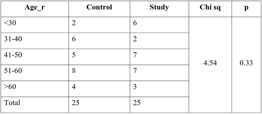
It is a common finding in studies of wound healing that complications are more prevalent in elderly patient: abdominal wound dehiscence is more common and has been shown that such patients also have a significantly higher incidence of dehiscence of colonic or colorectal anastomoses. The fact is, however, that surgical patients also have a higher incidence of malnutrition, major operations, vitamin deficiencies and various other systemic abnormalities and it is difficult to conclude that age alone is a factor affecting wound healing. Malnutrition
43
The amino acid methionine may have a key role in wound repair and that the adverse effects of malnutrition on wound healing may be reversed by the administration of methionine alone. Methionine is involved in the synthesis of the sulphatemucopolysaccharides of wound tissue and methionine and cystine are essential nutrients for the survival of fibroblasts in tissue culture. A deficiency of methionine would therefore provide a neat explanation for the failure of synthesis of collagen and mucopolsaccharides in malnourished subjects.
Vitamin Deficiency
Ascorbic acid deficiency is a significant factor in the healing of wound in surgical patients. Surgical trauma causes a fall in leucocyte ascorbic acid levels but these changes are unrelated to the severity of surgical trauma, blood loss, or blood transfusion and they may simply be obligatory features of the metabolic response to trauma; some of the ascorbic acid lost from leucocytes may be utilized in the surgical wound. The lowest levels of leucocyte ascorbic acid are found in the elderly and there may be a case for the use of ascorbic acid supplements in elderly patients.
44
Vitamin D is an essential in calcium metabolism and in the formation of new bone. In rickets, severe deficiency of the vitamin interferes with bone growth and a soft collagenous matrix of osteoid tissue is laid down instead of calcified bone. However, deficiencies of vitamin D are rarely encountered in civilized communities and there is no evidence that vitamin D deficiency affects the healing on bone in surgical patients.
Zinc Deficiency
It has been suggested that a deficiency of zinc has adverse effects on the healing of wounds in man. Zinc is required for several enzymatic reactions in the human body and it stabilizes lysosomal and cell membranes probably by inhibition of lipid peroxidases; the enzymes DNA – polymearse, reverse transcriptase and lysyl oxidase and zinc dependent. A deficiency of zinc has adverse effects on cell multiplication, fibroplasias, collagen synthesis and the epithelial covering of wounds but significant levels of zinc deficiency affecting wound healing in man are probably found only in severe burns, and in the management of intestinal fistulas. Patients required prolonged parenteral nutrition therapy in the absence of a normal diet should be given zinc supplements but the recommended dosage of zinc should not be exceeded since high serum levels of zinc are associated with toxic effects.
Trauma hypovolemia and hypoxia
45
the additional trauma of an extensive skin incision resulted in impaired healing of the abdominal wound. Zederfeldt suggested that the effects of remote trauma and hypovolemia on the breaking strength of abdominal wounds in rabbits and he reported that both factors had adverse effects on wound healing. Zedereldt suggested that the effects of trauma and hypovolemia on wound healing had a common basis and that tissue hypoxia was the final common factor affecting wound repair. Oxygen is an essential factor in the hydroxylation of the amino acid proline and lysine during the synthesis of collagen and further experimental studies have confirmed that a low tissue PO2 has adverse effects on wound healing. Further possible effect of remote trauma and wound healing is the adverse effect it may on the immunological defenses.
It has been shown that remote trauma and hypovolemia increased the susceptibility of experimental animals to staphylococcal and pseudomonas infections. However, according to Conolly and his colleagues, tissue hypoxia may be the most likely explanation for the susceptibility of traumatized or shocked animals to wound infection. Finally it has been suggested that postoperative changes in ascorbic acid availability may be a significant factor in the pathogenesis of wound failure in traumatized subjects but this seems unlikely since there is no significant correlation between the severity of trauma and postoperative changes in leucocyte ascorbic acid.
46
during surgical operations and the incidence of abdominal wound dehiscence and dehiscence of colonic anastomoses. All the same, these observations do not prove the tissue hypoxia is the final factor causing these complications of wound healing and there may be other factors involved. Indeed, in experimental studies of the effects of trauma on the healing of colonic anastomoses, it was found that the postoperative intraperitoneal infection was the factor responsible for an increased incidence of anastomotic dehiscence in traumatized animals.
47
tissue chambers and the tissue capillary oxygen gradients or conditions of oxygenation in these wounds may be quite different from those in sutured surgical incisions.
Anemia
Studies of surgical patients have suggested that anaemia may be involved in the pathogenesis of wound complications; an increased incidence of dehiscence of abdominal wounds and colonic anastomoses has been reported in anaemic patients. However, it is possible that other factor such as malnutrition or the type of surgery performed in anaemic patients were actually responsible for the failure of wound healing.
Malignant Disease
48 Jaundice
In retrospective clinical studies of abdominal wound healing, Reitamo and Moller and Keill and other found no convincing evidence that jaundice was involved in the pathogenesis of abdominal wound dehiscence. However, these studies included very small numbers of jaundiced patients and in a careful prospective study of abdominal wound healing. Ellis and Heddle found that jaundiced patients had a significantly higher incidence of wound dehiscence and incisional hernia compared with anicteric patients undergoing laparotomy for various abdominal disorders.
49
biliary obstruction and it was found that they had a significantly higher incidence of abdominal failure compared with anicteric patients ; wound dehiscence or incisional hernia occurred in 27.1% of jaundice patients and in 4.3% of anicteric patients. However, there was no correlation between wound dehiscence or incisional hernia and the depth of jaundice or plasma bilirubin, preoperative liver enzymes or plasma albumin levels and the factor which did seem to determine the outcome of abdominal wound healing was the presence or absence of malignant disease. Wound dehiscence or incision hernia occurred in 12 of 22 patients with malignant disease (59.1%) but these complications did not occur in 26 patients with jaundice resulting from benign pathology.
The findings of experimental studies indicate that jaundice has adverse effects on wound repair but the clinical significance of these findings is uncertain and it appears that other factors may account for the failure of wound healing in surgical patients; jaundiced patients with malignant disease are particularly at risk of complications of wound healing. The type of surgery involved in the management of patients with malignant disease may be a factor in the pathogenesis of wound complications.
Corticosteroids
50
early postoperative period; steroid therapy commenced several days after surgery has little or no effect on the healing of wound healing in experimental animals.
Clinical studies of wound healing in surgical patients have shown that wound dehiscence and sepsis are more frequent complications in patients receiving steroids at the time of operation. However, it is by no means certain that steroids are responsible for these complications for the patients receiving steroid therapy are frequency those who have serious disease, malnutrition and other factors which may affect wound healing.
Cytotoxic and Anti – Metabolite Drugs
There is increasing use of cytotoxic and anti – metabolic durg in medicine, transplantations surgery and in the management of malignant disease.
It seems probable by the nature of their action that the therapeutic use of these agents would interfere with wound repair and experimental studies in laboratory animals have produced some evidence to support this connection.
51
uraemia, we remain in the unhappy position of being unable to define the precise significance of the systemic abnormalities or to offer specific therapy which may remove the threat of wound complications. Further progress may depend on the development of improved techniques of local wound care.
The effect of bacterial infection on wound healing
The biochemistry of wound infection is complex. Delayed epithelial growth and migration, cellular necrosis and microvascular thrombosis are histological features of infected wounds and they result from the combined effect of bacterial toxins and the hostile chemical environment of the infected wound. The principle of biochemical abnormality in infected wound seems to be a disturbance of collagen metabolism, there is a constant process of synthesis and lysis of collagen in all wounds and to a lesser extent in unwounded tissue and this process may be affected in several ways by the presence of bacterial infection. Firstly, there is exaggerated lysis of wound collagen by collagenolytic enzymes, some of these are lysosomal enzymes present in polymorphonuclear lymphocytes in the infected wounds, others are enzymes which are normally present in tissues.31
52
process is not confined to the wound alone, it extends through the wound edges into unwounded tissues, the wound edges becomes soft and mechanically weak, and wound sutures will cut out the softened tissue resulting in the disruption of wounds closed by primary suture.
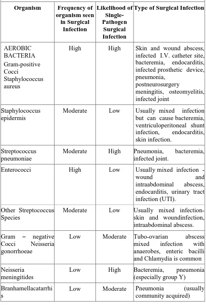
Etiology of Surgical Site Infection
No single factor is responsible for surgical site infection. Several factors are involved and the relative contribution of these factors varies greatly in different types of surgery. The vast majority of wound infections are endogenous. They are self-infections resulting from contamination of wound by bacteria carried by the host either on the body surface or more commonly within hollow viscera. A smaller proportion of wound infections are exogenous. They are cross infections by bacteria derived from another source and they may occur in the operating room or in the hospital ward.
Wound infection may be primary or secondary. Primary wound infection is the result of bacterial contamination of the wound occurring during surgery. Secondary wound infection occurs within the postoperative environment when bacteria gain access to the wound either through the wound suture line or through another portal such as a drainage tube or drainage track. The majority of wound infections are primary of type.
SURGICAL MICROBIOLOGY32
53 Bacteria
Bacteria can be classified according to staining characteristic with Gram stain (positive or negative), shape (cocci, rods, spirals) and sensitivity to Oxygen (aerobic, facultative, anaerobic) or according to the combination of these characters.
Gram positive cocci
Staphylococci and some streptococci species are the Gram positive cocci of interest to surgeons because of their ability to cause primary surgical infections and post operative infections. Staphylococci may be coagulase positive or coagulase negative.
Staphylococcus aureus is the most common pathogen isolated from wound infections. A major factor in its pathogenicity is coagulase productions, although the mechanism by which coagulase production increases virulence is not known. Most coagulase positive staphylococci should be resistant to penicillin and require treatment by a penicillinase resistant antibiotic. Extensive use of penicillinase resistant Beta lactam antibiotics during past 2 decades has encouraged emergence of Methicillin resistant staphylococcus aureus (MRSA). Coagulase negative staphylococci are the most common organisms recovered in nosocomial bacteremia and are frequently associated with clinically significant infections of intravascular devices.
54
discoloration around colonies containing intact red blood cells, beta hemolysis, complete clearing of the area around colonies and destructions of red blood cells; and gamma hemolysis.
Group A streptococci can cause infections of almost any organ although skin, subcutaneous tissue and pharynx are the most frequently affected areas. Streptococci are important pathogens because of their ability to cause post operative infections including cellulitis, wound infection, endocarditis, urinary tract infection and bacteremia. Enterococci are commonly recovered as a part of the normal flora of the gastrointestinal tract and the vagina. Enterococcal bacteremia has a poor prognosis in combination with intra abdominal or pelvic infections and is found most often in patients who here been hospitalized for long time.
Aerobic and facultative anaerobic gram negative bacilli33
Numerous gram negative rods that can cause human disease have been identified, but only a few are of surgical importance. The genera Escherhia, Klebsiella, Proteus, Enterobacter, frequently can be cultured from patients with intra abdominal and pelvic peritonitis and abscess, post operative wound infection, pneumonia and urinary tract infection.
55 Anaerobic Bacteria33
Anaerobic bacteria require reduced oxygen for growth. Virtually all anaerobic infections arise endogenously. The cell wall of anaerobic bacteria is important in abscess formation. The genus Clostridium is most virulent of all anaerobes. C.Dfficile cause pseudomembranous colitis and occurs in patients on antimicrobial therapy.
Fungi
Fungi are the most primitive eukaryote organism and are classified as protists. Because of their cell wall similarity to mammalian cells they are not sensitive to antibacterial agents, and many antifungal agents are toxic to human cells.
In surgical patients opportunists cause most infections. Candida albicans and other candida species are by far the most common. They cause infection in patients treated with broad spectrum antibiotics and steroids. These infection can be treated by stopping antimicrobial, correcting host defences and therapy with amphotericin B or one of the azole antifungal agents.
Viruses34
56
PHYSIOLOGY AND PATHO PHYSIOLOGY OF SURGICAL SITE INFECTION
Physiology35
The unique feature of all surgical infection is tissue necrosis. In post traumatic surgical infection tissue necrosis is induced by technical or other physical trauma, while in primary surgical infection tissue the pathophysiological process induces necrosis. Inflammation is the response to tissue necrosis, leading to the events visible at surface, which were well described by Celsius and refined by Galen as rubor, tumor, calor, dolor and functiolaesa. These symptoms describe the host response and when controlled and properly regulated, result in elimination of necrotic material and prepare the way for tissue repair.
The magnitude of inflammatory response and of its symptoms is dependent on the burden of tissue injury and on the number and pathogenicity of invading organisms. If toxins or other bacterial products continuously destroy tissue, or exceed the capability of the host to confine the challenge of body integrity, the inflammatory process will continue and may then result in multisystem malfunction.
Local Phase of Infections
57
necrotic tissue is an excellent medium for bacterial growth. Bacterial in turn release toxins and invade the surrounding tissue, which causes the host to respond with further inflammation in an attempt to confine the infection.
If tissue injury and number of bacteria exceed the capability of the host to terminate an infection locally an abscess may form. During infections inflammatory process spread centrifugally and fibrin deposition occur to confine the infection faster than bacterial toxins can destroy the tissue, and a pyogenic membrane is formed. An abscess is characterized by high pressure, low pH and low oxygen tension and is an ideal environment for multiplication of anaerobic bacteria36. Antibiotics poorly permeate it. The best treatment of an abscess is drainage. The local host defenses are so intensely concentrated around the abscess capsule that additional antibiotic therapy, except for the period immediately before and during drainage is rarely indicated.
Systemic Phase of Infection
When local circumscription of infection is not possible, either by bacteria or by abscess formation micro organisms eventually invade the blood stream and may reach distant organs. Non toxin producing, mostly non multiplying bacteria can sometimes be isolated by blood culture, but these cause no or only mild systemic symptoms, bacteremia may however progress to systemic disease especially in immune compromised and post-operative patients.
58
ensure. SIRS (systemic inflammatory response syndromes) is the clinical symptomatic state resulting from host response to septicemia. Septic shock is a state of acute circulatory failure identified by presence of persistent arterial hypotension despite adequate fluid resuscitation without other identifiable causes. Septic shock is the most severe manifestation of infection, occurring in approximately 40% of patients with severe sepsis, it has an attendant mortality rate of 60% to 80%37. If sepsis is not treated immediately patient may die immediately of septic shock or later following multisystem organ failure.
Pathogenesis of Wound Infection38,39
Factors involved in the pathogenesis of wound infection are 1. Nature of Surgery
2. Exogenous infection (cross-infection) 3. Infecting organism
4. Host resistance Nature of Surgery
59
infection may occur in 40-60% of such cases. Clean contaminated surgery refers to operations in which the surgical procedure includes exposure of the wound to bacterial contamination. Operations on the hollow viscera are included in this category ; operations on the biliary tract, gastrointestinal surgery and the surgery of the urinary tract. Widely differing accounts of the incidence of wound sepsis in clean contaminated operations have been descried. The incidence overall is probably of the order 10-20% but as one would expect, operations on the colon and the rectum are associated with the highest incidence of wound infection and the infection rates in excess of 50% have been reported in some series of colorectal operations.
In clean surgery, there are no special septic hazards inherent in the surgical procedure and wound infection results either from contamination by organisms from the patient's own skin surface or by exogenous contamination from the environment. Included in this category are most plastic, neurosurgical, orthopaedic and cardiovascular operations as well as breast surgery, herniorraphy and a variety of minor surgical procedures in general surgical practice. Infection rates of 2-4% have been reported in such operations.
Exogenous Infection
60
operations are associated with an increased incidence of wound sepsis. Cross infection also occurs in the surgical ward and ward design is a significant factor in the pathogenesis of wound infection. Infection is more common in the traditional open type of ward than in modern surgical units which include patient’s segregation and positive pressure ventilation. Patients are exposed to risk of cross infection in the surgical ward ; bacterial colonization of the wound may occur following surgery and the patient may acquire pathogenic strains of hospital bacteria following his admission to the hospital. In the later instance, these organisms may subsequently contaminate the wound during the surgical operation.
The risk of contaminating the patients wound occurs briefly within the operating room and the evidence or lack of it suggests that probably very few cases of wound infection are attributable to this factor. This is no doubt largely due to the insistence upon high standards of asepsis within the operating room environment.
The density of airborne contamination is a consequence of dispersal or shedding of bacteria by the operating room staff and it is thus affected by the number of staff in the operating room, the type of clothing worn, the activity or movement of the staff, and the nature of the ventilation system. Staff harboring pathogenic bacteria, present a special hazard and several outbreaks of staphylococcal wound infection have been attributed to this source.
61
mode of wound contamination appears to be an unusual cause of wound infection although it has been shown that wet operating gowns permit the transfer of bacteria from the surgeon's skin and that 5% surgeons gloves are perforated by the end of the surgical operation40.
Infecting Organisms41
Most bacteria recovered from the surgical wounds are opportunistic pathogens; they are commensal organisms normally found in the hollow viscera or on the skin surface and they give rise to wound infections when they are inoculated in the wound in sufficient numbers.
Most endogenous infections result from spillage of bacteria into the wound during operations on the biliary tract or gastrointestinal tract; and the different types of wound infection in abdominal surgery reflects the differing bacteria flora of the abdominal viscera. Infections following gastric surgery are caused by the oral commensals, diptheroid species and occasionally coliform bacteria. The bacterial content of the stomach increases in pathological states of hypochlorhydria or pyloric obstruction. Wound infections following biliary surgery are usually caused by aerobic bacteria including Escherichia coli, Streptococcus faecalis and Klebsiellaaerogenes. In contrast, the colon the rectum contain large numbers of nonsporing anaerobic organisms in addition to aerobic bacteria and this is reflected by the types of wound infection which complicate the surgery of the colon and the rectum.
62
when wounds are contaminated with aerobic bacteria and nonsporing anaerobes. The bacteriology of exogenous infections is less predictable but in most instances the infecting organism is an opportunistic pathogen which reaches the wound by airborne contamination. Usually such infections are caused by a single bacterial species.
Minority of surgical wound infections are caused by pathogenic bacteria, and most of these are staphylococcal. Certain phage types of staphylococcus aureus have been responsible for several minor epidemics of wound infection. These infections tend to be severe and they may have very serious consequences in clean surgical operations. Thus the repair of hernias, corrective or cosmetic plastic surgery and open orthopedic operations on joints or long bones may result in failure and infections in cardiovascular surgery may result in hospital deaths.
Host Resistance
63
Any factor which interferes with the blood supply will affect the local inflammatory response and favour bacterial growth. This may occur in several ways. In some cases, extensive tissue destruction is responsible particularly in traumatic wounds. In other cases, surgical technique is at fault. The surgeon may unwittingly produce tissue necrosis either by rough handling of the tissues or by strangulation of tissues during the knotting of ligatures or by excessive use of surgical diathermy.
Other local factors which affect host resistance are haematomas, seromas, and foreign bodies. Collections of blood or serum tend to occur when there is dead space in the wound. The most common foreign bodies are sutures and certain suture materials are more likely to propagate wound infections. Generally, bulky braided suture materials are more likely to cause trouble than fine monofilament sutures. Tissue fluid is drawn into space between the multiple fibers of braided sutures by a process of capillary attraction and bacteria may also be deposited in this location safe from the wound macrophages and free to multiply.
64
heavily contaminated wounds and the risk of secondary infection is an accepted hazard. In closed drainage systems, the drainage is connected to a collecting receptacle and the drainage may be aided by the use of vacuum suction. The risk of secondary infection of the wound is reduced as such drains are commonly used to remove collections of blood or serum rather than to drain infected material. In clean surgical operations, wound drainage may be desirable if collections of blood or serum are probable, but the drainage system should be closed; increased rates of wound sepsis have been reported when open drainage systems are used in clean surgical operations.
65
Systemic drug treatment may affect the host defences. The corticosteroid and cytotoxic drugs depress the immune response to infection and they may interfere with the early phase of wound repair. Steroids have cytotoxic effects on the reticuloendothelial system. They delay cellular repair and they have adverse effects on phagocytosis and it would be surprising perhaps if they did not propagate wound infection.
Similar problems are encountered in determining the effect of age on host resistance. Elderly patients have a higher incidence of wound infection, but these patients also have a higher incidence of malignant disease, malnutrition, major operations and heavily contaminated wounds. The total avoidance of wound infection in surgical operations is a desirable goal but one which currently remains beyond our reach. Nevertheless, the incidence of infection may be significantly reduced with care and attention to the factors which are involved in its pathogenesis and infection in clean surgery should be encountered very rarely.
RISK FACTORS FOR DEVELOPMENT OF SURGICAL SITE INFECTIONS42
Patient Factor Older age
Immunosuppression Obesity
Diabetes Mellitus
66 Malnutrition
Peripheral vascular disease Anaemia
Radiation
Chronic skin disease Recent operation Local Factor
Poor skin preparation
Contamination of instruments Inadequate antibiotic prophylaxis Prolonged procedure
Local tissue necrosis Hypoxia, Hypothermia Microbial Factors Prolonged hospitalization Toxin secretion
67
Methods used in Prevention of Surgical Site Infection43
1. Endogenous infections - Reduce bacterial content of hollow viscera.
Prevent access of bacteria to wound Mechanical cleansing of wound Prophylactic antibiotics
2. Exogenous infection - Aseptic technique Design of surgical wards
Isolation of infected patients
Non-woven operating room clothing Laminar flow operating room ventilation Prophylactic systemic antibodies
3. Host resistance - Meticulous surgical technique Delayed primary suture of contaminated wounds
The measures which may lead to a reduced incidence of wound infection are summarized above and to a large extent they follow naturally from the identification of the factors which cause infection.
Endogenous infection
68
wound infection is therefore concerned with the prevention of wound contamination or with the use of techniques which may prevent the infective sequel of wound contamination.
Wound contamination may be limited either by achieving a temporary reduction in the bacterial content of the hollow viscera and skin or by using mechanical methods which prevent bacterial access to the wound. Most of the evidence suggests that former method is more effective in practice.
Antiseptic preparation of the skin is a necessary prelude to the surgical incision and it results in a temporary reduction in the numbers of viable organisms resident in the skin; effect of skin preparation is partly due to the mechanical washing and partly due to the antimicrobial properties of the antiseptic wash. Complete sterilization of the skin is impossible but a satisfactory reduction in the skin flora is achieved with a 0.5% solution of chlorhexidine in 70% alcohol or l% iodine in 70% alcohol.
69
Antimicrobial agents may be used locally by topical application or systematically in the prevention of infection in contaminated or potentially contaminated wounds. Topical agents may be either antiseptic solutions or antibiotics. Antiseptic solutions have generally proved to be ineffective with possible exception of povidoneiodine. Systemic antibiotics are effective in the prevention of wound infection when therapeutic blood levels are achieved during the surgical operation; treatment started as prophylactic measure after the operation is probably of little value. Systemic treatment may be used either on a short term or on a long term basis. There are two distinct disadvantages associated with the prophylactic use of antibiotics. First, it has been shown that increased use of antibiotics results in an increased incidence of antibiotic resistant organisms in the hospital environment and this is inevitable consequence of long term antibiotic therapy. However, there is no evidence that short term therapy is associated with this risk. The second problem is the hazard of pseudomembranous colitis. The factors involved in the pathogenesis of this disease are obscure but it is associated with broad spectrum antibiotics lincomycin and clindamycin have been associated with a particularly high incidence of this disease but no broad spectrum antibiotic regimen may be exempted from this complication. Recent research has suggested that pseudomembranous colitis results from the suppression of the normal bowel flora and overgrowth of toxigenic strain of clostridium difficle.
Exogenous infection
70
no-touch techniques in the dressing of surgical wounds all of which are designed to prevent the transfer of bacteria to the surgical wound. The available evidence suggests that such measures are relatively effective in prevention of wound infection and air borne bacterial contamination of the surgical wound appears to be more important causes of wound infection.
It has been shown that traditional open ‘nightingale’ wards are associated with the higher incidence of wound sepsis compared with wards based on race-track principle. In the latter type of ward, clean and dirty areas are physically separated, air currents are controlled by positive pressure ventilation and patients are segregated in single rooms or in small units.
Patients who have clinical infections caused by the pathogenic bacteria such as staphylococcus aureus, Shigella or Salmonella must be isolated and barrier nursed. Ideally the general hospital should include an infectious disease unit in which such cases can be nursed.
71
should be dry and occlusive: ideally they should also be non adherent so that fibrin coagulum of the wound suture line is undisturbed if early removal of the dressing is necessary. However, wound dressings should not be disturbed until sutures are removed unless there is a valid reason for an earlier inspection.
In the operating room, cross infection is chiefly determined by the shedding or air borne dispersal of the bacteria by the operating