EFFICACY OF HAND INSTRUMENT AND DIFFERENT
ROTARY INSTRUMENTS TO REMOVE GUTTA-PERCHA
FROM ROOT CANALS-AN IN VITRO STUDY
A Dissertation submitted
in partial fulfillment of the requirements
for the degree of
MASTER OF DENTAL SURGERY
BRANCH – IVCONSERVATIVE DENTISTRY AND ENDODONTICS
THE TAMILNADU DR. MGR MEDICAL UNIVERSITY
CHENNAI – 600 032
DECLARATION BY THE CANDIDATE
I hereby declare that this dissertation titled
“COMPARATIVE
COMPUTED TOMOGRAPHIC ANALYSIS OF EFFICACY OF
HAND
INSTRUMENT
AND
DIFFERENT
ROTARY
INSTRUMENTS TO REMOVE GUTTA-PERCHA FROM ROOT
CANALS-AN IN VITRO STUDY” is a bonafide and genuine research
work
carried
out
by
me
under
the
guidance
of
Dr.K.AMUDHALAKSHMI, Associate Professor,
Department Of
Conservative Dentistry and Endodontics, Tamil Nadu Government Dental
College and Hospital, Chennai-600003.
This is to certify that
Dr. F.AJAY,
Post Graduate student
(2013-2016) in the Department of Conservative Dentistry and Endodontics,
TamilNadu Government Dental College and Hospital, Chennai- 600003
has done this dissertation titled
“COMPARATIVE COMPUTED
TOMOGRAPHIC
ANALYSIS
OF
EFFICACY
OF
HAND
INSTRUMENT AND DIFFERENT ROTARY INSTRUMENTS TO
REMOVE GUTTA-PERCHA FROM ROOT CANALS-AN IN
VITRO STUDY”
under my direct guidance and supervision in partial
fulfillment of the regulations laid down by the Tamil Nadu Dr.M.G.R
Medical University Chennai -600032, for M.D.S., Conservative Dentistry
and Endodontics (Branch IV) Degree Examination .
Dr.K.AMUDHALAKSHMI , M.D.S. Associate Professor & Guide
Department of Conservative Dentistry and Endodontics. Tamil Nadu Government Dental College and Hospital
HEAD OF THE INSTITUTION
This is to certify that the dissertation titled“COMPARATIVE COMPUTED TOMOGRAPHIC ANALYSIS OF EFFICACY OF HAND INSTRUMENT AND
DIFFERENT ROTARY INSTRUMENTS TO REMOVE GUTTA-PERCHA
FROM ROOT CANALS-AN IN VITRO STUDY” is a bonafide research work done by Dr. AJAY F, Post Graduate student (2013-2016) in the Department of Conservative Dentistry & Endodontics under the guidance of Dr. K. AMUDHALAKSHMI, M.D.S, Associate Professor and Guide, Department Of Conservative Dentistry & Endodontics, Tamil Nadu Government Dental College and Hospital, Chennai-600003.
Dr. M. KAVITHA, M.D.S. Dr. B.SARAVANAN, M.D.S, Ph.D. Professor & HOD, Principal
Dept of Conservative Dentistry & Endodontics
I wish to place on record my deep sense of gratitude to my mentor DR.M.KAVITHA M.D.S., for the keen interest, inspiration, immense help and
expert guidance throughout the course of this study as Professor & Head of the Department of Conservative Dentistry and Endodontics, Tamilnadu Govt. Dental College and Hospital, Chennai.
It is my immense pleasure to utilize this opportunity to show my heartfelt gratitude and sincere thanks to DR. K.AMUDHALAKSHMI M.D.S, Associate Professor & Guide, Department of Conservative Dentistry and Endodontics, Tamilnadu Govt. Dental College and Hospital, Chennai for her guidance, suggestions, source of inspiration and for the betterment of this dissertation.
I sincerely thank DR. B. RAMAPRABHA M.D.S, Professor for her support and encouragement throughout this dissertation.
I take this opportunity to convey my everlasting thanks and sincere gratitude to Dr.B.SARAVANAN, M.D.S, Ph.D, Principal, Tamilnadu Government Dental College and Hospital, Chennai for permitting me to utilize the available facilities in this institution.
My extended thanks to DR.S.JAIKAILASH MDS, DNB, Professor, DR.D.ARUNA RAJ M.D.S., Associate Professor and all Assistant Professors, Dr.G.Vinodh M.D.S., Dr.A.Nandhini M.D.S., Dr.P.Shakunthala M.D.S., Dr.M.S.Sharmila M.D.S., Dr.M.Sudharshana Ranjini M.D.S.,
guidance throughout this study.
I whole heartedly thank Bharat Scans for their support in completing my dissertation.
I specially thank Biostatistician, Dr.Junaid Mohammed MDS for all his statistical guidance and help.
My special thanks to My Parents, My Sister and My In-Laws for their moral support and encouragement in pursuing a career in dentistry.
I whole heartedly thank My Dear Wife for all her moral support, patience & guidance and to My Beloved Daughters.
I also thank my dear colleagues, seniors and juniors for their timely help and support
TITLE OF DISSERTATION “COMPARATIVE COMPUTED TOMOGRAPHIC ANALYSIS OF
EFFICACY OF HAND
INSTRUMENT AND DIFFERENT
ROTARY INSTRUMENTS TO
REMOVE GUTTA-PERCHA FROM
ROOT CANALS-AN IN VITRO
STUDY”
PLACE OF THE STUDY Tamil Nadu Government Dental College & Hospital, Chennai- 3.
DURATION OF THE COURSE 3 YEARS
NAME OF THE GUIDE DR. K.AMUDHALAKSHMI
HEAD OF THE DEPARTMENT DR. M. KAVITHA
I hereby declare that no part of dissertation will be utilized for gaining financial assistance or any promotion without obtaining prior permission of the Principal, Tamil Nadu Government Dental College & Hospital, Chennai – 3. In addition I declare that no part of this work will be published either in print or in electronic media without the guide who has been actively involved in dissertation. The author has the right to preserve for publish of the work solely with the prior permission of Principal, Tamil Nadu Government Dental College & Hospital, Chennai – 3.
This agreement herein after the “Agreement” is entered into on this day Dec 2015 between the Tamil Nadu Government Dental College and Hospital represented by its Principal having address at Tamil Nadu Government Dental College and Hospital, Chennai - 600 003, (hereafter referred to as, ‘the college‘)
And
MRS. DR. K.AMUDHALAKSHMI M.D.S aged 46 years working as Associate Professor in Department of Conservative Dentistry & Endodontics at the college, having residence address at AE 86-7th street,Tenth Main Road, Anna Nagar, Chennai – 40 ( ‘herein after referred to as the Principal Investigator’)
And
MR.DR.AJAY F aged 38 years currently studying as Post Graduate student in Department of Conservative Dentistry & Endodontics, Tamil Nadu Government Dental College and Hospital, Chennai 3 (herein after referred to as the PG student and coinvestigator‘).
Whereas the PG student as part of her curriculum undertakes to research on “COMPARATIVE COMPUTED TOMOGRAPHIC ANALYSIS OF EFFICACY OF HAND INSTRUMENT AND DIFFERENT ROTARY INSTRUMENTS TO REMOVE GUTTA-PERCHA FROM ROOT CANALS-AN IN VITRO STUDY” for which purpose the Principal Investigator shall act as principal investigator and the college shall provide the requisite infrastructure based on availability and also provide facility to the PG student as to the extent possible as a Co-investigator.
Whereas the parties, by this agreement have mutually agreed to the various issues including in particular the copyright and confidentiality issues that arise in this regard.
Now this agreement witnesseth as follows
1. The parties agree that all the Research material and ownership therein shall become the vested right of the college, including in particular all the copyright in the literature including the study, research and all other related papers.
2. To the extent that the college has legal right to do so, shall grant to license or assign the copyright so vested with it for medical and/or commercial usage of interested persons/entities subject to a reasonable terms/conditions including royalty as deemed by the college.
the copyright, Confidential information and know – how - generated during the course of research/study in any manner whatsoever, while shall sole vest with the college. 5. The PG student and Principal Investigator undertake not to divulge (or) cause to be divulged any of the confidential information or, know-how to anyone in any manner whatsoever and for any purpose without the express written consent of the college. 6. All expenses pertaining to the research shall be decided upon by the Principal Investigator/ Coinvestigator or borne solely by the PG student. (co-investigator) 7. The college shall provide all infrastructure and access facilities within and in other institutes to the extent possible. This includes patient interactions, introductory letters, recommendation letters and such other acts required in this regard.
8. The Principal Investigator shall suitably guide the Student Research right from selection of the Research Topic and Area till its completion. However the selection and conduct of research, topic an area of research by the student researcher under guidance from the Principal Investigator shall be subject to the prior approval, recommendations and comments of the Ethical Committee of the College constituted for this purpose.
9. It is agreed that as regards other aspects not covered under this agreement, but which pertain to the research undertaken by the PG student, under guidance from the Principal Investigator, the decision of the college shall be binding and final.
10. If any dispute arises as to the matters related or connected to this agreement herein, it shall be referred to arbitration in accordance with the provisions of the Arbitration and Conciliation Act 1996.
In witness where of the parties herein above mentioned have on this day, month and year herein above mentioned set their hands to this agreement in the presence of the following two witnesses.
College represented by its Principal PG Student
Witnesses Student Guide 1.
TABLE NO
TITLE PAGE
NO
1 VOLUME OF MATERIAL OBTURATED IN CANAL
38
2 VOLUME OF MATERIAL IN CANAL AFTER
RETREATMENT
39
3 REMOVAL EFFICIENCY (VALUES IN PERCENTAGE)
40
4 MEAN AND STANDARD DEVIATION OF THE CALCULATED REMOVAL EFFICIENCY (PERCENTAGE) OF THE 4 GROUPS
41
5 ONE WAY ANOVA OF REMOVAL EFFICIENCY OF THE 4 GROUPS
41
6 PAIRWISE COMPARISON OF THE REMOVAL EFFICIENCY OF THE FOUR GROUPS
42
7 TIME IN MINUTES FOR RETREATMENT FOR THE 4 GROUPS
44
8 MEAN AND STANDARD DEVIATION OF THE TIME TAKEN FOR RETREATMENT FOR 4 GROUPS
45
9 ONE WAY ANOVA OF THE TIME TAKEN FOR RETREATMENT FOR FOUR GROUPS
45
10 MULTIPLE COMPARISONS OF THE TIME TAKEN FOR RETREATMENT FOR THE 4 GROUPS USING TUKEY POST HOC TEST
GRAPH NO TITLE PAGE NO
1 COMPARISON OF PERCENTAGE OF FILLING MATERIAL RETRIEVED
43
2 COMPARISON OF TOTAL TIME FOR RETRIEVAL
EDTA Ethylene diamine Tetraacetic acid
NiTi Nickel titanium
CT Computed Tomography
GP
Gutta-percha
ProTaper R ProTaper retreatment file
Aim
To determine the efficiency of rotary NiTi instrumentation with ProTaper retreatment file, Mtwo retreatment file, DRaCe retreatment file and, hand instrument with Hedstrom file without a solvent
Methodology
Forty straight single rooted premolars were prepared with ProTaper Universal instruments up to F3 size and root canals were obturated with a F3 ProTaper gutta-Percha points and a epoxy resin based sealer by using the single cone technique. The samples with the obturation material were divided in to four groups each containing 10 samples for volume analysis using Spiral CT. Group 1 retreated with ProTaper retreatment file, Group II retreated with Mtwo retreatment file, Group III retreated with DRaCe retreatment file and group IV retreated with Hedstrom file without solvent. A second Spiral CT was done for all the specimens and the volume of remaining material in each root canal was estimated by three dimensional volume rendering software to calculate the removal efficiency. Data were analyzed statistically.
Results
D RaCe group had the highest mean removal efficiency followed by M two R, Pro Taper R. Least mean removal efficiency was observed among Hedstrom file. Group III Group II Group I > Group IV. However this difference was not found to be statistically significant (p=0.06). Lowest mean time taken for retreatment was by DRaCe Group followed by Mtwo and ProTaper while highest mean total time for retreatment was observed with Hedstrom file.
Conclusions
All root canals had residual filling material after retreatment. Additional instrumentation is required to enhance cleaning efficiency during retreatment. Gutta-percha with epoxy resin based sealer can be removed from canal without solvent by rotary NiTi file and removal with Hedstrom file is tedious and time consuming.
S.NO TITLE PAGE NO
1 INTRODUCTION 1-4
2 AIM AND OBJECTIVES 5
3 REVIEW OF LITERATURE 6-19
4 MATERIALS AND METHODS 20-36
5 RESULTS 37-49
6 DISCUSSION 50-58
7 SUMMARY 59-60
8 CONCLUSION 61
1
Persistent or secondary intra radicular infection is a major cause of post treatment disease. Various cause of treatment failure includes poor aseptic technique, missed canal, inability to prepare the canal to the length, procedural error, poor obturation, poor restoration and coronal microleakage and resistant bacteria. Therefore, nonsurgical root canal retreatment is indicated when the initial procedure has failed and can be corrected by improving root canal disinfection and debridement, and placing a permanent and homogeneous filling.
Non surgical endodontic retreatment is the treatment of choice for endodontic failure. It replaced apical surgery in case of treatment failure. Recognition of persistent infection in root canal space is the main cause of such failure. Persistent apical periodontitis caused mainly by microorganism viable in primary root canal treatment. Cleaning and disinfection of entire root canal system is required to remove the infection. To facilitate complete disinfection of canal complete removal of obturating material and access to apical foramen should be regained. So that irrigating solution and intracanal medicament can contact with root canal wall. But it provides greatest clinical difficulties.
2
instruments like ProTaper retreatment file,Mtwo retreatment file, DRaCe retreatment file, reciprocating NiTi system R Endo, Self adjusting file.
Retreatment is more time-consuming compared with initial root canal treatment. More efficient and more rapid techniques for removal of root filling materials would be an advantage.
Use of nickel titanium instrument system used for removal of obturating material is efficient and possibility of removing gutta-percha without solvents. Use of solvent can form a thin film of gutta-percha on the wall of root canal and it can reduce the action of intra canal medicament and root canal sealer43.Rotary instruments can be used without solvents. It also have the advantage of elimination of apical extrusion of gutta-percha by excessive dissolution and non utilisation of potential harmful solvent13.So in this study retreatment procedures are carried out without solvents to evaluate the efficiency of NiTi rotary retreatment files and Hedstrom file.
3
ProTaper Universal retreatment system is able to remove large amounts of gutta-percha through spirals running around the instruments, which produce both cutting and softening actions. The negative cutting angle and the absence of radial land exert a cutting action as opposed to a planning action on the gutta-percha.17
Mtwo retreatment file consists of two instruments with Mtwo R1 and Mtwo R2. Both instruments have an S shaped cross section as do the files of basic sequence but they have a shorter pitch length to enhance advancement of the file in to the filling material. Mtwo retreatment has active tip for all retreatment instruments Mtwo R25/.05 and Mtwo R15/.05.53
D-RaCe Retreatment files have been introduced recently. These retreatment instruments, DR1 and DR2, were designed with alternating cutting edges as well as a triangular cross section. DR1, have an active working tip to facilitate initial penetration of the filling material. Very limited studies are there to compare the efficiency of newer retreatment file without solvent and there are limited study that compare the efficiency of ProTaper retreatment file, DRaCe retreatment file, Mtwo retreatment file and hand instrument Hedstrom file without solvent to remove gutta-percha with epoxy resin sealer (AH plus) up to our knowledge .So these instruments are selected for this study.
4
Computed Tomography (CT) has been used in in this study because it enables 3-dimensional evaluation of the root canal system before and after instrumentation. It has the advantage of less invasive method for quantitative assessment of removal of filling material and it avoids the displacement of filling material during cleavage.4,30,32,61
5 AIM
To Compare the efficacy of hand instrument and different rotary instruments to remove gutta-percha from root canals by Computed Tomographic analysis.
OBJECTIVES
1. To determine the efficiency of hand instrument with Hedstrom file, rotary instrumentation with ProTaper retreatment file, Mtwo retreatment file, DRaCe retreatment files without a solvent
2. To determine the volume of filling material before and after retreatment using Multislice Spiral CT
6 RETREATMENT
Bergenholtz G etal4 (1979) carried out in vivo study in a 660 previously root filled tooth with endodontic failure following a 2-year observation. He concluded that Seventy-eight percent of the cases with pathologic lesion present periapically prior to retreatment either completely healed or displayed an obvious size-reduction of the process. Retreatments carried out because of technical inadequacies alone were successful in 94% of the cases. It was concluded that endodontic retreatment whenever possible is the method of choice when treating defective endodontic fillings complicated with pathologic processes periapically
Wilcox LR et al59 (1987) studied endodontic retreatment and evaluation of gutta-percha and sealer removal. He uses heated hand plugger, chloroform solvent, and reinstrumented with kflex file. He concluded that all methods of removal left debris in the canals. Sealer accounted for the greatest percentage of debris in canals.
Jeng,HW et al 23 (1987) studied the removal of hard paste fillings from the root canal by ultrasonic instrumentation and concluded that this technique was found to be safe and effective. It allowed the retreatment of the involved teeth without the need for surgical intervention.
Stamos DE et al49 (1988) in his study on retreatodontics and ultrasonics stated that ultrasound can simplify retreatment in endodontic failures.
7
nonsurgical retreatment offers a more favorable long-term outcome and should be considered as a primary treatment approach when possible compared with endodontic surgery.
Roggendorf MJ et al39 (2010) stated that residual sealer may act as shelter for bacteria, particularly in the apical canal level where it is the most abundant. In this study he concluded that retreatment of the canal to two sizes beyond the prefilling size is warranted to eliminate most of the residue in the apical level of the canal.
Zuolo et al64 ( 2013 ) compared the efficacy of reciprocating and rotary techniques with that of hand files for removing gutta-percha and sealer from root canals concluded that the reciprocating technique was the most rapid method for removing gutta-percha and sealer followed by the rotary technique and hand file technique.
Pawar SS et al 33(2014) stated AH plus as an epoxy resin-based sealer has greater adhesion to root dentin. AH Plus has better penetration in to the micro-irregularities because of its creep capacity and long setting time which increases the mechanical inter locking between sealer and root dentin. But there is inadequate bonding between the sealer and the gutta-percha point allowing fluid leakage at this interface.
8
Silva RV et al45 (2015) studied the filling effectiveness and dentinal penetration of endodontic sealers by a stereo and confocal laser scanning microscopy and he stated that AH plus sealer have ability to adapt to the root canal and penetrate in to the dentinal tubules.
HAND INSTRUMENTATION AND NITI ROTARY RETREATMENT
Lim VS et al26(2000) studied the effectiveness of ProFile .04 taper rotary instruments in endodontic retreatment concluded in his study that ProFile with or without chloroform seemed to be a viable alternative retreatment method for removing gutta-percha with roths sealer.
Nusair KMB32 (2002) studied gutta-percha retreatment and effectiveness of nickel-titanium rotary instruments versus stainless steel hand files and concluded that NiTi rotary and hand instrument were similar in material remaining after retreatment, but stainless steel hand file was a bit faster.
Zmener O et al63 (2006) studied retreatment efficacy of hand versus automated instrumentation in oval-shaped root canals and concluded that completely clean root canal walls were not produced with any of the techniques investigated.
Gergi R et al16(2007) stated that all instruments left filling material inside the root canal. ProTaper and R-Endo rotary instruments were inadequate for the complete removal of filling material from the root canal system.
9
concluded that ProTaper and K3 were found to be effective and faster in removing gutta-percha.
Hammad M et al 19(2008) in his study teeth were scanned with a micro–Computed Tomography scan, and then root fillings were removed by using ProTaper retreatment files and hand K-files. The study showed that all tested filling materials were not completely removed during retreatment by using hand or rotary files. Gutta-percha was more efficiently removed by using hand K files.
Giuliani V et al17 (2008) concluded in his study that ProTaper retreatment files left significantly cleaner root canal walls than the K file hand instruments and the ProFile rotary instruments. Time required to remove gutta-percha is less in NiTi rotary retreatment system with respect to the Hedström files.
Tasdemir T et al52 (2008) studied resilon and epiphany ,guttaflow obturation system, endotwin obturation system , gutta-percha with AH Plus sealer. The filled canals were retreated by using Mtwo retreatment instruments and the mean time for retreatment of the fillings performed by using guttaflow and endotwinn methods were removed much more quickly compared with the gutta-percha with AH plus sealer.
10
Gu L.-S. et al18 (2008 ) stated in his study that ProTaper Universal rotary retreatment system removed gutta-percha more efficiently compared with other traditional techniques in maxillary anterior teeth. The better performance of ProTaper Universal retreatment instruments may be attributable to their design. D1, D2 and D3 have three progressive tapers and lengths. He also noted that the specific flute design and rotary motion of the ProTaper Universal retreatment instruments tend to pull gutta-percha into the file flutes and direct it towards the orifice. Furthermore, it is possible that the rotary movements of engine driven files produce a certain degree of frictional heat which might plasticize gutta-percha. The plasticized gutta-percha would thus present less resistance and be easier to remove penetration and removal of the softened filling material. He also concluded that no procedural errors were observed in the Hedstrom group while retreatment with rotary NiTi systems resulted in a high incidence of procedural errors
Pirani C et al34 (2009) studied the effectiveness of three different retreatment techniques in canals filled with compacted gutta-percha or thermafil by Scanning Electron Microscope. Author observed that a small amount of smear layer compacted and spread only in several portions of dentin root walls was observed in all retreatment techniques and noted Ultrasound and M-Two retreatment technique created a smear layer similar to that produced by manual K files.
11
Huang X et al21 (2009) evaluated the amount of apical debris during endodontic retreatment by using the ProTaper Universal rotary retreatment system and found ProTaper retreatment file produced significantly less amount of apical extrusion than Hedstrom file resulted in cleaner canal walls in the apical third compared with the engine driven NiTi rotary systems
Unal C et al57(2009) compared the retreatment efficacy of hand instruments, ProFile R-Endo (MicroMega), and ProTaper Universal retreatment files in curved molar root canals and reported higher efficacy for hand instruments and ProFile NiTi instruments than ProTaper Universal retreatment file.
Bramante CM et al6 (2010) studied heat release, time required, and cleaning ability of Mtwo R and ProTaper universal retreatment systems in the removal of filling material and stated that none of the techniques removed the root fillings completely. Filling material removal with ProTaper retreatment file was faster but caused more heat release. Mtwo retreatment produced less heat release than the other techniques but was the least efficient in removing gutta-percha and sealer.
Dadresanfar B etal11 (2011) studied efficacy of two rotary systems in removing gutta-percha and sealer from the root canal walls and concluded that Mtwo retreatment without the use of solvent was more efficient in material removal.
12
the Mtwo retreatment instruments have the potential to exhibit a better clinical performance with regard to torsional fracture.
Shemesh H et al43 (2011) studied damage to root dentin during retreatment procedures and evaluated ProTaper retreatment file and Hedstrom file and concluded that retreatment procedures result in more defects than initial treatment. When assessing the outcomes of endodontic retreatment, the substantial damage to the root canal walls should be considered.
Abramovitz et al1 ( 2011) concluded in his study that the two-stage procedure using ProTaper Universal retreatment files followed by the Self adjusting file may effectively remove root canal filling material from curved canals of mandibular molars.
Rodig T et al37( 2012) concluded that root canals retreated with D-RaCe instruments showed significantly less remaining filling material compared with Hedstrom files and ProTaper instruments. He also noted in his study during removal and repreparation, both rotary NiTi systems removed approximately twice as much dentin as the Hedstrom files. Retreatment time in rotary retreatment system is less due to Plasticization of gutta-percha.
13
Ersev H et al12(2012) stated that ProTaper Universal retreatment instruments were as safe and effective as hand instruments in reaching the working length. No solvent was used to prevent any interference with the removability of the sealers and its evaluation in his study. He concluded in his study that Activ GP was more effectively removed from the root canals than AH Plus with hand instrumentation.
Silva BMD et al 44 (2012) compared the effectiveness of ProTaper, D-RaCe, and Mtwo retreatment files with and without supplementary instruments in the removal of root canal filling material. He concluded that all root canals had residual filling material after retreatment even when additional instruments were used. Amongst groups in which additional instrumentation was used, the ProTaper Universal Retreatment system was the most effective system especially when compared with D-RaCe.
Mollo A et al30(2012) stated in his study that all instrumentation techniques left gutta-percha and sealer remnants inside the root canals. Ni-Ti systems were significantly faster than the manual technique and significantly more effective in removing gutta-percha particularly from the middle and apical thirds of the root canal. R-Endo instrumentation was significantly more effective than Mtwo retreatment files in removing gutta-percha from the middle and apical thirds.
14
cutting efficiency. He concluded that the use of the xylene solvent with passive ultrasonic agitation did not statistically improve the removal of the obturation material.
Topcuoglu HS et al53(2014) evaluated apical crack initiation and propagation in apical dentin after use of retreatment instruments and additional instrumentation in retreatment. Finding from this study was that additional instrumentation with NiTi rotary instruments showed incidence of apical crack and propagation but the hand file group showed none. This is because both rotary instruments have an active rotating movement that may cause more friction between the files and canal walls inside the root canal and may thereby increase the incidence of cracks in which root fractures can begin to form. A further cause may be that the ProTaper (.06 taper) and Mtwo (.04 taper) instruments have a taper that is larger than hand files (.02 taper) and remove more apical dentin compared with when hand files are used. It has been stated that the more root dentin that is removed, the greater the risk of root defects.
Rodig T et al38 ( 2014) concluded in his study that Flexmaster NiTi rotary files were significantly faster than Hedstrom File, but were associated with a higher risk of instrument fracture. He concluded in his study that root canals retreated with Hedstromfile had significantly less remaining filling material compared with FlexMaster instrument
15
Ustun,Y et al58 (2014) concluded in his study that NiTi systems, both with reciprocating and rotational movement caused dentinal defects during retreatment procedures. Additionally the Reciproc system was associated with significantly more cracks in the middle and coronal part of the roots than the ProTaper system.
Topcuoglu HS et al53 (2014) study the apical extrusion of debris during retreatment using ProTaper, D-RaCe, and R-Endo rotary nickel titanium retreatment instruments and hand files. He concluded that the rotary NiTi retreatment systems used in his study caused less apical extrusion of debris than hand files during the removal of root canal filling material.
Keles A et al24 (2015) studied the removal of filling materials from oval-shaped canals using laser irradiation by Micro-Computed Tomographic Study and noted Er:YAG laser application after the use of rotary instruments had a significantly higher removal of filling remnants. The application of lasers in retreatment procedures relies mainly on the thermal effect of irradiation which presents evidence to improve the removal of filling remnants.
16
Garg A et al15 (2015) concluded in his study that D-RaCe and Mtwo required significantly less time than R-Endo and hand file. Hand file took maximum time, which was significantly slower than all groups. and DRaCe removes faster than Mtwo retreatment. However, D-RaCe and Mtwo retreatment time was statistically insignificant.
Saglam BC et al41(2015) evaluated surface alterations in different retreatment nickel-titanium files ProTaper retreatment files, R-endo files, and Mtwo retreatment files and found that all three NiTi rotary retreatment file systems showed significant deteriorations after three and five uses and noted that retreatment files always have a tendency to break off after the third time they have been used.
RETREATMENT WITHOUT SOLVENTS
Takahashi CM50et al (2009) studied the effectiveness of ProTaper universal rotary
retreatment system for gutta-percha removal with or without a solvent and concluded that all of the techniques proved helpful for the removal of endodontic filling material, and they were similar in material remaining after retreatment but the ProTaper Universal rotary retreatment system without chloroform was faster.
Horvath et al20 ( 2009 ) found that solvents led to more gutta-percha and sealer remnants on root canal walls and inside dentinal tubules. It is postulated that the softening effect of chloroform on gutta-percha results in inadvertent distribution of gutta-percha from the canal walls in the form of a film on the canal surface.
17
instrumentation. Small amounts of a solvent may be helpful in some cases for passive removal of gutta-percha. Over-instrumentation or apical extrusion of filling material, solvent needs to be avoided
Fariniuk LF et al 13(2011) compared the use of nickel-titanium rotary instruments for root filling removal and root canal retreatment. An important aspect of this method is the possibility of removing the root filling material without using gutta-percha solvents. Eliminating the use of solvents may avoid the formation of a thin film of gutta-percha on the walls of the root canal. Such film might reduce the action of intracanal medicaments and the adhesion of the root canal sealer to the canal walls on the retreatment therapy. Other advantages of rotary instruments are the non-utilization of potential carcinogenic products and the elimination of possible apical extrusion of gutta-percha by excessive dissolution of this material. The frictional heat generated by rotary instruments may soften the gutta-percha and this way the working length is easily reached.
Ma J et al 28 (2012) found that canals filled by using the continuous wave of
condensation technique had more remaining material in the apical part of the canals than those filled by using the cold lateral condensation technique. This study also noted less time was required in the non solvent groups to achieve satisfactory gutta-percha removal and root canal refinement than in the solvent groups..
18
Canakci BC et al 7( 2015) studied the sealer solvent in retreatment. Resosolv (Pierre Rolland, Merignac, France) is a solvent specifically produced for resin-based sealers, such as AH Plus (DentsplyDeTrey, Konstanz, Germany). EndosolvE (Septodont, Paris, France) is a solvent specifically for eugenol based sealers, Guttasolv (Septodont) is an eucalyptol based solvent for softening gutta-percha and concluded that the amount of apically extruded debris and the duration of retreatment were reduced by the use of a solvent specific to the sealer.
SPIRAL COMPUTED TOMOGRAPHY
Nandini S et al31 ( 2006 ) studied the removal efficiency of calcium hydroxide intra canal medicament with two calcium chelators by volumetric analysis using Spiral CT and noted in his study that volume analysis gives a more accurate measure than surface area measurement. Using Spiral CT, three-dimensional volume measurements are possible without sectioning the specimens and thus avoiding the loss of material during sectioning.
Dall'Agnol C et al10 (2008) used Computed Tomography for assessing the removal efficiency of different techniques for removal of root canal filling material and concluded in his study that none of the techniques evaluated was effective in providing complete removal of filling material from the root canal.
19
Anbu et al2 (2010) analyzed volumetric analysis of root fillings using Spiral Computed Tomography and measured the filled volume of gutta-percha with AH plus sealer in each canal using Spiral Computed Tomography and the percentage of obturated volume (POV) was calculated.
Yadav P et al60 (2013) compared ProTaper retreatment file, Mtwo retreatment and Hedstrom files in gutta-percha removal from root canal walls using Computed Tomography and concluded in his study that Mtwo and ProTaper retreatment files left less gutta-percha and sealer than Hedstrom file. However, complete removal of filling materials was not achieved by the three systems investigated. All retreatment techniques used in this study left some filling material inside the root canal. The mean volume of remaining filling materials in the canal were less with the Mtwo retreatment. Mtwo retreatment file is efficient than ProTaper retreatment system.
Mittal N et al29 ( 2014) used Computed Tomography for assessment of the efficacy
20 ARMAMENTARIUM ( Fig no 3).
1. Micromotor straight handpiece (NSK,Japan)
2. Diamond disc (DFS Germany)
3. Endobloc (Dentsply Mailefer Switzerland)
4. K-File No 10, No15, No20 (Access, Dentsply Mailefer Switzerland)
5. Gear reduction hand piece with a torque controlled electric motor (XSmart Dentsply Maillefer Switzerland)
6. Mixing Pad and plastic spatula
7. ProTaper Universal System (Dentsply Maillefer, Ballaigues, Switzerland)
8. MTwo retreatment file (VDW, Munich, Germany)
9. DRaCe retreatment file (FKGDentaire Switzerland)
10. Hedstrom file (Mani Inc, Japan)
MATERIAL ( Fig no 1, 4 )
0.5% ChloramineT (SDFCL, India)
Normal saline (Baxter, India)
3% Sodium hypochlorite (Vensions India)
21
EDTA and Carbamideperoxide (Endoprep-RC,Anabond Stedman, India)
ProTaper paper points ((Dentsply Maillefer, Ballaigues, Switzerland)
ProTaper gutta-percha points ((Dentsply Maillefer, Ballaigues, Switzerland)
AHplus sealer (Dentsply DeTrey, Konstanz, Germany)
Cavit G(3M ESPE, St.Paul USA)
EQUIPMENTS (Fig no 10)
22 PROCEDURE
23
The samples are numbered and the samples with the obturation material were divided in to four groups each containing 10 samples for volume analysis by three dimensional volume rendering linux software using Spiral CT (fig.no.11) and to receive one of the four retreatment protocols,
Groups Method and specification Manufacturer
I (Fig no
13)
ProTaper Retreatment Files (D1D2, and D3)
D1-ISO size 30,taper 0.09.Length16mm D2-ISOsize 25 taper 0.08 length -18mm D3-ISOsize 20,taper 0.07 ,length-22mm
(Dentsply Maillefer Switzerland)
II (Fig no
14)
Mtwo R1 ISO size 25 taper 0.05 length -21mm.
Mtwo R2 ISO size 15 taper 0.05, length-21mm
(VDW, Munich, Germany)
III (Fig no
15)
D-RaCe retreatment File DR1(ISO size 30, 0.10 taper length 15mm- 8mm active tip DR2 (ISO size 25, 0.04 taper
Length -25mmm-16mm safety tip
(FKG Dentaire Switzerland)
IV (Fig No.16)
24 Retreatment
All rotary NiTi instruments were used with a torque and speed-controlled motor (X-Smart, Dentsply Maillefer) at the torque and speed recommended by the manufacturer for each specific system used. Root fillings were removed using the following techniques
GROUP I-(PROTAPER RETREATMENT FILE)
ProTaper R instruments (fig no.13) were used with a constant speed of 500 rpm, as suggested by the manufacturer. The instruments were used with a brushing action against the canal walls in a crown-down direction until the working length was reached. D1 was used to remove the filling from the coronal third of the root. D2 was used to remove the filling from the middle third of the root. D3 was used to remove the filling from the apical third of the root.
GROUPII- (MTWO RETREATMENT FILE)
Mtwo R1 25/0.05 with a constant speed of 280rpm and torque of 1.2Ncm was used to retreat the coronal and middle thirds of the root. Mtwo R215/0.05 with torque of .3Ncm was used to reach the working length. Use Mtwo Retreatment file (fig.no14) without downward pressure. Gradually remove the root canal filling with circumferential filing movements.
GROUP-III (DRACE RETREATMENT FILE)
25
Ncm for the cervical third and beginning of the middle third and DR2 (size 25, 0.04 taper) at a speed of 600 rpm and a torque of 1 Ncm to the working length. The DR2 instrument was used with light apical pressure until the working length was reached.
GROUP IV (HAND INSTRUMENT HEDSTROM FILE)
Canals were reinstrumented with Hedstrom files (mani), sizes 30, 25, 20, and 15 (fig.no.16) in a circumferential, quarter-turn, push-pull, filing motion to remove filling material until the working length was achieved. Retreatment was judged complete when no gutta-percha or sealer was detected on the instrument surfaces.
The time needed for the procedure was measured with a stop watch for each sample.
EVALUATION:
Canal Wall Cleanliness:
A second Spiral CT (fig.no18) was done for all the specimens and the volume of remaining material in each root canal was estimated as before (fig.no.19). The removal efficiency was calculated using the formula
{(a-b)x100/a}
where a was the volume of material obturated in the root canal and
26 Total time for retreatment:
The time required for reaching the working length and for removal of Gutta-percha obturated with epoxy resin sealer (AH Plus; Dentsply, Germany) until the completion of retreatment were noted
The following results were obtained from our study and were tabulated:
Volume of the obturated material
Volume of the remaining obturated material
Time taken for retrieval of the obturated material by stop watch
27
Group IV
Hedstrom file
(n=10) Group II
Mtwo R
(n=10) Group I
PROTAPER R (n=10)
Total time for retreatment calculated for each tooth using stop watch
Volume of remaining material in each root canal was estimated using
Spiral CT
Group III
DRaCe
28
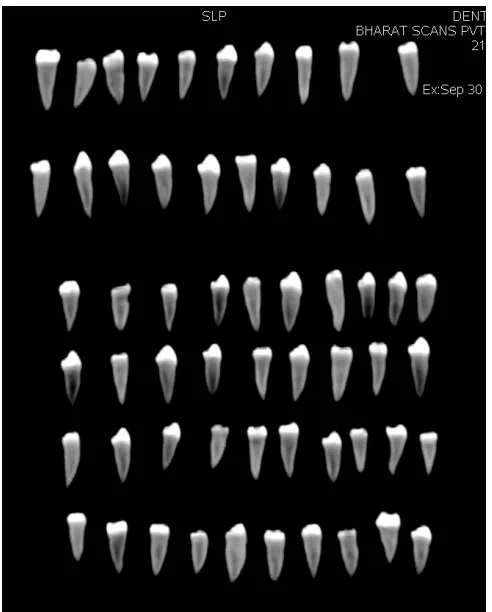
[image:42.595.189.435.379.685.2]Fig No.1 Extracted Mandibular Premolar
29
Fig No.3 Armamentarium.
30
Fig No.5 Restorative Material
31
Fig No.7 Mixing of AH Plus Sealer Fig No.8 Obturation with ProTaper
guttapercha Point
[image:45.595.146.439.334.622.2]32
33
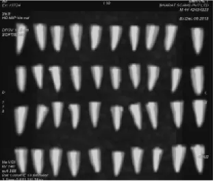
[image:47.595.160.474.89.355.2]Fig No.11 Spiral Computed Tomography Scan after obturation
34
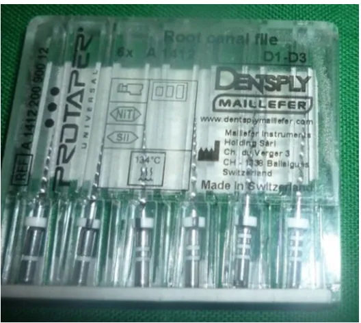
Fig No.13 ProTaper retreatment file
35
Fig No.15 DRaCe retreatment file
[image:49.595.234.399.88.271.2]Fig No.16 Hedstrom file
36
Fig No.18 CT image after retreatment
37 Statistical analysis:
Data obtained in the present study was assessed for normality using Shapiro Wilks test for normality of data and the data was found to be parametric in distribution.
The following statistical tests were carried out in our study:
One way ANOVA for comparison of mean difference between the four groups.
Tukeys Post Hoc Test for pairwise comparison between the groups.
pvalue of ≤ 0.05 was used to determine significance.
The data obtained were tabulated and analysed using SPSS software version 16.
The volume of material obturated in canal for the four groups in cm3 is shown in Table. 1
The volume of the material in the canal after retreatment for the four groups is represented in Table. 2.
The removal efficiency of the 4 groups in percentage were tabulated in Table 3.
Mean and standard deviation of the removal efficiency of the four groups are shown in Table 4
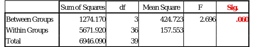
The results of the one way ANOVA comparing the percentage removal efficiency among the four groups is represented in Table 5.
38
The time taken for removal of obturating materials is shown in Table 7. Mean and standard deviation of the time taken for retreatment for the four
groups are shown in Table 8.
The results of the one way ANOVA comparing the time taken for retreatment for the four groups are shown in Table 9.
[image:52.595.100.529.314.679.2] The Multiple comparisons of the time taken for retreatment for the 4 groups using Tukey Post hoc test is tabulated in table 10
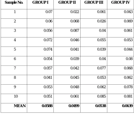
Table 1 : VOLUME OF MATERIAL OBTURATED IN CANAL in cm3
Sample No. GROUP I GROUP II GROUP III GROUP IV
1 0.07 0.022 0.061 0.043
2 0.06 0.068 0.026 0.069
3 0.056 0.087 0.04 0.061
4 0.072 0.046 0.055 0.053
5 0.074 0.041 0.039 0.044
6 0.054 0.039 0.04 0.08
7 0.057 0.042 0.077 0.068
8 0.041 0.045 0.053 0.062
9 0.053 0.048 0.062 0.078
10 0.051 0.061 0.085 0.081
39
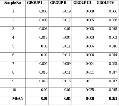
Table 2 : VOLUME OF MATERIAL IN CANAL AFTER RETREATMENT in cm3
Sample No. GROUP I GROUP II GROUP III GROUP IV
1 0.008 0.019 0.008 0.006
2 0.003 0.017 0.005 0.038
3 0.003 0.01 0.008 0.018
4 0.017 0.004 0.003 0.003
5 0.03 0.011 0.006 0.016
6 0.02 0.011 0.006 0.044
7 0.005 0.009 0.004 0.035
8 0.015 0.011 0.011 0.017
9 0.018 0.015 0.011 0.017
10 0.02 0.01 0.025 0.015
40
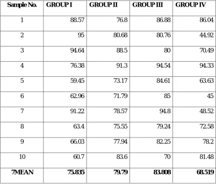
Table 3 : REMOVAL EFFICIENCY (Values in Percentage)
Sample No. GROUP I GROUP II GROUP III GROUP IV
1 88.57 76.8 86.88 86.04
2 95 80.68 80.76 44.92
3 94.64 88.5 80 70.49
4 76.38 91.3 94.54 94.33
5 59.45 73.17 84.61 63.63
6 62.96 71.79 85 45
7 91.22 78.57 94.8 48.52
8 63.4 75.55 79.24 72.58
9 66.03 77.94 82.25 78.2
10 60.7 83.6 70 81.48
41
Table 4 : MEAN AND STANDARD DEVIATION OF THE CALCULATED
REMOVAL EFFICIENCY (PERCENTAGE) OF THE 4 GROUPS
Canal Wall Cleanliness
N Mean Std. Deviation
Protaper 10 75.8350 15.03392
M2 10 79.7900 6.35352
Drace 10 83.8080 7.34278
H file 10 68.5190 17.60428
Total 40 76.9880 13.34559
Table 5 : ONE WAY ANOVA OF REMOVAL EFFICIENCY OF THE 4 GROUPS
ANOVA Canal Wall Cleanliness
Sum of Squares df Mean Square F Sig. Between Groups 1274.170 3 424.723 2.696 .060 Within Groups 5671.920 36 157.553
Total 6946.090 39
[image:55.595.98.532.386.468.2]42
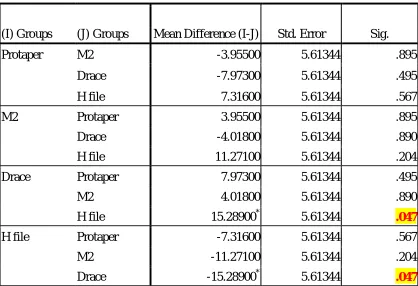
Table 6 : PAIRWISE COMPARISON OF THE REMOVAL EFFICIENCY OF
THE FOUR GROUPS
Tukey HSD
Canal Wall Cleanliness
(I) Groups (J) Groups Mean Difference (I-J) Std. Error Sig.
Protaper M2 -3.95500 5.61344 .895
Drace -7.97300 5.61344 .495
H file 7.31600 5.61344 .567
M2 Protaper 3.95500 5.61344 .895
Drace -4.01800 5.61344 .890
H file 11.27100 5.61344 .204
Drace Protaper 7.97300 5.61344 .495
M2 4.01800 5.61344 .890
H file 15.28900* 5.61344 .047
H file Protaper -7.31600 5.61344 .567
M2 -11.27100 5.61344 .204
Drace -15.28900* 5.61344 .047
43 GRAPH 1
COMPARISON OF PERCENTAGE OF FILLING MATERIAL RETRIEVED
0
10
20
30
40
50
60
70
80
90
REMOVAL EFFICIENCY
GROUPS
Pro Taper R
M Two R
D RaCe
44
Table 7 : TIME IN MINUTES FOR RETREATMENT FOR THE 4 GROUPS
Sample No. GROUP I GROUP II GROUP III GROUP IV
1 7.43 5 6.2 13.36
2 8.25 6.5 5.3 12
3 9.3 4 5.2 11.5
4 8.23 5.2 5 13.5
5 8.3 6.27 5 10.5
6 7 6.3 5.2 9
7 7 6 6 10.5
8 8 6 6.5 11
9 7.5 6.3 5.5 10.2
10 8 6.2 5.8 11.2
45
Table 8 : MEAN AND STANDARD DEVIATION OF THE TIME TAKEN FOR
RETREATMENT FOR 4 GROUPS
Parameter
N Mean Std. Deviation
Timeinminutes Protaper 10 7.9010 .69700
M2 10 5.7770 .79482
Drace 10 5.5700 .52715
H file 10 11.2760 1.39395
Total 40 7.6310 2.48321
Table 9 : ONE WAY ANOVA OF THE TIME TAKEN FOR RETREATMENT
FOR 4 GROUPS
ANOVA Sum of
Squares df
Mean
Square F Sig. Timeinminutes Between
Groups 210.440 3 70.147 84.045 .000 Within
Groups 30.047 36 .835
[image:59.595.104.526.367.502.2]46
Table : 10 MULTIPLE COMPARISONS OF THE TIME TAKEN FOR RETREATMENT FOR THE 4 GROUPS USING TUKEY POST HOC TEST
Tukey HSD
Dependent Vaiable (I) Groups (J) Groups
Mean
Difference (I-J) Std. Error Sig. Timeinminutes Protaper M2 2.12400* .40857 .000
Drace 2.33100* .40857 .000
H file -3.37500* .40857 .000
M2 Protaper -2.12400* .40857 .000
Drace .20700 .40857 .957 H file -5.49900* .40857 .000
Drace Protaper -2.33100* .40857 .000
M2 -.20700 .40857 .957
H file -5.70600* .40857 .000
H file Protaper 3.37500* .40857 .000
M2 5.49900* .40857 .000
Drace 5.70600* .40857 .000
47 GRAPH 2
COMPARISON OF TOTAL TIME FOR RETRIEVAL FOR FOUR GROUPS
0
2
4
6
8
10
12
TIME IN
MINUTES
GROUPS
Pro Taper R
M Two R
D RaCe
48
Result interpretation
Removal efficiency:
Intergroup comparison:
With respect to removal efficiency it was observed that DRaCe group had the highest mean removal efficiency (83.84 ± 7.34) followed by M Two R (79.79 ± 6.35), Pro Taper R (75.83 ± 15.03). Group III Group II Group I > Group IV (Graph 1) .Least mean removal efficiency was observed among H files (68.52 ± 17.60). However this difference was not found to be statistically significant (p=0.06).
Pairwise comparison:
Pair wise comparison with Tukey’s Post Hoc test revealed that that there was a significant mean difference between DRaCe and H files groups. (p=0.04)
Time duration:
Intergroup:
49 Pairwise comparison:
50
Treatment options for the retention of endodontically treated teeth with post treatment disease are limited. They included non surgical retreatment, surgical treatment (including intentional replantation), or a combination of both procedures. Torabinejad M et al 562009 reported that non surgical retreatment showed a higher rate of success (83.0%) compared with endodontic surgery (71.8%) at each recall visit. These findings suggest nonsurgical retreatment offers a more favorable long-term outcome and should be considered as a primary treatment approach when possible compared with endodontic surgery.
Nonsurgical endodontic retreatment is often indicated as the first choice to eliminate or reduce microbial infection when initial root canal treatment fails8. The retreatment aims to completely remove the filling material from the canal system to allow effective cleaning, shaping, and filling of the root canal. It is important to remove as much obturating materials as possible during retreatment to uncover remnants of necrotic tissue or microorganisms that might be responsible for endodontic failure.46
Gutta-percha in conjunction with sealers is the most common root filling material.30AH Plus is a epoxy resin based sealer used in our study which has strong sealing ability due to great adhesion to dentin and consequent reduced bacterial leakage.62
51
nickel-titanium instruments) and other methods like heat, ultrasonic instruments, lasers and solvent.
Solvent was not used in this study with the purpose to eliminate a possible confounding factor. Solvents are potentially harmful. Softened root filling material by solvents could be pushed further into the irregularities along the root canal walls and dentinal tubules, making its removal more difficult.20 NiTi rotary system can be used for removing gutta-percha without solvents. Use of solvents can form a thin film of gutta-percha on the wall of root canal and it can reduce the action of intracanal medicament.13
It has been shown that the use of rotary nickel-titanium (NiTi) instruments for the removal of root fillings during retreatment is safer. 71 Rotary techniques are faster because of the motion, inherent speed, greater taper, flute design and active tip of the rotary files. Rotary files in motion generate frictional heat, which plasticizes and softens gutta-percha. The rotary instruments convey debris toward the cervical portion of the canal because of their cross-sectional shape 18and it is possible to remove gutta-percha and AHplus sealer without solvent.
52
ProTaper retreatment file, Mtwo retreatment file, DRaCe retreatment file. and compared with Hedstrom file without solvent by Computed Tomography.
Evaluation of the effectiveness of removal and identification of remnants of the filling material under clinical conditions is difficult. In experimental studies, this can be monitored by splitting the root and examining the two halves under a microscope. In research, Computed Tomography, micro-CT, Scanning Electron Microscopy, Clearing techniques, and stereomicroscopy have also been used for identification of remnants of filling material and root canal cleanliness. We used Computed Tomography analysis in our study. It can provide three dimensional volume analysis with precisely outlining filling material by a dedicated software. It is non invasive and volume in cm3 can be calculated before and after retreatment.
All instruments should be used in specific speed and low torque for maximum efficiency. The low-torque hand piece approved to increase tactile sensitivity, give better control of rotary instrumentation, and reduced the risk of instrument separation. This study uses gear reduction hand piece driven by electric motor where speed and torque can be controlled may contribute to better efficiency. If we use low speed than recommended by manufacturer does not results in efficient removal of obturating material.
53
part of root canal and D3-ISOsize 20, taper 0.07 used to remove obturating material from apical part of root canal.
Ma J et al28 ( 2012) concluded in his study ProTaper retreatment file leaves less residual filling when it is used without solvent, mean percentage is 6.04% than ProTaper with solvent and supplemental instrumentation with ProTaper finishing file was done in this study.
In our study, it is observed that ProTaper retreatment file can reach working length without solvent but leaves more residual filling than other rotary files because it is not supplemented by additional instrumentation.
Giuliani V et al17 (2008) stated that ProTaper Universal retreatment system performed better than Hedstrom files and also observed that ProTaper retreatment file require less time with respect to Hedstrom file which is in accordance with our study.
T. Rodig et al38 (2014) concluded that root canals retreated with Hedstrom File had significantly less remaining filling material compared with FlexMaster instrument, it is not in accordance with our study. The reason could be previous study used solvent with Hedstrom file. But in our study, H file is used without solvent. To standardize last apical size of Hedstrom file used in our study is No 30 may be reason for more residual filling.
54
ability than Hedstrom file. Retreatment instruments have specially designed for retreatment purpose with flute design, active cutting tip for initial instrument penetration and rotary files in motion generate frictional heat, which plasticizes and softens gutta-percha makes retreatment faster than Hedstrom file.
Mtwo retreatment file consists of Mtwo R1 and Mtwo R2. Both instruments have an S shaped cross section. Mtwo retreatment has active tip for all retreatment instruments and a constant helical angle.
Bramante CM et al6 (2010) compared Mtwo retreatment file and ProTaper retreatment file without solvent and found that ProTaper retreatment file was better than Mtwo retreatment file since ProTaper R leaves lesser filling residue. Mtwo retreatment is better than ProTaper retreatment in our study.
Dadresanfar B et al 11 (2011) studied Efficacy of two rotary systems in removing gutta-percha and sealer from the root canal wall and concluded that Mtwo R without the use of solvent was more efficient in material removal compared to ProTaper retreatment file which is in accordance with our study.
Yadav P et al60 (2013) concluded in his study, that the mean volume of remaining filling materials in the canal were less with the Mtwo retreatment and ProTaper rotary retreatment systems compared to hand instruments and Mtwo retreatment file is efficient than ProTaper retreatment system which is also in accordance with our study.
55
10% tapering capable of 1/3 coronal cleaning. DR2 is a 25mm in length, size 25 file with 4% tapering for 2/3 apical cleaning.
Rodig T et al 37(2012) concluded in his study that DRaCe instruments were associated with significantly less residual filling material than ProTaper Universal Retreatment instruments and Hedstrom file, No difference was found between ProTaper retreatment and Hedstrom file in removal of gutta percha. Our study also revealed that DRaCe is more efficient in removing obturating material than ProTaper retreatment, Mtwo retreatment but statistically not significant.
This finding may be attributed to the alternating cutting edges that prevent the screw effect, favoring penetration in to the filling material.24 The flute area of these instruments allows coronal extrusion of filling material and the smooth instrument surface created by a special electrochemical treatment, which might also contribute to the superior sharpness of these instruments.11,20
Colaco AS and Pai VAR 9(2015 ) concluded in their study that DRaCe is better than ProTaper retreatment file and Hedstrom file which is in accordance with our study. Contrary to this study Silva BMD et al 44 (2012) studied the effectiveness of ProTaper, DRaCe, and Mtwo retreatment files with and without supplementary instruments in the removal of root canal filling material. They stated in their study that DRaCe is less efficient when it is used alone without supplementation than Mtwo retreatment file.
56
All groups after retreatment demonstrated some residual filling material on the canal wall in this study .The apical diameters of ProTaper D3 instrument ISO size 20, MtwoR2 instrument ISO size 25, DRaCe DR2 ISO size 25 respectively. These retreatment instruments, which are designed to reach the working length, may not provide complete removal of root canal filling material from the apical third7. The purpose of study is to find efficiency of retreatment file so additional instrumentation not used in this study. Additional measures such as the supplemental instrumentation with combination of rotary and manual techniques and newer research are recommended to enhance the removal of gutta-percha during endodontic retreatment.
It is observed from the study that epoxy resin sealer is not completely removed from canal wall which is in accordance with study Wilcox LR et al59 (1987), reason may be there is no chemical attachment between gutta-percha and epoxy resin based sealer. There is more tendency for sealer to remain inside the canal wall, so to enhance complete removal of sealer newer techniques are required.
57 Time
The engine driven rotary NiTi file required less time for retreatment than Hedstrom files. This is probably caused by the gutta-percha plasticization resulting from rotation of the instrument4. In the present study all types of rotary NiTi instruments were significantly faster than hand files in removing gutta-percha, while DRaCe and Mtwo retreatment file instrument systems required less time than ProTaper retreatment file.
Garg A et al 15 (2015) concluded in his study that DRaCe and Mtwo required significantly less time than R-Endo and hand file. Hand file took maximum time, which was significantly slower than all groups and DRace removes faster than Mtwo retreatment. However DRaCe and Mtwo retreatment time was statistically insignificant.
Rotary retreatment techniques were more efficient than manual techniques in gutta-percha removal. Among these techniques, the rotary DRaCe retreatment system was most efficient, whereas the manual use of H-files was least efficient in our study. Removal efficiency between DRaCe, Mtwo retreatment, and ProTaper are not statistically significant.
58
59
Forty extracted human mandibular premolar were selected and used for this study. Crowns were resected at the cemento enamel junction using a diamond disc. 40 specimens were randomly divided in to four group of 10 teeth. Teeth were prepared with ProTaper Universal instruments up to F3 size and root canals were obturated with a F3 ProTaper gutta-percha points and a AH Plus sealer by using the single cone technique. Each specimen is mounted for the purpose of taking Spiral CT. Volume of obturation material in each tooth was estimated using Spiral CT. Each group received one of the four treatment protocols.
GroupI -ProTaper retreatment file,
Group II-Mtwo retreatment file,
Group III-D-RaCe retreatment file,
Group IV-Hedstrom file
60
required to enhance cleaning efficiency during retreatment. Gutta-percha with epoxy resin based sealer can be removed from canal without solvent by rotary NiTi file and removal with Hedstrom file is tedious and time consuming.
The working time required for complete retrieval of obturated material from the root canal was significantly longer for Hedstrom file compared to Rotary Niti retreatment file.
Among the mechanical NiTi rotary retreatment files used in our study, DRaCe required less duration for retrieval comparing to Mtwo and ProTaper retreatment files and the difference is statistically not significant.
61
Within the limitation of this study following conclusion were drawn.
Mechanical rotary NiTi retreatment systems are more efficient than manual instrumentation.
DRaCe is more efficient in removing gutta-percha and epoxy resin based sealer in comparison to Mtwo retreatment file and ProTaper retreatment file but difference is not statistically significant.
Manual hand instrument using Hedstrom file without solvent is the least efficient in removing obturation material and the difference with D-Race being statistically significant.
The working time required for retrieval of obturated material from the root canal was significantly longer for Hedstrom file compared to Rotary Niti retreatment file.
i
1. Abramovitz, Bonar SR, Baransi B, Kfir A. The effectiveness of a self-adjusting file to remove residual gutta-percha after retreatment with rotary files. Int Endod J 2012; 45: 386–392.
2. Anbu R, Nandini S, Velmurugan N. Volumetric analysis of root fillings using spiral Computed Tomography: an in vitro study. Int Endod J 2010 Jan;43(1):64-68.
3. Barletta FB, de Mello Rahde N, Limongi O, Moura AAM, Zanesco C, Mazocatto G. In Vitro Comparative Analysis of 2 Mechanical Techniques for
Removing Gutta-Percha during Retreatment. J Can Dent Ass 2007;73(1):65a-65e.
4. Bergenholtz G, Lekholm U, Milthon R, Heden G, Odesjö B, Engström B. Retreatment of endodontic fillings. Scand J Dent Res 1979 Jun;87(3):217-224. 5. Betti LC, Bramante CM, Moraes IG, Bernardineli N, Garcia RB. Efficacy of ProFile .04 taper series 29 in removing filling materials during root canal retreatment—an in vitro study. Oral Surg Oral Med Oral Pathol Oral Radiol Endod 2009;108:e46-e50.
6. Bramante CM, Fidelis NS, Assumpção TS, Bernardineli N, Garcia RB, Bramante AS, de Moraes IG.Heat release, time required, and cleaning ability
of MTwo R and ProTaper universal retreatment systems in the removal of filling material. J Endod 2010 Nov;36(11):1870-1873.
ii
8. Chevigny CD, Dao TT, Basrani BR, Marquis V , Farzaneh F, Abitbol S, Friedman S. Treatment outcome in endodontics: theToronto study—phases 3
and 4: orthograde retreatment. J Endod 2008;34:131–137.
9. Colaco AS and Pai VAR. Comparative Evaluation of the Efficiency of