PERSPECTIVE
Securing the future of research computing in
the biosciences
Joanna LengID1, Massa ShouraID2, Tom C. B. McLeish3, Alan N. RealID4,
Mariann HardeyID4,5, James McCafferty6, Neil A. Ranson7,8, Sarah A. HarrisID7,9*
1 School of Computing, University of Leeds, Leeds, United Kingdom, 2 School of Pathology, Stanford
University, Palo Alto, California, United States of America, 3 Department of Physics, University of York, York, United Kingdom, 4 Advanced Research Computing, University of Durham, Durham, United Kingdom,
5 School of Business, University of Durham, Durham, United Kingdom, 6 Information Services Division,
UCL, London, United Kingdom, 7 Astbury Centre for Structural and Molecular Biology, University of Leeds, Leeds, United Kingdom, 8 School of Molecular and Cellular Biology, University of Leeds, Leeds, United Kingdom, 9 School of Physics and Astronomy, University of Leeds, Leeds, United Kingdom
Author summary
Improvements in technology often drive scientific discovery. Therefore, research requires sustained investment in the latest equipment and training for the researchers who are going to use it. Prioritising and administering infrastructure investment is challenging because future needs are difficult to predict. In the past, highly computationally demand-ing research was associated primarily with particle physics and astronomy experiments. However, as biology becomes more quantitative and bioscientists generate more and more data, their computational requirements may ultimately exceed those of physical sci-entists. Computation has always been central to bioinformatics, but now imaging experi-ments have rapidly growing data processing and storage requireexperi-ments. There is also an urgent need for new modelling and simulation tools to provide insight and understanding of these biophysical experiments. Bioscience communities must work together to provide the software and skills training needed in their areas. Research-active institutions need to recognise that computation is now vital in many more areas of discovery and create an environment where it can be embraced. The public must also become aware of both the power and limitations of computing, particularly with respect to their health and personal data.
Overview
Research computing is the innovative use of computer hardware and software to enhance sci-entific research. Here, we discuss the exciting progress in the biosciences that can be made by embracing computation, in particular because of the recent upsurge in the use of cryo-electron microscopy (cryo-EM) and cryo-electron tomography (cryo-ET) for structure determination at multiple biological length-scales. Breakthroughs in experimental biophysical tools for auto-mated data collection are providing the biosciences, from molecular biology up to the level of cells and tissues, with a torrent of microscopy and informatics data that needs robust software and fast hardware for data processing and a suite of new simulation and modelling tools. The computational challenge of image processing and of integrating experimental information a1111111111 a1111111111 a1111111111 a1111111111 a1111111111 OPEN ACCESS
Citation: Leng J, Shoura M, McLeish TCB, Real
AN, Hardey M, McCafferty J, et al. (2019) Securing the future of research computing in the
biosciences. PLoS Comput Biol 15(5): e1006958. https://doi.org/10.1371/journal.pcbi.1006958
Editor: Anna R. Panchenko, National Institutes of
Health, UNITED STATES
Published: May 16, 2019
Copyright:©2019 Leng et al. This is an open access article distributed under the terms of the Creative Commons Attribution License, which permits unrestricted use, distribution, and reproduction in any medium, provided the original author and source are credited.
Funding: JL would like to thank the UK Engineering
from diverse sources may prove to be the major bottleneck to gaining scientific understanding. As computation becomes ubiquitous, bioscience researchers will need stronger computational skills, which has implications for provision of training. The rapid pace of growth of computa-tion in the biosciences requires that our research community urgently address these issues. We conclude by making predictions about the directions of bioscience computing and the actions required to secure its future.
Introduction
Until recently, only physical scientists routinely needed expertise in supercomputing and the management of large datasets. Bioinformatics and bioimaging are now providing the biologi-cal sciences with a torrent of data. New simulation and modelling tools, underpinned by com-putation, are essential to provide insight and understanding. Given this key principle, we describe the exciting scientific discoveries and understanding that computation will inspire within the biosciences. Although computation also plays a vital role in areas such as ecology, evolution, and population dynamics, here, we focus on molecular biology, bioinformatics, and biomaterials, as these areas are arguably experiencing the most rapid current expansion. We conclude with speculations on the future directions of computing in the biosciences, which highlight the urgent importance of long-term investment in people and infrastructure for bio-science computation.
Scientific background
Building on the past: The ‘omics’ revolution
Genomic DNA sequencing (genomics), quantification of RNA expression levels (transcrip-tomics), microbiome characterisation, and metabolomics studies are providing increasingly more information about how molecular-level changes affect organisms. Omics data present a particular challenge concerning their size and in allowing open access to a global research community. To be searchable and readily accessible, these datasets require the very highest standards of curation. For example, the Encyclopedia of DNA Elements (ENCODE) database now contains approximately 13,000 datasets from nematodes, flies, mice, and humans, total-ling over 500 TB of data [1], and the European Molecular Biology Laboratory–European Bio-informatics Institute (EMBL–EBI), run collaboratively across 16 partner countries, currently totals 120 PB in size. This figure is projected to reach the exabyte scale in 2022 [2]. These resources represent an invaluable shared global resource and thus require new international agreements between government funding agencies to safeguard their capture, curation, and maintenance over the long term [3].
Such a wealth of resources brings new challenges: How do researchers find (and trust) the information that they need, and how do they combine different datasets to make connections that can answer biological questions? A survey of the 2018 online molecular biology database collection reported 82 new databases and 84 updates to previously published computational biology database resources, whereas only 47 databases were discontinued [4]. These databases span biological disciplines as diverse as genomics, transcriptomics and proteomics, evolution-ary analysis, metabolomics, and chemical biology. Recent updates to the Reactome Pathway Knowledgebase [5], which contains molecular details of all known signal transduction and transport pathways, have focused on providing users with diagrammatic and graphical repre-sentations of their queries so that complex metabolic relationships can be more readily under-stood and communicated. The design of interactive software tools that enable exploration of massive datasets will continue to be an active area of research in bioscience computation. For informatics, datasets are presently maintained by a mature network of international
(https://wellcome.ac.uk/home) grants 108466/Z/ 15/Z and 208395/Z/17/Z. The funders had no role in study design, data collection and analysis, decision to publish, or preparation of the manuscript.
Competing interests: The authors have declared
collaborations with a robust community infrastructure. For nascent experimental tools, how-ever, such as those under development for bioimaging, this is not always the case.
Current challenges: The imaging revolution
One of the notable challenges for the biosciences today is to connect from the omics (molecu-lar) level through to the whole organism. Although omics quantifies which molecules are pres-ent, it does not show where they are. New imaging tools, such as cryo-EM, cryo-ET, and superresolution light microscopy, now allow us to visualise biological systems from the level of a single protein molecule to cells and tissues. This will allow us to connect the molecular and cellular levels for the first time, revealing details of processes such as assembly and disassembly of cellular structures, the operation of enzyme-controlled chemical factories, the protein trans-port network, and cell regulation strategies. The Electron Microscopy Database (EMDB), which provides a public archive of 3D electron microscopy reconstructions, grew from 640 entries in 2015 to 4,431 by the end of 2016 and is projected to contain 10,000 entries by 2020 [6]. For comparison, there are already over 139,000 atomic models for biological macromole-cules in the Protein Data Bank (PDB). The complementary Electron Microscopy Public Image Archive (EMPIAR) database contains raw electron microscopy images [7], and discussion of the need for equivalent archives for emerging 3D cellular imaging techniques, including 3D scanning electron microscopy and soft X-ray tomography, have been initiated [8][9][10]. The provision of these resources builds on best practice acquired through the curation of omics and atomistic structural data (e.g., the PDB). The importance of gaining an international con-sensus on common file formats, which is vital for software interoperability (as is nonpropri-etary software), functionality, and usability, has already been established. The bioimaging community faces additional challenges owing to the large size and multiscale nature of the datasets. Curating, sharing, and integrating these data will require new storage, networking, software, and skills infrastructure.
Single-particle cryo-EM imaging is now providing atomic-resolution structural data for macromolecular complexes that have eluded X-ray crystallography [11][12]. Processing and curating this wealth of information requires robust, user-friendly software [6], high-perfor-mance computing (HPC), and bespoke data storage facilities [6]. Cryo-EM facilities can gener-ate over 10 Tb of image data per day per microscope, and potentially>160 Tb each year will need archiving for 10 years (seeS1 Supporting Information) to satisfy the open-data require-ments of funding bodies. As the next generation of detectors become available, this will increase by a factor of around 6 (e.g., in the transition from the Gatan K2 to K3 detector [13]), requiring an equivalent uplift in the data storage and analysis pipelines. Only major research facilities have previously had to tackle the problems of understanding and controlling for con-tinuous data production. This is an urgent issue in many cryo-EM facilities. In response, there is an important emerging industry set around the products and services that are designed to make data more portable and widely accessible, with the expectation that the researcher also stands as an expert ‘software analyst’. Many electron microscopy facilities now do significant data processing concurrently with data collection to speed up processing of the datasets, but it is clear that a substantial, continual investment into networking and storage infrastructures will be required in the future.
nm in thickness, in which the molecular resolution at the surface is approximately 10Å[14]. Soft X-rays now achieve resolutions of<50 nm and can be used for 3D reconstructions of whole cryopreserved cells [15]. Correlated microscopies, in which data from distinct modali-ties are combined to give complementary information, are now used to identify specific mole-cules of interest within 3D cellular landscapes [16] by labelling them with a fluorescent tag or bar-coded strands of DNA [17]. Adaptive optics combined with lattice light-sheet microscopy in transparent living cells has revealed the subcellular dynamics of processes as diverse as the nanoscale diffusion of clathrin-coated pits, cancer cell metastasis, and the motility of axons, which involved mining and visualisation of around half a terabyte of raw data [18]. Much of the future bottleneck for understanding this new wealth of biological information is computa-tional [18]. Although microscope suppliers do already provide bespoke software tools for visu-alising and processing 3D microscopy data, such as the Amira program [19], bioimaging is evolving so rapidly that the future software needs of this community are unknown. As a result, new programming tools, particularly for correlative microscopies and the segmentation of noisy volumetric datasets, now need to be developed concurrently with the experiments that rely on them and be implemented close to the science. The Image Data Resource, for example, combines experimental results from multiple independent imaging modalities for reanalysis [20]. A broad awareness of the growing computational needs of the biosciences is now neces-sary to ensure that the required hardware, software, and technical expertise is available locally.
Requirments for the future: Biomolecular modelling and simulation
Computational tools have been developed at all length-scales in the biosciences, but integrating between these different regimes remains a challenge. Examples in which this challenge has been embraced include the Virtual Cell Software Environment [21], which provides a ‘biol-ogy-orientated’ tool for spatial–temporal modelling and visualisation of biochemical pathways. The European ‘Virtual Physiological Human’ (VPH) project has undertaken to construct a ‘digital representation of the human body and its relevant physiological systems’, with the long-term aim of using computational physiology in biomedical research and clinical practice [22]. In biomechanics, the aspiration is for simulations to speed up the design cycle for medical implants and devices before experimental prototypes are built. In 2016, the United States Food and Drug Administration (FDA) agency issued guidance on the use of computational studies to evaluate the safety and effectiveness of medical devices [23]. Multiscale models of the heart that span from the length-scale of ion channels up to whole-organ models [24] have already been integrated with imaging data from individual patients in research towards personalised surgery [25]. The concept of a ‘Digital Twin’, which is a 4D in silico copy of a physical system, and which has been widely adopted in mechanical engineering for applications as diverse as sensors to power plants, will similarly become an integral component of industrial tissue engi-neering [26].
simulation timescales that can be explored, even with specialist HPC resources. This has, in turn, inspired radical developments in bespoke hardware for MD (e.g., the Anton chip [29] [30]). Biomolecular simulation has a mature software stack that is actively developed and maintained by the community as an international priority. This facilitated the porting of the most popular community codes to graphical processing units (GPUs). As well as the improve-ments in speed, the relatively low cost and easy availability of GPUs have broadened the avail-ability of such tools. Molecular modelling software developers know that usavail-ability is key to user engagement and are investing considerable effort into making their software (which has traditionally been limited to the HPC community) more user friendly. The importance of bringing high-quality computational tools into the everyday repertoire of experimental biolo-gists has been emphasised by the computational biology community [31]. In the molecular biosciences, in silico screening has already been integrated into the rational drug design pipe-line. As their predictive power improves, the hope is that in the future, biomolecular simula-tions will replace animal testing, and patients will be treated with personalised medicines designed using bespoke computer models.
We therefore need a deeper understanding of how changes at the molecular level propagate through to cells and tissues. Statistical approaches, such as those used in systems biology, have proven particularly powerful for identifying correlations in complex datasets that can be used pre-dictively. For example, Bayesian methods have been developed that can assess functional assign-ments to unknown genes—for example, by analysis of hormonal networks described by multiple (e.g., of order 10) experimentally measured rate constants [32]. Machine learning is now being used to extract patterns and correlations from elaborate datasets, and in image processing [33] [34][35], drug design [36], and omics analysis (for a review, see [37]). Identification of correla-tions in large datasets is valuable for hypothesis generation and provides new understanding when combined with complementary tools, such as simulation and experimentation. However, machine learning cannot explain the underlying, causative interactions, which limits its predictive power to within the scope of the data supplied [38]. Computer modelling is therefore essential for applications such as personalised medicine because the number of genetic mutations possible is so vast that the circumstances of individual patients will never be captured in any dataset.
The bioimaging revolution is providing data that is inherently multiscale. To understand how the structures we observe ultimately give rise to biological function requires the development of new models that integrate datasets collected at different spatial and temporal resolutions. For example, the CellView visualisation tool, which is implemented within a game engine, uses the latest GPU-based algorithms to construct and render enormous biological scenes (approximately 15 billion atoms) [39], such as an entire mycoplasma cell [40]. Modelling will also be required to integrate imaging and informatics. The enormous data sizes required to comprehensively map cellular components at an atomistic level (seeS1 Supporting Information) implies that abstrac-tion is essential. The challenge is therefore to capture the detail necessary to understand how a single amino acid substitution can give rise to disease yet build a model that is computationally tractable. Multiscale simulation tools capable of coupling different levels of chemical detail into integrated models are therefore essential for bioscience model building [41]. Multiscale, integra-tive modelling is one of the ‘grand challenges’ of computational chemistry and physics. In the biosciences, it may transform the field into a fully quantitative discipline.
Organisation, structures, and skills
Evolution of research computing technologies and services
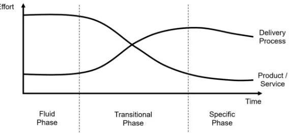
provided inS1 Supporting Information) tools will also evolve. All research continuously gen-erates new and innovative techniques and technologies that disrupt existing approaches. Many technology disruptors follow similar evolutionary paths over time, and there are models to track and predict this.Fig 1shows the well-established model put forward by Abernathy and Utterback [42]. For any new technology, early efforts concentrate on establishing core capabili-ties and features. As the technology matures, the feature set becomes both canonical and com-moditised. Thus, development and operations effort shifts more towards usability and efficiency.
Although improvements in usability and efficiency in the ‘transitional’ and ‘specific’ phases do not necessarily push back the frontiers of research, such developments can massively ‘democratise’ a new approach, as is currently happening in cryo-EM (as demonstrated by the increase in depositions to the EMDB discussed previously). Improvements in service delivery often significantly reduce costs, reducing barriers to adoption and increasing uptake. When a research computing tool set becomes entirely mainstream, increased standardisation for data sharing and workflows further improve access and efficiency, enabling data to be combined in new ways and providing novel insights and understanding.
Bioscience computation is following these trends. In the ‘fluid phase’, when a new algo-rithm is first conceived (for example, for image processing, biomolecular modelling, or infor-matics analysis) it will most likely be implemented by an individual research team. The focus is on developing the core functionality of the software so that new biological questions can be addressed. It is only when the computational tool has been validated that usability and sustain-ability improve and the tool enters the ‘transitional phase’. A computational technology in the ‘specific phase’ is fully mature, often with so many users that the cost of providing it needs to be considered at an institutional level. Examples include standard scientific software such as Matlab and core infrastructure hardware (e.g., GPUs and computing blades).
[image:6.612.204.489.522.653.2]Other aspects of research computing follow this evolutionary path too, with interesting con-sequences. Research ‘grand challenges’, such as the development of the bespoke Anton chip for biomolecular simulation, are located at the far left ofFig 1, as these involve highly innova-tive (and often expensive) computational tools accessible to only a few researchers at their con-ception. New techniques in the ‘fluid phase’ will most likely be funded by short-term academic research grants, whereas technologies in the ‘specific phase’ often receive international, national, or institutional support, mainly through professional information technology (IT) services. This has a profound influence on the working cultures of computing experts
Fig 1. Evolution of research computing technologies, based on the Abernathy-Utterback curve. Innovations in the
fluid phase undergo churn, eventually yielding a dominant design. In the transitional phase, delivery processes become more important than feature sets. Then, in the specific phase, the innovation is well established, and effort is mainly devoted to efficient operation.
supporting technology at each of the various phases, as academic research and IT services have different priorities and career structures (seeS1 Supporting Informationfor a compar-ison between research computing and enterprise IT). Most attrition occurs in the ‘transi-tional phase’. Research groups rarely possess the expertise to transform an academic code into widely usable software. Therefore, new types of research computing experts are needed to meet the growing demand for good scientific software, and such roles are becoming part of the basic fabric of a research-intensive institution [43]. Software developers and systems administrators responsible for research computing hardware are necessarily clustered around the intersection of the two curves inFig 1. The dynamic nature of the evolution of technology necessitates a fully integrated approach between innovative research teams working on the left of the curve (‘fluid phase’) and operational IT teams working on the right (‘specific phase’). This is particularly vital for the biosciences because research com-puting needs to evolve rapidly to keep up with experimental data production. For example, academic bioscience software can be brought to a broader user base by exploiting the mod-ern software development processes of enterprise IT. Conversely, academic research often stretches IT capabilities and drives innovation to meet those challenges. Close collaboration with researchers will enable professional IT services to anticipate future problems based on the experience of the early adopters if both sides are willing to learn and adapt. The relation-ship needs to be bidirectional so that future service provision is informed and relevant, with biological researchers and IT professionals working as partners to reach new and innovative solutions together. Only this collaborative approach is capable of providing bioscientists with the computing tools and skills that they increasingly need with the urgency required to keep pace with experimental advances.
Building computational skills for the biosciences
Software engineers understand the importance of making software easy to use, thus reducing training requirements. However, much of the biosciences software, such as NAMD for MD simulations [44], is extremely sophisticated, so expert knowledge is needed to exploit its full potential. As the quantity and complexity of bioscience data grow, researchers also need to write software of their own. Consequently, python and other high-level programming lan-guages have grown in popularity, and many software packages such as Visual Molecular Dynamics (VMD) [45] provide an internal scripting language for advanced users. Although PhD students and postdocs rely on software development, principal investigators (PIs) may not always be so aware of its intrinsic value because research computing tools are evolving so fast it can be challenging for academic teaching staff (and managers of industrial research teams) to keep up with developments. The consequence of this is that grant applications to improve software usability are often outperformed by standard proposals that address a spe-cific biological research question.
We propose that the research community will require some or all of the following three ele-ments of computational knowledge (ranked by sophistication):
1. An understanding of hardware so that systems and software can be configured most effec-tively and data can be transferred across the network efficiently and securely
2. Practical skills in programming or use of high-level utilities such as databases so that raw data can be processed before importing into specialist research software
Currently, much application-specific software training is delivered by the research commu-nities themselves. Many of the world-class computational bioscience facilities, such as EMBL– EBI, are also centres of excellence for training [2], which brings biological researchers into continual contact with software developers and is key to providing usable and relevant compu-tational tools. Much of this activity is funded by research councils that understand that sup-porting communities to develop good software is critical to their science. Less experienced researchers need training from world experts to use these packages robustly, which implies an understanding of the underlying science, an awareness of the potential pitfalls, and the knowl-edge of the limitations and significant sources of error. However, the growing demand for soft-ware applications training now requires this to be provided by local institutions.
Researchers who are developing code need additional software development skills, such as understanding how to use code repositories and how to design robust test suites. It is currently unlikely that a biosciences researcher will acquire these skills during an undergraduate degree. Consequently, research students and postdocs need to find training at their institution, attend an external course, or draw upon the experience of their local research team. In the United Kingdom, the Software Sustainability Institute (SSI) boasts that it has ‘trained over 5,000 new learners in the basics of software engineering’ [46], which potentially has a massive scientific impact given the high proportion of researchers who require software for their research. As partners of the US Software Carpentry Foundation, the SSI join with a global network of insti-tutions with a staggering diversity of scientific interests, including a strong representation from data-intensive bioscience disciplines [47]. Ensuring that research-intensive organisations provide up-to-date research computing training is essential.
Bioscience computation in the cloud
The cloud has the potential to revolutionise research in the biosciences. The porting of biosci-ences software onto GPUs combined with secure, on-demand access to such hardware in the cloud now provides the opportunity to embed biomolecular modelling and data analytics in far more areas of discovery. The need for good biosciences software, and for people with the skills to write and use it, is therefore set to explode. Although in the future cloud providers may well see commercial value in installing and testing software with a sufficiently large user base, currently cloud computing devolves the responsibility for installing and testing software and choosing the associated hardware platform from centralised facilities to the user. Bioscien-tists will need additional computational expertise, particularly in DevOps/ResOps (seeS1 Sup-porting Information) and support to benefit. Current financial models for cloud-based computing also charge for data ingress/egress, which may slow uptake of applications such as cryo-EM image processing, which is very data intensive. Added to this, the scale of data involved is leading to the physical shipping of hard drives as the only currently practical way of transferring data at this scale [48]. Upgraded network infrastructure and new cost models would therefore be required to remove these roadblocks.
small simulations) research. Research pioneers will continue to demand bespoke hardware facilities until computations sufficiently sophisticated to answer their research questions run efficiently without them. The complexity of biology implies that this will not occur in the fore-seeable future. Therefore, universities, national supercomputing facilities, and the commercial cloud providers will all be required to expand the boundaries of computation and to develop the tools that will ultimately become standard as they become more mainstream.
An even more fundamental change in bioscience computation may arise from new ‘cloud native’ working practices (e.g., the ResOps approach from the EBI [50]). Applications within the cloud need to work independently of the hardware platform, have a higher tolerance for failure, and be able to respond to changes in price. Software ‘containers’ allow for this, provid-ing everythprovid-ing needed to run the code. Containers thus facilitate sharprovid-ing and increased repro-ducibility. As these technologies evolve, the community will need to continue to engage with these new computational tools.
Discussion
Embedding computational thinking within bioscience culture
In molecular biology, materials science, and increasingly, social sciences and humanities, com-putation has become an essential part of the experimental pipeline. The integrity of results relies on both the software and the way the user employs it. Researchers therefore need a deep understanding of the computational aspects of their experiments and the science underlying these tools. Concerns about researchers’ use of software and our readiness to believe the answers it provides are not new in the biosciences [51] and have been reawakened in the light of increasing levels of automation in macromolecular crystallography, which encourages reli-ance on ‘magical black boxes’ [52]. The discussion will intensify as machine learning algo-rithms enter scientific workflows. We must keep questioning how our computational tools are solving a particular problem for us rather than focusing only on the broader research agenda.
Scientifically correct and user-friendly community software is essential to the productivity of researchers. However, software developers face a completely different design challenge to engineers building software platforms for automation of tasks such as payroll or goods deliv-ery, in which complexity must be hidden from users. In research software, the package should communicate the full range of options in an intuitive manner, inform users of the choice of defaults, and provide easy access to detailed explanations of input variables, with caveats, through links to online manuals, tutorials, and research papers. Wherever possible, informa-tive error checking and validation procedures should be built into the workflow. These must give understandable and practical advice or risk being ignored by users. Creating software that engages users appropriately is an enormous challenge, but the benefits can be dramatic. This is amply shown in the rapid increase in cryo-EM structure depositions, which have been enabled in part by rapid improvements in software functionality and ease of use [53][54][55]. The architectural equivalent of software platforms for organisations is a transport hub, such as a railway station or an airport, where providing the most efficient route to the final destination is critical. For research software, the design principles should mimic those of professional library services, in which engagement, exploration, and education are paramount.
the data using a different approach. This external validation will improve scientific rigor, as com-mon mistakes will be identified, corrected, and then avoided in the future. Moreover, by being interactive, these archives would be engaging and educational and enable large-scale collaborative bioscience projects across multiple international sites.
A subtle and informed discussion is necessary on what ‘open data’ means. Providing public access to datasets for a decade following the completion of a research project is challenging. Many experimental and computational projects now create volumes of data vastly greater than what was envisaged when the 10-year standard was created, and such datasets may be beyond the limit that is practical to curate. Examples are particle physics experiments, cryo-EM data-sets, biomolecular simulations, X-ray free-electron lasers (XFELS) (seeS1 Supporting Informa-tion), and supercomputer simulations for cosmology. However, it is also clear that
preservation of raw datasets can enable invaluable new insights, such as the extraction of dynamic information from cryo-EM [56]. The effectiveness of open-data policies in practice will depend critically on curation because reuse is impossible if the data cannot be found, and protocols will be unreproducible unless they are clearly explained. Data can be much easier to preserve than executable software, which can have hidden dependencies in both software and hardware that are not recorded and may be difficult to recreate. This is a problem that research computing and curation experts need to resolve.
Predictions for the future
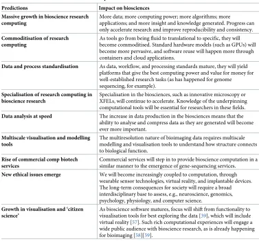
Given the above, we make the following predictions on the future of research computing in the biosciences (seeTable 1).
Surveying the current landscape and anticipated future directions leads us to the following three requirements to secure the future of research computing in the biosciences:
1. Investment in the e-infrastructure environment: The analysis, curation, and sharing of data will require robust and sustained investment in networking, data storage, HPC, software, support, and people at the local, national, and international levels.
2. Importance of collaborative research communities: Communities are vital. They ensure the interoperability of datasets and software. They define ‘grand challenge’ problems that require collaboration. They deliver bespoke training and share expertise. Communities advocate and influence, securing the investment necessary.
3. Computation is an integral part of the scientific method: The understanding gained from experimental biology arises from computation, either through analysis, modelling, or both. Computational thinking needs to be embedded within the culture of biology, creating a research computing landscape that is resilient enough to accommodate a continual flow of disruptive methodology and analysis.
Conclusions
novel technology and the people who support it. The closer integration between research puting and enterprise IT needed to deliver computational tools to an increasingly varied com-munity of researchers requires that institutions recognise the contribution that supportive IT service provision makes to research. The success of any research computing initiative in the biosciences will be judged by the novelty and depth of the biological questions it answers; therefore, performance metrics for people, projects, and processes should be designed to sup-port research.
More broadly, growth in cheap, online public access to personalised biological datasets, such as genome sequencing [60], microbiome analysis [61], and biometrics collected from wearable sensor technologies (e.g., fitness trackers and social media apps to record diet), along with the classification of personalised data characterised as the ‘quantified self’ [62], will fur-ther fuel the omics revolution. These innovations will define the research questions asked by biologists, not only in response to public demand but also from legislators trying to regulate these new industries. In July 2014, the European Commission led a consultation on medical devices and mobile health (mHealth) apps and proposed a code of conduct [63]. In parallel, manufacturers are in the process of marketing smart technology–enabled medical devices to allow healthcare providers access to patient data, including remote monitoring and cloud-Table 1. Predictions for the future of biocomputation.
Predictions Impact on biosciences Massive growth in bioscience research
computing
More data; more computing power; more algorithms; more
applications; and more insight and knowledge generated. Progress can only accelerate research and improve reproducibility and consistency.
Commoditisation of research computing
As tools go from being fluid to translational to specific, they will become commoditised. Standard hardware models (such as GPUs) will become more pervasive, and software reuse will happen more through containers and cloud applications.
Data and process standardisation As data, workflow, and processing standards mature, they will yield platforms that give the best computing power and value for money for well-established research tasks (as has happened for genome sequencing, for example).
Specialisation of research computing in bioscience research
Specialisation in the biosciences, such as innovative microscopy or XFELs, will continue to accelerate. Knowledge of the underpinning computational tools will be essential for researchers in these fields.
Data analysis at speed The increase in data production in the biosciences means that the ability to analyse and compress data as they are generated will become ever more important.
Multiscale visualisation and modelling tools
The multiresolution nature of bioimaging data requires multiscale modelling and visualisation tools to understand how structure connects to biological function.
Rise of commercial comp biotech services
Commercial services will step in to provide bioscience computation in a similar manner to the emergence of gene-sequencing services.
New ethical issues emerge We will become increasingly coupled to computation, through wearable sensor technologies, virtual reality, and implantable devices. The long-term consequences for society will require a broad interdisciplinary base to assess, e.g., neuroscience, genomics, psychology, physiology, and computer science.
Growth in visualisation and ‘citizen science’
As bioscience software matures, focus will shift from functionality to visualisation tools for best exploring the data [39], which will include virtual reality [57]. Such rich computational experiences will engage a wide public audience with bioscience research, as is already happening for bioimaging [58][59].
Abbreviations: GPU, graphical processing unit; XFEL, X-ray free-electron laser.
[image:11.612.198.575.84.435.2]based data-sharing systems. Although the outcomes are too revolutionary to be foreseen, with-out doubt, computational tools will be vital to the analysis and interpretation of individuals’ data. As the world becomes ever more technologically empowered, we must remember to engage mindfully with computation and the answers that it produces to make certain that we are informed more often than we are misled.
Understanding the molecular choreography that allows cells to work, how this is affected by a disease, and our relationships with other living organisms will influence societal attitudes to health and lifestyle, medicine, and our impact on the environment. The imaging revolution, combined with informatics, physical modelling, and visualisation, will lead to profound new insights. The aim of ‘cellular cartography’ is to chart out the whole atlas of the cell, in which all structural and omics information is unified within a single multidimensional, multiscale computational framework [57]. Realising this ambition will place computation at the very cen-tre of biological research and will, therefore, drive a massive uptake of computational tools and skills by bioscientists.
Supporting information
S1 Supporting Information. This contains two case studies that quantify the future computational needs (e.g., networking, compute, and storage) of key areas of structural biology. The first, ‘Quantitative estimates for the computational requirements of single particle cryo-EM studies’, focuses on imaging, and the second, ‘Data storage sizes for an atomistic map ofC.elegans’, focuses on computer modelling and simulation, including a discussion of coarse graining. The final appendix, ‘Research Computing, Enterprise IT and bioscience computation support’, compares the approaches of IT services and research computing and describes how they can work together to support bioscientists.
(PDF)
Acknowledgments
Thanks to Arend Dijkstra (University of Leeds), Andy Fire (Stanford University), Idan Gab-dank (Stanford University), Molly Gravett (University of Leeds), Izzy Jayasinghe (University of Leeds), Peter Jimack (University of Leeds), Julia Mahamid (European Molecular Biology Laboratory), Marlene Mengoni (University of Leeds), Haruki Nakamura (Osaka University), Arwen Pearson (Hamburg), Stefan Przyborski (University of Durham), William Sharrock (University of Manchester), Rebecca Thompson (University of Leeds), and Jeremy Yates (Uni-versity College London).
References
1. Davis CA, Hitz BC, Sloan CA, Chan ET, Davidson JM, Gabdank I, et al. The Encyclopedia of DNA ele-ments (ENCODE): data portal update. Nucleic Acids Research. 2018; 46:D794–D801.https://doi.org/ 10.1093/nar/gkx1081PMID:29126249
2. Cook CE, Bergman MT, Cochrane G, Apweiler R, Birney E. The European Bioinformatics Institute in 2017: data coordination and integration. Nucleic Acids Res. 2018; 46:D21–D9.https://doi.org/10.1093/ nar/gkx1154PMID:29186510
3. Anderson WP. Global Life Science Data Resources Working G. Data management: A global coalition to sustain core data. 2017; 543:7644.
4. Rigden DJ, Fernandez XM. The 2018 Nucleic Acids Research database issue and the online molecular biology database collection. Nucleic Acids Research. 2018; 46:D1–D7.https://doi.org/10.1093/nar/ gkx1235PMID:29316735
5. Fabregat A, Jupe S, Matthews L, Sidiropoulos K, Gillespie M, Garapati P, et al. The Reactome Pathway Knowledgebase. Nucleic Acids Res. 2018; 46:D649–D55.https://doi.org/10.1093/nar/gkx1132PMID:
6. Patwardhan A. Trends in the Electron Microscopy Data Bank (EMDB). Acta Crystallogr D Struct Biol. 2017; 73:503–8.https://doi.org/10.1107/S2059798317004181PMID:28580912
7. Iudin A, Korir PK, Salavert-Torres J, Kleywegt GJ, Patwardhan A. EMPIAR: a public archive for raw electron microscopy image data. Nat Methods. 2016; 13(5):387–8.https://doi.org/10.1038/nmeth.3806
PMID:27067018
8. Patwardhan A, Ashton A, Brandt R, Butcher S, Carzaniga R, Chiu W, et al. A 3D cellular context for the macromolecular world. Nat Struct Mol Biol. 2016; 21(10):841–5.
9. Kuhlbrandt W. Biochemistry. The resolution revolution. Science. 2014; 343(6178):1443–4.https://doi. org/10.1126/science.1251652PMID:24675944
10. Ellenberg J, Swedlow JR, Barlow M, Cook CE, Sarkans U, Patwardhan A, et al. A call for public archives for biological image data. Nature Methods. 2018; 15:849–854. https://doi.org/10.1038/s41592-018-0195-8PMID:30377375
11. Thompson RF, Iadanza MG, Hesketh EL, Rawson SD, Ranson NA. Collection, pre-processing, and on-the-fly analysis of data for high-resolution, single-particle cryo-electron microscopy. Nature Protocols. 2018; 14:100–118.
12. Scheres SH. RELION: implementation of a Bayesian approach to cryo-EM structure determination. J Struct Biol. 2012; 180(3):519–30.https://doi.org/10.1016/j.jsb.2012.09.006PMID:23000701 13. Gatan [Internet]. c2018 [cited 2019 Apr 26]. K3 Camera. Available from:
http://www.gatan.com/k3-camera.
14. Earnest TM, Watanabe R, Stone JE, Mahamid J, Baumeister W, Villa E, et al. Challenges of Integrating Stochastic Dynamics and Cryo-Electron Tomograms in Whole-Cell Simulations. J Phys Chem B. 2017; 121(15):3871–81.https://doi.org/10.1021/acs.jpcb.7b00672PMID:28291359
15. Ekman AA, Chen JH, Guo J, McDermott G, Le Gros MA, Ca L. Mesoscale imaging with cryo-light and X-rays: Larger than molecular machines, smaller than a cell. Biol Cell. 2017; 109(1):24–38.https://doi. org/10.1111/boc.201600044PMID:27690365
16. Kopek BG, Paez-Segala MG, Shtengel G, Sochacki KA, Sun MG, Wang Y, et al. Diverse protocols for correlative super-resolution fluorescence imaging and electron microscopy of chemically fixed samples. Nat Protoc. 2017; 12(5):916–46.https://doi.org/10.1038/nprot.2017.017PMID:28384138
17. Jayasinghe I, Clowsley AH, Lin R, Lutz T, Harrison C, Green E, et al. True Molecular Scale Visualization of Variable Clustering Properties of Ryanodine Receptors. Cell Rep. 2018; 22(2):557–67.https://doi. org/10.1016/j.celrep.2017.12.045PMID:29320748
18. Liu TL, Upadhyayula S, Milkie DE, Singh V, Wang K, Swinburne IA, et al. Observing the cell in its native state: Imaging subcellular dynamics in multicellular organisms. Science. 2018; 360:6386.
19. Thermo Fisher Scientific [Internet]. c2018 [cited 2018 Apr 15]. Amira Software for the Life Sciences. Available from:https://www.fei.com/software/amira-for-life-sciences/.
20. Williams E, Moore J, Li SW, Rustici G, Tarkowska A, Chessel A, et al. Image Data Resource: a bio-image data integration and publication platform. Nature Methods. 2017; 14:775–781.https://doi.org/10. 1038/nmeth.4326PMID:28775673
21. Loew LM, Schaff JC. The Virtual Cell: a software environment for computational cell biology. Trends in Biotechnology. 2001; 19(10):401–6.https://doi.org/10.1016/S0167-7799(01)01740-1PMID:11587765 22. Viceconti M, Hunter P. The Virtual Physiological Human: Ten Years After. Annu Rev Biomed Eng.
2016; 18:103–23.https://doi.org/10.1146/annurev-bioeng-110915-114742PMID:27420570
23. Food and Drug Administration (FDA). Reporting of Computational Modeling Studies in Medical Device: Submissions Guidance for Industry and Food and Drug Administration Staff. FDA; 2010 [cited 2018 Jun 5]. Available from:https://www.fda.gov/downloads/MedicalDevices/DeviceRegulationandGuidance/ GuidanceDocuments/UCM381813.pdf.
24. Colman MA, Castro SJ, Perez Alday EA, Hancox JC, Garratt C, Zhang H. Recent progress in multi-scale models of the human atria. Drug Discovery Today: Disease Models. 2014; 14:23–32.
25. Trayanova NA, Chang KC. How computer simulations of the human heart can improve anti-arrhythmia therapy. J Physiol. 2016; 594(9):2483–502.https://doi.org/10.1113/JP270532PMID:26621489 26. Geris L, Lambrechts T, Carlier A, Papantoniou I. The future is digital: in silico tissue engineering.
Cur-rent Opinion in Biomedical Engineering; 2018; 6:92–98.
27. Gray A, Harlen OG, Harris SA, Khalid S, Leung YM, Lonsdale R, et al. In pursuit of an accurate spatial and temporal model of biomolecules at the atomistic level: a perspective on computer simulation. Acta Crystallogr D Biol Crystallogr. 2015; 71:162–72.https://doi.org/10.1107/S1399004714026777PMID:
28. Huggins DJ, Biggin PC, Da¨mgen MA, Essex JW, Harris SA, Henchman RH, et al. Biomolecular simula-tions: From dynamics and mechanisms to computational assays of biological activity. Wiley Interdisci-plinary Reviews: Computational Molecular Science. 2018; 9:e1393.https://doi.org/10.1002/wcms.1393 29. Shaw DE, Dror RO, Salmon JK, Grossman JP, Mackenzie KM, Bank JA, et al. Millisecond-scale Molec-ular Dynamics Simulations on Anton. In: Proceedings of the Conference on High Performance Comput-ing NetworkComput-ing, Storage and Analysis. SC ’09. New York, NY, USA: ACM; 2009 [cited 2019 Apr 26]. p. 39:1–39:11. Available from:http://doi.acm.org/10.1145/1654059.1654099.
30. Dror RO, Dirks RM, Grossman JP, Xu H, Shaw De. Biomolecular simulation: a computational micro-scope for molecular biology. Annu Rev Biophys. 2012; 41:429–52. https://doi.org/10.1146/annurev-biophys-042910-155245PMID:22577825
31. Cui Q, Nussinov R. Making biomolecular simulations accessible in the post-Nobel Prize era. PLoS Comput Biol. 2014; 10:8.
32. Vernon I, Liu J, Goldstein M, Rowe J, Topping J, Lindsey K. Bayesian uncertainty analysis for complex systems biology models: emulation, global parameter searches and evaluation of gene functions. BMC Syst Biol. 2018; 12:1.https://doi.org/10.1186/s12918-017-0484-3PMID:29291750
33. Wu J, Ma YB, Congdon C, Brett B, Chen S, Xu Y, et al. Massively parallel unsupervised single-particle cryo-EM data clustering via statistical manifold learning. PLoS ONE. 2017; 12:8.
34. Langlois R, Pallesen J, Ash JT, Nam Ho D, Rubinstein JL, Frank J. Automated particle picking for low-contrast macromolecules in cryo-electron microscopy. J Struct Biol. 2014; 186(1):1–7.https://doi.org/ 10.1016/j.jsb.2014.03.001PMID:24607413
35. Zeng X, Leung MR, Zeev-Ben-Mordehai T, Xu MA. Convolutional autoencoder approach for mining fea-tures in cellular electron cryo-tomograms and weakly supervised coarse segmentation. J Struct Biol. 2018; 202(2):150–60.https://doi.org/10.1016/j.jsb.2017.12.015PMID:29289599
36. Chen H, Engkvist O, Wang Y, Olivecrona M, Blaschke T. The rise of deep learning in drug discovery. Drug Discov Today; 2018; 23:1241–1250.https://doi.org/10.1016/j.drudis.2018.01.039PMID:
29366762
37. Lin E, Lane H. Machine learning and systems genomics approaches for multi-omics data. Biomark Res. 2017; 5:2.https://doi.org/10.1186/s40364-017-0082-yPMID:28127429
38. Jordan AM. Artificial Intelligence in Drug Design-The Storm Before the Calm? ACS Medicinal Chemistry Letters. 2018; 9:1150–1152.https://doi.org/10.1021/acsmedchemlett.8b00500PMID:30613315 39. Le Muzic M, Autin L, Parulek J, Viola I. cellVIEW: a Tool for Illustrative and Multi-Scale Rendering of
Large Biomolecular Datasets. Eurographics Workshop Vis Comput Biomed. 2015; 2015:61–70.https:// doi.org/10.2312/vcbm.20151209PMID:29291131
40. Klein T, Autin L, Kozlikova B, Goodsell DS, Olson A, Groller ME, et al. Instant Construction and Visuali-zation of Crowded Biological Environments. IEEE Trans Vis Comput Graph. 2018; 24(1):862–72.
https://doi.org/10.1109/TVCG.2017.2744258PMID:28866533
41. Voth GA. A Multiscale Description of Biomolecular Active Matter: The Chemistry Underlying Many Life Processes. Accounts of Chemical Research. 2017; 50(3):594–598.https://doi.org/10.1021/acs. accounts.6b00572PMID:28945406
42. Abernathy WJ, Utterback JM. Patterns of Industrial Innovation. Technical Review. 1978; 8(2).
43. Brett A, Croucher M, Haines R, Hettrick S, Hetherington J, Stillwell M, et al. Research Software Engi-neers: State of the Nation Report 2017. Swindon, UK: Engineering and Physical Sciences Research Council (EPSRC); 2017.
44. Phillips JC, Braun R, Wang W, Gumbart J, Tajkhorshid E, Villa E, et al. Scalable molecular dynamics with NAMD. J Comput Chem. 2005; 26(16):1781–802.https://doi.org/10.1002/jcc.20289PMID:
16222654
45. Humphrey W, Dalke A, Schulten K. VMD: Visual molecular dynamics. Journal of Molecular Graphics. 1996; 14(1):33–8. PMID:8744570
46. software.ac.uk [Internet]. The Software Sustainability Institute; c2018 [cited 2019 Apr 26]. Available from:https://www.software.ac.uk/.
47. software-carpentry.org [Internet]. The Software Carpentry Foundation; c2018 [cited 2019 Apr 26]. Avail-able from:https://software-carpentry.org/.
48. Microsoft Azure [Internet]. c2018 [cited 2019 Apr 26]. Import/Export Pricing. Available from:https:// azure.microsoft.com/en-gb/pricing/details/storage-import-export/.
49. Microsoft Azure [Internet]. c2018 [cited 2019 Apr 26]. Big Compute: HPC & Batch. Available from:
https://azure.microsoft.com/en-us/solutions/big-compute/.
51. Kleywegt GJ, Jones TA. Where freedom is given, liberties are taken. Structure. 1995; 3(6):535–40. PMID:8590014
52. Kleywegt GJ, Jones TA. Homo Crystallographicus—Quo Vadis? Structure. 2002; 10(4):465–72. PMID:
11937051
53. Fernandez-Leiro R, Scheres SHW. A pipeline approach to single-particle processing in RELION. Acta Crystallographica Section D. 2017; 73(6):496–502.https://doi.org/10.1107/S2059798316019276
PMID:28580911
54. Punjani A, Rubinstein JL, Fleet DJ, Brubaker MA. cryoSPARC: algorithms for rapid unsupervised cryo-EM structure determination. Nature Methods. 2017; 14(3):290–6.https://doi.org/10.1038/nmeth.4169
PMID:28165473
55. Grant T, Rohou A, Grigorieff N. cisTEM, user-friendly software for single-particle image processing. eLife. 2018; 7:e35383.https://doi.org/10.7554/eLife.35383PMID:29513216
56. Frank J. New Opportunities Created by Single-Particle Cryo-EM: The Mapping of Conformational Space. Biochemistry. 2018; 57:6.
57. Horwitz R, Johnson GT. Whole cell maps chart a course for 21st-century cell biology. Science. 2017; 356(6340):806–7.https://doi.org/10.1126/science.aan5955PMID:28546174
58. Zooniverse [Internet]. c2018 [cited 2019 Apr 26]. Etch a cell. Available from:https://www.zooniverse. org/projects/h-spiers/etch-a-cell.
59. Bruggemann J, Lander GC, Su AI. Exploring applications of crowdsourcing to cryo-EM. J Struct Biol. 2018; 203:37–45.https://doi.org/10.1016/j.jsb.2018.02.006PMID:29486249
60. 23andme.com [Internet]. 23 and Me; c2018 [cited 2019 Apr 26]. Available from:https://www.23andme. com.
61. uBiome.com [Internet]. uBiome; c2018 [cited 2019 Apr 26]. Available from:https://ubiome.com/.
62. Lupton D. The quantified self. Hoboken, NJ: John Wiley and Sons; 2016.