White Rose Research Online URL for this paper: http://eprints.whiterose.ac.uk/139693/
Version: Accepted Version
Article:
Camilleri, M, Chedid, V, Ford, AC orcid.org/0000-0001-6371-4359 et al. (7 more authors) (2018) Gastroparesis. Nature reviews Disease primers, 4 (1). ARTN 41. 41-. ISSN
2056-676X
https://doi.org/10.1038/s41572-018-0038-z
This article is protected by copyright. This is an author produced version of a paper published in Gastroparesis. Uploaded in accordance with the publisher's self-archiving policy.
[email protected] https://eprints.whiterose.ac.uk/
Reuse
See Attached
Takedown
If you consider content in White Rose Research Online to be in breach of UK law, please notify us by
Accepted for publication 1st October 2018
gim/c/c/gastroparesis revised Nat Rev Dis Primers/Gastroparesis_v4_clean_9_28_18.docx
9-28-18
Gastroparesis
Michael Camilleri1, Victor Chedid1, Alexander C. Ford2, Ken Haruma3, Michael Horowitz4, Karen L. Jones5, Phillip A. Low6, Seon-Young Park7, Henry P. Parkman8,
Vincenzo Stanghellini9
1Division of Gastroenterology and Hepatology, Mayo Clinic, Rochester, MN, USA
2Leeds Gastroenterology Institute, Leeds Teaching Hospitals NHS Trust, Leeds, UK, and Leeds
Institute of Biomedical and Clinical Sciences, University of Leeds, Leeds, UK
3Department of Internal Medicine 2, General Medical Center, Kawasaki Medical School,
Okayama, Japan
4Endocrine and Metabolic Unit, Royal Adelaide Hospital, Adelaide, SA, Australia
5National Health and Medical Research Council of Australia Centre of Research Excellence in
Translating Nutritional Science to Good Health, University of Adelaide, Adelaide, SA, Australia
6Department of Neurology, Mayo Clinic, Rochester, MN, USA
7Division of Gastroenterology, Chonnam National University School of Medicine, Gwangju,
Republic of Korea
8GI Section, Department of Medicine, Temple University School of Medicine, Philadelphia, PA
9Department of Digestive Diseases, Policlinico S.Orsola-Malpighi, University of Bologna,
Bologna, Italy
Abstract
Gastroparesis is a disorder characterized by delayed gastric emptying of solid food in the absence
of a mechanical obstruction of the stomach, resulting in the cardinal symptoms of early satiety,
postprandial fullness, nausea, vomiting, belching and bloating. Gastroparesis is now recognized
gastric accommodation. The overlap between upper gastrointestinal symptoms makes the
distinction between gastroparesis and other disorders, such as functional dyspepsia, challenging.
Thus, a confirmed diagnosis of gastroparesis requires measurement of delayed gastric emptying
via an appropriate test, such as gastric scintigraphy or breath testing. Gastroparesis can have
idiopathic, diabetic, iatrogenic, post-surgical, or post-viral aetiologies. The management of
gastroparesis involves correcting fluid, electrolyte and nutritional deficiencies, identifying and
treating the cause of delayed gastric emptying (for example, diabetes mellitus); and suppressing
or eliminating symptoms with pharmacological agents as first line therapies. Several novel
pharmacologic agents and interventions are currently in the pipeline and show promise to help
tailor individualized therapy for patients with gastroparesis.
[H1] Introduction
A key function of the stomach is to produce acid and facilitate the peptic digestion of food. In
addition to this, the main motor functions of the stomach are accommodation, which allows the
delivery and storage of food, followed by trituration (grinding of food into fragments) and
emptying of solid food. Gastroparesis is a chronic disorder that is characterized by delayed
emptying of the stomach after eating (gastric emptying), in the absence of any mechanical
obstruction, particularly pyloric stenosis1. Cardinal symptoms include early satiety after eating,
postprandial fullness, nausea, vomiting, belchingand bloating. The syndrome is caused by
neuromuscular dysfunction that leads to delayed gastric emptying. To elaborate, the basic
mechanisms that lead to gastroparesis involve derangements in extrinsic neural control
(particularly vagal function), dysfunction of the intrinsic nerves and interstitial cells involved in
local control of gastrointestinal muscle function, and the loss of function of smooth muscles.
Gastroparesis can be idiopathic, associated with diabetes mellitus, can occur after a
medical intervention (iatrogenic or post-surgical), may be associated with neurological disorders
or may occur after a viral or bacterial infection, such as Salmonella gastroenteritis2 .
Interestingly, Helicobacter pylori infection does not seem to influence gastric emptying or
accommodation, but may be associated with heightened sensitivity in patients with functional
dyspepsia(a disorderassociated with accelerated or delayed gastric emptying, impaired gastric
accommodation and heightened sensitivity in the upper gastrointestinal tract)3,4. Rarely, specific
dysautonomia that results in a generalized motility disorder including gastroparesis5. In addition, many other conditions such as Parkinson’s disease, collagen vascular diseases (such as systemic sclerosis), chronic intestinal pseudo-obstruction, and other conditions can lead to gastroparesis or
delayed gastric emptying(Box 1). All of these causesultimately induce gastroparesis through
induction of neuromuscular dysfunction.
In recent years, suggestions have been made to change the definition of gastroparesis to “gastroparesis and related disorders”, therefore recognizing the disorder as part of a broader spectrum of gastric neuromuscular dysfunction6. There is symptom overlap between
gastroparesis and postprandial distress syndrome, which is one of the recognized types of
functional dyspepsia7. Functional dyspepsia may be associated with accelerated or delayed
gastric emptying, impaired gastric accommodation and heightened sensitivity in the upper gut8,9.
With the availability of measurements of gastric volume with scintigraphy, single photon
emission computed tomography (SPECT), or MRI, disorders of gastric accommodation, which
result typically from functional dyspepsia or prior gastric surgery (such as fundoplication or
vagal injury or vagotomy)can be differentiated from gastroparesis. Thus, the term gastroparesis
should be restricted to disorders in which upper gastrointestinal symptoms are associated with
delayed gastric emptying.
In this Primer, we cover mechanisms of gastric innervation and emptying, before
describing how these pathways are perturbed in gastroparesis. We review epidemiological data
and diagnosis of gastroparesis and the effective management of this disorder. Finally, we discuss
future research directions.
[H1] Epidemiology
Describing the global epidemiology of gastroparesis is challenging, as some symptoms of
gastroparesis, such as upper abdominal pain or discomfort, belching, bloating, and early satiety
overlap with those of functional dyspepsia10,11. One important aetiology of gastroparesis is
diabetes mellitus; in one tertiary referral series, diabetes mellitus accounted for almost 1/3 of all
cases of gastroparesis.12Notably, symptoms attributable to gastroparesis are reported by 5–12%
of patients with diabetes mellitus13.In clinical practice, there are approximately equal numbers
of patients with type 1 and type 2 diabetes being evaluated for upper gastrointestinal symptoms,
studies among patients with diabetes and age-stratified and sex-stratified controls showed a
similar prevalence of upper gastrointestinal (GI) symptoms in both type 1 and 2 diabetes, in
studies from the United States and Australia14,15. Thus, documentation of delayed gastric
emptying via gastric scintigraphy or breath testing is required in order to distinguish between
gastroparesis and functional dyspepsia. As a result, most natural history studies of gastroparesis
have been conducted in referral populations, with very few community-based studies. In
addition, most epidemiological data describing the burden of gastroparesis are from the United
States.
[H2] Incidence and prevalence
A population-based study in Minnesota estimated that the age-adjusted incidence of
gastroparesis during a 10-year period was 2.4 cases per 100,000 person-years for men, and 9.8
cases per 100,000 person-years for women; prevalence was estimated to be 9.6 cases per 100,000
individuals for men, and 37.8 cases per 100,000 individuals for women16. Some individuals with
typical symptoms of gastroparesis may never undergo confirmatory testing; one study estimated
that as many as 1.8% of the general population may have gastroparesis, but only 0.2% are
diagnosed.17 Presumably, this relates to a lack of awareness of the disorder, and existing
diagnostic confusion caused by an overlap between the symptoms of gastroparesis and functional
dyspepsia. The same studyreported that consultation rates were similar between those with
symptoms typical for gastroparesis and those with functional dyspepsia, however, those with
gastroparesis-like symptoms were unlikely to undergo a gastric emptying test17. Given that
current estimates of prevalence are based on clinical data obtained from patients who sought
medical attention, these estimates may be too low, as they could be impacted by
healthcare-seeking behavior among patients with symptoms suggestive of gastroparesis.
[H2] Risk factors
The reason for the higher incidence and prevalence of gastroparesis in women is unclear.
However, stomach motility is dependent on neuronal nitric oxide synthesis, and this pathway
may be regulated by estrogen.18,19 Few studies existconcerning the effect of body mass on
gastroparesis. In one study of patients with type 2 diabetes, obesity was associated with an
demonstrated an association between higher body mass index and delayed gastric emptying on
scintigraphy in a cohort of 140 Indian patients with type 2 diabetes.6 Interestingly, it has been
reported that almost 50% of patients with idiopathic gastroparesis are overweight or obese, and
that symptom patterns differ according to body mass; patients who were obese or overweight had
statistically significant lower scores for loss of appetite and inability to finish a meal, but
statistically significantly worse scores for gastroesophageal reflux-type symptoms compared to
patients of a healthy weight11 . The role of other modifiable risk factors, such as smoking or
alcohol, in gastroparesis is unproven even though sizeable proportions of patients with diabetes
mellitusand control individuals used tobacco or alcohol in the same epidemiological study.11 In
one longitudinal follow-up study conducted among 262 patients with gastroparesis, treated
according to the current standard of care in the USA, a history of smoking was significantly
associated with no improvement in symptoms during 48 weeks of follow-up21.
[H3] Diabetic gastroparesis.
Among a historical cohort of patients with diabetes mellitus and control individuals (269 with
type 1 diabetes; 409 with type 2 diabetes; and 735 control individuals) the cumulative
proportions who developed gastroparesis (based on delayed gastric emptying by standard
scintigraphy or gastroparesis symptoms for >3 months plus a physician diagnosis of
gastroparesis or food retention on endoscopy or upper gastrointestinal radiology) over a 10-year
period were 5.2% of patients with type 1 diabetes, 1.0% of patients with type 2 diabetes, and
0.2% of control individuals.22 By contrast, in referral populations of patients with type 1 or type
2 diabetes mellitus, the prevalence of individual symptoms suggestive of gastroparesis is
between 25–53%23, the cardinal symptoms occur in 10%,24,25 and a clinical diagnosis of
gastroparesis is made in almost 5% of patients.13 The presence of presumed gastroparesis in
patients with diabetes mellitus is associated with other end-organ damage, including retinopathy
and peripheral neuropathy, higher mean levels of glycosylated haemoglobin (HbA1c, which is a
measure of glycemic control over the prior three months), and lower socioeconomic status.13,24
The seminal studies of the long-term complications of type 1 diabetes are the Diabetes
Control and Complications Trial (DCCT) and its follow-up, Epidemiology of Diabetes
Interventions and Complications (EDIC). A recent report indicates that delayed gastric emptying
cross-sectional studies.27 Improvements in glycemic control in patients with diabetes mellitus are
associated with decreases in the incidence of microvascular complications and it would not be
surprising if the incidence of diabetic gastroparesis were also to decrease. In agreement with this,
individuals with longstanding type 2 diabetes without evidence of autonomic impairment are
usually reported to have normal gastric emptying28.
Among patients with diabetes mellitus, once delayed gastric emptying is established, it
may persistfor up to 25 years of follow-up, despite evidence of improved glycemic control.29-31
Interestingly, hospitalization rates and emergency department consultations for gastroparesis
appear to be on the rise,32-34 which is possibly owing to both an increase in awareness of
gastroparesis as a potential cause of upper GI symptoms and the increased prevalence of both
type 1 and type 2 diabetes.35 Hospital admissions for exacerbation of symptoms in patients with
gastroparesisare influenced by glycemic control, infection rates (most frequently urinary tract
infections), and poor adherence with or intolerance of medications36. There are regional
variations in inpatient management of patients with diabetic gastroparesis, which are likely to
reflect local differences in healthcare delivery.37 Mortality during a hospital stay has been
estimated at 1.2%, mostly related to comorbidities.38 During a 16-year period in the United
States, mean hospital charges per patient for management of gastroparesis increased by 160%,
and the national bill increased over 10-fold.34
[H2] Life expectancy
In terms of the effect of gastroparesis on life expectancy, data are conflicting. Studies conducted
in referral populations demonstrate no effect of delayed gastric emptying on mortality among
patients with diabetes mellitus, after 12 years of follow up in one study39 and 25 years in a
second study.31 However, in a community-based survey in Minnesota, USA, among individuals
with gastroparesis of mixed etiology (typcially documented by symptoms and delayed gastric
emptying by scintigraphy or gastric food retention on imaging reports), survival was lower than
expected for age- and sex-matched individuals without gastroparesis.16
Although there have been advances in understanding the mechanisms and pathophysiology of
gastroparesis, there are still significant gaps in knowledge, inconsistencies across studies,
potential differences between different aetiological groups (for example, diabetic versus
idiopathic) and therefore individualization of therapy is currently best achieved by carefully
identifying functional impairment rather than cellular mechanisms. One example is the
recognition of concomitant reduction in gastric accommodation among patients presenting with
symptoms suggestive of gastroparesis.
Gastroparesis and impaired gastric accommodation result from neuromuscular
dysfunction of the stomach. Triturationof food in the stomach grinds food into fragments; food
fragments are liquefied by a combination of gastric acid digestion and antral contractions; these
contractions establish high liquid shearing forces and propel food particles against the closed
pylorus before ~1–2mm sized particles are emptied into the duodenum.40,41 Vagal innervation of
the stomach by the vagus afferent nerve is essential for the gastric accommodation of consumed
food. The antral contractions that are essential for triturating solid food and gastric emptying are
mediated by extrinsic vagal innervation and intrinsic cholinergic neurons. In addition, intrinsic
inhibitory mechanisms, such as nitrergic neurons, facilitate pyloric relaxation and intragastric
peristalsis. Nitrergic neurons are pivotal for the relaxation of GI muscle ahead of a contraction,
and they are responsible for descending inhibition ahead of the upstream contraction, which is
induced by excitatory neurons, such as cholinergic and tachykininergic neurons.42 These
inhibitory and excitatoryneural effects are transmitted through interstitial cells of Cajal (ICC)
and possibly other fibroblast-like cells that have pacemaker function, and to smooth muscle cells
in GI muscles, which causes the muscular layer of the stomach to behave as a multicellular
electrical syncytium, so that coordinated contractions, which initiate in the proximal stomach and
involve the entire circumference of the stomach, can propagate towards the antropyloric region.
This electrical syncytium consists of smooth muscle cells, ICCand fibroblast-like cells, which
are positive for platelet-derived growth factor receptor alpha (PDGFR ).43 The ICCs and
PDGFR -positive cells are considered to be pacemaker cells in the GI tract and possess the
ability to transmit electrical signals (Figure 1). In gastroparesis, delayed gastric emptying is
associated with antral hypomotility and, in some patients, with pyloric sphincter dysfunction
[H2] Intrinsic neuropathy
Recent studies have explored the histopathological features and expression of neurotransmitters
in the intrinsic mechanisms involved in gastric motor function (Figure 2). Light microscopy
examination of full thickness gastric biopsies from 20 patients with idiopathic gastroparesis, 20
patients with diabetic gastroparesis, and 20 controls individuals undergoing gastric bypass
surgery, demonstrated no statistically significantdifferences between diabetic and idiopathic
gastroparesis in ninemorphological endpoints; these were protein gene product9.5 (expressed in
the cytoplasm of neurons and serving as a marker of nerve cells), vasoactive intestinal peptide
(an inhibitory neurotransmitter), substance P (an excitatory neurotransmitter), tyrosine
hydroxylase (an enzyme expressed in adrenergic neurons), protein S100 (a marker of glia),
mast/stem cell growth factor receptor Kit (a cell surface marker of ICC), CD45 (cell surface
marker of lymphocytic immune cells) and CD68 (a cell surface marker for monocytic immune
cells), and smoothelin (a marker of smooth muscle cells). However, there were reduced numbers
of inhibitory neuronsexpressing neuronal nitric oxide synthase (nNOS) in patients with
gastroparesis, with decreased nNOS neurons in 40% of patients with idiopathic gastroparesis and
in 20% of patients with diabetic gastroparesis compared to control individuals.The reduction in
nNOS-positive neurons may contribute to impaired gastric emptying by reducing the
coordination of gastric peristalsis that is essential to establish trituration of solid food in the
gastric antrum.44 Some, but not all reports have shown reductions in numbers ofICCs in the
body of the stomach of patients with both diabetic and idiopathic gastroparesis45,46; such a
reductionin ICCs would be expected to impair conduction of electrical activity through the
electrical syncytium and, therefore, interfere with coordinated gastric electrical rhythms,
peristalsis, trituration and gastric emptying. However, it is still unclear whether damage to ICCs
or reductions in their number results in symptom generation. In contrast to patients with diabetic
gastroparesis, biopsies from patients with idiopathic gastroparesis showed altered smooth muscle cell contractile protein expression and loss of PDGFR + cells without a significant change in the numbers of ICCs.46
The reduction in or damage to ICCs in the stomach of some patients with gastroparesis was
associated with reduction in the numbers of anti-inflammatory M2 macrophages, which normally
express mannose receptor (CD206) and heme oxygenase-1 (HO1), and mediate cell repair.45 The
reduction in M2 macrophages reduces the protection of neural tissue from the effects of
oxidative stress and inflammation, both of which are mechanisms involved in the
pathophysiology of diabetes mellitus,47 and may conceivably result in gastric intrinsic neural
dysfunction.
Studies have provided discordant information on immune cells in biopsies; reduced
numbers of M2 macrophages in one study,45 but numbers comparable with healthy tissuein a
separate study.46 These contradictory observations on M2 macrophages are complicated by the
fact that the function of the vagus nerve (which affects immune cell function) was not tested in
relation to the histopathological findings, and vagal neuropathy is associated with diabetic
gastroparesis. Indeed, efferent vagus signals activate and thereby release noepinephrine from splenic nerves, and the transmitter activates 2-adrenergic receptor ( 2AR) expressed on T cells, which activates T cells to release acetylcholine (Ach), which acts on the 7 nicotinic
acetylcholine receptor ( 7 nAChR) on macrophages and other immune cells. Ultimately, the
complete vagus stimulated pathway suppresses the release of pro-inflammatory cytokines.48
Interestingly, another study found that counts of ICCs were inversely correlated with 4-hour
gastric retention in patients with diabetic gastroparesis (that is slower gastric emptying was
associated with higher numbers of ICC in the circular muscle per field), and myenteric immune
infiltrate was associated with overall clinical severity and nausea in patients with idiopathic
gastroparesis.49 The relationship between hyperglycemia, oxidative metabolism, ICCs, and
gastric emptying is the subject of ongoing research.50
[H3] Heme oxygenase 1.
HO1 attenuates the overall production of reactive oxygen species. In gastric tissues obtained
from animal models (tested predominantly in non-obese diabetic mice), loss of HO1 leads to
increased oxidative stress, loss of Kit expression (implying predominantly a loss of ICCs) and
emptying. Expression of HO1 is low in the muscularis propria of stomach under normal
conditions, whereas HO1 is markedly upregulated in muscularis propria resident macrophages
after diabetes develops in a nonobese diabetic mouse model.51 These observations led to the
hypothesis that when macrophages are not producing HO1 to reduce oxidative stress (a frequent
consequence of diabetes, for example, in the causation of neuropathy), the intrinsic mechanisms
that are responsible for normal motor function are damaged. Interestingly, alterations in the
activity of HO1 that may result from variation in a gene controlling its synthesis supports the
potential association of impaired HO1 function and development of gastroparesis. For the
HMOX1 gene promoter, sequences containing longer polyGT repeats had lower transcriptional
activity than sequences with fewer repeats52,53; therefore, shorter polyGT repeat alleles result in
higher expression of HO1 protein. A recent study showed that polyGT alleles in the HO1 gene
(HMOX1) are longest in patients with type 2 diabetes and gastroparesis, and the promoters were
longer in patients with gastroparesis (idiopathic or diabetic) compared to control individuals.54
However, in all the patient groups with gastroparesis studied, allele lengths were longer in
African Americanscompared to other ethnic groups, and the differences in the proportion of
African Americans in the groups may have accounted for at least some of the differences
between patients with gastroparesis and control individuals. It is still unclear whether this genetic
variant is actually more prevalent in patients with gastroparesis and how it might contribute to
dysfunction of the intrinsic mechanisms that impair gastric emptying.
[H3] PDGFR + fibroblasts.
Recent insights suggest there may be abnormalities in PDGFR + fibroblasts in patients with
gastroparesis. These fibroblasts were reduced in numberin gastric biopsy samples from patients
with idiopathic gastroparesis with increased numbers ofCD68+ monocytes, but no change in the
numbers of ICCs in one study46. By contrast, a second study found that PDGFR fibroblast-like
cells were not altered in distribution or overall numbers in idiopathic or diabetic gastroparesis.55
In summary, the underlying neuromuscular and neuroimmune mechanisms of gastroparesis are
[H2] Diabetic Gastroparesis
Diabetic gastroparesis is multifactorial with contributions from hyperglycemia, extrinsic (vagal)
denervation, and intrinsic neural denervation (discussed above). In general, the mechanisms do
not appear to differ between type 1 and type 2 diabetes, probably because gastroparesis tends to
occur years after diagnosis with diabetes mellitus typically in association with other features
such as neuropathy (Box 2)and oxidative stress that occur in both type 1 and 2 diabetes.
[H3] Hyperglycemia.
The role of hyperglycemia in the pathophysiology of diabetic gastroparesis is unclear. This has
been evaluated in epidemiological studies, in human studies with experimental acute
hyperglycemiaand in natural history studies of diabetes control. There is epidemiological
evidence of associations of hyperglycemia with upper GI symptoms,56 and studies document
poor glycemic control in 36% of patients admitted to a hospital in the U.S. for exacerbations of
diabetic gastroparesis.36 Kidney and pancreas transplant probably have significant beneficial
impacts on gastric emptying and associated GI symptoms, which suggests that control of
hyperglycemia may be beneficial for gastroparesis.57
In human experiments that imposed acute hyperglycemia, typically 8mmol/L
(144mg/dL) (typically associated with glucose clamp studies (in which the blood glucose is
increased and maintained by infusion of glucose) there were definite effects on gastric functions;
an inhibition of antral contractility and delayedgastric emptying during hyperglycemic clamp
compared to euglycemia, and a dose-dependent slowing of gastric emptying with hyperglycemia
(even in the range observed postprandially) when compared to euglycemia. Similarly, clamping
blood glucose at 250mg/dL was associated with induction of gastric dysrhythmias in healthy
human volunteers.5859,60 This is in contrast to the effect of insulin-induced hypoglycemia
(defined as blood glucose ~2.6 mmol/L) which markedly accelerates gastric emptying, possibly
through stimulation of vagal function.This acceleration of gastric emptying is likely to be
important in the counter-regulation of hypoglycemia.61
Natural history studies provide uncertain evidence of the relationship between glycemic
control and gastric emptying. In a study of 129 patients, HbA1cwas not a statistically significant
predictor of abnormal) gastric emptying of solids using scintigraphy (discussed below).62 In
type 2 diabetes.63 By contrast, in the DCCT/EDIC cohort comprising exclusively patients with
type 1 diabetes, gastroparesis was associated with relatively worse glycemic control compared to
diabetic patients with better glycemic control as assessed by HbA1c26 It is also uncertain whether
improving chronic hyperglycemia over a relatively short term results in a meaningful
improvement in gastric emptying. Improved glycemic control has, hitherto, been reported to not
be associated with any change in gastric emptying in patients with delayed gastric emptyingin
type 1 or type 2 diabetes,64,65 apart from one uncontrolled study.66 Further studies are required to
appraise the relationship of long-term hyperglycemia and gastric emptying.
[H2] Iatrogenic and surgical gastroparesis
The most common surgical association with gastroparesis is with fundoplication and bariatric
procedures and the most common iatrogenic associations are with µ-opioid agonists and
hypoglycemic agents such as amylin analogs (for example, pramlintide) or glucagon-like
peptide-1 (GLP-1) analogs or agonists (for example, liraglutide and exenatide) but not
dipeptidyl peptidase IV inhibitors such as vildagliptin and sitagliptin which increase GLP-1,
improves glycemia without delaying gastric emptying.67Medications used in the treatment of
Parkinsonism including levodopa and anticholinergic medications may also cause iatrogenic
gastroparesis.
[H3] Post-surgical gastroparesis.
Post-surgical gastroparesis is generally caused by to damage to or entrapment of the vagus nerve,
and this occurs most commonly with fundoplication or bariatric surgical interventions, as truncal
vagotomy for peptic ulceration is now rarely performed. Although laparoscopic sleeve
gastrectomy has been associated with aberrant distal ectopic pacemaking in the human
stomach68, there was no evidence that this caused delay in gastric emptying; in fact, the typical
effect of LSG is acceleration of gastric emptying.69
Rarer forms of post-surgical gastroparesis result from Billroth I and II gastrectomy (which are
rarely performed nowadays for treatment of peptic ulceration) sometimes accompanied by
vagotomy, as well as partial esophagectomy for esophageal cancer or heart transplantation which
[H3] Opioids.
Mu- Opioids such as codeine, oxycodone and morphine, have pharmacological effects
throughout the GI tract. They decrease gastric emptying and stimulate pyloric tone, inhibit
propulsion, increase the amplitude of non-propulsive segmental contractions, increase fluid
absorption in the small and large intestine, increase anal sphincter tone, and impair reflex
relaxation of the anal sphincter in response to rectal distention.70 At the cellular level, opioids
inhibit adenylate cylase and nerve terminal Ca2+ channels and activate K+ channels, leading to an
inhibition of Ach release from enteric interneurons and purine/nitric oxide release from
inhibitory motor neurons.71 In the stomach, opioids stimulate pyloric sphincter tone and phasic
pressure, and inhibit gastric motility such as antral contractility,72,73 resulting in impaired gastric
emptying, postprandial nausea and early satiety.70 Many recent series of patients with
gastroparesis show relatively high prevalence of concomitant treatment with opioids and central
neuromodulator drugs, such as antidepressants (74-77).
The effects of opioids on gastroparesis are illustrated by the report from Temple University in
223 patients with gastroparesis: 70.9% not taking opioids, 9.9% taking opioids only as needed,
19.3% on chronic scheduled opioids for at least 1 month; of the latter group, 8.1% were on
opioids for gastroparesis and/or stomach pain. The patients on chronic scheduled
opioidscompared to non-opioid controls with gastroparesis had higher symptom severity, lower
employment rate, and higher rates of hospitalizations over 1 year, and worse outcomes with
treatment for gastroparesis with prokinetics agents and gastric electrical stimulation.78,79 These
data have to be assessed in the context that higher hospitalization rates and resource utilization
are common to chronic opioid users in general and are not unique to those with gastroparesis.
[H2] Post-viral
Post-infectious dyspepsia has been described in the literature, but the evidence is rather weak as
it is based on presence of symptoms such as myalgia during the acute onset of symptoms rather
than serological or tissue demonstration of viral etiology. Gastroparesis has been rarely
associated with specific viral infections, for example, Epstein-Barr virus, norovirus, herpes virus
and cytomegalovirus usually in association with the development of autonomic dysfunction such
as postural hypotension or abnormal sweating; this form of post-viral gastroparesis in the setting
generally associated with a good prognosis when patients are followed for ~1 year.80,81 The few
available literature reports do not provide data on the typical time lag from virus infection to
development of gastroparesis, or whether there are any genetic or acquired factors that
predispose to the development of post-viral gastroparesis.
[H2] Other causes of gastroparesis
Gastric neuromuscular disorders may result from extrinsic denervation, intrinsic neuropathy,
disorders of pacemaker cells or of smooth muscle. Smooth muscle disorders (myopathic
disorders) may be infiltrative (for example, scleroderma) or degenerative (for example, hollow
visceral myopathy, amyloidosis, and rarely, mitochondrial cytopathy). Myopathic disorders are
invariably associated with a component of more generalized motility disorder affecting other
regions of the gut, for example, small bowel, lower esophageal sphincter (LES) and esophagus.
Moreover, scleroderma is associated with systemic features such as CREST (calcinosis,
Raynaud’s, esophageal, sclerodactyly and telangiectasia) syndrome, and there may be external
ophthalmoplegia or skeletal muscle involvement in mitochondrial cytopathy. The degeneration
of smooth muscle cells and/or surrounding fibrosis is considered to be the mechanism underlying
the impairment of gastric emptying in these disorders.
[H1] Diagnosis, screening and prevention
In general, the severity of gastroparesis is assessed by the degree of nutritional impairment or
weight loss, or the degree of delay to gastric emptying (for example, the proportion of food
retained in the stomach at 4 hours), which will be impacted by the method and meal used to
assess the overall function of the stomach. Several symptom severity scales also exist to assess
clinical signs and symptoms.
[H2] Clinical Signs and Symptoms
The clinical symptoms of gastroparesis include nausea, vomiting, early satiety, postprandial
fullness, bloating, belching and upper abdominal discomfort which may overlap with symptoms
observed in functional dyspepsia and accelerated gastric emptying.82,83 Several symptom severity
the Gastroparesis Symptom Index (GCSI), which is based on the comprehensive Patients
Assessment of Upper Gastrointestinal Disorders-Symptoms (PAGI-SYMP)84, and the revised
GCSI-Daily Diary (GCSI-DD).85 These scales have been used in clinical trials to assess the
effects of treatment in clinical studies of gastroparesis. However, the scales have not been
utilized for assessment of symptoms in clinical practices, such as deciding whether patients
should undergo diagnostic studies of diverse gastric functions.
[H3] Clustered symptoms.
The cardinal symptoms of gastroparesis typically occur in combination, not as individual
symptoms. In a cohort of 483 patients with type 1 and 2 diabetes mellitus, upper GI symptoms
occurred in clusters, for example, pain with early satiety and heartburn; heartburn with bloating,
early satiety, nausea and vomiting; and regurgitation with bloating, nausea and vomiting.86 The
symptoms of idiopathic and diabetic gastroparesis are similar, although vomiting and early
satiety are more frequent in diabetic gastroparesis and pain is more frequent in idiopathic
gastroparesis.74
[H3] Nausea and vomiting.
Nausea is a highly prevalent symptom in patients with gastroparesis, irrespective of aetiology,
and it is frequently associated with vomiting.74,75 As mentioned above, vomiting occurs more
often in diabetic gastroparesis than in idiopathic gastroparesis. However, in patients with
idiopathic gastroparesis from the NIH Gastroparesis Cohort of 393 patientsa severe delay in
gastric emptying was associated with more severe symptoms of vomiting.11
[H3] Pain.
In the NIH Consortium Gastroparesis Cohort, the predominant symptoms were pain and/or
discomfort in 21% of patients (in two-thirds of these patients, this pain was graded at moderate
or severe) and nausea and/or vomiting in 44% of patients. The presence of pain was not
associated with the results of gastric emptying test, or with presence of diabetic neuropathy or
control of diabetes.87
Severe early satiety and postprandial fullness are common in both diabetic and idiopathic
gastroparesis, and severity is associated with the severity of other gastroparesis symptoms, that
is, body weight, gastric emptying, and the volume of water that a patient is able to consume at a
constant rate in a water load test.76
[H3] Bloating.
Bloating is prevalent in patients with gastroparesis, with 41% of patients reporting mild
symptoms and 14% of patients reporting severe symptoms. Bloating severity relates to female
sex and heavier body mass.11 However, gastric emptying did not correlate with bloating
severity.77
[H2] Diagnosis
Patients with postprandial upper abdominal symptoms such as nausea, vomiting, postprandial
fullness, bloating and epigastric pain are candidates for gastric motility tests. Patients must first
undergo an upper gastrointestinal endoscopy; if this test does not reveal a cause for the
symptoms, patients can begin motility and functional investigations. The most relevant
functional test is a measurement of gastric emptying. Gastric emptying is best assessed with
scintigraphy88,89 conducted over at least 3 hours; stable isotope breath test90 is also approved by
regulatory agencies; these tests are well validated, and data from healthy control individuals are
available (Figure 3). When results from tests of gastric emptying are inconclusiveparticularly in
patients with prominent postprandial fullness or early satiety, measurements of gastric
accommodation are also indicated. Impaired gastric accommodation is diagnosed with validated
methods where available such as SPECT91 and MRI,92 or with screening tests such as the
proximal stomach size on the gastric scintiscan (taken immediately after radiolabeled meal
ingestion),93 or by a water load or nutrient drink test.94
Table 1 shows some of the more widely used tests to measure gastric motility in
suspected gastroparesis.
[H3] Gastric emptying scintigraphy.
Gastric emptying scintigraphy is a functional test involving the ingestion of a solid meal
multicenter studies in the United States, Canada, and Europe, using Western style meals, usually
scrambled eggs (Figure 3); it is important to assess the amount of food ingested when applying
normal values, and therefore the meals need to be of sufficient calorie and fat content to serve as a minor “stress test” for the stomach, but not so large as to be impossible to be consumed by symptomatic patients.88,95 Gastric emptying scintigraphy is relatively reproducible with good
concordance correlation coefficient (0.54~0.83) between two repeated studies.96 Although this
Western style meal has the advantage of good tolerability by the majority of symptomatic
patients, it has some limitations. First, Western style meals are not familiar to most Asian
patients. Therefore, some Asian centers have modified the components of the test meal, instead
using rice-based meals composed of steamed rice, a microwaved egg and water (267 kcal: 57%
carbohydrate; 23% fat and 19% protein), which contain a calorie and fat content that is
intermediate between the Eggbeaters meal (255kcal, 2% fat) and the 2 scrambled egg meal
296kcal, 30% fat) typical of the Western style meals,88,89 to measure gastric emptying by
scintigraphy. Using this modified meal, the upper limits of gastric retention at 2 hours and 4
hours post consumption (95th percentile) were 49.8% and 8.8%, respectively97, which is similar
to values (60% at 2 hours; 10% at 4 hours) obtained using the Eggbeaters meal in consensus
guidelines.88 Given the possible effects of glycemia on gastric emptying, it is recommended that
blood glucose should be less than 10mmol/L or 180mg/dL in the fasting period before starting
the gastric emptying test.
Second, a few patients may not be able to tolerate any solid food. Moreover, the low fat
and calorie content of the test meal may not adequately test gastric motility, and may fail to
identify impaired gastric motor function in some patients. Therefore, alternative test meals have
been proposed, such as a liquid nutrient meal (Ensure Plus® meal)which has a very similar
gastric emptying profile when compared to a solid meal of comparable caloric content in healthy
participants, but these alternative test meals have yet to be clinically validated in male or female
patients with gastroparesis98.Moreover, the operational definition of gastroparesis relies on a
delay in emptying solid foods, and not liquids.
The timing of scintigraphy imaging after consumption of a radiolabelled meal is also of
importance. For estimating solid phase gastric emptying, data shows that imaging up to 4 hours
after meal consumption detects more patients with delayed gastric emptying than imaging over
power exponential curve fitting, or more simply by linear interpolation, as solid foods empty
with a relatively linear pattern in the postlag phase, that is the phase after trituration of solids has
been completed, and the solid phase of the meal starts to empty linearly from the stomach. This
simplification with scans obtained hourly for up to 4 hours reduces costs and provides a relevant
parameter that appraises overall gastric emptying, such as the percentage of the meal emptied at
2, 3, and 4 hours after consumption; in addition, linear interpolation between the percentages
emptied at 1-4 hours allows estimation of gastric emptying half-time (T1/2).100
[H3] Stable isotope breath test.
The gastric emptying breath test incorporates a stable isotope, 13C, in a substrate such as octanoic
acid or spiruluna platensis (blue-green algae) This noninvasive method is easy to perform, with
similar cost to scintigraphy, and does not involve exposing patients to ionizing radiation;
therefore, these tests are possible to use in pregnant or breast-feeding women, and in children.
The principle underlying this test, which has been clinically validated as an alternative to gastric
emptying scintigraphy, is that the rate of gastric emptying of the 13C substrate incorporated in the
solid mealis reflected by breath excretion of 13CO2.90,101 The test is conducted over a 4 hour
period after an 8h fast. Pre-meal breath samples are collected, patients eat a special test meal, and
after consuming the meal, additional breath samples (typically every 30 minutes) are collected at
specified times. Thus, as the meal empties from the stomach, the medium chain triglyceride
(octanoic acid) or the amino acids in spirulina (which contains 50%– 60% protein, 30% starch,
and 10% lipid) rapidly undergo digestion, absorption, and metabolism to produce 13CO2 which is
exhaled in the breath. Since the rate limiting step of all of these processes is the rate of gastric
emptying, the cumulation of 13CO2 in breath reflects the rate of gastric emptying. Confounders
that may influence the test results are changes in endogenous CO2 excretion caused by physical
activity such as walking and malabsorption, pancreatic exocrine insufficiency, significant lung or
liver disease, or cardiac failure (Figure 3).102
[H3] Wireless motor capsule.
A wireless motor capsule (WMC) (SmartPillTM) has been approved by the U.S. FDA for the
evaluation of gastric emptying and colonic transit in patients with suspected slow transit
GI tract.103 The completion of gastric emptying is demonstrated by an abrupt change in pH into
alkaline range due to WMC passage into the duodenum. Gastric emptying by WMC correlated
moderately with simultaneous gastric emptying of a low-fat meal measured by concurrent
scintigraphy.104 However, there was only 52.5% agreement with scintigraphy,105 and further
validation in patients with gastroparesis is required.105
[H3] Gastroduodenal manometry.
Antroduodenal manometry is the intraluminal measurement of the pressure activity in the distal
stomach and duodenal loop during fasting and postprandially. The technique, which is conducted
at very few centers, can be used to assess gastric and/or small intestinal motility disturbances. In
the postprandial period, a distal antral contraction pressure of <40mmHg is suggestive of a
myopathic disorder, and reduced frequency of normal amplitude distal antral contractions is
suggestive of myopathic or neuropathic dysfunction106. Interestingly, manometry studies of 102
patients with gastroparesis showed abnormalities suggestive of neurogenic derangement in the
proximal small bowel, especially in patients with documented delayed gastric emptying107. These
data confirm earlier observations of small intestinal involvement in the neuropathic process in
some patients who present with upper GI symptoms suggestive of gastroparesis.108
At centers that perform antroduodenal manometry, the finding of antral hypomotility and failure
to respond to prokinetics and anti-emetics would be an indication for drainage of the stomach
with percutaneous endoscopic gastrostomy, support of nutrition with jejunal feeding and, if
unsuccessful in maintaining nutrition, institution of long-term total parenteral nutrition
[H2] Differential Diagnosis
The symptoms of gastroparesis are nonspecific and may result from other sensory or motor
disorders of the upper GI tract, including impaired gastric accommodation. A study of 1287
patients with upper GI symptoms enrolled at a tertiary care center over ~10 years measured
gastric emptying by scintigraphy and gastric accommodation by SPECT, and found there were
approximately equal numbers of patients with either delayed gastric emptying, or impaired
definition, gastroparesis is undistinguishable from functional dyspepsia with delayed gastric
emptying. Approximately 25–35% of patients with dyspeptic symptoms are estimated to have
delayed gastric emptying.9,10 Functional dyspepsia is reviewed in another primer article.7
Severe gastroparesis must be clinically differentiated from chronic intestinal
pseudo-obstruction (CIPO). The two pathological disorders are characterized by similar clinical
manifestations, gastrointestinal motor abnormalities and some form of underlying neuromuscular
disorder.44,107,109 The main difference between patients with gastroparesis and patients with CIPO
is that patients with CIPO have episodes resembling mechanical intestinal obstruction. Correct
diagnosis is essential, as patients with CIPO are more frequently exposed to useless and
potentially dangerous surgical procedures.
Other conditions to differentiate from gastroparesis are rumination syndrome,110,111
cannabinoid hyperemesis syndrome (CHS),112 and cyclic vomiting syndrome (CVS)113. Chronic
unexplained nausea and vomiting CUNV) or chronic nausea and vomiting syndrome (CNVS) are
identified as symptom subgroups of functional upper gastrointestinal disorders with unknown
prevalence and overlap of symptoms with gastroparesis and functional dyspepsia9.
Patients with rumination syndrome may present with effortless, repetitive regurgitation,
chewing or sucking and re-swallowing or spitting of previously ingested food. The disorder is
not associated with nausea, but weight loss can occur and rumination can be mistaken for
vomiting. The diagnosis is primarily based on careful history and clinical observation, and the
manifestations are similar in adults and adolescents.110,111 A recent review made a series of
recommendations regarding clinical management of rumination syndrome: Clinicians strongly
should consider rumination syndrome in patients who report consistent postprandial
regurgitation.: The presence of nocturnal regurgitation, dysphagia, nausea, or symptoms
occurring in the absence of meals does not exclude rumination syndrome, but makes it less
likely. Diaphragmatic breathing with or without biofeedback (given by speech therapists,
psychologists, gastroenterologists, and other health practitioners) is the first-line therapy in all
cases of rumination syndrome. Objective testing for rumination syndrome with postprandial
high-resolution esophageal impedance manometry can be used to support the diagnosis, but
Both CHS and CVS are characterized by recurrent episodes of severe nausea and
vomiting, often associated with abdominal pain, in the absence of any underlying recognizable
cause other than the use of cannabis in CHS. The main clinical feature that distinguishes CHS
and CVS from gastroparesis is that both disorders are characterized by a substantial absence of
symptoms between episodes. Delayed gastric emptying is strongly associated with vomiting,115
and has been reported in patients with CHS116. By contrast, normal or even accelerated gastric
emptying is considered to represent a supportive criterion for the diagnosis of CVS.117 Notably,
compulsive hot bathing or showering behavior is a clinical feature that has been traditionally
considered of diagnostic value for CHS,112 but this behaviour may also be present in patients
with CVS118 and helps to distinguish both conditions from gastroparesis. CHS can be
distinguished from CVS if there is resolution of nausea and vomiting episodes following
cessation of cannabis use. Common and uncommon causes of nausea and vomitingare discussed
in detail elsewhere.9,119
[H2]Prevention
There are few known preventive strategies specific to gastroparesis. The enhanced, long term
control of hyperglycemia in patients with diabetes mellitus can prevent the occurrence of
diabetic neuropathy. In addition, the risk of post-surgical gastroparesis can be improved by
pyloroplasty, which is an incision of the pyloric region that increases the diameter of the
gastroduodenal junction and removes any impediment to flow, to enhance gastric emptying in
patients undergoing gastric surgery. Finally, choice of medication may help to prevent iatrogenic
gastroparesis (for example, opioid associated gastroparesis) by the use of alternative drugs that
achieve the same effect.
[H1] Management
Management of gastroparesis involves correcting fluid, electrolyte, and nutritional deficiencies;
identifying and treating the cause of delayed gastric emptying (for example, diabetes mellitus);
medications that stimulate gastric motor activity, antiemetic drug therapy and
non-pharmacological measures such as endoscopic or surgical intervention or gastric electrical
stimulation. Here we focus on a general treatment strategy that is based on the severity of the
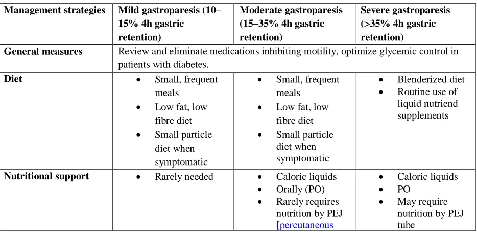
objective gastric retention at 4 hours (management is reviewed in detail in ref. 119)(Table 2).
[H2] Dietary Modifications
Dietary modifications represent the first line of treatment for gastroparesis and are generally used
for all patients, regardless of disease severity.Oral intake is preferable for nutrition and
hydration in patients with gastroparesis. As patients often have early satiety, they are
recommended to eat small meals and to avoid foods high in fat and indigestible fibres, because
these delay gastric emptying.1,77 When small meals are eaten as part of the gastroparesis diet,
more frequent meals, such as three meals per day plus two snacks, are often needed to maintain
caloric intake. Patients are advised to consume liquids such as soups, as the gastric emptying of
caloric liquids or homogenized solids is often preserved in patients with gastroparesis, who can
tolerate smaller sizes of such meals ingested more frequently rather than large meals three times
per day1. Importantly, a high-fat diet, with solid meals increases the severity and frequency of
symptoms among patients with gastroparesis;120 by contrast, a small particle size diet reduces
upper GI symptoms (nausea, vomiting, bloating, postprandial fullness, regurgitation and
heartburn) in patients with diabetic gastroparesis.121
[H2] Pharmacology
If a gastroparesis-suitable diet fails to manage symptoms, patients may be treated medically with
pharmacological agents including prokinetic and antiemetic medications. The clinical efficacy of
pharmacological agents for symptoms of nausea and vomiting is questionable, based on analysis
of data of 425 patients.122 Gastric prokinetic medications increase the rate or amplitude of
stomach contractions and, thus, increase the rate of gastric emptying. Medications currently
approved (though not in all countries) include metoclopramide, domperidone, and
erythromycin.123
Metoclopramide (a 5-HT4 agonist,5-HT3 and dopamine D2 antagonist), has both
prokinetic and antiemetic actions; however, it can cause both acute and chronic CNS side effects
reversible or irreversible, and may be less prevalent than 1 in 1000,124, in contrast to the
estimated 1–10% risk previously suggested in a guideline.125 In the United States,
metoclopramide is approved for diabetic gastroparesis for up to 12 weeks duration. A nasal spray
formulation of metoclopramide in gastroparesis has demonstrated efficacy in females, not in
male patients.126
Another dopamine receptor antagonist, domperidone, exhibits gastric prokinetic as well
as antiemetic properties via action on the area postrema, which is the vomiting center present in
the brainstem.Domperidone does not readily cross the blood–brain barrier; therefore, this drug is
much less likely to cause extrapyramidal side effects than metoclopramide. However,
domperidone (like the macrolide erythromycin, which is also used as a prokinetic) is associated
with prolongation of the cardiac QTc interval.Domperidone is not currently approved in the
United States, but is available in many other countries in Europe and Asia. Oral erythromycin, a
pure prokinetic agent that acts on motilin receptors, produced an improvement in symptoms in
43% of patients;127 however, a third of patients experience loss of the long term efficacy of erythromycin (mean 11 months’ follow up)128 due to tachyphylaxis.129 Erythromycin is not approved for the treatment of gastroparesis in any country and is used off-label, typically for a
short period of less than a month.
Some patients with post-surgical gastroparesis or diabetic gastroparesis may have
impaired gastric accommodation in addition to impaired gastric emptying130. In such patients,
erythromycin is contra-indicated as it reduces gastric accommodation, and the 5-HT1A agonist,
Buspirone, is prescribed to enhance gastric accommodation and relieve symptoms, though this
recommendation is based on relatively small clinical trial.131
[H3] New prokinetic drugs.
Several promising new prokinetic agents are in the pipeline for the treatment of gastroparesis.
Relamorelin is a ghrelin receptor agonist that stimulates gastric body and antral contractions,
accelerates gastric emptying, and has been shown in phase IIA and IIB clinical studies to
increase gastric emptying of solids and reduce the symptoms of gatroparesis, particularly nausea,
fullness, bloating and pain.132,133 Relamorelin is currently being tested in phase III trials which
should also provide information on the optimal subcutaneous dose of this treatment. In addition,
countries, other than the United States, for the treatment of chronic constipation. The drug
accelerates gastric emptying and was shown in a preliminary report to relieve symptoms in 28
patients with idiopathic gastroparesis.134
[H3] New drugs for impaired gastric accommodation.
Acotiamide has fundus-relaxing and prokinetic properties owing to the ability of this drug to
antagonize the inhibitory muscarinic type 1 and type 2 autoreceptors on cholinergic nerve
endings and to inhibit acetylcholinesterase. The drug enhances gastric accommodation and
emptying135 and relieves dyspeptic symptoms,136 and it is approved in Japan for treatment of
functional dyspepsia. However, there are currently no registered trials with acotiamide in
gastroparesis.
[H3] Approved Drugs Used Off Label.
Several drugs that are approved for other conditions are used by clinicians ‘off-label’ to treat the
symptoms of gastroparesis. Although not proven efficacious in a randomized, controlled trial in
patients with gastroparesis,137 nortriptyline (a tricyclic antidepressant) is used for relief of pain.
In a study conducted in patients with functional dyspepsia, amitriptyline (a tricyclic
antidepressant as well as a muscarinic receptor antagonist) improved symptoms in patients with
dyspeptic symptoms who did not have delayed gastric emptying138 and it modestly improved
sleep quality.139
Mirtazapine, an antidepressant with central adrenergic and serotonergic activity with
direct anti-emetic activity possibly related to 5-HT3 antagonist activity,140 provides symptom
relief for patients with functional dyspepsia and weight loss, a condition with substantial overlap
with gastroparesis. However, mirtazapine is not actually approved for treatment of functional
dyspepsia. Encouragingly, an open-label study of mirtazapine in patients with gastroparesis was
associated with improvements in nausea, vomiting, retching and loss of appetite.141 Another drug
that is used off-label to treat upper gastrointestinal symptoms in functional dyspepsia is
buspirone, an anxiolytic medication and 5-HT1A agonist, which is used to treat anxiety; it
enhances gastric accommodation and reduces postprandial symptoms in patients with functional
dyspepsia.131 Last, aprepitant, a neurokinin antagonist approved for use for the treatment of
gastroparesis and related disorders.142 It does not alter gastric emptying, but increases fasting and
postprandial gastric volumes.143
[H2] Pyloric Intervention
As mentioned above, delayed gastric emptying in gastroparesis is associated with antral
hypomotility and, in some patients, with pyloric sphincter dysfunction in the form of
pylorospasm; it is important to note that this intervention is not performed for pyloric stenosis.144
Botulinum toxin blocks the exocytosis of acetylcholine in cholinergic nerve endings, thereby
blocking the increased tone or spasm of the pyloric sphincter. An open-label study using
intra-pyloric botulinum type A (Botox) injection in 179 patients with gastroparesis was associated
with a decrease in gastroparesis symptoms at 1–4 months in 92 patients (51.4%). An improved
response was observed in those who received a higher dose, in females, in those aged <50 years,
and in patients without diabetes mellitus or post-surgical gastroparesis.145 Two double-blind
studies showed an improvement in gastric emptying, but a similar reduction in severity of
symptoms compared to placebo.146,147 Botulinum toxin injections do not result in sustained
improvement in the symptoms of gastroparesis, but may provide temporary relief, lasting on
average 3 months. Further studies are necessary to work out the specific patients who may most
benefit from the use of this treatment; it is also still unclear whether a positive clinical response
to botulinum toxin injection is valid for selecting patients for more permanent interventions of
the pylorus, which are discussed next.
Pyloroplasty (to widen the pylorus and prevent spasm ) or pyloromyotomy (an incision in
the wall of the pylorus by endoscopic intervention, referred to as peroral pyloroplasty or gastric
POEM [per-oral endoscopic myotomy]) performed surgically or endoscopically (Table 3), are
procedures being offered to patients who are refractory to other treatments, including
pharmacological approaches. The literature currently does not provide insight on the proportion
of patients who are refractory to other treatments and undergo pyloric interventions. The basic
rationale for this approach is the observation of pylorospasm in an unknown proportion of
patientswith gastroparesis, particularly diabetic gastroparesis.144 However, it is unclear whether
Table 3.148-156 Clearly, controlled studies are required to assess the efficacy of pyloric
interventions. Meanwhile, the algorithm in Figure 4, has been proposedto help guide the
selection of patients for pyloric interventions, using measurement of pyloric sphincter
abnormalities (Endoflip®; Endoluminal Functional Lumen Imaging Probe) or the symptomatic
response to pyloric botox injections.119 However, it is important to note that Endoflip measures
stiffness or compliance at the pylorus rather than active contractions or sphincter tone, and it is
as yet unproven whether the response to intra-pyloric injection of botulinum toxin is sufficient to
predict efficacy of pyloromyotomy.
[H2] Gastric Electrical Stimulation
Gastric contractility depends on the underlying basal electrical rhythm, which is relayed through
the gastric pacemaker cells. Therefore, a novel method for gastroparesis has been considered;
that as in the heart, an artificial pacemaker might capture the electrical rhythm of the stomach
and drive the contractile frequency. Unfortunately, there is as yet no clinical device that has been
able to entrain the basal electrical rhythm of the human stomach, although this has been achieved
in experimental animal models, and therefore it has not yet been possible to test a pacemaker
system in the stomach with the same objective as that achieved in the heart.
Gastric electrical stimulation was originally developed to enhance gastric emptying,
however, the technique has evolved to become a high frequency stimulation that appears to
interfere with sensory transduction to the brain and thus provides a treatment for refractory
symptoms in gastroparesis. Based on the initial studies that have shown an improvement in
symptoms, particularly in patients with diabetic gastroparesis, the gastric electric neurostimulator
was granted approval from the FDA under the Humanitarian Device Exemption, for the
treatment of chronic intractable (drug refractory) nausea and vomiting secondary to gastroparesis
of diabetic or idiopathic aetiology in patients aged 18–70 years. In 151 patients with refractory
gastroparesis treated at a single center, gastric electric stimulation at least moderately improved
symptoms in 43%.157 The response in patients with diabetes mellitus was better than in patients
with gastroparesis from other aetiologies. Patients with symptoms of nausea, loss of appetite, and
early satiety were the best responders. Although there are a number of open-label studies
suggesting the efficacy of gastric electrical stimulation in treatment of gastroparesis, particularly
caution in recommending gastric electrical stimulation outside of research studies. This
recommendation is made on the basis of insufficient efficacy of gastric electrical stimulation in
the few controlled trials comparing “stimulation switched off” versus “stimulation on” gastric
electrical stimulation treatment as well as symptom regression to the mean, that is the natural
tendency for symptoms to improve from the high level at the time of initiation of treatment to a
later time, while the patient was receiving stimulator treatment.96,97
[H2] Diabetes and gastroparesis
The rate of gastric emptying has a major impact on the glycemic response to
carbohydrate-containing meals in healthy individuals and in patients with diabetes mellitus, particularly in the
initial postprandial increment.27 Notably, the delayed gastric emptying that characterizes
gastroparesis in patients with diabetes mellitus can affect the postprandial blood glucose
response. Furthermore, postprandial glycemic excursions make a major contribution to ‘overall’
glycemic control as assessed by HbA1c. Therefore, impaired postprandial glycemic control
represents an important target for management in patients with diabetic gastroparesis. In patients
treated with insulin, delayed gastric emptying may result in a mismatch of the timing of
exogenous, preprandial insulin and the actual delivery of nutrients, including carbohydrates,
from the stomach to the small intestine. In a study involving 11 patients with type 1 diabetes
mellitus, less insulin was required to be administered to the five patients with gastroparesis
(compared to the 6 patients without gastroparesis) to achieve euglycemia during the first 120
minutes after a meal, and more insulin was needed by the patients with gastroparesis between
180–240 minutes.158 Furthermore, delayed gastric emptying in patients with type 1 diabetes
mellitus has recently been reported to be associated with an overall increase in blood glucose
during the day, which may reflect the discordance between the timing of the preprandial insulin
and the later absorption of food due to delayed gastric emptying.159
Patients with diabetic gastroparesis frequently exhibit labile blood glucose with periods
of marked hyperglycemia and frequent hypoglycemia, particularly postprandially. No long-term
studies exist that document the benefit of maintaining optimal glycemia in patients with diabetic
gastroparesis. Therefore, the recommendation to strive for near normal blood glucose levels in
patients with diabetic gastroparesis derives mainly from studies conducted in healthy volunteers
results in delayed gastric emptying.1 Nevertheless, optimizing glycemic control can also be
beneficial in gastroparesis, as shown in a recent multicenter pilot study, in which continuous
subcutaneous insulin infusion with insulin pump therapy, with continuous glucose monitoring,
reduced hyperglycemia and HbA1c levels in patients with diabetic gastroparesis.160 Patients also
showed associated improvements in gastroparesis symptoms and in tolerance of nutrients, which
were maintained for the 24-week phase of intensive monitoring and therapy.
[H1] Quality of Life
Quality of life (QOL) in patients with gastroparesis is impaired compared with the general
population,161 and to a level that is similar to patients with other chronic medical and
psychological disorders.162 In one large study of 335 patients with gastroparesis conducted in the
United States, the average impairment of disease-specific QOL, measured using the Patient
Assessment of Upper Gastrointestinal Disorders Quality of Life (PAGI-QOL) questionnaire, was
moderate.77
The degree of impairment in QOL relates to the duration and severity of symptoms.163
The cardinal symptoms of gastroparesis are nausea and vomiting. In one study, nausea appeared
to be of a similar severity in patients with either idiopathic or diabetic gastroparesis, and
increasing severity of nauseawas associated with impaired quality of life on the PAGI-QOL.164
These results have been replicated in another study,75 but in this patient cohort vomiting was
more severe among those with diabetic gastroparesis as compared to idiopathic gastroparesis. An
increasing severity of vomiting, irrespective of gastroparesis etiology, correlated negatively with
both disease-specific quality of life, according to the PAGI-QOL, and generic quality of life,
using the short-form 36 (SF-36).
Bloating and upper abdominal pain or discomfort also impact QOL. Greater severity of
bloating was associated with progressive impairments in disease-specific quality of life on the
PAGI-QOL, and physical and mental components of the SF-36.77 Moreover, greater severity of
upper abdominal pain and discomfort was also associated with statistically significantly higher
levels of impairment on the PAGI-QOL and the SF-36.87 In another study, there was a negative
correlation between abdominal pain severity and QOL, but there was no correlation between pain