Int. J. Electrochem. Sci., 8 (2013) 10526 - 10539
International Journal of
ELECTROCHEMICAL
SCIENCE
www.electrochemsci.org
Rapid Screening Method for Assessing Total Phenolic Content
Using Simple Flow Injection System with Laccase
based-biosensor
Maliwan Amatatongchai*, Wongduan Sroysee, Saowanee Laosing and Sanoe Chairam
Department of Chemistry and Center of Excellence for Innovation in Chemistry, Faculty of Science, Ubon Ratchathani University, Ubonratchathani, 34190, Thailand.
*
E-mail: [email protected]; [email protected]
Received: 10 June 2013 / Accepted: 8 July 2013 / Published: 1 August 2013
A sensitive and selective amperometric biosensor for evaluation of total phenolic content (TPC) was developed by immobilizing laccase (Lac) on the glassy carbon electrode modified with matrix nanocomposites. The nanocomposites composed of NH2 -functionalized carbon nanotubes (CNT-NH2), gold nanoparticles (AuNPS) and Bovine serum albumin (BSA). In order to increase the stability of the biosensor Lac was covalently immobilized in nanocomposites matrix by using glutaraldehyde (Glu). The rapid screening method of TPC assay was proposed based on amperometric detection of gallic acid on the laccase-based biosensor in flow injection analysis. The developed biosensor GC/CNT-NH2/AuNPs/Lac-BSA shows an excellent activity for gallic acid. The developed system also demonstrates good stability of the immobilized enzyme in spite of the hydrodynamic conditions. The system provides an impressively good precision (%RSD =3.1) for 20 L injections of 6 µM gallic acid (n = 30). Under the flow rate used of 1.5 mL min-1, throughput of sample is 42 samples h-1. The method was successfully applied to TPC assay in plant extracts and tea infusions.
Keywords: Laccase biosensor; flow injection analysis; total phenolic content, nanocomposites
1. INTRODUCTION
stress describes the over production of reactive oxygen species (ROS), for example H2O2, OH and O2
-, and reactive nitrogen species (RNS) such as NO and NO. A number of previous studies have
suggested that antioxidant play a key role in the prevention of chronic diseases such as neurodegenerative diseases, cancers, cardiovascular diseases as well as inflammation and aging [1-7].
Since fruits and vegetables are major sources of antioxidants, the ability to measure antioxidant capacity in these natural sources is timely challenge. Antioxidant power or capacity can be evaluated both in vivo [8, 9] and in vitro [10-11]. Typically, in vitro experiments are employed in screening antioxidant capacity in foods and beverages. According to recent review articles [11-13], in vitro methods for evaluating antioxidant capacities are either based on scavenging capacity against a biologically relevant oxidant (e.g. ROS and RNS), or on scavenging capacity against selected non-biological oxidants (such as DPPH, ABTS+, FRAP and FC assays).
In order to fulfill the need for rapid and automated evaluation of antioxidant capacity in a large number of samples, a range of flow-based techniques have been presented. For example, a scavenging DPPH assay was carried out in a flow injection (FI) format using spin resonance [14] and amperometry [15, 16] for analyzed detection. Additionally, a FI system with on-line electrogeneration of ABTS+ has been shown to yield a throughput of 32 sample h-1[17].
Polyphenols are also one of the most important classes of natural antioxidants widespread in vegetables, fruit and medicinal plants. More recently, electrochemical approaches have effectively been used for evaluation of polyphenols. Biosensor using laccase have been reported to provide very stable amperometric response to polyphenolic compounds [18]. Flow injection (FI) with amperometric detection on laccase based biosensor was developed for total phenolic content (TPC) in cultivated plants [19] and wine [20]. One of the main benefits to consider in the development of an amperometric biosensor is the possibility of operating within the low potential range, i.e. between -0.2 and 0.05 V Vs Ag/AgCl [21, 22] to avoid the interferences from sample matrices.
2. EXPERIMENTAL
2.1 Reagent and Chemical
Laccase (Tramestes versicolor, 21.8 U mg-1), HAuCl4.H2O (Au > 48%), bovine serum albumin (BSA) solution and 50% glutaraldehyde (Glu) were purchased from Sigma-Aldrich (St. Louis, USA). NH2 functionalized carbon nanotubes (CNT-NH2) used in this study were purchased from Nanolab inc. (MA, USA), (> 95% purity, 15±5 nm diameter, length 1-5 micron). Catechin, caffeic acid, gallic acid, chlorogenic acid and Trolox (6-hydroxy-2,5,7,8-tetramethylchroman-2-carboxylic acid) were purchased from Acros Organic (Geel, Belgium). 1 mM standard solutions of water-soluble antioxidants (Trolox and gallic acid) were prepared using deionized-distilled water. Other antioxidants were prepared in absolute ethanol. Gold nanoparticles (AuNPs) were synthesized according to the method previously described by McFarland et al. [23].
2.2 Preparation of laccase biosensor
Scheme 1. Illustrating the diagram of phenolic biosensor (GC/CNT-NH2/AuNPs/Lac-BSA) preparation and the corresponding reaction generated the amperometric signal.
CNT-NH2 was added to 1 mL of DMF. The suspension was then sonicated for 3 h. Twenty µL of CNT-NH2 mixture was casted on the surface of GC electrode. The surface was dried at ambient temperature in a fume hood. Ten µL of 1 mM AuNPs solution was deposited on the GC/CNT-NH2 electrode, and the electrode was dried at room temperature. After that, 20 µL of laccase-BSA solution (mixture of 4.5 mg mL-1 laccase and 0.05% BSA) was deposited on the GC/CNT-NH2/AuNPs electrode. The GC/CNT-NH2/AuNPs/Lac-BSA was dried for approximately 30 min at room temperature. Finally, 15 µL of 2.5% glutaraldehyde was deposited on GC/CNT-NH2/AuNPs/Lac-BSA electrode and then dried at ambient temperature.
[image:4.596.94.513.524.692.2]Basically, total phenolic content in plant extracts was related to the amperometric signal due to the reduction of the quinine obtained as a product of the enzymatic oxidation of phenolic compounds [24, 25]. The reaction which involves gallic acid and laccase on electrode surface is described in Scheme 1. Laccase oxidized gallic acid with the concomitant reduction of molecular oxygen to water. Therefore, total phenolic content (TPC) was evaluated by measuring the reduction current of gallic acid.
2.3 Electrochemical measurement Cyclic voltammetry
An eDAQ potentiostat (model EA161, Australia), equipped with the e-corder 210 and e-Chem v2.0.13 software, was used for all the cyclic voltammetric studies. The active surface area of glassy carbon or GC electrode (CH Instrument, USA) was approximately 0.07 cm2. A home made three-electrode cell, comprising of a working three-electrode (GC/CNT-NH2/AuNPs/Lac-BSA three-electrode), a reference electrode (Ag/AgCl (sat.)) and a counter electrode (platinum wire) was employed. Measurements were performed using a citric buffer (0.1 M, pH 6.0) as supporting-electrolyte solution.
Simple flow injection system with laccase based-biosensor
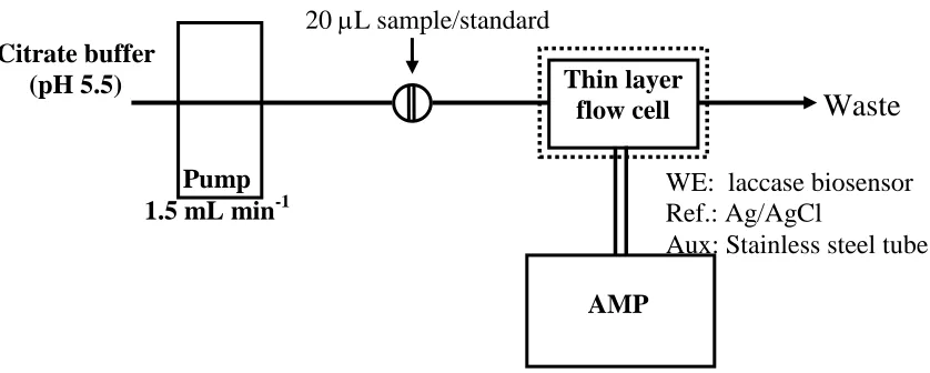
Figure 1. FI manifold for evaluation of TPC with amperometric detection using the developed laccase biosensor (GC/CNT-NH2/AuNPs/Lac-BSA) as working electrode (WE). Optimal condition: potential, -0.05 V (vs Ag/AgCl); carrier, 0.1 M citrate buffer solution (pH = 5.5); flow rate, 1.5 mL min-1.
AMP
20 L sample/standard
WE: laccase biosensor Ref.: Ag/AgCl
Aux: Stainless steel tube
Citrate buffer (pH 5.5)
Waste
Pump 1.5 mL min-1
Figure 1 depicts schematic diagram of the FI system for amperometric detection (AMP) of antioxidant at the laccase based biosensor. A Shimadzu pump (model LC-10 AD, Japan) was used for the liquid flow. A Rheodyne injector (model 7725, USA) fitted with 20 µL sample loop was used for injecting antioxidant standards and samples. An eDAQ potentiostat (EA161), equipped with the e-corder 210, Chart v5.5.11 software and thin layer flow cell with three electrodes system (CH Instruments, USA) was used for amperometric measurements. The laccase biosensor (GC/CNT-NH2/AuNPs/Lac-BSA electrode) was used as a working electrode, Ag/AgCl as a reference electrode and a stainless steel tube as a counter electrode. The electrode area was utilized at 0.06 cm2.
2.4 Preparation of plant extract
Thai vegetables were purchased from the local supermarkets in Ubon Ratchathani Province, Thailand. Six vegetables (Asiatic ennywort, Bael leaves, Cabbage, Ginger, Guduchi and Limnocharis flava Buch) and two teas (Jasmine and Mulberry tree tea) were selected as samples for TPC assay.
The extraction scheme for fruits and leaves were previously described in detail elsewhere [16, 26]. In brief, fresh vegetable leaves were dried in an oven to constant weight (40 oC, 48 h), followed by powdering in mortar. Three consecutive extractions with hexane (3x25 mL) were performed on 2.5 g portions of plant powder to remove both pigments and lipids. Centrifugation at 3,000 rpm was carried out for 5 min and the organic solvent discarded. A 10 mL aliquot of 2% (w/v) of aqueous sodium metabisulphite was then added to the solid residue, followed by 25 mL methanol:water (80:20, v/v). The supernatant was separated out and the solid was further extracted two times with the methanol:water solution. The 3 methanolic fractions were pooled together before filtering through a 0.25 micron cellulose membrane prior to the determination of TPC.
Two tea samples are commercial products. Infusions were prepared by adding 50 mL of deionized water (100 oC) to the 1.40 g of herbal tea material. The infusions were stirred for 2 min. The filtrate solutions were cooled to room temperature.
2.5 The spectrophotometric Folin-Ciocalteau method
The spectrophotometric Folin-Ciocalteau method was employed for assess total phenolic content. This method was adopted from the previous study [27-28]. Vegetable extract or tea infusion was quantitatively put into a 10 mL volumetric flask, 0.5 mL of Folin-Ciocalteau reagent (FCR) and 1.5 mL of 20% Na2CO3 solution was added. After refilling with distilled water to the mark and thorough agitation the reaction mixture was left to stand for 20 min and then measured on the spectrophotometer at the wavelength of 765 nm against the blank. Total phenolic content (TPC) was expressed as mg gallic acid equivalent in g of dried sample as average from three parallel determinations.
2.6 Method validation
buffer before injection if necessary. External calibrations of gallic acid were adopted. The results were compared with the values obtained from FCR method.
3. RESULTS AND DISCUSSION
3.1 Cyclic voltammetry study of antioxidants
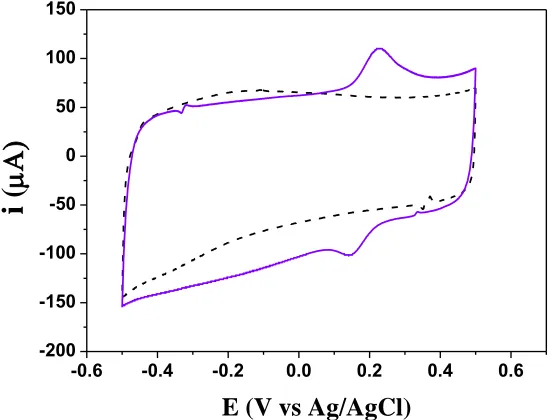
[image:6.596.146.422.331.541.2]Cyclic voltammetry was performed to assess the electrochemical behaviors of the developed biosensor (GC/CNT-NH2/AuNPs/Lac-BSA) toward gallic acid. Cyclic voltammogram (CV) of the GC/CNT-NH2/AuNPs/Lac-BSA electrode in the presence of gallic acid was depicted in Figure 2. The CV shows the characteristic of quasi-reversible reaction of gallic acid. An increase of the cathodic current and the decrease of anodic peak in the presence of immobilized enzyme into the electrode were observed. These results were attributed to catalytic electrochemical reaction [22, 29].
Figure 2. Cyclic voltammograms of 0.02 mM gallic acid at GC/CNT-NH2/AuNPs/Lac-BSA electrode. Background current (0.1 M citrate buffer, pH 5.0) is also shown as dotted line. Scan rate was fixed at 100 mV s-1.
3.2 Optimization of the laccase biosensor
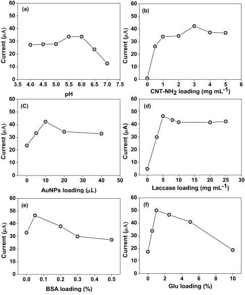
In order to optimize the performance of the proposed laccase biosensor toward phenolic detection, the effect of pH and the effect of CNT-NH2, AuNPs, laccase, BSA as well as Glu loadings were examined. The responses of 0.02 mM gallic acid were recorded in 0.1 M citrate buffer solution with different pHs (4.0-7.0) at the GC/CNT-NH2/AuNPs/Lac-BSA electrode. Figure 3a shows that reduction current of gallic acid increase up to pH 5.0-6.0, and decrease as increasing pH greater than 6.0. The decrease of response at pH higher than 6.0 is attributed to the decrease of laccase activity.
-0.6 -0.4 -0.2 0.0 0.2 0.4 0.6 -200
-150 -100 -50 0 50 100 150
i
(
According to Roy et al. [30], at higher pH, the OH- will inhibit T2/T3 site of laccase to bind with substrate resulting in decreasing of the reduction current.
Figure 3. Effect of (a) pH and effect of (b) MWCNT-NH2, (c) AuNPs, (d) laccase, (e) BSA and (f) Glu and loadings on the reduction current of 0.02 mM gallic acid.
0 1 2 3 4 5 6
0 10 20 30 40 50 60 C u rr e n t (
CNT-NH2 loading (mg mL-1)
(b)
3.5 4.0 4.5 5.0 5.5 6.0 6.5 7.0 7.5 0 10 20 30 40 50 60 C u rr e n t ( pH (a)
0 10 20 30 40 50
0 10 20 30 40 50 60 C u rr e n t (
AuNPs loading (L)
(C)
0 5 10 15 20 25 30
0 10 20 30 40 50 60 C u rr e n t ( A )
Laccase loading (mg mL-1)
(d)
0.0 0.1 0.2 0.3 0.4 0.5
0 10 20 30 40 50 60 C u rr e n t (
BSA loading (%)
(e)
0 2 4 6 8 10
0 10 20 30 40 50 60 C u rr e n t (
Glu loading (%)
[image:7.596.51.537.138.723.2]
The effect of the CNT-NH2 loading on the reduction current of the biosensor was evaluated by casting different amounts of CNT-NH2 suspension on the GC electrode surface. Figure 3b illustrates the effect of CNT-NH2 loading on the intensity of the reduction current of 0.02 mM gallic acid. The signal increased when increasing the amount of CNT-NH2 and reached the plateau when 3 mg. mL-1 of CNT-NH2 (20 µL) was used. Therefore, 3 mg. mL-1 of CNT-NH2 was selected for the biosensor fabrication.
Figure 3c shows the effect of AuNPs loading on the intensity of the reduction current of 0.02 mM gallic acid. The signal increased when increasing the amount of AuNPsand reached the plateau when 20 µL of AuNPs was casted. Therefore, this condition was selected for the biosensor fabrication. Biosensor with CNT-NH2 -AuNPs conducting binding nanocomposites showed significantly improved the corresponding responses when compared with the biosensor without the binding nanocomposites similarly to the previous report from Sarma et al.[31].
As previously noted, the immobilization of the enzymes on the electrode surface is considered as one of the critical steps that controls the effectiveness of the enzyme electrodes [31]. According to Kulys, albumins can prevent laccase inactivation and can increase the reaction yield of phenolic oxidation [32]. Therefore, effect of laccase, BSA and Glu on the response of the biosensor was also investigated. As shown in Figure 3d, the reduction current increased when increasing the amount of laccase and then reached a plateau at about 8 mg. mL-1 laccase. Studies on the effect of BSA and Glu loading on the reduction current showed that the concentration of 0.05 % BSA and 2.5% Glu were the optimum conditions for the biosensor fabrication (Figure 3e-3f).
3.3 Optimum potential for amperometric detection
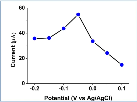
Figure 4. Influence of the applied potential on the GC/CNT-NH2/AuNPs/Lac-BSA biosensor response for 0.02 mM gallic acid.
The proposed amperometric method for evaluation of antioxidant activity is based on the electrochemical monitoring of reduction signal from antioxidant at the laccase-modified electrode. Hydrodynamic voltametric in flow injection system (Figure 1) was employed to find the optimum
-0.2 -0.1 0.0 0.1
0 20 40 60
C
u
rr
e
n
t
(
[image:8.596.158.390.474.647.2]
potential for the CNT-NH2/AuNPs/Lac-BSA electrode. As shown in Figure 4, the reduction current slightly rises in potential rage of -0.2 up to -0.05 V and decreases rapidly above -0.05 V. The potential at -0.05 V provides the maximum peak area and therefore chosen for further work. The electrocatalytic activity of enzyme laccase incorporated with electrical conducting nanocomposites (CNT-NH2 and AuNPs) and biocompatible matrix (BSA) enable the biosensor effectively detection at low potential.
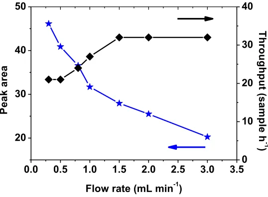
3.3. Optimum flow rate for antioxidant detection
[image:9.596.169.438.385.583.2]The effect of flow rate on the biosensor response was optimized by injection of 20 µL of gallic acid standard into the carrier stream to achieve satisfactory sensitivity and sample throughput. Figure 5 shows that the response decreases with increasing flow rate from 0.25 to 3.0 mL min-1. In enzyme-based flow injection assay, it is usual that the FI peak height or peak area decreased as the flow rate increased. This behaviour is consistent with previously published data [33]. The authors attributed this behaviour to the need for slow passage of the sample plug to the enzyme reaction that take place in a high extent. Conversely, increasing flow rate increases sample throughput. To balance between response and sample throughput, the flow rate of 1.5 mL min-1 was chosen.
Figure 5. Effect of the flow rate on laccase biosensor response and sample throughput.
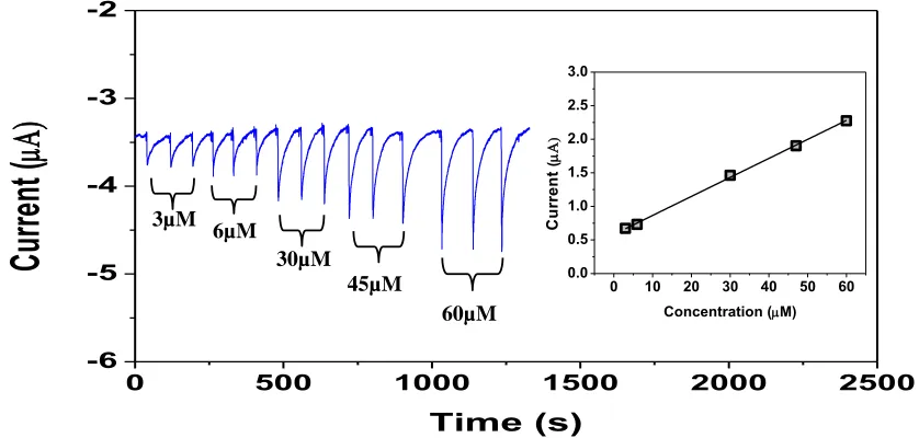
3.4. Analytical features
Analytical performance of the FI method with amperometric detection at the developed laccase biosensor for assessing total phenolic content (TPC) was examined. Examples of the FI grams obtained from replicate injection of five gallic acid standards and the corresponding calibration curve were shown in Figure 6.
0.0 0.5 1.0 1.5 2.0 2.5 3.0 3.5 20
30 40 50
Flow rate (mL min-1)
P
e
a
k
a
re
a
0 10 20 30 40
T
h
ro
u
g
h
p
u
t (
s
a
m
p
le
h
-1
Figure 6. Examples of the FI grams obtained from replicate injection of five gallic acid standards. The inset shows the linear relationship between the signal of gallic acid and the concentration.
Performance characteristics of the developed FI method investigated on five polyphenols summarize in Table 1.
Table 1. Performance characteristics of the developed antioxidant biosensor for polyphenol determination.
Substrate Linearity range
(M)
Sensitivity
(A M-1)
r2 LODa (M) %RSDb
Caffeic acid 0.3– 45 0.753 0.997 0.49 2.40
Catechin 1.7 – 30 0.142 0.997 0.20 2.60
Gallic acid 3.0– 60 0.169 0.999 0.71 1.70
Chlorogenic acid 1.5 – 30 0.207 0.995 0.10 2.20
Trolox 2.0– 35 0.879 0.997 0.10 2.16
a Calculated from 3 (n=7) of the signal form the lowest concentration of each working range.
bCalculated from the peak area obtained from 20 µL injections (n=71) of the lowest concentration of each working range.
For all tested polyphenols (caffeic acid, catechin, gallic acid, chlorogenic acid and trolox), linearity calibrations were performed. The developed FI method provides good precision (%RSD < 2.7). The limits of detections are in M level.
The developed system also demonstrates good stability of the immobilized enzyme in spite of the hydrodynamic conditions. The system provides an impressively good precision (%RSD =3.1) for
0 500 1000 1500 2000 2500
-6 -5 -4 -3 -2
Cu
rre
nt
(
Time (s)
0 10 20 30 40 50 60
0.0 0.5 1.0 1.5 2.0 2.5 3.0
C
u
rr
e
n
t
(
Concentration (M)
3µM 6µM
30µM 45µM
[image:10.596.91.510.80.280.2] [image:10.596.62.529.451.599.2]
20 L injections of 6 µM gallic acid (n = 30). Under the flow rate used of 1.5 mL min-1
, throughput of sample is 42 samples h-1.
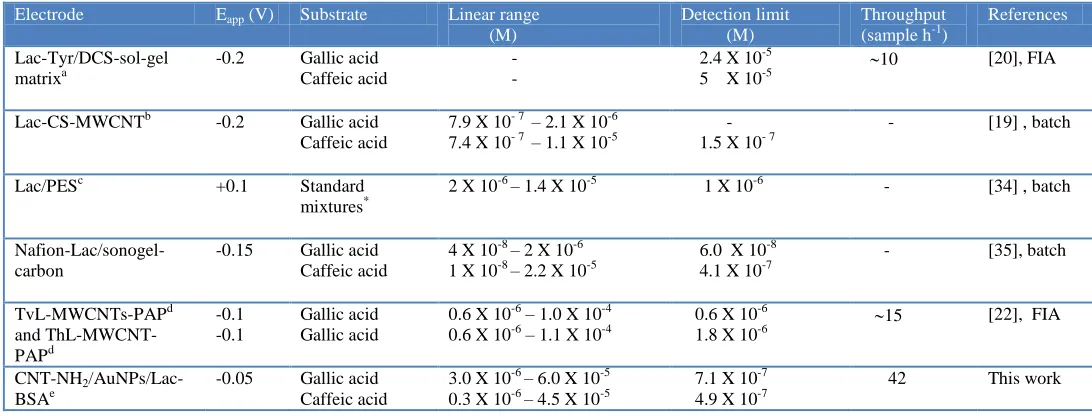
[image:11.596.24.570.210.418.2]Analytical performance of the proposed laccase biosensor towards polyphenols detection compared with various modified electrodes was shown in Table 2.
Table 2. Comparison of analytical performance of the proposed laccase biosensor towards polyphenols determination with previously reported modified electrodes.
Electrode Eapp (V) Substrate Linear range
(M)
Detection limit (M)
Throughput
(sample h-1)
References
Lac-Tyr/DCS-sol-gel matrixa
-0.2 Gallic acid Caffeic acid
- -
2.4 X 10⁻⁵ 5 X 10⁻⁵
10 [20], FIA
Lac-CS-MWCNTb -0.2 Gallic acid
Caffeic acid
7.9 X 10- 7– 2.1 X 10⁻⁶ 7.4 X 10- 7– 1.1 X 10⁻⁵
- 1.5 X 10- 7
- [19] , batch
Lac/PESc +0.1 Standard
mixtures*
2 X 10⁻⁶ – 1.4 X 10⁻⁵ 1 X 10⁻⁶ - [34] , batch
Nafion-Lac/sonogel-carbon
-0.15 Gallic acid Caffeic acid
4 X 10⁻⁸ – 2 X 10⁻⁶
1 X 10⁻⁸ – 2.2 X 10⁻⁵ 6.0 X 10 -8
4.1 X 10⁻⁷
- [35], batch
TvL-MWCNTs-PAPd and ThL-MWCNT-PAPd -0.1 -0.1 Gallic acid Gallic acid
0.6 X 10⁻⁶ – 1.0 X 10⁻⁴ 0.6 X 10⁻⁶ – 1.1 X 10⁻⁴
0.6 X 10⁻⁶
1.8 X 10⁻⁶
15 [22], FIA
CNT-NH2
/AuNPs/Lac-BSAe
-0.05 Gallic acid Caffeic acid
3.0 X 10⁻⁶ – 6.0 X 10⁻⁵ 0.3 X 10⁻⁶ – 4.5 X 10⁻⁵
7.1 X 10⁻⁷ 4.9 X 10⁻⁷
42 This work
Lac = Laccase, Tyr = Tyrosinase, DCS = diglycerysilane; CS = chitosan, MWCNT multiwalled carbon nanotubes, PES = polyethersulfone, TvL = Trametes versicolor, ThL = Trametes hirsuta, PAP = polyazetidine prepolymer , CNT-NH2 = amino-functionalized multiwalled carbon nanotubes; AuNPs = gold nano particles, BSA = bovine serum albumin, Eapp = Applied potential,
a
Graphite screen printed electrode b
Gold (Au) electrode c
Platinum (Pt)electrode
d
Screen Printed Electrode e
Glassy carbon electrode (GCE)
Table 2 shows the analytical performances of the proposed phenolic biosensor compared with various modified electrodes. This suggests that the performance characteristics of the proposed phenolic biosensor being comparable or better to other previously reported modified electrodes. Compared with the previous flow injection system [20, 22], this current FIA system gave higher sample throughput (up to 42 sample h-1). Accordingly the system is suitable for high throughput screening of TPC in remote environments, and as such was utilized in the screening of functional plant foods including vegetables, herbs and fruits.
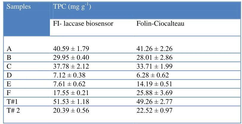
3.6. Performance on plant extract and tea applications
[image:12.596.93.500.205.413.2]
spectrophotometric method [27, 28] (Folin-Ciocalteau method). For the proposed FI method, TPC could be determined using the calibration graph of gallic acid (Figure 5) and expressed as mg gallic acid equivalent in g of dried sample as shown in Table 3.
Table 3. Comparison between the total phenolic content (TPC) from Thai vegetable extracts and tea infusions analyzed by the developed method (FI-laccase biosensor) and Folin-Ciocalteau method.
Samples TPC (mg g-1)
FI- laccase biosensor Folin-Ciocalteau
A 40.59 ± 1.79 41.26 ± 2.26
B 29.95 ± 0.40 28.01 ± 2.86
C 37.78 ± 2.12 33.71 ± 1.99
D 7.12 ± 0.38 6.28 ± 0.62
E 7.61 ± 0.62 14.19 ± 0.51
F 17.55 ± 0.21 25.88 ± 3.69
T#1 51.53 ± 1.18 49.26 ± 2.77
T# 2 20.39 ± 0.56 22.52 ± 0.97
Expressed as mean ± standard deviation obtain from triplicate determination A= Asiatic ennywort, B= Bael leaves, C= Cabbage, D= Ginger, E= Guduchi, F= Limnocharis flava Buch, T# 1= Jasmine tea, T# 2= Mulberry tree tea
The analyzed TPC were compared with the values measured by the Folin-Ciocalteau method, using paired t-test [36]. The results indicated that the TPC as determined from the two methods agree significantly well with each other (t observed = 0.692, t critical = 2.364 at P = 0.05).
4. CONCLUSION
ACKNOWLEDGEMENTS
Financial supports from the Center of Excellence for Innovation in Chemistry: Postgraduate Education and Research Program in Chemistry (PERCH-CIC), Commission on Higher Education, Ministry of Education, National Research Council of Thailand (NRCT) are gratefully acknowledged.
References
1. B. Halliwell, Biochem. Pharmacol. 49 (1995) 1341.
2. M. Valko, D.Leibfritz, J. Moncol, M. T. D. Cronin, M. Mazur, J. Telser, Int. J. Biochem. Cell. B 39 (2007) 44.
3. C. Kaur, H. C. Kapoor, Int. J. Food Sci. Technol. 36 (2001)703.
4. J. A. Vinson, X. Liang, J. Proch, B. A. Hontz, J. Dancel, N. Sandone, Adv. Exp. Med. Biol. 505 (2002) 113.
5. İ. Gülçin, J. Med. Food 14 (2011) 975. 6. İ. Gülçin, Arch. Toxicol. 86 (2012) 345.
7. B. Halliwell, J. M. C. Gutteridge, Free Radicals in Biology and Medicine; Clarendon Press: Oxford, UK, (1998).
8. R. L. Prior, G. Cao, Free Radic. Biol. Med. 27 (1999) 1173. 9. M. Serafini, D. del Rio, Redox Rep. 9 (2004) 145.
10. J. Cheorun, J. Seok-Moon, K. Soo-Yeon, P. Eunju, L. Seung-Cheol, Int. J. Food Sci. Technol. 43 (2008) 400.
11. L. M. Magalhaes, M. A. Segundo, S. Reis and J. L. F. C. Lima, Anal. Chim. Acta. 613 (2008) 1. 12. L. M. Magalhaes, M. Santos, M. A. Segundo, S. Reis and J. L. F. C. Lima, Talanta, 77, (2009).
1559.
13. J. Sochor, J. Dobes, O. Krystofova, B. Ruttkay-Nedecky, P. Babula, M. Pohanka, T. Jurikova, O. Zitka, V. Adam, B. Klejdus and R. Kizek, Int. J. Electrochem. Sci., 8 (2013)8464.
14. H. Ukeda, Y. Adachi and M. Sawamura, Talanta, 58(6) (2002)1279.
15. S. Milardovic, D. Ivekovic and B. S. Grabaric, Biochemistry 68 (2005) 175-180.
16. M. Amatatongchai, S. Laosing, O. Chailapakul and D. Nacapricha, Talanta 97 (2012) 267. 17. D. Ivekovic, S. Milardovic, M. Roboz and B. S. Grabaric, Analyst 130(5) (2005) 708. 18. S. S. Rosatto, R.S. Freire, R.S. N. Duran and L. T. Kubota, Quim. Nova 24 (2001)24. 19. M. Diaconu, S. C. Litescu and G. L. Radu, Sens Acturators B 145 (2010) 800.
20. M. R. Montereali, L. Della Seta, W. Vastarella, R. Pilloton, J. Mol. Catal. B: Enzyme 64 (2010) 189.
21. Y. Tan, W. Deng, B. Ge, Q. Xie, J. Huang and S. Yao, Biosens. Bioelectron. 24 (2009) 2225. 22. M. Di Fusco, C. Tortolina, D. Deriu and F. Mazzei, Talanta 81 (2010) 235.
23. A. D. McFarland, C. L. Haynes, C.A. Mirkin, R.P. Van Dyne and H.A. Godwin, J. Chem. Educ.,81 (2004) 544A.
24. G. Marko-Varga, J. Emneus, L. Gorton, T. Ruzgas, Trends Anal. Chem. 14 (1995)127.
25. A. I. Yaropolov, A. N. Kharybin, J. Emneus, G. Marko-Vagar, L. Gorton, Anal. Chim. Acta 308 (1995)137.
26. S. Silva, L. Gomes, F. Leitao, A. V. Coelho and L. V. Boas, Food Sci. Technol. Int., 12;5 (2006) 385.
27. P. Cunniff, Official Methods of Analysis, vol. 1, 16th ed., AOAC International, Alington, VA, USA, 1995.
28. O. Folin, W. Denis, J. Biol. Chem. 12 (1912) 239.
30. J. J. Roy, T. E. Abraham, K. S. Abhijith, P. V. S. Kumar, M. S. Thakur, Biosens. Bioelectron. 21 (2005) 206.
31. A. K. Sarma, P. Vatsyayan, P. Goswami, S. D. Minteer, Biosens. Bioelectron. 24 (2009) 2313. 32. J. Kulys, K. Krikstopaitis, A. Ziemys and P. Scheneider, J. Mol. Catal. B: Enzym.18 (2002) 99. 33. C. Fernández-Sánchez, T. Tzanov, G. M. Gübitz, A. Cavaco-Paulo, Bioelectrochemistry. 58
(2002) 149.
34. S. A. S. S. Gomes, J. M. F. Nogueira, M. J. F. Rebelo, Biosens. Bioelectron. 20 (2004) 1211. 35. M. ElKaoutit, I. Naranjo-Rodriguez, K. R. Temsamani, M. P. Hernández-Artiga, D. Bellido-Milla,
J. L. H.-H. d. Cisneros, Food Chem. 110 (2008) 1019.
36. J. N. Miller and J. C. Miller, Statistics and Chemometrics for Analytical Chemistry, 5th ed. Pearson Education Limited, Essex, 2005.